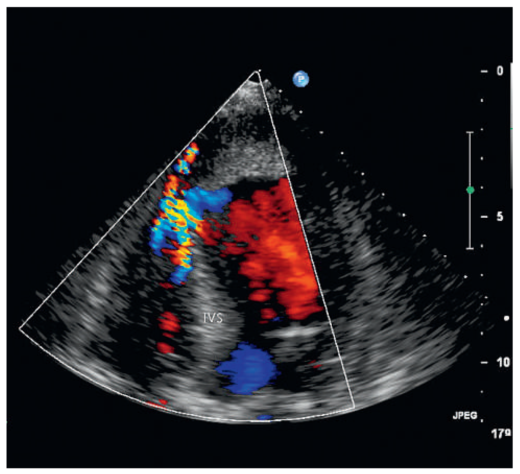
A 87-year-old female patient was hospitalised for decline in general health status, fatigue and oedema following a fall two weeks earlier. On no occasion did the patient experience chest pain or shortness of breath. The blood pressure and heart rate on admission were 113/79 mm and 80 bpm Hg respectively. Cardiopulmonary auscultation revealed a harsh, loud holosystolic murmur most audible along the left sternal border and radiating to the base, apex and right parasternal area, with accentuation of the second heart sound and crepitus rales across all lung fields. Bilateral ankle oedema was present without distention of the jugular veins. The ECG was compatible with subacute anterior and inferior myocardial infarction. Echocardiography showed severely depressed left ventricular function in the presence of apical and anteroseptal dyskinesia and septal rupture with bidirectional shunt on colour Doppler. In addition, an apical thrombus and severe aortic stenosis with severe pulmonary hypertension were noted. In light of the patient’s age and the echocardiographic findings it was decided not to institute active treatment, and during the echocardiographic examination the patient developed profound cardiogenic shock and died.
Aggressive reperfusion therapy – in particular with primary percutaneous coronary intervention – has reduced the incidence of mechanical complications of myocardial infarction. It is known that in elderly patients myocardial infarction may occur with few or no symptoms. The peculiarity of our case is that not only the myocardial infarction was completely silent but the septal rupture occurred in total absence of pain. In addition, a simultaneous complication of myocardial infarction was observed, i.e., an apical thrombus.
Key words: postinfarction mechanical complications; myocardial infarction; ventricular septal rupture; apical thrombosis
Figure 1.
Apical four chamber view of echocardiography showing apical thrombus. AT = apex with apical thrombus; LV = left ventricle; IVS = interventricular septum; RV = right ventricule.
Figure 1.
Apical four chamber view of echocardiography showing apical thrombus. AT = apex with apical thrombus; LV = left ventricle; IVS = interventricular septum; RV = right ventricule.
Figure 2.
Colour Doppler with ventricular septal rupture (left to right shunt). IVS = interventricular septum.
Figure 2.
Colour Doppler with ventricular septal rupture (left to right shunt). IVS = interventricular septum.
Figure 3.
Parasternal short axis showing bidirectional shunt: right to left shunt (A), left to right shunt (B). LV = left ventricle; IVS = interventricular septum; RV = right ventricle.
Figure 3.
Parasternal short axis showing bidirectional shunt: right to left shunt (A), left to right shunt (B). LV = left ventricle; IVS = interventricular septum; RV = right ventricle.


