Next Article in Journal
Practical Aspects and Challenges in the Echocardiographic Assessment of Right Ventricle and Its Function
Previous Article in Journal
Blocking Bug–Complete Atrioventricular Block in Chronic Chagas Disease
 
 
Cardiovascular Medicine is published by MDPI from Volume 28 Issue 1 (2025). Previous articles were published by another publisher in Open Access under a CC-BY (or CC-BY-NC-ND) licence, and they are hosted by MDPI on mdpi.com as a courtesy and upon agreement with Editores Medicorum Helveticorum (EMH).
Article

Balloon Angioplasty Using the “GRIP™” Scoring Balloon for Treatment of Coronary In-Stent Restenosis—Immediate and 12-Month Clinical Outcomes

by
Serban Puricel
1,†,
Linus Schoepke
2,†,
Mario Togni
1,
Markus Oberhänsli
1,
Aris Moschovitis
2,
Bernhard Meier
2,
Stephan Windecker
2 and
Stéphane Cook
1,*
1
Swiss Cardiovascular Center, Cardiology, University Fribourg, CH-1708 Fribourg, Switzerland
2
Swiss Cardiovascular Center, CH-3010 Bern, Switzerland
*
Author to whom correspondence should be addressed.
S. Puricel and L. Schoepke contributed equally to this article.
Cardiovasc. Med. 2012, 15(12), 354; https://doi.org/10.4414/cvm.2012.00128
Submission received: 19 September 2012 / Revised: 19 October 2012 / Accepted: 19 November 2012 / Published: 19 December 2012

Abstract

Objectives: We sought to determine the efficacy and safety of a scoring balloon for the treatment of in-stent restenosis (ISR). Background: The optimal therapy for ISR is not yet properly established and subject to many discussions. The GRIP™ Balloon (Acrostak, Switzerland) might be of interest for the treatment of certain types of ISR. Methods: Between 2003 and 2009, 157 patients who were treated for ISR (182 lesions, 164 interventions) with the GRIP™ balloon were retrieved from our database and followed clinically. The safety endpoint was the occurrence of immediate coronary complication (such as perforation or dissection). The efficacy endpoint was freedom from major adverse cardiac events (MACE: cardiac death, myocardial infarction, and target lesion revascularisation) at 12-month clinical follow-up. Results: Mean age was 65 ± 11 years and 82% were men. A focal ISR was found in 54% (n = 93) of lesions. Additional stenting was performed in 22% of lesions after angioplasty with the GRIP™ balloon. There was no perforation as immediate coronary complication whereas localised dissection was identified in five patients (3%). Survival was 98% at 12 months. MACE occurred in 13% of patients (n = 21). Target lesion revascularisation (TLR) needed to be performed in 17 (11%). Myocardial infarction occurred in 2 (1%), and stent thrombosis occurred in 1 (1%) patient. MACE rates were higher in patients with diffuse ISR (20%) compared with focal ISR (7%) (p = 0.02). Conclusions: Balloon angioplasty with the GRIP™ balloon for ISR can be safely and successfully performed, and leads to good clinical outcome in patients presenting with focal ISR.

Introduction

In-stent restenosis (ISR) used to be responsible for almost one million revascularisation procedures each year worldwide [1]. The successful launch of drug-eluting stent (DES) in 2002 decreased the need for revascularisation after stenting from 15–25% to currently 5–10%, and modified the restenotic pattern to more focal ISR [2,3,4,5,6,7]. Stable and troponin-negative unstable angina are the standard clinical presentations of ISR but 9–19% of patients present with myocardial infarction or death due to stent thrombosis [8,9,10,11,12,13].
Several therapeutic options have been recommended for the treatment of ISR: use of rotational atherectomy, cutting balloon, drug-eluting balloon (DEB), additional DES implantation, endobrachytherapy, or coronary artery bypass grafting (CABG) [14,15,16,17,18]. Yet, based on two studies [PACCOCATH-I and –II and PEPCAD-II] [15,19], DEB has been recommended for treatment of restenosis after BMS in the 2010 ESCguidelines on myocardial revascularisation [class IIa recommendation, level of evidence B] [20].
Because conventional balloons tend to slip out of the lesion during ISR-angioplasty, some balloons were specifically designed to address this issue and avoid potential consequences of balloon slippage, such as geographical miss, dissection, or perforation. The GRIP™ balloon (Acrostak, Switzerland, Figure 1) has 4 lines of knobs on its surface that aim to prevent balloon slippage and could even help to crush tender hyperplastic tissue.
The purpose of this retrospective study was to evaluate the immediate results and the 12-month clinical outcome after angioplasty with GRIP™ balloons for ISR.

Materials and methods

Between 2003 and 2009, 157 patients who were treated for ISR (182 lesions, 162 interventions) with the GRIP™ balloon at our institution were retrieved from the database. Included were patients at least 18 years of age that had clinical evidence of stable or unstable angina and presented a restenotic lesion in a stented coronary artery. Clinical exclusion criteria were acute myocardial infarction in the previous 48 hours, moderate to severe renal failure (defined as creatinine clearance of 30–60 ml/min and <30 ml/min respectively), a known or presumed hypersensitivity to heparin, antiplatelet drugs and hypersensitivity to contrast media that was uncontrollable with pre-medication. Patients that presented with a thrombus in the restenotic segment were equally excluded, as were those unable or unwilling to provide written, informed consent or to participate in follow-up.
Medical records including medical history, information regarding the index procedure such as implanted stent type, clinical presentation, lesion characteristics, information regarding the ISR procedure, and clinical outcome up to twelve months were reviewed. An informed written consent was obtained from every subject included in this scientific work. The revascularisation risk was estimated using the surgical logistic EuroSCORE (European system for cardiac operative risk evaluation) [21].

Quantitative angiography

Coronary angiograms were recorded before, during, and immediately after the intervention. Digital angiograms were analysed with the use of automated edgedetection system (CAAS II, Pie Medical Imaging). Quantitative measurements included the diameter of the reference vessel, minimal luminal diameter, and percent diameter of stenosis (defined as the diameter of the reference vessel [RVD] minus the minimal luminal diameter [MLD], divided by the reference diameter and multiplied by 100). Quantitative measurements were performed before and after each angioplasty device used.

Follow-up

Data collected during clinical follow-up were: death, cardiac death (CD), non-fatal acute myocardial infarction (MI), target lesion revascularisation (TLR), and stent thrombosis (ST). Data were retrieved from the hospital’s electronic database. In addition to medical records, death records were carefully reviewed. Followup angiography was performed at operator discretion.

Definitions

The index procedure was defined as PCI with stent implantation that led to ISR. ISR was defined as any luminal narrowing >50% within the stent. ISR pattern was classified according to the Mehran classification and was considered focal if <10 mm (type I) or diffuse if >10 mm of length (type II, III and IV) [22]. All ISR procedures were ischaemia-driven.
The safety endpoint was the occurrence of immediate coronary complication (perforation or dissection). Dissections were categorised using the NHLBI classification [23]. The efficacy endpoint was a composite of cardiac death, myocardial infarction and target lesion revascularisation (major adverse cardiac event, or MACE) at 12-month follow-up. Death was classified as either cardiac or non-cardiac, according to the Academic Research Consortium (ARC) definition [24]. Deaths that could not be classified were considered cardiac. The diagnosis of myocardial infarction after the intervention was established whenever new Q-waves of at least 0.4 seconds duration in at least 2 contiguous leads appeared on the electrocardiogram with an elevated creatine kinase MB fraction level, or in the absence of pathologic Q waves, an elevation in creatine kinase levels to more than twice the upper limit of normal with an elevated creatine kinase MB or troponin I level. Target lesion revascularisation was defined as any repeat percutaneous or surgical intervention of the target lesion or within 5 mm of the stent edges. Stent thrombosis was defined according to ARC into definite, probable, and possible, and categorised into early (<30 days), late, and very late (>1 year) [24].

Statistical analysis

Statistical analysis was performed using dedicated software (SPSS 18, SPSS Inc, Chicago, IL, USA). For continuous variables QQ plots were computed to check for a normal distribution. Normally distributed variables were analysed using parametric tests and nonnormally distributed data using non-parametric tests. Continuous variables are expressed as mean ± standard deviation or median with 25–75% interquartile range and differences were compared using Student’s-t test or Mann Whitney U test. Categorical variables are expressed as counts and percentages. Cumulative survival-free of MACE was depicted by computation of Kaplan-Meier curves. We further computed a binary logistic regression model with respect to MACE in order to show which variable would be able to predict their occurrence at 12-month follow-up.

Results

From 162 interventions included in the study, 10 were excluded from the analysis due to missing medical records. Of the remaining 152 interventions with a total of 172 lesions, 93 were classified as focal (Mehran class I) and 79 as diffuse (Mehran class II, III and IV).

Baseline data

Baseline clinical characteristics are summarised in Table 1. Mean age was 65 ± 11 years and 82% were men. Dyslipidaemia, arterial hypertension, and a positive family history of coronary artery disease were frequently encountered. Diabetes mellitus was found in 38 patients (23%). No statistically significant difference in baseline characteristics was found between patients suffering from focal and patients with diffuse ISR.

Index procedure

The index procedure was performed for de novo lesions in 88% (n = 151) of the cases, ISR in 11% (n = 20), and spontaneous dissection in 1% (n = 1). Bare metal stents (BMS) were used in 33% (n = 57) and DES in 58% (n = 99) of lesions. Information regarding implanted stent type was missing in 9% of cases.

ISR procedure

Median time from index procedure to ischaemia-driven ISR procedure was 24 [9–55] months without statistically significant difference between patients treated with BMS (27 [8–74]) and DES (19 [9–46], p = 0.2), as well as between focal (29 [9–54]) and diffuse ISR (16 [8–55], p = 0.3).
The most common presentation of ISR was stable angina (59%). Focal ISR was found in 54% (n = 93) of lesions. According to Mehran and colleagues [22], type IA was found in 1% (n = 2), type IB in 14% (n = 24), type IC in 37% (n = 64), and type ID in 2% (n = 3). Diffuse ISR was found in 46% (n = 79). According to Mehran’s classification, 32% (n = 56) were classified as type II, 8% (n = 13) as type III (proliferative), and 6% (n = 10) as type IV (chronic total occlusion).
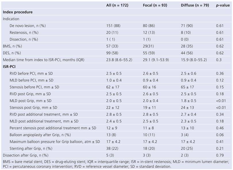
Quantitative angiography data are given in Table 2. Intervention with the GRIP™ balloon improved the mean degree of stenosis from 62 ± 17% to 22 ± 12% (p =<0.001) and was the only treatment in 66% (n = 114). An additional intervention was performed in the remaining: in 8% (n = 13) of the cases additional balloon angioplasty with noncompliant balloons was performed; in 22% (n = 38) additional stent implantation (DES 94%) was done, and in 4% (n = 7) both techniques were performed. These additional interventions led to a further reduction in mean degree of stenosis to 12 ± 9% (p = 0.08). After Grip intervention, MLD (2.0 ± 0.4 mm vs 1.8 ± 0.5 mm, p = 0.003) was higher and percent stenosis (19 ± 11% vs 24 ± 13%) lower in focal compare to diffuse lesions (p = 0.008). These differences did not persist at the end of the ISR procedure (p = 0.18 and p = 0.46 respectively, Table 2).
Patients who underwent revascularisation for recurrent (second) ISR presented more frequently a history of revascularisation [PCI (100%, n = 20); CABG (40%, n = 8] than those with naïve (first) ISR [PCI (21%, n = 33; p <0.001); CABG (12%, n = 19; p = 0.001].
No significant difference was noticed with regard to risk factors, clinical presentation, procedural characteristics, complications or clinical outcome except for a higher preprocedural % stenosis in the recurrent-ISR group (68 ± 14% vs 61 ± 17%; p = 0.06) and a reduced postprocedural %stenosis (4 ± 6% vs 13 ± 9%, p = 0.02) compared to the naïve-ISR group.

Safety outcome

No perforation was seen as immediate coronary complication. A dissection was seen in five patients (3%) after GRIP™ balloon inflation. All dissections were classified as localised (NHLBI type A and B) and successfully covered by stent implantation.

Clinical follow up

Clinical follow-up was available in 160 patients (93%). Mean follow-up was 771 ± 565 days. Follow-up angiography was available in 73 cases (42%). The 12-month clinical outcome is summarised in Table 3. Survival was 98%. MACE were achieved by 13% of patients (n = 21).
Median time to secondary endpoint was 148 days [IQR 105–225]. Cumulative survival free of MACE is depicted in Figure 2. TLR was performed in 17 patients (11%): 13 (8%) were treated by re-PCI and 4 (3%) underwent CABG. Myocardial infarction occurred in 2 patients (1%) and definite stent thrombosis occurred in 1 patient (1%). Most of patients suffering from MACE had diffuse stenosis (n = 15 vs 6, p = 0.02). This trend was even more germane to patients treated solely with the GRIP™ balloon and is depicted in Figure 2B.

ISR prediction

Binary logistic regression showed that diffuse restenosis was associated with occurrence of MACE at tewlve months (OR 3.4; 95%CI: 1.2–9.9, p = 0.03).

Discussion

Plain balloon angioplasty to treat ISR has been intensively investigated for over a decade and has been associated with suboptimal outcome as illustrated by recurrent restenosis rates of to 22–54% at six months [25,26]. With the successful introduction of DES, the pattern of ISR evolved to more focal lesion. Moreover new interventional modalities have emerged like scoring balloons that decrease the risk of balloon slippage and allow greater luminal gain than conventional angioplasty balloons. Therefore, balloon dilatation with scoring balloons like the GRIP™ balloon has been considered as the first-line treatment of ISR at our institution.
In the present observational study we found that: (1) Balloon angioplasty with GRIP™ balloons is safe. (2) When post-procedural result is good, plain balloon angioplasty is effective to treat focal ISR with 12-month MACE of 7%. (3) When post-procedural result is insufficient, additional stent placement (sandwich technique) is justified and does not negatively influence clinical outcome. (4) Diffuse ISR is associated with worse outcome. As expected patients treated for a recurrent-ISR had more frequently a history of previous revascularisation at the index procedure. The differences observed for patients with recurrent-ISR compared to patients with naïve ISR might translate a higher tolerance to ischaemia in patients with recurrent-ISR as suggested by higher degrees of stenosis at presentation. One can speculate that the better postprocedural result in this latter group might translate the operator’s intention to avoid further TLR.
A scoring balloon has elements placed outside of the balloon in order to reduce the occurrence of slippage during inflation for ISR and to increase the acute luminal gain. To date, three scoring balloons have been introduced in Europe [GRIP™ (Acrostak), Lacrosse NSE (Goodman) and AngioSculpt (Angioscore/Biotronik)], but no data are available regarding their safety and efficacy to treat ISR in humans. The main concern of the scoring balloon is that the elements on its surface could lead to higher complication rates such as dissection, perforation, or entrapment of the scoring elements [27]. In the present study, no entrapment or perforation was observed with the GRIP™ balloon. Moreover, dissections were rarely observed (five cases) and the incidence was considered within the anticipated range of 10%–13% incidence based on previous publications [25,28].
According to the current guidelines, DEB should be considered as first-line treatment for ISR after BMS. Paclitaxel-eluting balloons have been found superior to plain old balloon angioplasty (POBA) for the treatment of restenosis after BMS in the randomised PACCOCATH trial (5-year TLR 9% vs 39%, p <0.001) [29]. In the PEPCAD-II trial were non-inferior to paclitaxeleluting stents with regard to clinical endpoints such as TLR (3-year follow-up: 8% vs 19%, psuperiority = 0.07) [30] and achieved a significantly lower rate of late lumen loss. Similarly, Habara and colleagues demonstrated encouraging results in patients with ISR after sirolimus-eluting treated by DEB vs POBA (6-month TLR 4% vs 42%, p = 0.003) [31]. Rittger and colleagues reports similar results in DES-ISR with a significantly lower rate of late lumen loss, TLR and MACE at six months in patients treated by DEB (TLR 15.3% vs 36.8%, p = 0.005; MACE 16.7% vs 50.0%, p = <0.001) [32]. Clinical long-term data with DEB for restenosis are however, still scarce.
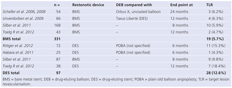
TLR was 11% in our population, clearly higher than the initial reported TLR in patients treated with DEB for BMS restenosis (5%–6%) [15,19,33]. Yet, when compared to TLR in patients treated with DEB for in-DES restenosis, these rates are within the expected range (4%–18%) [31,32,34]. Table 4 depicts TLR rates for relevant DEB trials. As noted in the introduction conventional angioplasty was always performed prior to DEB application in restenotic patients. Maybe the adjunction of a scoring balloon prior to DEB could yield to higher luminal gain and eventually even more favourable clinical outcomes.
In contrast, the anti-slip effect of GRIP™ balloons works well and is associated with effective angioplasty, especially in focal lesions. The efficacy endpoint in our sample was reached by a similar percentage of patients as previously reported [3,8,10,35,36,37,38]. For instance, the figures are close to the recent publication of Tagliareni and colleagues in 213 patients undergoing PCI for ISR [39]. As expected, patients with focal lesions had a better clinical outcome than patients with diffuse ISR. This was solely due to lower TLR in the focal group. In line with this, Rathore et al. reported that a focal pattern of ISR after sirolimus-eluting stent implantation was an independent predictor of lower recurrent restenosis rate [40]. In addition, Cosgrave et al. reported a recurrence rate following PCI for ISR in drug-eluting stent from 18% in patients with focal lesion and 51% in patients with diffuse lesion [41]; the incidence of TLR at 14 months was close to our findings with 10% and 23%, respectively. Interestingly, TLR rates were similar in patients treated with POBA or sandwich drug-eluting stent implantation (focal lesion: 11% vs 9%, p = 0.6; diffuse lesion: 24% vs 23%, p = 1.0) [3]. Finally, Mishkel et al. reported higher rates of TLR (28.2%) and MACE (42.9%) at 12-month in 92 patients who were mostly treated by sandwich DES (84%) for ISR [42].
With the current therapeutical armamentarium including scoring, cutting, and drug-eluting balloons, as well as effective drug-eluting stents, such results underline that ISR treatment should be tailored to and based on ISR pattern (focal versus diffuse), presence or absence of stent fracture, and type of stent implanted (BMS, DES).

Limitations

The present study was designed as a retrospective analysis and therefore lacks randomisation and intention-to-treat data. Since no sample size calculations were performed, we acknowledge that our results may be affected by a type II error.

Conclusions

Balloon angioplasty with the GRIP™ balloon for ISR can be safely and successfully performed, and leads to an especially good clinical outcome in patients presenting with focal ISR.

Funding / potential competing interests

This work was partly supported by the Fonds Scientifique Cardiovasculaire (FSC, Fribourg). No financial support and no other potential conflict of interest relevant to this article were reported.

Abbreviations

BMSBare metal stent
DEBDrug-eluting balloon
DESDrug-eluting stent
ISRIn-stent restenosis
MACEMajor adverse cardiac events
MLDMinimum lumen diameter
PCIPercutaneous coronary intervention
RVDReference vessel diameter
STStent thrombosis
TLRTarget lesion revascularisation

References

  1. Komatsu, R.; Ueda, M.; Naruko, T.; Kojima, A.; Becker, A.E. Neointimal tissue response at sites of coronary stenting in humans: Macroscopic, histological, and immunohistochemical analyses. Circulation. 1998, 98, 224–233. [Google Scholar] [CrossRef]
  2. Lee, M.S.; Pessegueiro, A.; Zimmer, R.; Jurewitz, D.; Tobis, J. Clinical presentation of patients with in-stent restenosis in the drug-eluting stent era. J Invasive Cardiol. 2008, 20, 401–403. [Google Scholar] [PubMed]
  3. Mehilli, J.; Byrne, R.A.; Tiroch, K.; Pinieck, S.; Schulz, S.; Kufner, S.; et al. Randomized trial of paclitaxelversus sirolimus-eluting stents for treatment of coronary restenosis in sirolimus-eluting stents: The isar-desire 2 (intracoronary stenting and angiographic results: Drug eluting stents for in-stent restenosis 2) study. J Am Coll Cardiol. 2010, 55, 2710–2716. [Google Scholar] [CrossRef] [PubMed]
  4. Kastrati, A.; Mehilli, J.; von Beckerath, N.; Dibra, A.; Hausleiter, J.; Pache, J.; et al. Sirolimus-eluting stent or paclitaxel-eluting stent vs balloon angioplasty for prevention of recurrences in patients with coronary instent restenosis: A randomized controlled trial. JAMA. 2005, 293, 165–171. [Google Scholar] [CrossRef] [PubMed]
  5. Zahn, R.; Hamm, C.W.; Schneider, S.; Zeymer, U.; Nienaber, C.A.; Richardt, G.; et al. Incidence and predictors of target vessel revascularization and clinical event rates of the sirolimus-eluting coronary stent (results from the prospective multicenter german cypher stent registry). Am J Cardiol. 2005, 95, 1302–1308. [Google Scholar] [CrossRef]
  6. Zahn, R.; Hamm, C.W.; Schneider, S.; Richardt, G.; Kelm, M.; Levenson, B.; et al. Coronary stenting with the sirolimus-eluting stent in clinical practice: Final results from the prospective multicenter german cypher stent registry. J Interv Cardiol. 2010, 23, 18–25. [Google Scholar] [CrossRef]
  7. Kastrati, A.; Dibra, A.; Mehilli, J.; Mayer, S.; Pinieck, S.; Pache, J.; et al. Predictive factors of restenosis after coronary implantation of sirolimusor paclitaxel-eluting stents. Circulation. 2006, 113, 2293–2300. [Google Scholar] [CrossRef]
  8. Bossi, I.; Klersy, C.; Black, A.J.; Cortina, R.; Choussat, R.; Cassagneau, B.; et al. In-stent restenosis: Long-term outcome and predictors of subsequent target lesion revascularization after repeat balloon angioplasty. J Am Coll Cardiol. 2000, 35, 1569–1576. [Google Scholar] [CrossRef]
  9. Chen, M.S.; John, J.M.; Chew, D.P.; Lee, D.S.; Ellis, S.G.; Bhatt, D.L. Bare metal stent restenosis is not a benign clinical entity. Am Heart J. 2006, 151, 1260–1264. [Google Scholar] [CrossRef]
  10. Rathore, S.; Kinoshita, Y.; Terashima, M.; Katoh, O.; Matsuo, H.; Tanaka, N.; et al. A comparison of clinical presentations, angiographic patterns and outcomes of in-stent restenosis between bare metal stents and drug eluting stents. EuroIntervention. 2010, 5, 841–846. [Google Scholar] [CrossRef]
  11. Assali, A.R.; Moustapha, A.; Sdringola, S.; Denktas, A.E.; Willerson, J.T.; Holmes, D.R., Jr.; Smalling, R.W. Acute coronary syndrome may occur with in-stent restenosis and is associated with adverse outcomes (the presto trial). Am J Cardiol. 2006, 98, 729–733. [Google Scholar] [CrossRef] [PubMed]
  12. Walters, D.L.; Harding, S.A.; Walsh, C.R.; Wong, P.; Pomerantsev, E.; Jang, I.K. Acute coronary syndrome is a common clinical presentation of in-stent restenosis. Am J Cardiol. 2002, 89, 491–494. [Google Scholar] [CrossRef] [PubMed]
  13. Nayak, A.K.; Kawamura, A.; Nesto, R.W.; Davis, G.; Jarbeau, J.; Pyne, C.T.; et al. Myocardial infarction as a presentation of clinical in-stent restenosis. Circ J. 2006, 70, 1026–1029. [Google Scholar] [CrossRef]
  14. Adamian, M.; Colombo, A.; Briguori, C.; Nishida, T.; Marsico, F.; Di Mario, C.; et al. Cutting balloon angioplasty for the treatment of in-stent restenosis: A matched comparison with rotational atherectomy, additional stent implantation and balloon angioplasty. J Am Coll Cardiol. 2001, 38, 672–679. [Google Scholar] [CrossRef] [PubMed]
  15. Scheller, B.; Hehrlein, C.; Bocksch, W.; Rutsch, W.; Haghi, D.; Dietz, U.; et al. Treatment of coronary in-stent restenosis with a paclitaxel-coated balloon catheter. N Engl J Med. 2006, 355, 2113–2124. [Google Scholar] [CrossRef]
  16. Guidon, A.; Cook, S.; Berger, A.; Goy, J.J. Long-term clinical outcome after sirolimus-stent implantation for in sirolimuseluting stent restenosis. Clinical Medicine: Cardiology. 2008, 2, 161–163. [Google Scholar]
  17. Waksman, R.; White, R.L.; Chan, R.C.; Bass, B.G.; Geirlach, L.; Mintz, G.S.; et al. Intracoronary gamma-radiation therapy after angioplasty inhibits recurrence in patients with in-stent restenosis. Circulation. 2000, 101, 2165–2171. [Google Scholar] [CrossRef]
  18. Dangas, G.D.; Claessen, B.E.; Caixeta, A.; Sanidas, E.A.; Mintz, G.S.; Mehran, R. In-stent restenosis in the drug-eluting stent era. J Am Coll Cardiol. 2010, 56, 1897–1907. [Google Scholar] [CrossRef]
  19. Scheller, B.; Hehrlein, C.; Bocksch, W.; Rutsch, W.; Haghi, D.; Dietz, U.; et al. Two year follow-up after treatment of coronary in-stent restenosis with a paclitaxel-coated balloon catheter. Clin Res Cardiol. 2008, 97, 773–781. [Google Scholar] [CrossRef]
  20. Wijns, W.; Kolh, P.; Danchin, N.; Di Mario, C.; Falk, V.; Folliguet, T.; et al. Guidelines on myocardial revascularization. Eur Heart J. 2010, 31, 2501–2555. [Google Scholar] [CrossRef]
  21. Roques, F.; Michel, P.; GoldstonNashef, S.A. The logistic euroscore. Eur Heart J. 2003, 24, 881–882. [Google Scholar] [CrossRef] [PubMed]
  22. Mehran, R.; Dangas, G.; Abizaid, A.S.; Mintz, G.S.; Lansky, A.J.; Satler, L.F.; et al. Angiographic patterns of in-stent restenosis: Classification and implications for long-term outcome. Circulation. 1999, 100, 1872–1878. [Google Scholar] [CrossRef] [PubMed]
  23. Rogers, J.H.; Lasala, J.M. Coronary artery dissection and perforation complicating percutaneous coronary intervention. J Invasive Cardiol. 2004, 16, 493–499. [Google Scholar] [PubMed]
  24. Cutlip, D.E.; Windecker, S.; Mehran, R.; Boam, A.; Cohen, D.J.; van Es, G.A.; et al. Clinical end points in coronary stent trials: A case for standardized definitions. Circulation. 2007, 115, 2344–2351. [Google Scholar] [CrossRef]
  25. Bauters, C.; Banos, J.L.; Van Belle, E.; Mc Fadden, E.P.; Lablanche, J.M.; Bertrand, M.E. Six-month angiographic outcome after successful repeat percutaneous intervention for in-stent restenosis. Circulation. 1998, 97, 318–321. [Google Scholar] [CrossRef][Green Version]
  26. Eltchaninoff, H.; Koning, R.; Tron, C.; Gupta, V.; Cribier, A. Balloon angioplasty for the treatment of coronary in-stent restenosis: Immediate results and 6-month angiographic recurrent restenosis rate. J Am Coll Cardiol. 1998, 32, 980–984. [Google Scholar] [CrossRef]
  27. Sanchez-Recalde, A.; Galeote, G.; Martin-Reyes, R.; Moreno, R. Angiosculpt ptca balloon entrapment during dilatation of a heavily calcified lesion. Rev Esp Cardiol. 2008, 61, 1361–1363. [Google Scholar] [CrossRef]
  28. Reimers, B.; Moussa, I.; Akiyama, T.; Tucci, G.; Ferraro, M.; Martini, G.; et al. Long-term clinical follow-up after successful repeat percutaneous intervention for stent restenosis. J Am Coll Cardiol. 1997, 30, 186–192. [Google Scholar] [CrossRef][Green Version]
  29. Scheller, B.; Clever, Y.P.; Kelsch, B.; Hehrlein, C.; Bocksch, W.; Rutsch, W.; et al. Long-term follow-up after treatment of coronary in-stent restenosis with a paclitaxel-coated balloon catheter. JACC Cardiovasc Interv. 2012, 5, 323–330. [Google Scholar] [CrossRef]
  30. Unverdorben, M. Trend towards lower tlr and mace rates in patients treated with deb compared to des. EuroPCR 2011.
  31. Habara, S.; Mitsudo, K.; Kadota, K.; Goto, T.; Fujii, S.; Yamamoto, H.; et al. Effectiveness of paclitaxel-eluting balloon catheter in patients with sirolimus-eluting stent restenosis. JACC Cardiovasc Interv. 2011, 4, 149–154. [Google Scholar] [CrossRef]
  32. Rittger, H.; Brachmann, J.; Sinha, A.M.; Waliszewski, M.; Ohlow, M.; Brugger, A.; et al. A randomized, multicenter, single-blinded trial comparing paclitaxel-coated balloon angioplasty with plain balloon angioplasty in drug-eluting stent restenosis: The pepcad-des study. J Am Coll Cardiol. 2012, 59, 1377–1382. [Google Scholar] [CrossRef] [PubMed]
  33. Unverdorben, M.; Vallbracht, C.; Cremers, B.; Heuer, H.; Hengstenberg, C.; Maikowski, C.; et al. Paclitaxel-coated balloon catheter versus paclitaxelcoated stent for the treatment of coronary in-stent restenosis. Circulation. 2009, 119, 2986–2994. [Google Scholar] [CrossRef] [PubMed]
  34. Toelg, R. Paclitaxel releasing balloon with an inert bthc excipient: Sixmonth results on 1’064 patients of the international delux registry; EuroPCR: Paris, France, 2012. [Google Scholar]
  35. Appleby, C.E.; Khattar, R.S.; Morgan, K.; Clarke, B.; Curzen, N.; Neyses, L.; et al. Drug eluting stents for the treatment of bare metal in-stent restenosis: Long-term outcomes in real world practice. EuroIntervention. 2011, 6, 748–753. [Google Scholar] [CrossRef] [PubMed]
  36. Beijk, M.A.; Claessen, B.E.; Koch, K.T.; Henriques, J.P.; Baan, J.; Vis, M.M.; et al. One-year clinical outcome after treatment of bare-metal stent in-stent restenosis with the paclitaxel-eluting stent in an unselected cohort. Int J Cardiol. 2010, 145, 608–609. [Google Scholar] [CrossRef]
  37. Steinberg, D.H.; Gaglia, M.A., Jr.; Pinto Slottow, T.L.; Roy, P.; Bonello, L.; De Labriolle, A.; et al. Outcome differences with the use of drug-eluting stents for the treatment of in-stent restenosis of bare-metal stents versus drug-eluting stents. Am J Cardiol. 2009, 103, 491–495. [Google Scholar] [CrossRef]
  38. Pohl, T.; Kupatt, C.; Steinbeck, G.; Boekstegers, P. Angiographic and clinical outcome for the treatment of in-stent restenosis with sirolimus-eluting stent compared to vascular brachytherapy. Z Kardiol. 2005, 94, 405–410. [Google Scholar] [CrossRef]
  39. Tagliareni, F.; La Manna, A.; Saia, F.; Marzocchi, A.; Tamburino, C. Longterm clinical follow-up of drug-eluting stent restenosis treatment: Retrospective analysis from two high volume catheterisation laboratories. EuroIntervention. 2010, 5, 703–708. [Google Scholar] [CrossRef]
  40. Rathore, S.; Terashima, M.; Katoh, O.; Matsuo, H.; Tanaka, N.; Kinoshita, Y.; et al. Predictors of angiographic restenosis after drug eluting stents in the coronary arteries: Contemporary practice in real world patients. EuroIntervention. 2009, 5, 349–354. [Google Scholar] [CrossRef]
  41. Cosgrave, J.; Melzi, G.; Biondi-Zoccai, G.G.; Airoldi, F.; Chieffo, A.; Sangiorgi, G.M.; et al. Drug-eluting stent restenosis the pattern predicts the outcome. J Am Coll Cardiol. 2006, 47, 2399–2404. [Google Scholar] [CrossRef][Green Version]
  42. Mishkel, G.J.; Moore, A.L.; Markwell, S.; Shelton, M.C.; Shelton, M.E. Longterm outcomes after management of restenosis or thrombosis of drugeluting stents. J Am Coll Cardiol. 2007, 49, 181–184. [Google Scholar] [CrossRef]
Figure 1. Computer simulated image of the GRIP™ scoring balloon (Acrostak, Switzerland) with 4 lines of knobs on its surface that protect against balloon slippage (by courtesy of Acrostak, Winterthur, Switzerland).
Cardiovascmed 15 00354 g001
Figure 2. Survival free of MACE at 12 months according to lesion pattern in the whole population (A), and in patients treated solely with GRIP™ balloon (B). ISR = in-stent restenosis; MACE = major adverse cardiac events.
Cardiovascmed 15 00354 g002
Table 1. Baseline data.
Cardiovascmed 15 00354 i001
Table 2. Data on index procedure and ISR PCI.
Cardiovascmed 15 00354 i002
Table 3. Clinical outcome of patients undergoing PCI with the GRIP™ balloon for ISR at 12-month.
Cardiovascmed 15 00354 i003
Table 4. Rates of TLR in randomised controlled DEB-trials and registries.
Cardiovascmed 15 00354 i004