Next Article in Journal
A Dangerous Spike: Cause or Coincidence?
Previous Article in Journal
Coronary Steal Phenomenon Due to Ipsilateral Upper Extremity Arteriovenous Fistula in a Patient with Internal Thoracic Artery Bypass Graft—An Alternative Treatment Strategy
 
 
Cardiovascular Medicine is published by MDPI from Volume 28 Issue 1 (2025). Previous articles were published by another publisher in Open Access under a CC-BY (or CC-BY-NC-ND) licence, and they are hosted by MDPI on mdpi.com as a courtesy and upon agreement with Editores Medicorum Helveticorum (EMH).
Interesting Images

Heart Failure in a Patient with Multi-Organ Sarcoidosis

by
Marc Buser
1,
Theresa Dellas Buser
2,
Jens Bremerich
2 and
Daniel Tobler
1,*
1
Department of Cardiology, University Hospital Basel, Petersgraben 4, CH-4051 Basel, Switzerland
2
Department of Radiology, University Hospital Basel, CH-4051 Basel, Switzerland
*
Author to whom correspondence should be addressed.
Cardiovasc. Med. 2012, 15(11), 329; https://doi.org/10.4414/cvm.2012.00124
Submission received: 21 August 2012 / Revised: 21 September 2012 / Accepted: 21 October 2012 / Published: 21 November 2012

Case Report

A 53-year-old woman was admitted to our emergency department with progressive shortness of breath during exertion for three months. She had been well before without any remarkable diseases in her medical history and she had never been on regular medication. Physical examination revealed leg oedema, jugular venous congestion and bilateral inspiratory crackles on auscultation of the lungs consistent with biventricular congestive heart failure of unknown aetiology. BNP was markedly increased at a level of 2,812 ng/l. An ECG showed sinus-tachycardia but otherwise was normal.
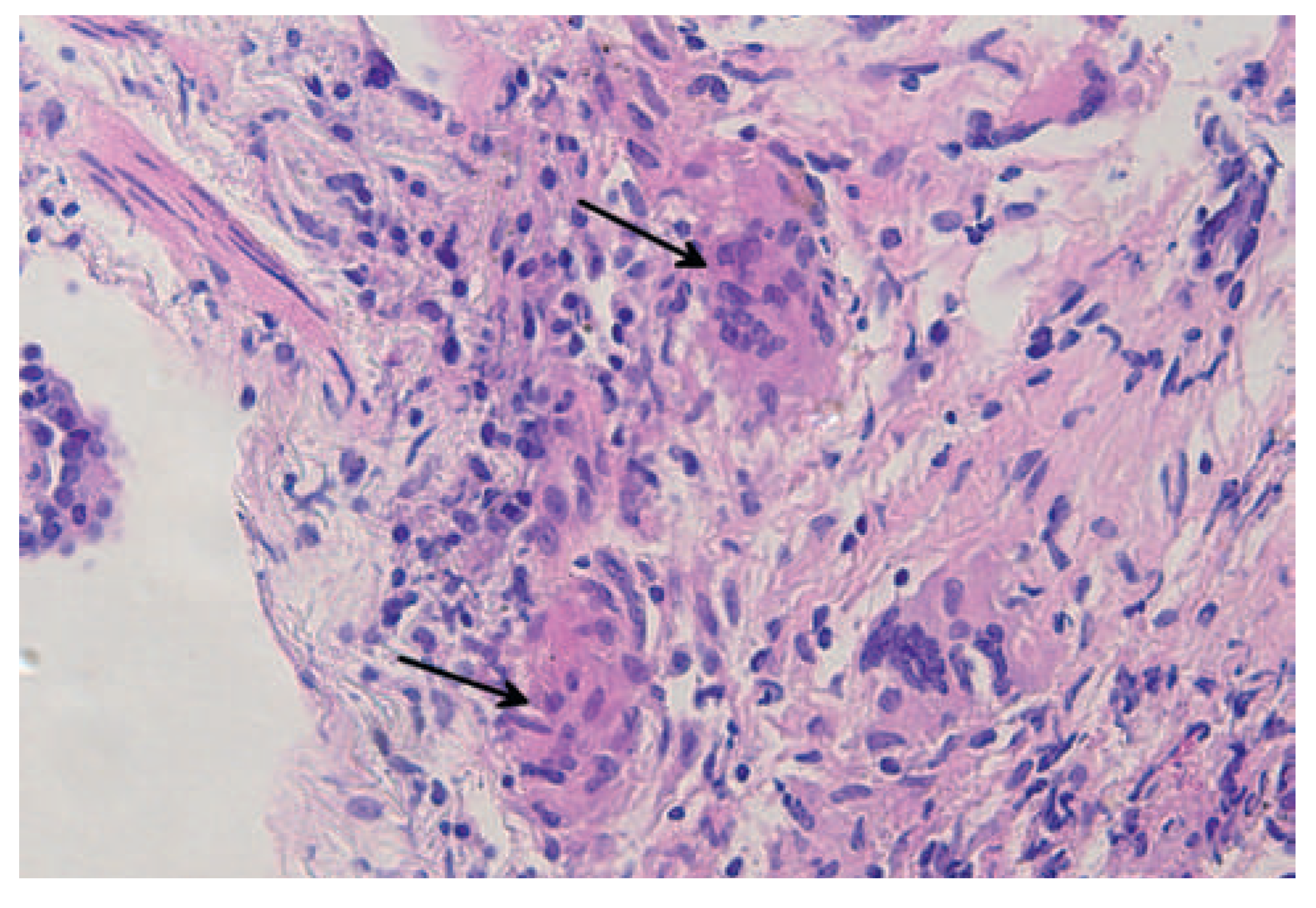
Echocardiography (Figure 1) revealed dilated cardiomyopathy with severely reduced left ventricular ejection fraction of 20% and diffuse wall motion abnormalities. There was secondary mitral regurgitation stage II. End-systolic pulmonary pressure was elevated (sPAP 50 mm Hg). A chest radiograph (Figure 2A) showed cardiomegaly and diffuse reticulonodular opacifications with patchy consolidation in the right upper lobe and left mid zone. The right hilum was retracted. These findings were confirmed by computer tomography of the chest (Figure 2B), which also revealed mediastinal lymphadenopathy and extensive sub-pleural micro-nodularity, most prominent in the mid and lower lung zones. Moreover, traction bronchiectases in the apices were noted, particularly in the right upper lobe with associated retractile cavitations. Involvement of the pleura was suggested by pleural effusion. The heart was enlarged and surrounded by pericardial effusion. Based on these findings a diagnosis of pulmonic sarcoidosis stage IV with cardiac involvement was suggested. Bronchial biopsy was performed to affirm the diagnosis. Histology revealed multiple non-caseating granulomas consistent with pulmonic sarcoidosis (Figure 3). Cardiac MRI (Figure 4) showed an impaired global cardiac function and reduced anterolateral, septal and apical myocardial contractibility as well as multiple stippled areas of delayed contrast enhancement, particularly in regions of impaired contractility, consistent with myocardial sarcoidosis. Additional granulomatous lesions were found in peripheral bones. Treatment was started with initial high dose steroids as well as a standard heart failure therapy. On the 6-month follow-up visit, the patient was asymptomatic and echocardiography revealed normalisation of the left ventricular dimensions and systolic function.
This case is a striking example of multi-organ involvement of sarcoidosis with heart failure symptoms due to cardiac involvement leading to the initial presentation. In our case, the typical imaging features of mediastinal lymphadenopathy, perilymphatic micro-nodules and upper lobe predominance alongside typical cardiac manifestations pointed to the diagnosis [1]. Clinical manifestations of cardiac involvement are observed in about 5% of all sarcoidosis patients, although in biopsy studies a cardiac infiltration can be found in up to 25% [2]. When present, congestive heart failure with dilated cardiomyopathy, conduction abnormalities, tachyarrhythmias and even sudden cardiac death are the most common clinical manifestations [2]. Whereas these findings by itself are not indicative of cardiac infiltration of sarcoidosis, concomitant pulmonary involvement strongly suggests the diagnosis.

Conflicts of Interest

No financial support and no other potential conflict of interest relevant to this article were reported.

References

  1. Koyama T, Ueda H, Togashi K, Umeoka S, Kataoka M, Nagai S. Radiologic manifestations of sarcoidosis in various organs. Radio Graphics 2004, 24, 87–104. [Google Scholar]
  2. Kim JS, Judson MA, Donnino R, Gold M, Cooper LT Jr, Prystowsky EN, et al. Cardiac sarcoidosis. Am Heart J. 2009, 157, 9. [Google Scholar] [CrossRef] [PubMed]
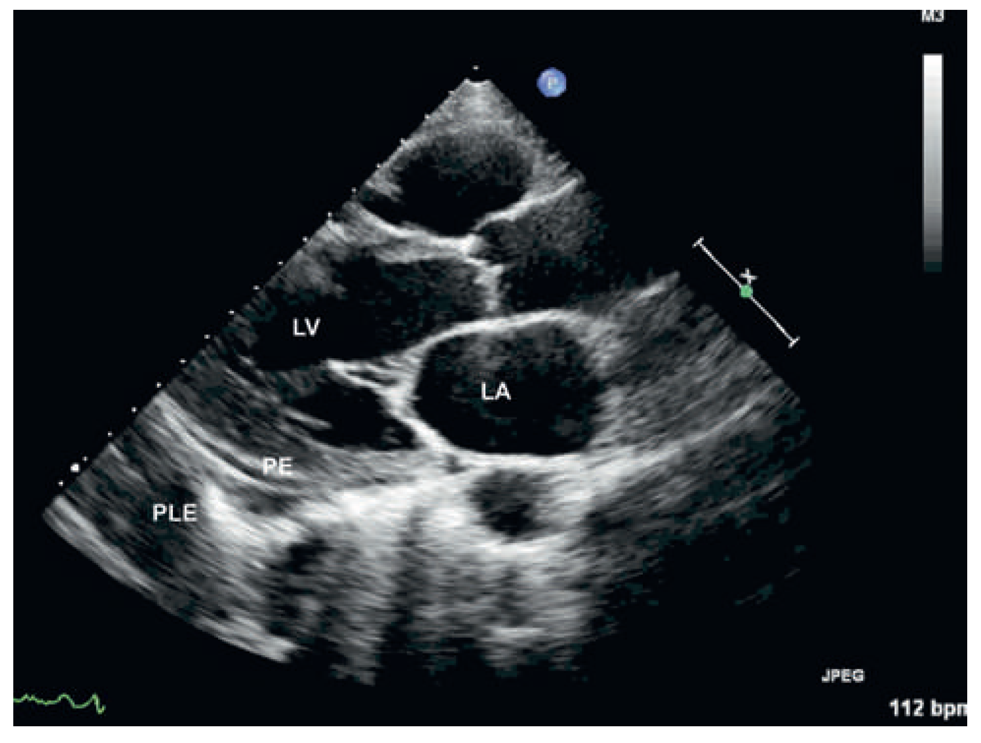
Figure 1. Parasternal long axis view showing a dilted left ventricle (LV) and atrium (LA), small pericardial effusion (PE) and pleural effusion (PLE).
Cardiovascmed 15 00329 g001
Figure 2. Pulmonary findings. (A) Chest x-ray with cardiomegaly and a diffuse reticulonodular opacifications as well as patchy consolidations in the right upper lobe and left mid zone. (B) Chest CT (lung window) confirming extensive sub-pleural micronodularity (large arrow), bronchiectasis and scarring in the apices with associated retractile cavitations (small arrow).
Cardiovascmed 15 00329 g002
Figure 3. Histologic image of non-caseating granulomas (arrow).
Cardiovascmed 15 00329 g003
Figure 4. Cardiac MRI in short axis view revealing stippled areas of late contrast enhancement with non-ischaemic pattern (arrow shows contrast enhancement in the anterior wall segment).
Cardiovascmed 15 00329 g004