Next Article in Journal
Werbung mit Ungeschicktem «Wording»
Previous Article in Journal
Neo Snowman in X-Ray
 
 
Cardiovascular Medicine is published by MDPI from Volume 28 Issue 1 (2025). Previous articles were published by another publisher in Open Access under a CC-BY (or CC-BY-NC-ND) licence, and they are hosted by MDPI on mdpi.com as a courtesy and upon agreement with Editores Medicorum Helveticorum (EMH).
Interesting Images

Phantom Tumour of the Lung

by
Thomas Sebastian Gilhofer
* and
Lars C. Huber
Spital Lachen, CH-8853 Lachen, Switzerland
*
Author to whom correspondence should be addressed.
Cardiovasc. Med. 2011, 14(11), 327; https://doi.org/10.4414/cvm.2011.01625
Submission received: 23 August 2011 / Revised: 23 September 2011 / Accepted: 23 October 2011 / Published: 23 November 2011

Case report

A 87-year-old woman was admitted to our emergency department with progredient exertional dyspnoea. Medical history was remarkable for atrial fibrillation and hypertensive cardiopathy. Prior to admission, the patient did not take the prescribed diuretic drugs and gained 1 kilogram of weight within few days. Vital signs showed normotensive blood pressure (120/63 mm Hg) and optimal rate control of the atrial fibrillation (HR 75/min). Physical examination revealed neither jugular venous distension nor peripheral oedema. Auscultation of heart and lungs was normal. Chest X-ray showed a wedge-shaped effusion within the right interlobar fissure and a large, oval density in the right lower lobe (Figure 1). Levels of brain natriuretic peptide (BNP) were elevated to 747 ng/l (<50 ng/l). Despite lack of typical physical findings in this patient we thus interpreted the actual clinical detoriation as congestive heart failure [1]. Radiologic follow-up after enforced diuretic therapy and adaptation of heart failure therapy revealed complete regression (Figure 2), unmasking the hypodense lesion as phantom tumour of the lung. Since serial measurements of BNP have not clearly been associated with a better outcome, levels were not checked during follow-up [2].
Localised interlobar effusion in heart failure with prompt resolution upon treatment has been described initially as vanishing tumour of the lung by Gefter et al. [3]. Others have reviewed the clinical significance of this finding, showing that phantom tumours are commonly found within the transverse fissure and less frequently within the oblique fissure, occasionally within both [4,5]. A vanishing tumour remains an important differential diagnosis in patients with congestive heart failure presenting with dyspnoea and an unclear mass lesion of the lung; appropriate therapy with diuretics and ACE inhibitors should thus be established before proceeding with further diagnostics and might unravel such lesion as hypervolemic overload in heart failure patients.

References

  1. Maisel, A.S.; Krishnaswamy, P.; Novak, R.M.; McCord, J.; Hollander, J.E.; Duc, P.; et al. Rapid Measurement of B-type natriuretic peptide in the emergency diagnosis of heart failure. NEJM. 2002, 347, 161–7. [Google Scholar] [CrossRef] [PubMed]
  2. Nohria, A.; Mielniczuk, L.M.; Stevenson, L.W. Evaluation and monitoring of patients with acute heart failure syndromes. Am J Cardiol. 2005, 96, 32G. [Google Scholar] [CrossRef] [PubMed]
  3. Gefter, W.I.; Boucot, K.R.; Marshall, E.W. Localized interlobar effusion in congestive heart failure; vanishing tumor of the lung. Circulation. 1950, 2, 336–43. [Google Scholar] [CrossRef] [PubMed]
  4. Haus, B.M.; Stark, P.; Shofer, S.L.; Kuschner, W.G. Massive pulmonary pseudotumor. Chest. 2003, 124, 758–60. [Google Scholar] [CrossRef] [PubMed]
  5. Buch, K.P.; Morehead, R.S. Multiple left-sided vanishing tumors. Chest. 2000, 118, 1486–9. [Google Scholar] [CrossRef] [PubMed]
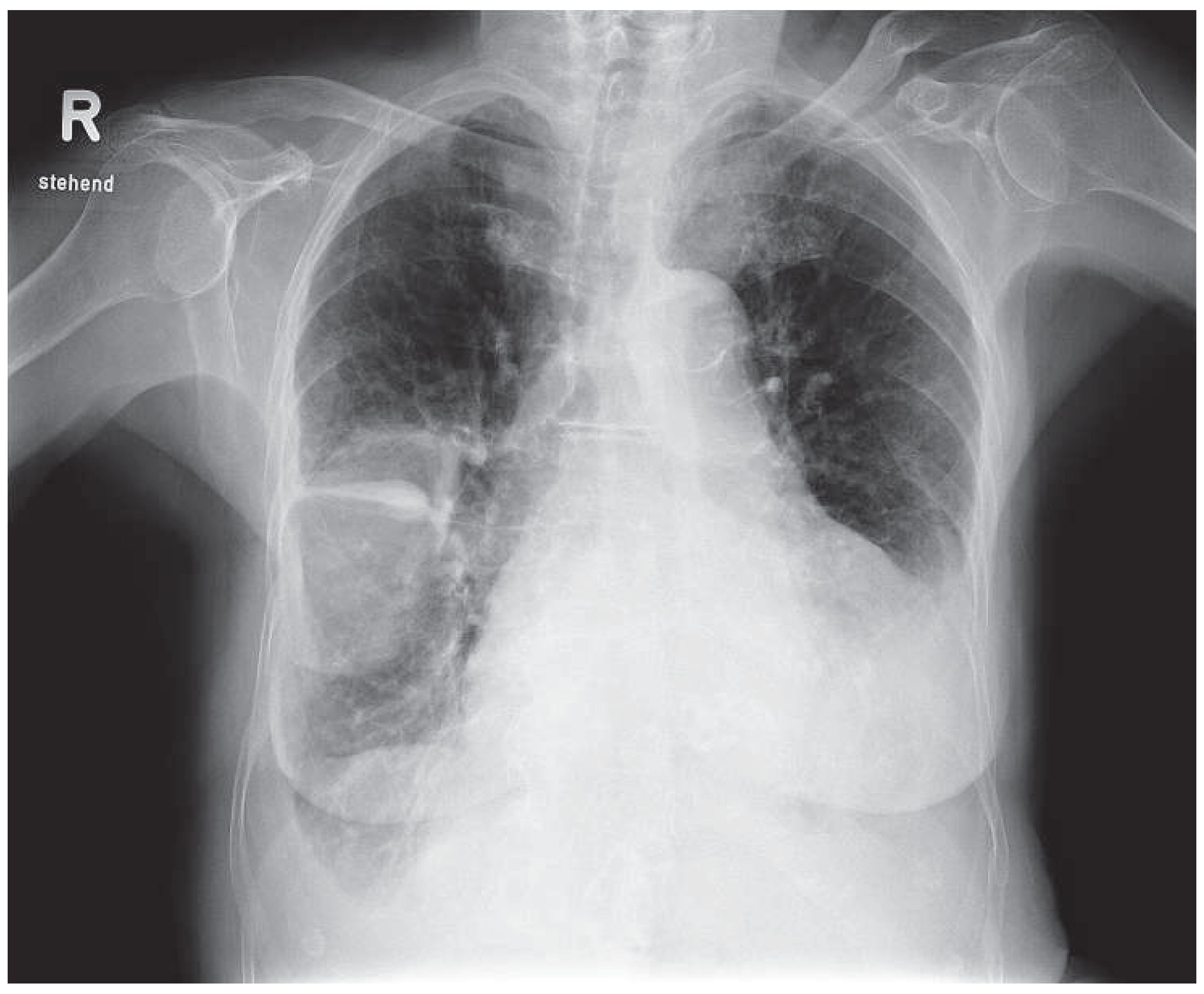
Figure 1. Chest x-ray obtained at admission revealing a large oval density in the lower lobe of the right lung and a wedge-shaped effusion within the transverse fissure.
Cardiovascmed 14 00327 g001
Figure 2. Follow-up imaging five days after treatment with diuretics and ACE-inhibitors showing complete regression.
Cardiovascmed 14 00327 g002