


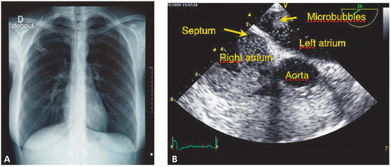
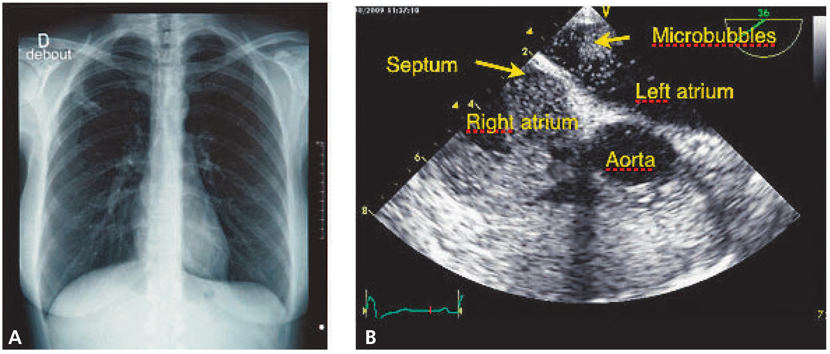
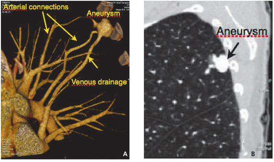
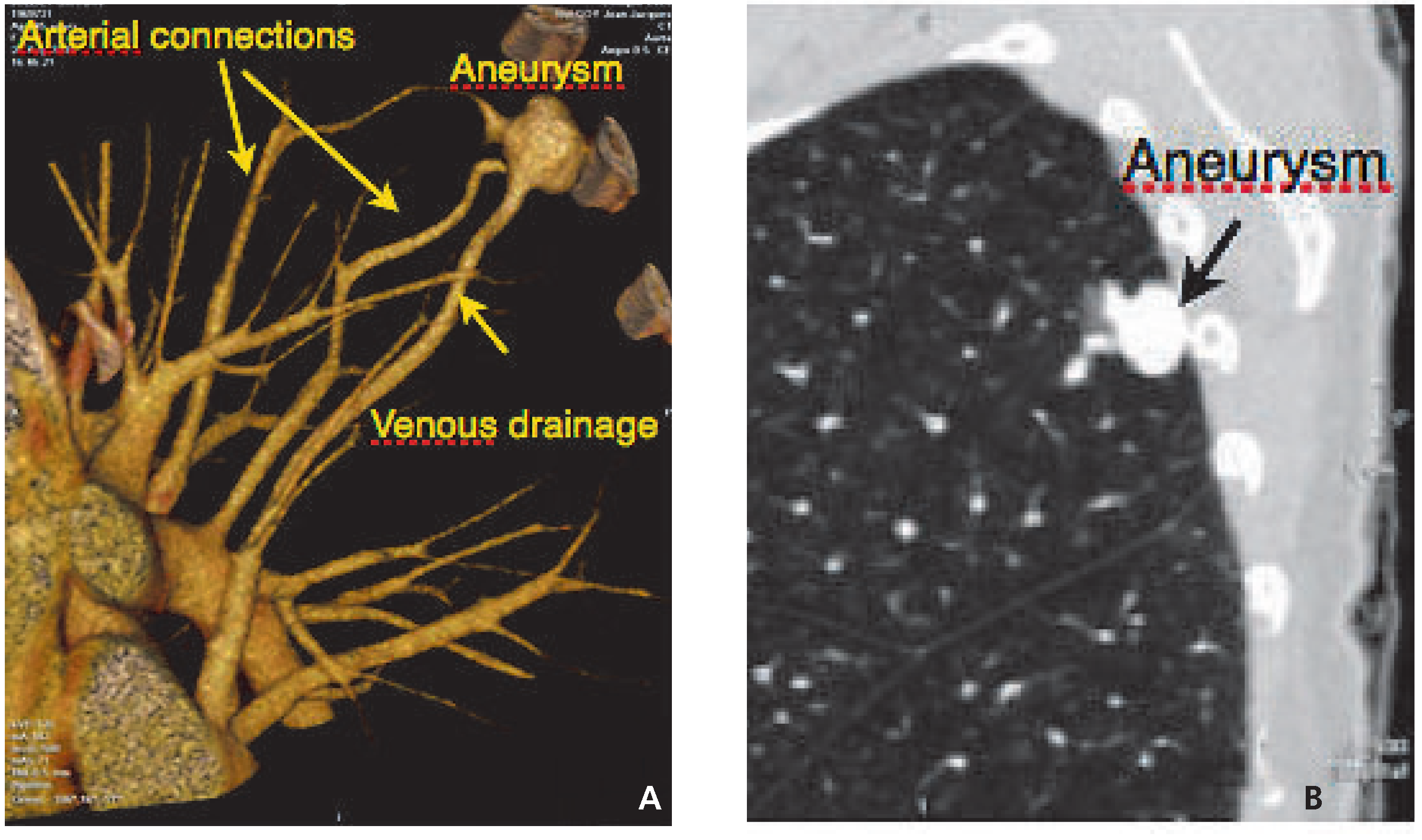
A 46-year-old woman patient was admitted for transient ischaemic attack. She was not taking medication and had no history of cardiac or vascular problems. On admission she still had mild peripheral dysaesthesia but had fully recovered within 2 h. The cardiovascular status was normal per baseline electrocardiogram and cardiac echocardiography. A right superior opacity was present on chest X-ray (Figure 1). Cryptogenic stroke through a patent foramen ovale (PFO) was suspected. A transoesophageal echocardiography (TEE) microbubble test showed a very early (after 2 cycles) presence of bubbles in the left atrium (positive microbubble test) but not arising from a PFO (Figure 1). It was absolutely clear that bubbles were coming from the right pulmonary veins. An abnormal pulmonary arte-riovenous (AV) connection was suspected and subsequently confirmed by thoracic CT scan. A peripheral AV aneurysm with 2 arterial connections and 1 venous branch draining into the left atrium was present (Figure 2). Due to its peripheral location, resection of the fistula by thoracoscopy was preferred to coil embolisation. The intervention was uneventful and the patient left hospital 2 days later. This case is remarkable since it confirms that a TIA due to paradoxical embolism may be due to right-to-left shunt without the existence or complicity of a PFO, and underlines the necessity of performing complete investigations even when TEE produces expected findings. In particular, rare causes of right-to-left shunt should be considered when microbubbles are seen in the left atrium suspiciously early or from an unusual atrial location during the provocation test.
Figure 1.
A Chest x-ray with abnormal superior opacity. B Transoesophageal echocardiography right (second cycle after occurrence of bubbles in the right atrium) showing microbubbles coming from the right superior portion of the left atrium and not through the atrial septum.
Figure 2.
A Three-dimensional CT reconstruction showing the aneurysm with its vascular connections. B Lateral view showing the peripheral location of the aneurysm.
© 2010 by the author. Attribution - Non-Commercial - NoDerivatives 4.0.