Next Article in Journal
Jahresbericht 2008 der Arbeitsgruppe «Erwachsene mit Angeborenem Herzfehler» (WATCH) der SGK
Previous Article in Journal
Jahresbericht 2008 der Arbeitsgruppe «Kardiovaskuläre Biologie» der Schweizerischen Gesellschaft für Kardiologie
 
 
Cardiovascular Medicine is published by MDPI from Volume 28 Issue 1 (2025). Previous articles were published by another publisher in Open Access under a CC-BY (or CC-BY-NC-ND) licence, and they are hosted by MDPI on mdpi.com as a courtesy and upon agreement with Editores Medicorum Helveticorum (EMH).
Interesting Images

My Heart Dances

by
Marcus Mutschelknauss
,
Hans Rickli
and
Peter Ammann
*
Cardiology Kantonsspital St. Gallen Rorschacherstrasse 95 CH-9000 St. Gallen, Switzerland
*
Author to whom correspondence should be addressed.
Cardiovasc. Med. 2009, 12(9), 251; https://doi.org/10.4414/cvm.2009.01442
Submission received: 18 June 2009 / Revised: 18 July 2009 / Accepted: 18 August 2009 / Published: 18 September 2009

Case presentation

A 33-year old woman was admitted for persistent palpitations over the past six years. The patient had no history of syncope, pre-syncope or family history of sudden cardiac death.
On admission the heart rate was approx. 160 bpm and blood pressure was 115/77 mm Hg. The remaining results from physical examination and laboratory tests were unremarkable. Echocardiography showed no evidence of relevant structural heart disease.
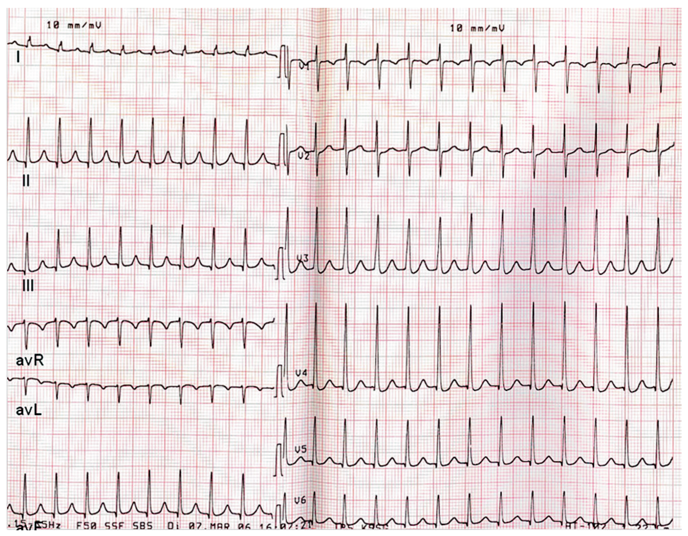
A 12-lead recumbent ECG obtained on admission is shown in Figure 1.
Figure 2 shows the 12-lead ECG recorded after assuming upright posture. The patient complained of palpitations during the ECG recording.

Question

What is the underlying rhythm in our patient in supine/standing position?

Discussion

This case illustrates atrial tachycardia (AT) originating from the crista terminalis. Initially the ECG at rest was interpreted as a normal sinus rhythm with AV block I. But in fact, as close observation of the T-wave shows, there is a second P-wave hidden in the T-wave, suggesting AT with 2:1 AV conduction.
Vagally induced 2:1 conduction in the supine position changed to 1:1 conduction in the upright position.
As shown in our patient, any fluctuation of sympathovagal balance (postural changes) produced palpitations due to 1:1 conduction of atrial tachycardia.
AT are either focal or macrorentrant. The predominant area of origin of right atrial tachycardia is the area along the crista terminalis. If AT arises from the superior portion of the crista terminalis, P-wave morphology may be similar to sinus P-wave. There is extensive literature on P-wave morphology and the site of atrial focus [1,2]. Different P-wave algorithms have been proposed for localisation of the site of AT. In our patient AT was localised in the right atrium, as predicted by a negative P-wave in lead V1. Positive P-waves in the inferior leads suggested superior origin from the crista terminalis. Differentiating the origin of the AT prior to ablation is necessary in order to plan for transseptal puncture for left atrial access if needed.
Successful mapping and radiofrequency ablation of focal right atrial tachycardia was performed in our patient, and the follow-up to date has been uneventful.

References

  1. Tang, C.W.; Scheinman, M.M.; Van Hare, G.F.; et al. Use of P wave configuration during atrial tachycardia to predict site of origin. J. Am. Coll. Cardiol. 1995, 26, 1315–1324. [Google Scholar] [CrossRef]
  2. The, A.; Kistler, P.M.; Kalman, J.M. Using the 12-Lead ECG to Localize the Origin of Ventricular and Atrial Tachycardias: Part 1. Focal Atrial Tachycardia. J. Cardiovasc. Electrophysiol. 2009, 1–4. [Google Scholar]
  3. Fox, D.J.; Tischenko, A.; Krahn, A.D.; et al. Supraventricular tachycardia: diagnosis and management. Mayo Clin Proc. 2008, 83, 1400–1411. [Google Scholar] [CrossRef] [PubMed]
Figure 1. 12-lead ECG in recumbent position.
Cardiovascmed 12 00251 g001
Figure 2. 12-lead ECG in upright position.
Cardiovascmed 12 00251 g002