Next Article in Journal
Coronary CT Angiography: Overuse, Underuse or Adequate Use?
Previous Article in Journal
Konservative Versus Invasive Strategie in der Behandlung der Chronisch Koronaren Herzkrankheit: Wir Brauchen sie Beide!
 
 
Cardiovascular Medicine is published by MDPI from Volume 28 Issue 1 (2025). Previous articles were published by another publisher in Open Access under a CC-BY (or CC-BY-NC-ND) licence, and they are hosted by MDPI on mdpi.com as a courtesy and upon agreement with Editores Medicorum Helveticorum (EMH).
Interesting Images

Angiographic Visualisation of a Right Atrial Myxoma †

by
Stéphane Noble
*,
Caroline Frangos
and
Jean-François Tanguay
Interventional Cardiology, Department of Medicine, Montreal Heart Institute, Montreal University, Montreal, QC, Canada
*
Author to whom correspondence should be addressed.
Korrigierte Version 5.9.2008.
Cardiovasc. Med. 2008, 11(7), 256; https://doi.org/10.4414/cvm.2008.01340
Submission received: 22 May 2008 / Revised: 22 June 2008 / Accepted: 22 July 2008 / Published: 22 August 2008

Case report

A 58-year-old woman presented with one year history of fatigue and gradually increasing exertional dyspnoea. During cardiac auscultation, an early diastolic sound compatible with a “tumour plop” could be heard. Transthoracic echocardiography (Figure 1, 1) showed a 5.5 × 3.2 cm pedonculated mass attached to the right side of the interatrial septum prolapsing into the tricuspid orifice during diastole. Preoperative coronary angiogram revealed no significant coronary artery disease, but demonstrated a mobile mass vascularised by the left and right coronary arteries (Figure 2 and Figure 3) and dynamic coronary angiogram 2,3). The patient underwent surgical resection of the mass and the postoperative course was uneventful. Pathological analysis confirmed the diagnosis of myxoma, the most common primary cardiac tumour in adults. Approximately 75% of myxomas occur in the left atrium, 15–20% in the right atrium and less often they may be found in the right or left ventricle [1]. The value of two-dimensional echocardiography in the diagnosis of atrial myxoma has been clearly established. In this case coronary angiography nicely demonstrates the movement of the tumour between the right atrium and ventricle. It also highlights that coronary angiogram may raise the suspicion of myxoma if not yet diagnosed, by showing a vascular blush in the tumour. Although common, the angiographic sign of “tumour vascularity” is not specific to myxoma. Selective coronary angiogram can be performed when appropriate to assess the risk of surgical resection.

Notes

1
ETT apical view showing a pedonculated mass prolapsing into the tricuspid orifice during diastole.
2
Left coronary artery injection (lateral view) showing in a beautiful manner the movement of the mass and its neovascularisation arising from a left auricular branch of the LCx.
3
Right coronary artery injection (antero-posterior cranial view) showing the large mobile mass being supplied by an AV nodal artery arising from the right coronary artery.

References

  1. Sabatine, M.; Colucci, W.; Schoen, F. Primary tumors of the heart. In Braunwald’s Heart Disease, 7th ed.; Libby, P., Bonow, R.O., Mann, D.L., Zipes, D.P., Eds.; Elsevier Saunders: Philadelphia, 2005; p. 1746. [Google Scholar]
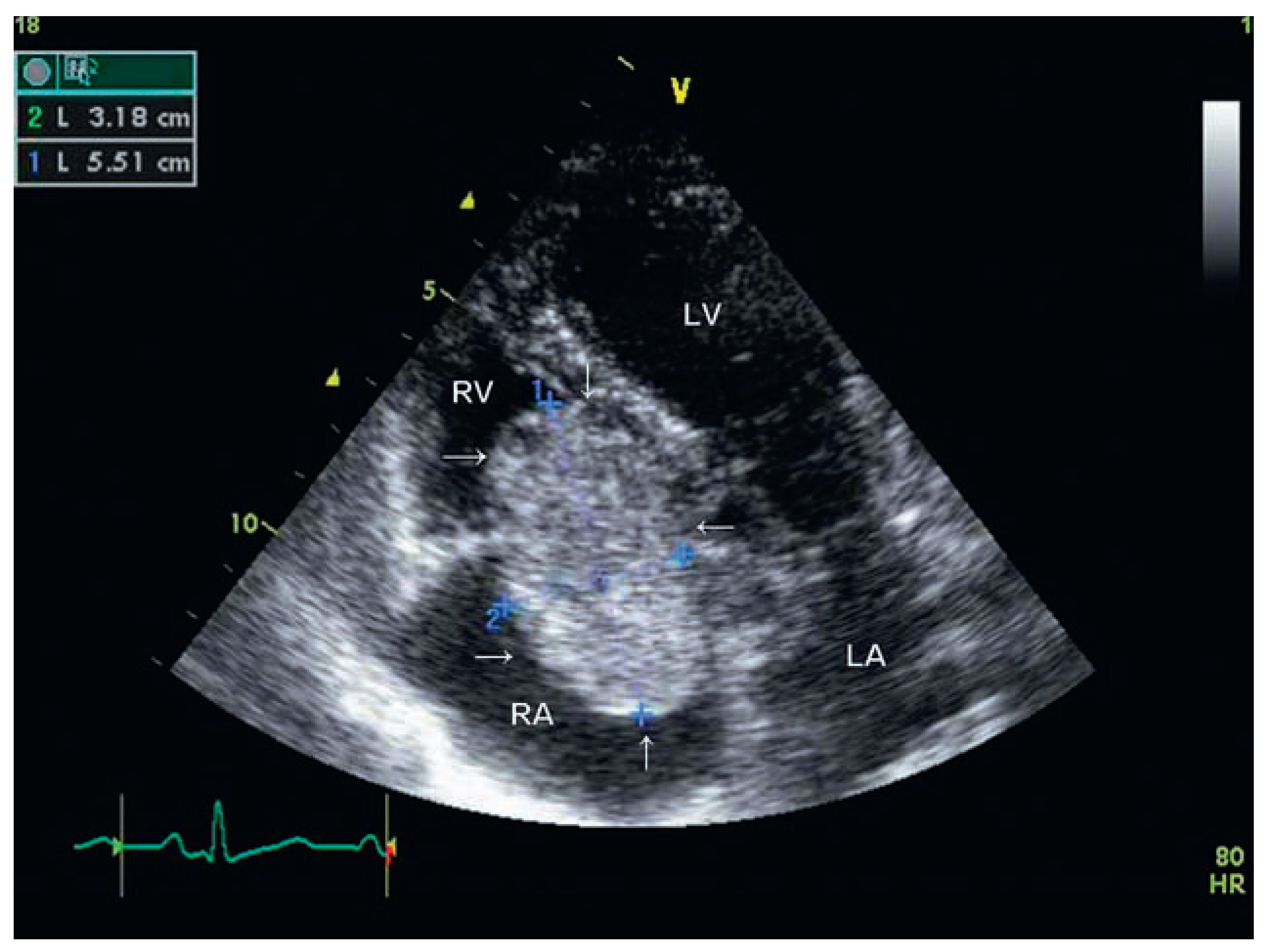
Figure 1. ETT apical view showing a right atrial mass (3.2 × 5.5 cm) compatible with a myxoma. RV = right ventricle; RA = right atrium; LV = left ventricle; LA = left atrium.
Cardiovascmed 11 00256 g001
Figure 2. Coronary angiogram (lateral view) showing left coronary artery injection with vascular blush in the mass (arrows). LAD = left anterior descending artery; LCx = left circumflex artery.
Cardiovascmed 11 00256 g002
Figure 3. Coronary angiogram (anteroposterior view) showing right coronary artery injection with localised vascular blush inside the mass (arrows).RCA = right coronary artery.
Cardiovascmed 11 00256 g003