Next Article in Journal
Der Perkutane Aortenklappenersatz
Previous Article in Journal
Regular Wide QRS Tachycardia Complicating Treatment for Atrial Fibrillation
 
 
Cardiovascular Medicine is published by MDPI from Volume 28 Issue 1 (2025). Previous articles were published by another publisher in Open Access under a CC-BY (or CC-BY-NC-ND) licence, and they are hosted by MDPI on mdpi.com as a courtesy and upon agreement with Editores Medicorum Helveticorum (EMH).
Interesting Images

Low Dose Hybrid Cardiac Imaging in a 61-Year-Old Male With Atypical Angina Pectoris1 †

by
Lars Husmann
1,
Ines Valenta
1,
Oliver Gaemperli
1,
Christophe A. Wyss
1 and
Philipp A. Kaufmann
1,2,*
1
Cardiovascular Center, University Hospital, 8091 Zurich, Switzerland
2
The Zurich Center for Integrative Human Physiology, University of Zurich, CH-8091 Zurich, Switzerland
*
Author to whom correspondence should be addressed.
Korrigierte Version 30. 5. 2008.
Cardiovasc. Med. 2008, 11(5), 168; https://doi.org/10.4414/cvm.2008.01327
Submission received: 23 February 2008 / Revised: 23 March 2008 / Accepted: 23 April 2008 / Published: 23 May 2008

Case report

A 61-year-old male patient with hypertension and dyslipidaemia reported recurrend episodes of chest discomfort at peak performance during bicycle exercise but also at rest and at night. Cardiac stress testing on a treadmill ergometer was negative and the patient was subsequently referred to hybrid cardiac imaging.
99mTc-Tetrofosmin single-photon emission computed tomography (SPECT) images were acquired using a routine one day adenosine stress/rest protocol (effective radiation dose 8.6 mSv), and fused with a low-dose computed tomography coronary angiography (CTCA), using prospective electrocardiogramgating [1] (1.5 mSv; performed with a Light-Speed VCT XT® scanner, GE Healthcare). CTCA revealed coronary artery disease with stenoses between 50–80% in all coronary arteries, but SPECT demonstrated a large reversible perfusion defect (ischaemia) only in the anterior myocardium. Therefore a culprit lesion could be identified with the fused SPECT/CTCA images in the proximal left anterior descending artery (LAD) (Figure 1). In this lesion coronary angioplasty with stent placement was subsequently performed.

Discussion

Hybrid cardiac imaging has previously been shown to reliably identify culprit lesions [2,3], but high radiation exposure from CTCA has remained an issue of concern. Our first experience indicates that low dose hybrid cardiac imaging using prospective ECG-gating for CTCA [1] is feasible with good image quality and accurate findings if heart rate is below 63 bpm.

References

  1. Husmann, L.; Valenta, I.; Gaemperli, O.; Adda, O.; Treyer, V.; et al. Feasibility of low-dose coronary CT angiography: first experience with prospective ECG-gating. Eur Heart J. 2008, 29, 191–197. [Google Scholar] [CrossRef] [PubMed]
  2. Gaemperli, O.; Schepis, T.; Kalff, V.; Namdar, M.; Valenta, I.; et al. Validation of a new cardiac image fusion software for threedimensional integration of myocardial perfusion SPECT and stand-alone 64-slice CT angiography. Eur J Nucl Med Mol Imaging. 2007, 34, 1097–1106. [Google Scholar] [CrossRef] [PubMed]
  3. Gaemperli, O.; Schepis, T.; Valenta, I.; Husmann, L.; Scheffel, H.; et al. Cardiac image fusion from stand-alone SPECT and CT: clinical experience. J Nucl Med. 2007, 48, 696–703. [Google Scholar] [CrossRef] [PubMed]
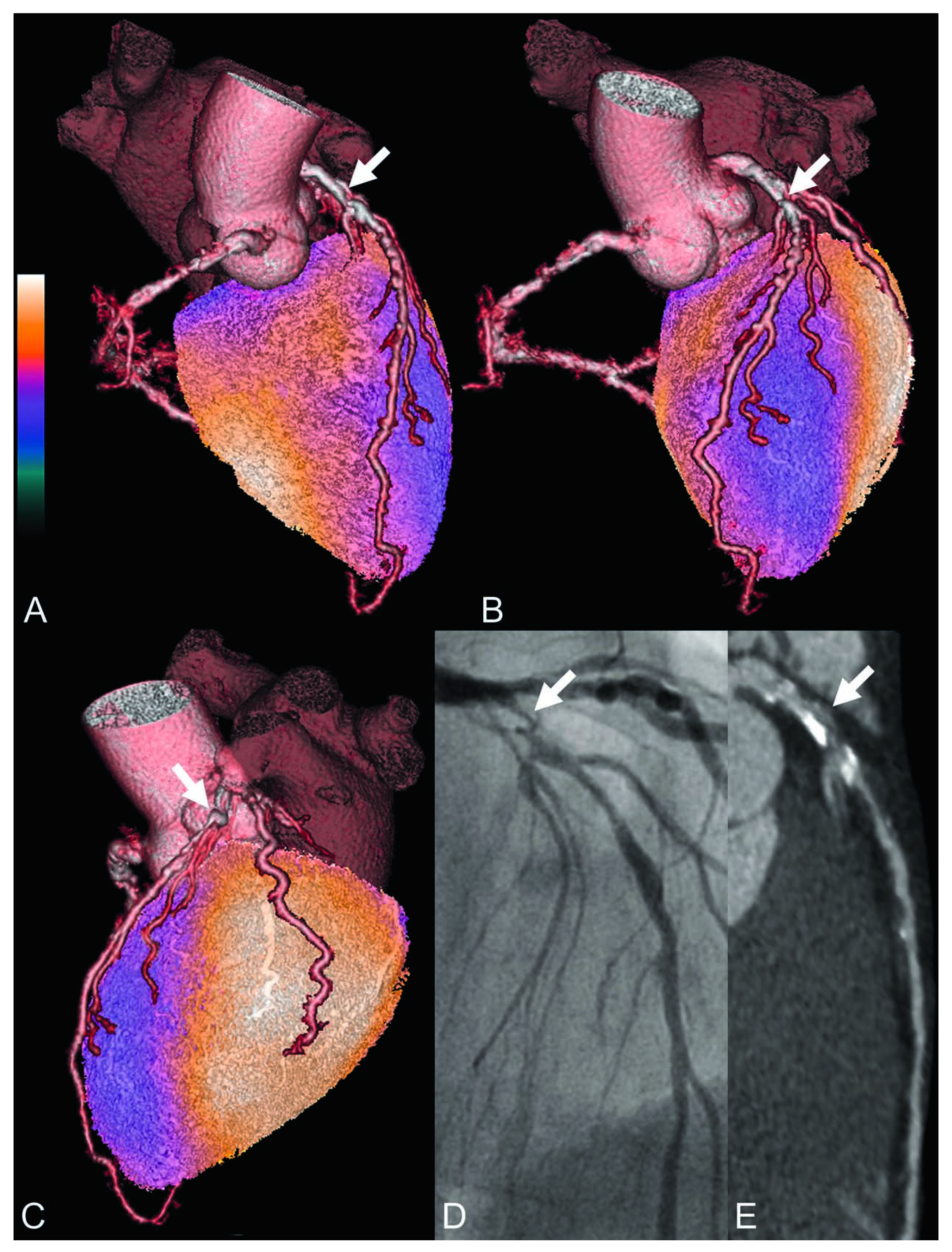
Figure 1. Fused SPECT/CTCA images (A–C) demonstrate a large perfusion defect in the anterior myocardium (bluish colours). A significant stenosis in the proximal LAD was identified as the culprit lesion (indicated by white arrows in the SPECT/CTCA images A–C, conventional coronary angiography D, and curved multiplanar CTCA reconstructions E).
Cardiovascmed 11 00168 g001