Next Article in Journal
Low Dose Hybrid Cardiac Imaging in a 61-Year-Old Male With Atypical Angina Pectoris1
Previous Article in Journal
European National Society Cardiovascular Journals
 
 
Cardiovascular Medicine is published by MDPI from Volume 28 Issue 1 (2025). Previous articles were published by another publisher in Open Access under a CC-BY (or CC-BY-NC-ND) licence, and they are hosted by MDPI on mdpi.com as a courtesy and upon agreement with Editores Medicorum Helveticorum (EMH).
Interesting Images

Regular Wide QRS Tachycardia Complicating Treatment for Atrial Fibrillation

by
Jens Seiler
*,
Joseph C. Lee
and
Kurt C. Roberts-Thomson
Cardiovascular Division, Brigham and Women’s Hospital, Boston, MA 02115, USA
*
Author to whom correspondence should be addressed.
Cardiovasc. Med. 2008, 11(5), 166; https://doi.org/10.4414/cvm.2008.01328
Submission received: 23 February 2008 / Revised: 23 March 2008 / Accepted: 23 April 2008 / Published: 23 May 2008

Case presentation

This 46-year-old gentleman presented with palpitations. His medical history is significant for a bicuspid aortic valve with moderate aortic regurgitation and an ascending aortic aneurysm of 5.2 cm. He developed atrial fibrillation (AF) four years ago, which had been well controlled on anti-arrhythmic drug therapy. His medications included flecainide 150 mg BID, metoprolol 12.5 mg QD, lisinopril 2.5 mg QD and simvastatin 20 mg QD.
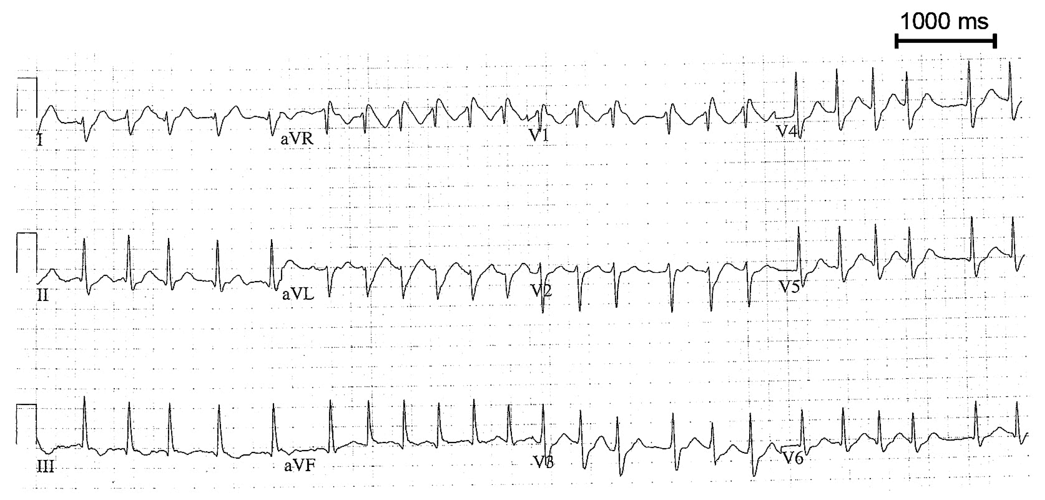
He developed shortness of breath and chest pain following exertion and was found in tachycardia of >200 beats per minute (bpm) and treated with metoprolol. He was noted to be in AF and cardioverted to sinus rhythm. The next day, the palpitations and chest pressure occurred again. He took an extra dose of 12.5 mg metoprolol. After treatment with a total of 10 mg metoprolol IV, his heart rate decreased approximately from 220 to 110 bpm on arrival in the emergency department. His electrolytes were within normal limits. While monitored, his symptoms recurred. An ECG was done (Figure 1). He was prepared for cardioversion, but converted spontaneously into coarse AF (Figure 2).
What is the most likely mechanism of the arrhythmia shown in Figure 1? What is the most probable cause?

Discussion

The tachycardia shown in figure 1 is a regular wide complex tachycardia (QRS width 162 ms) with a heart rate of 211 bpm. QRS morphology is that of a typical right bundle branch block (RBBB) pattern (rSR’ in V1 and Rs in V6) with right frontal plane axis. A RBBB also exists during AF. Initial ventricular depolarisation is rapid with a maximum precordial RS interval of 60 ms. There are no capture or fusion beats. P-waves are not clearly discernable. These surface ECG features strongly favor supraventricular tachycardia with RBBB aberration, but ventricular tachycardia cannot be excluded. Although a QRS width of >140 ms is suggestive for ventricular tachycardia with RBBB morphology, patients may have supraventricular tachycardia with a QRS width of >140 ms in the presence of preexisting bundle branch block and antiarrhythmic drug therapy [1]. Preexcited supraventricular tachycardia is unlikely due to the lack of a delta wave and morphologic stability of the QRS complex during AF.
Assuming that we are dealing with a regular supraventricular tachycardia with RBBB aberration, atrioventricular (AV)-nodal reentrant tachycardia, orthodromic AV-reciprocating tachycardia, ectopic atrial or junctional tachycardia and atrial flutter are possible entities. Class IC antiarrhythmic drugs cause new or modify preexisting atrial arrhythmias in 10% of patients. They are known to convert AF into slow atrial flutter which allows 1:1 AVconduction [2]. To prevent a rapid ventricular response during atrial flutter, co-administration of AV-nodal blocking agents is recommended [3]. This patient was treated with the class IC anti-arrhythmic drug flecainide and only a modest dose of metoprolol. The saw-tooth appearance of the baseline in the inferior leads supports a diagnosis of atrial flutter, although overlap with ventricular repolarisation makes the interpretation difficult. Furthermore, the reported drop of heart rate from 220 to 110 bpm after administration of an AV-nodal blokking agent is consistent with the conversion of atrial flutter from 1:1 AV-conduction to 2:1 AVconduction. Therefore, the most likely diagnosis in the present clinical setting is slow atrial flutter with 1:1 AV-conduction. This could be confirmed by vagal maneuvers or the administration of adenosine, resulting in transient slowing or blocking of AV-conduction, and thus revealing flutter waves.

Conflicts of Interest

There is no conflict of interest. Jens Seiler is recipient of a research grant from St. Jude Medical which is not related to the content of the present study. Kurt Roberts-Thomson is the recipient of a Postdoctoral Research Scholarship from the National Health and Medical Research Council (NHMRC) of Australia.

References

  1. Blomstrom-Lundqvist, C.; Scheinman, M.M.; Aliot, E.M.; Alpert, J.S.; Calkins, H.; Camm, A.J.; et al. ACC/AHA/ESC guidelines for the management of patients with supraventricular arrhythmias – executive summary: a report of the American College of Cardiology/American Heart Association task force on practice guidelines and the European Society of Cardiology committee for practice guidelines (writing committee to develop guidelines for the management of patients with supraventricular arrhythmias). Circulation. 2003, 108, 1871–1909. [Google Scholar]
  2. Feld, G.K.; Chen, P.S.; Nicod, P.; Fleck, R.P.; Meyer, D. Possible atrial proarrhythmic effects of class 1C antiarrhythmic drugs. Am J Cardiol. 1990, 66, 378–383. [Google Scholar] [CrossRef]
  3. Fuster, V.; Ryden, L.E.; Cannom, D.S.; Crijns, H.J.; Curtis, A.B.; Ellenbogen, K.A.; et al. ACC/AHA/ESC 2006 guidelines for the management of patients with atrial fibrillation: a report of the American College of Cardiology/American Heart Association task force on practice guidelines and the European Society of Cardiology committee for practice guidelines (writing committee to revise the 2001 guidelines for the management of patients with atrial fibrillation): developed in collaboration with the European Heart Rhythm Association and the Heart Rhythm Society. Circulation. 2006, 114, e257–354. [Google Scholar] [PubMed]
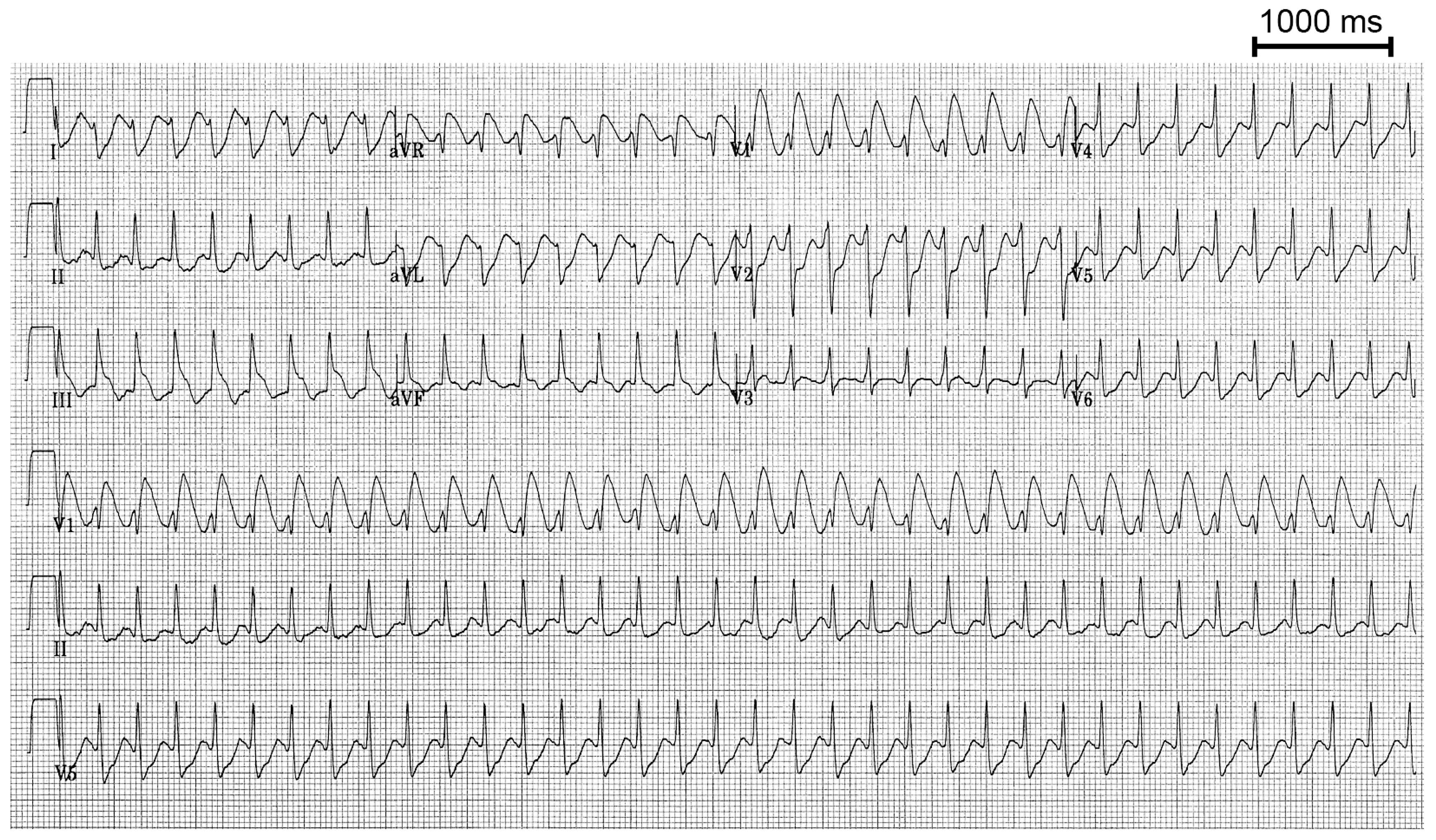
Figure 1. Wide QRS tachycardia during treatment of AF.
Cardiovascmed 11 00166 g001
Figure 2. Repeat-ECG after conversion into coarse AF.
Cardiovascmed 11 00166 g002