Next Article in Journal
Long-Term Follow-Up After Thoracic Radiotherapy: Symptomatic Heart Disease Is an Ominous Sign
Previous Article in Journal
Schweizer Richtlinien Zum Management von Pavk-Patienten Durch Den Facharzt
 
 
Cardiovascular Medicine is published by MDPI from Volume 28 Issue 1 (2025). Previous articles were published by another publisher in Open Access under a CC-BY (or CC-BY-NC-ND) licence, and they are hosted by MDPI on mdpi.com as a courtesy and upon agreement with Editores Medicorum Helveticorum (EMH).
Interesting Images

Transcoronary Ablation of Septal Hypertrophy in HOCM: Septal Collaterals May Cause Unwanted Inferior Myocardial Infarction

Swiss Cardiovascular Center Bern, University Hospital, Bern, Switzerland
*
Author to whom correspondence should be addressed.
Cardiovasc. Med. 2007, 10(12), 401; https://doi.org/10.4414/cvm.2007.01286
Submission received: 28 September 2007 / Revised: 28 October 2007 / Accepted: 28 November 2007 / Published: 28 December 2007

Case report

A patient with HOCM presented with worsening dyspnea on medical treatment with verapamil. Transcoronary ablation of the septal hypertrophy (TASH) via the 1st septal branch (SB) was performed. There were normal coronary arteries and a large 1st SB of the left anterior descending artery. It did not seem to receive or supply collaterals from or to the right coronary artery (RCA). The midventricular / LV outflow tract (LVOT) obstruction was severe (Figure 1). TASH was performed with slow injection of absolute alcohol ([AA] 96%) over 25 min. Following injection of 5 ml of AA over 20 min, there was no fall in ΔPmean. Therefore, an additional 3 ml was instilled, which lead to a decrease of resting ΔPmean to 15 mm Hg (Figure 2). At that time, the patient complained of severe chest pain and contrast injection into the SB revealed filling of the occluded RCA via septal collaterals. There were simultaneous inferior ST-segment elevations. So far, there has been a similar case report speculating on the possible cause of septal collaterals for inferior or apical myocardial infarction during TASH [1]. Due to the fact that 80% of individuals with normal coronary arteries have at least some preformed recruitable collateral vessels (1/4 of them have collaterals which prevent signs of ischaemia during brief vascular occlusion) [2], the likelihood is high that with septal balloon occlusion collaterals to the RCA are recruited. The present report provides the first proof of “collateral damage” during TASH.

References

  1. Chowdhary, S.; Galiwango, P.; Woo, A.; Schwartz, L. Inferior infarction following alcohol septal ablation: A consequence of collateral damage? Catheter Cardiovasc Interv. 2007, 69, 236–42. [Google Scholar] [CrossRef] [PubMed]
  2. Wustmann, K.; Zbinden, S.; Windecker, S.; Meier, B.; Seiler, C. Is there functional collateral flow during vascular occlusion in angiographically normal coronary arteries? Circulation. 2003, 107, 2213–20. [Google Scholar] [CrossRef] [PubMed]
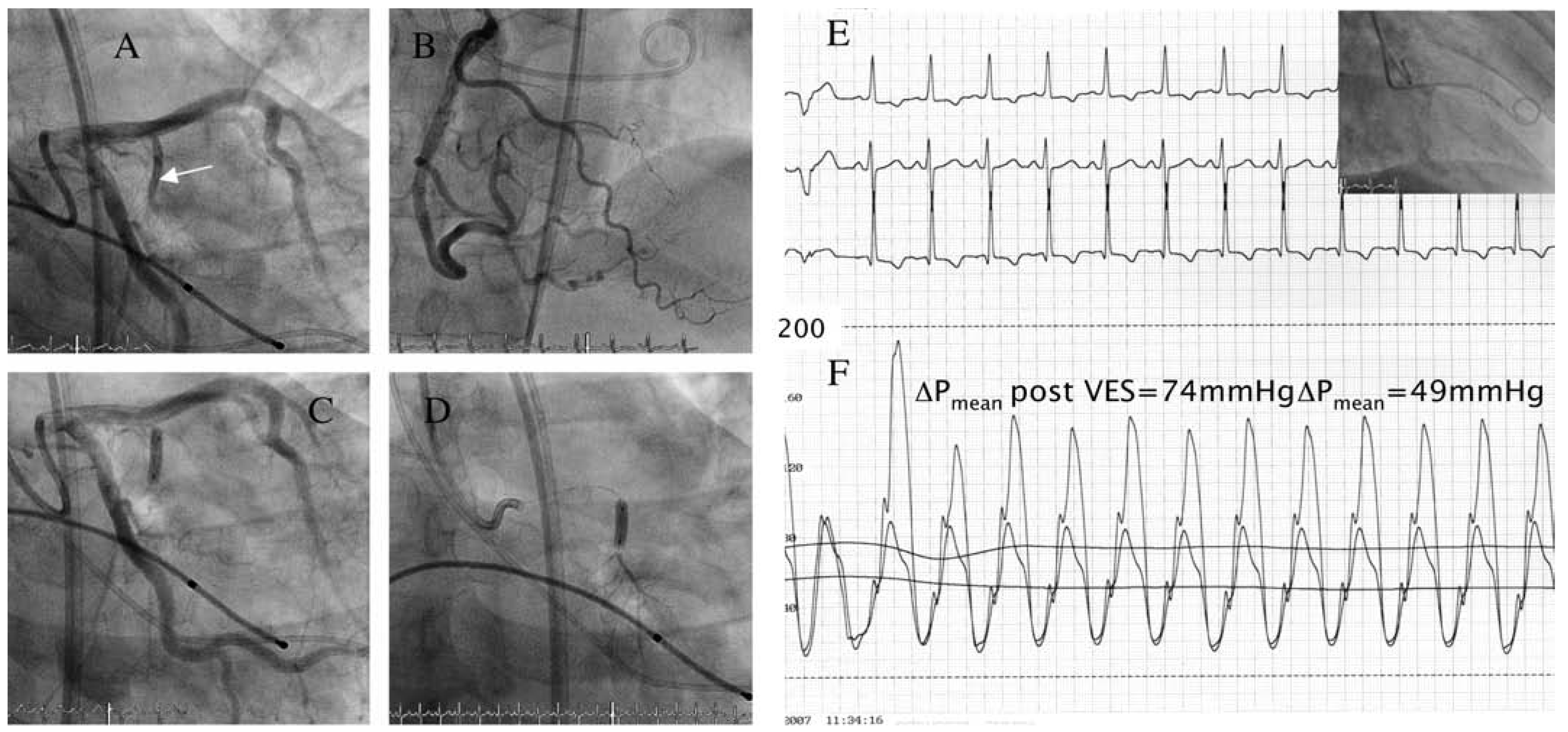
Figure 1. A shows the large-sized 1st septal branch (SB; arrow) of the left anterior descending artery; B depicts the small normal right coronary artery (RCA); C over-the-wire ballon occlusion of the 1st SB; D injection of contrast through the balloon; E ECG tracing during septal ablation; F documents the pressure gradient between left ventricular caivity and aorta (ΔPmean).
Cardiovascmed 10 00401 g001
Figure 2. A and B show retrograde filling of the RCA (arrows) from the septal injection; C Occlusion of the distal RCA (arrow); D ST-T changes in the ECG; E Reduction of the pressure gradient post alcohol ablation.
Cardiovascmed 10 00401 g002