Next Article in Journal
Variation on the Anchor Balloon Technique for Difficult Stent Delivery
Previous Article in Journal
Transcoronary Ablation of Septal Hypertrophy in HOCM: Septal Collaterals May Cause Unwanted Inferior Myocardial Infarction
 
 
Cardiovascular Medicine is published by MDPI from Volume 28 Issue 1 (2025). Previous articles were published by another publisher in Open Access under a CC-BY (or CC-BY-NC-ND) licence, and they are hosted by MDPI on mdpi.com as a courtesy and upon agreement with Editores Medicorum Helveticorum (EMH).
Article

Long-Term Follow-Up After Thoracic Radiotherapy: Symptomatic Heart Disease Is an Ominous Sign

by
Christine H. Attenhofer Jost
5,*,
Pia Huguenin
2,
René Prêtre
3,
Juraj Turina
1,
Burkhardt Seifert
4,
F. Wolfgang Amann
5,
Christoph Glanzmann
2,
Paul R. Vogt
3 and
Rolf Jenni
1
1
Division of Cardiology, University Hospital, Zurich, Switzerland
2
Department of Radiation Therapy, University Hospital, Zurich, Switzerland
3
Division of Cardiovascular Surgery, University Hospital, Zurich, Switzerland
4
Department of Biostatistics, University of Zurich, Zurich, Switzerland
5
Cardiovascular Center Zurich, Klinik im Park, Zurich, Switzerland
*
Author to whom correspondence should be addressed.
Cardiovasc. Med. 2007, 10(12), 387; https://doi.org/10.4414/cvm.2007.01285
Submission received: 28 September 2007 / Revised: 28 October 2007 / Accepted: 28 November 2007 / Published: 28 December 2007

Summary

Background: Thoracic radiotherapy (RT), especially with past technology, may affect the heart, but rarely leads to symptoms. In patients with symptomatic heart disease after RT, outcome seems to be dismal. Methods: In this observational descriptive study, clinical characteristics, findings of ECG, echocardiography, cardiac interventions and follow-up were analysed in patients with prior RT and symptomatic heart disease. The patients were identified in the echocardiography database during a ten year period. Results: There were 25 patients who had thoracic RT at a median age of 35 years (range: 9–59) for lymphoma (12 patients), breast cancer (9) or other cancer (4). At least likely inclusion of the heart in the target volume of previous RT was present in of 16 of 20 patients with detailed information on RT. Last follow-up was 24 years (range: 5–57) after RT at a median age of 56 years (range: 30–84). Symptoms (≥1 per patient) included: dyspnea (21 patients), angina (12) and/or heart failure (10). Three patients had prior myocardial infarction. The following disease was found: moderate valvular disease in 19 patients (76%), coronary artery disease in 12 (48%), abnormal ECG in 18 (72%), relevant conduction system disease in 9 (36%), restriction / constriction in 7 (28%), and pericardial effusion in 4 (16%). Cardiac surgery was necessary in 12 patients (death in 2 patients), percutaneous coronary interventions in 5 and pacemaker implantation in 3. Endocarditis occurred in 2 patients. During follow-up (21 ± 8 months), death occurred in 6 patients (24%) and was due to heart disease in 5 of them. Conclusions: If symptomatic heart disease develops in the long-term follow-up after RT, complex disease of valves, coronary arteries, conduction system, myocardium and pericardium is frequently observed. Cardiac interventions are often necessary; and heart disease may be a common cause of death in these patients. Careful assessment and evaluation of treatment options are needed in this patient group.

Introduction

Since 1902, radiation therapy is used for treatment of Hodgkin’s disease and lymphomas. Initially, the heart was considered to be relatively radio-resistant. In long-term follow-up of cured patients, cardiac damage was found to be more frequent than expected [1]. Pericardial disease (acute or delayed pericarditis) was the earliest described cardiac side-effect of thoracic radiotherapy (RT) with an incidence of 2.5–20% depending on the use of subcarinal blocks shielding the heart [2,3]. Before effective chemotherapy regimens became available, patients with a relapse often underwent re-RT for breast cancer and lymphomas. Clinically silent cardiac disease after RT has been found in up to 96% of the patients consisting of symptomatic or occult constrictive pericarditis (50–96%), coronary artery disease (13%), myocardial fibrosis (up to 50%) and left ventricular dysfunction (5%) [3,4,5]. Valvular abnormalities occur with a high prevalence (especially aortic valvular disease) even in asymptomatic patients [6]. Coronary artery disease is frequent and may typically manifest with ostial stenosis of the left main and right coronary arteries occurring at a mean time of 16 years after RT [2,7]. Conduction system disease may consist of bundle branch block, and first or higher degree atrioventricular block [6]. A history of prior RT increases the rate of cardiac mortality two- to threefold versus controls which were not radiated [8,9]. In one study, this was only seen after a follow-up of at least 15 years [10].
There is no doubt that improvement of knowledge and RT technique have diminished the risk of cardiac damage in cancer patients [11]. However, we still have to care for patients with symptomatic heart disease occurring many years after RT to the chest. In previous reports [12,13], cardiac disease in patients in long-term follow-up after RT for lymphoma was described, and the need for life-long cardiac follow-up and correction of cardiac risk factors other than RT was demonstrated [13]. Symptomatic cardiac disease in patients irradiated to the chest often seemed associated with a poor outcome.
We retrospectively analysed patients with symptomatic cardiac disease after thoracic RT focussing on symptoms, type of heart disease, frequency of combined disease of valves, pericardium, coronary arteries and conduction system, outcome of cardiac interventions and long-term follow-up.

Methods

Study group 

Among all 41 864 patients in the database referred for an echocardiographic exam to our echocardiography laboratory within 10 years, there were 25 patients (0.06%) who had both: a history of prior RT to the chest as well as cardiac symptoms. We reviewed the medical records of these patients focussing on symptoms, coronary risk factors, treatment, interventions and outcome. The median interval between RT and the echocardiographic exam in the 25 patients was 24 ± 13 years (range: 5–57). Age at RT was defined as the age of the patient when RT was started.

Echocardiography 

Transthoracic echocardiograms were performed using fully equipped commercially available ultrasound instruments (HP 5500 [Hewlett Packard Co., Andover, MA, USA] and Siemens Acuson Sequoia 250 [Mountain View, CA, USA]).
Comprehensive standard 2D and Doppler echocardiography was performed according to the guidelines of the American Society of Echocardiography [14]. Ejection fraction was calculated by biplane area length method in all patients.
Diastolic function was assessed as previously described [15]. Criteria for constrictive and restrictive pericarditis were used as published [16].
Valvular regurgitation was classified as trivial, mild, moderate or severe according to usual criteria [17]. Aortic stenosis was defined as mild with a mean gradient of 10–29 mm Hg and/or valve area of 1.5–1.9 cm2, moderate with a mean gradient of 30–49 mm Hg and/or valve are of 1.0–1.5 cm2, and as severe with a mean gradient of >50 mm Hg and/or valve area of less than 1.0 cm2. Echocardiographic signs for significant heart disease were defined as described previously for other conditions [18]: at least moderate mitral regurgitation, at least mild mitral stenosis, at least mild aortic regurgitation or stenosis, at least moderate tricuspid regurgitation or at least mild tricuspid stenosis, at least mild pulmonary stenosis, definite signs for constrictive pericarditis, restrictive filling pattern, a left ventricular ejection fraction of <50% and/or the presence of pericardial effusion.
Systolic pulmonary artery hypertension was defined as systolic pulmonary artery pressure of greater than 35 mm Hg

ECG analysis 

ECG tracings were all retrospectively reviewed. Conduction system disease was defined as complete right or left bundle branch block, and/or first, second or third degree atrioventricular block (AV block). Incomplete left anterior hemiblock was not included. Incidence of pacemaker implantations was noted.

Coronary angiography 

Routine coronary angiography and heart catheterisation according to standard criteria were performed where clinically indicated. Coronary artery disease was called significant in the presence of ≥50% stenosis of a major epicardial coronary artery by coronary angiography (or autopsy). If necessary, percutaneous transluminal coronary angioplasty with ballooning with or without stenting was performed as described before [19].

Definition of combined heart disease 

Combined heart disease was defined as the presence of significant valvular abnormalities and/or conduction system pathology (complete right or left bundle branch block, 1st, 2nd or 3rd degree atrioventricular block) and/or pericardial disease (constrictive/restrictive signs and/or pericardial effusion).

Data on radiation therapy 

The patients received their radiation treatment at different centres in Switzerland and most commonly in the 1970s and early 1980s. Data were collected as available and a radiation oncologist (P.H.) reviewed all available data and scored the inclusion of relevant parts of the heart in the target volume as unknown, unlikely, likely or certain.

Heart surgery 

The surgery and hospital records of the 12 patients who underwent cardiac surgery were carefully analysed focussing on intraoperative findings, type of surgery, complications and length of hospitalisation.

Statistical analysis 

Results are presented as mean values ± standard deviation or frequency expressed as a percentage for dichotomous or qualitative variables.

Results

Clinical characteristics 

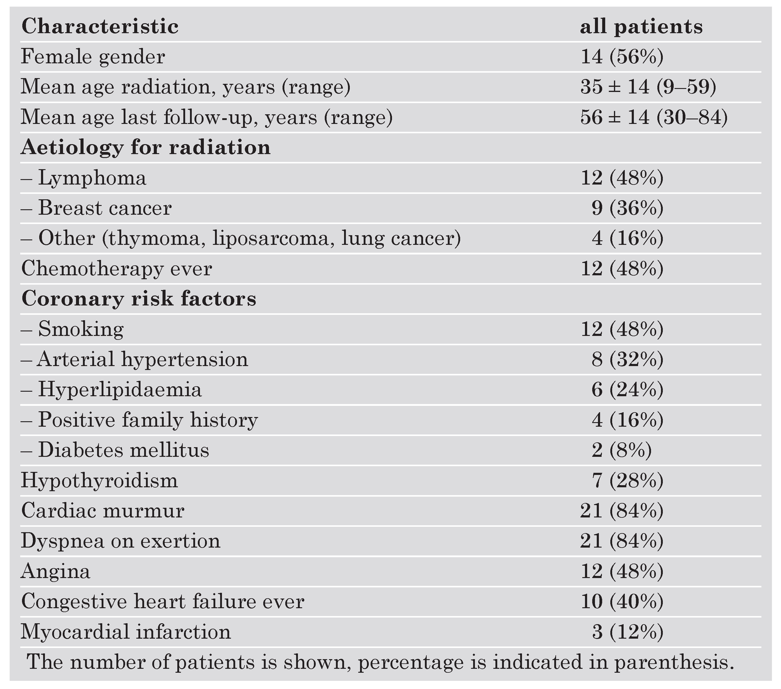
The clinical characteristics of the 25 patients are summarised in Table 1. RT was performed at a rather young age of 35 ± 14 years. The year of RT is shown in Figure 1. The distribution was as follows: 7 patients had RT between 1960 and 1970; 7 between 1971 and 1975, 2 between 1976 and 1980, 5 between 1981 and 1985, and only 4 patients 1986 or later. The two most common diseases treated with RT were lymphoma and breast cancer. At least one cardiovascular risk factor was present prior to RT in 18 patients (72%); the average number of coronary risk factors was 1.3. The distribution of coronary risk factors is shown further down in Table 1. Prior chemotherapy was administered in 12 patients (48%). Hypothyroidism requiring thyroid hormone replacement was noted in 7 patients (28%). A heart murmur was present in all patients with significant valvular heart disease. Dyspnea on exertion and angina were frequent. A history of heart failure was present in 7 of 12 patients with prior chemotherapy, compared to 8 of 13 patients following RT alone. A history of myocardial infarction was present in 3 patients (12%): it was clinically manifest in only one patient; in two patients a scar consistent with myocardial infarction was only discovered at coronary angiography or at autopsy, respectively.
Table 1. Summary of clinical characteristics of the 25 patients at the time of echocardiography.
Cardiovascmed 10 00387 i001
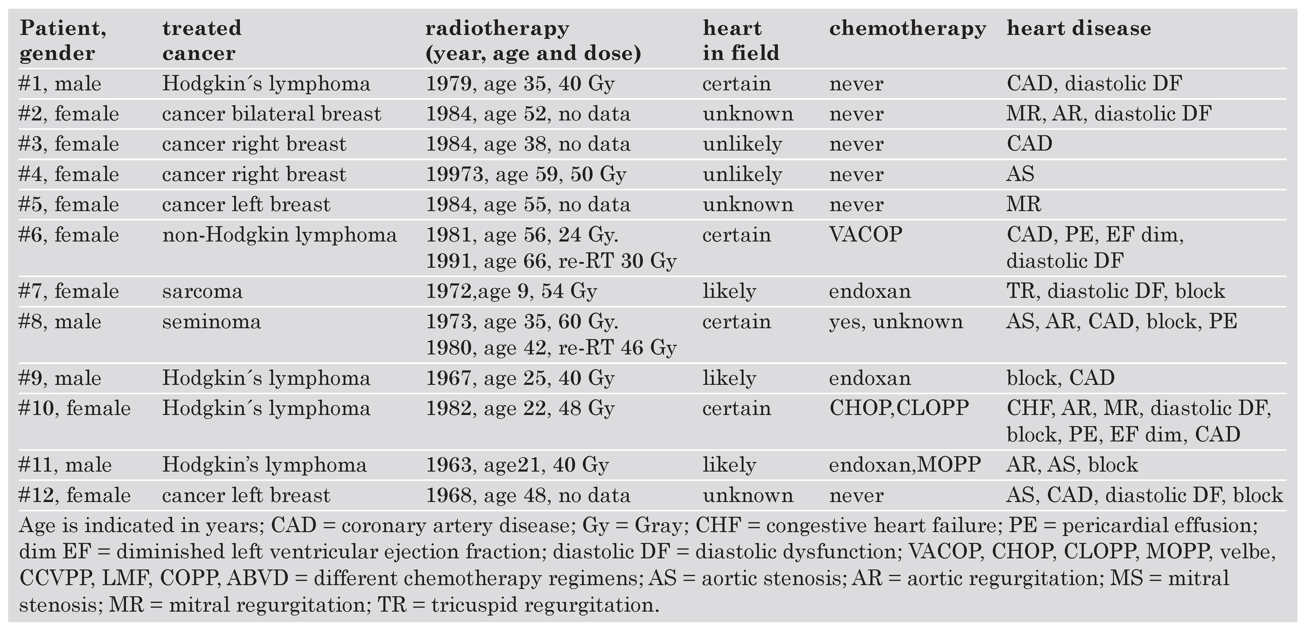
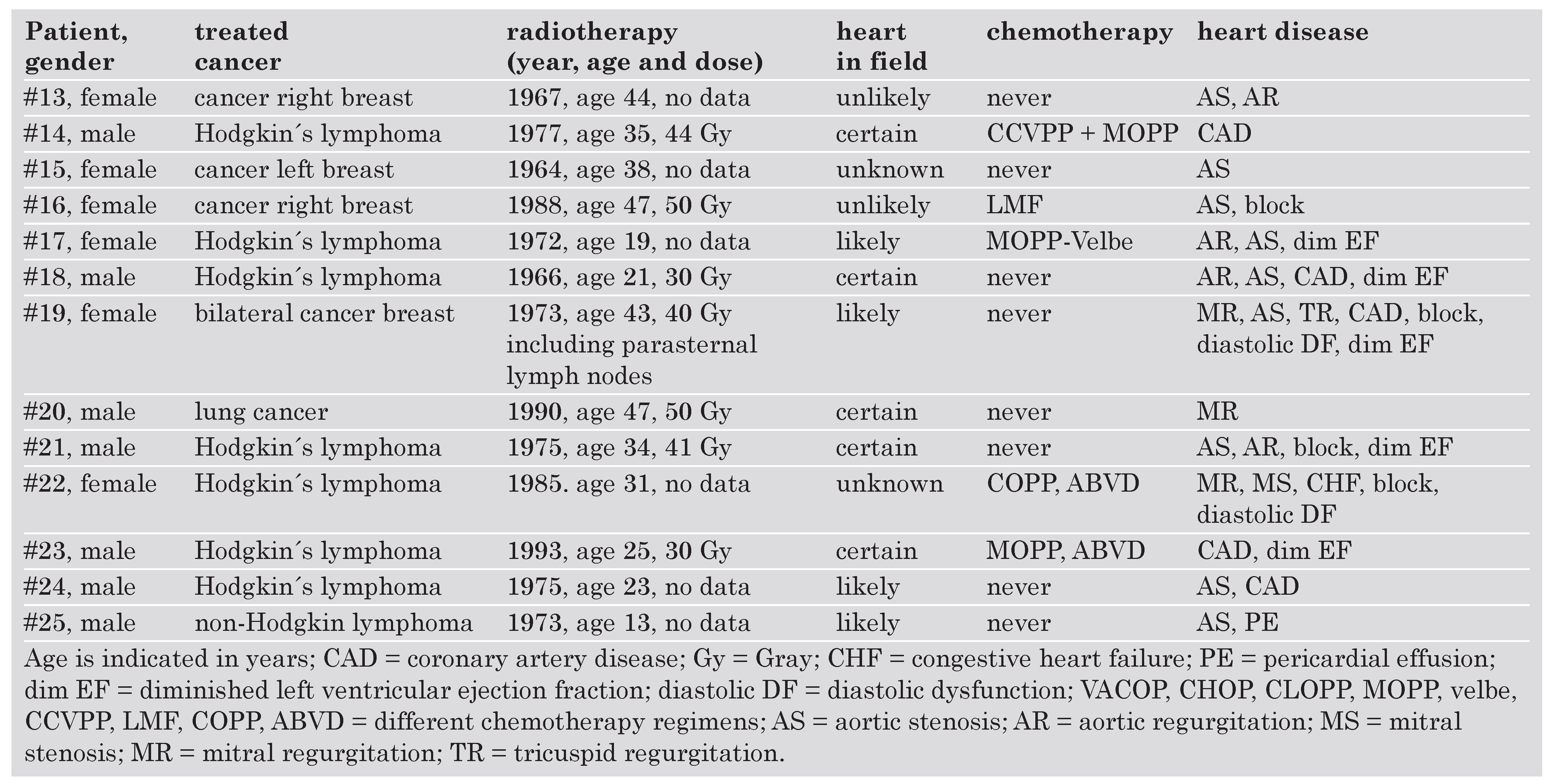
Patient characteristics including the type of tumor, treatment and types of heart disease are summarized in Table 2. In 16 of 20 patients with some or all available details on RT, target volume including the heart was likely or certain.
Table 2. Summary of the 25 patients including data on radiotherapy, chemotherapy and heart disease (part I).
Cardiovascmed 10 00387 i002
Table 2. Summary of the 25 patients including data on radiotherapy, chemotherapy and heart disease (part II).
Cardiovascmed 10 00387 i003
Associated heart disease (as shown in Table 2) included coronary artery disease in 12 patients (48%), valvular heart disease in 19 (76%), pericardial effusion in 4 (16%), diastolic dysfunction in 8 patients (32%), diminished left ventricular ejection fraction in 7 patients (28%), and signs of conduction system disease with complete right or left bundle branch block or atrioventricular block in 10 patients (40%).

Echocardiographic findings 

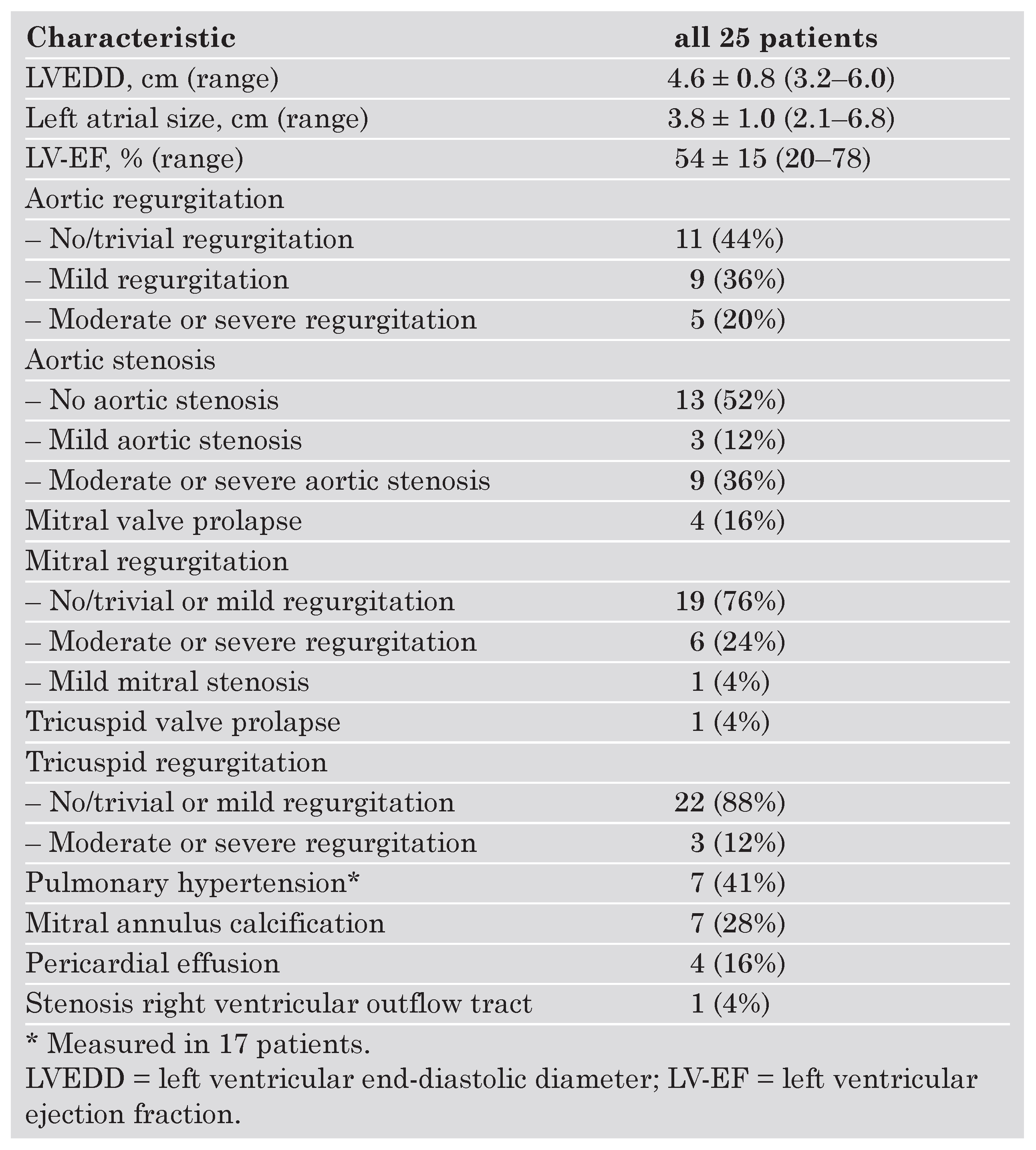
The detailed echocardiographic findings are shown in Table 3. The ejection fraction of the left ventricle ranged from 20 to 78% and was diminished (<50%) in 8 patients (32%).
Table 3. Echocardiographic findings in the 25 patients.
Cardiovascmed 10 00387 i004
Significant aortic valve disease with aortic regurgitation and/or stenosis was found in 17 patients (68%). The tricuspid valve was rarely affected. Pulmonary hypertension was quite frequent and was noted in 7 of 17 patients (41%) in whom it could be measured. One patient had stenosis of the right ventricular outflow tract at right heart catheterisation; findings of further evaluation with computed tomography were compatible as a calcific remnant of irradiated Hodgkin’s tissue.
Combined heart disease was found in 18 patients (72%). A typical example is patient #10 in whom Hodgkin’s disease was treated with a relatively high dose of radiotherapy (48 Gy) and chemotherapy at age 22 years. At age 37, she developed recurrent pulmonary oedema due to combined heart disease. She had severe mitral regurgitation, moderate aortic regurgitation and pericardial effusion as well as a significant calcific stenosis of her right coronary ostium. There was also complete left bundle branch block. At heart surgery, it was noted that her atria and the coronary sinus were scarred and small, the aortic valve was fibrotic; also, the pericardium was severely fibrotic with changes typical of constriction. Aortic and mitral valve replacement as well as coronary artery bypass grafting with one bypass to the right coronary artery were performed. She died postoperatively due to diastolic heart failure. At autopsy, severe fibrotic changes of pericardium and pleura and scarring of the posterior wall of the left ventricle and septum were found.
Another impressive example is a 43 year old woman (#22) who developed severe heart disease with constrictive pericarditis, severe tricuspid regurgitation and complete atrioventricular block 12 years after RT and chemotherapy for Hodgkin’s lymphoma. One year after RT, she had transient pneumonia. At cardiac surgery 12 years later (tricuspid valve repair, pericardectomy), severe adhesions over the big, partially calcified vessels, involving the superior vena cava and anonymous vein, were noted, which made surgery very difficult. The patient barely survived the operation and postoperative recovery was lengthy.
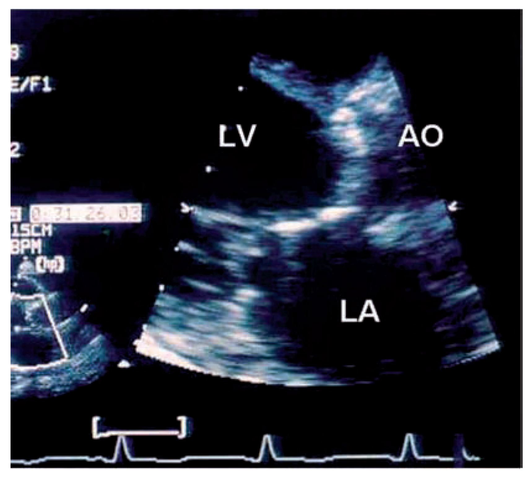
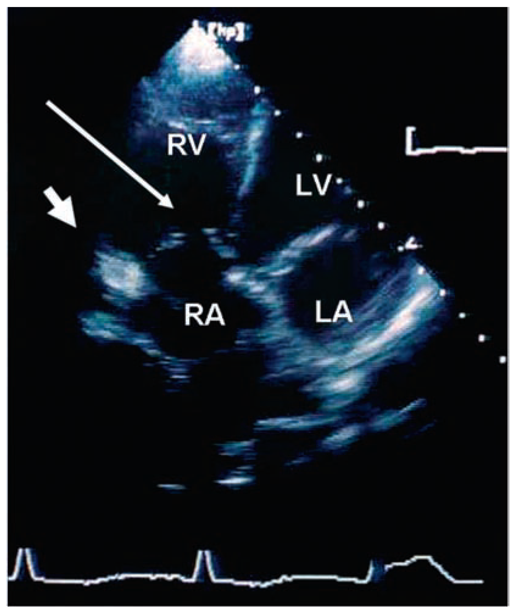
A typical echocardiographic example of a young woman with significant heart disease 15 years after RT (patient #10) is shown in Figure 2. The mitral valve and aortic valve are calcified, shrunk, stenotic, typically, left atrial size was not increased. Intraoperatively, tissue of both atria and coronary sinus was shrunk. In Figure 3, an example of a 35 year old woman with moderate tricuspid regurgitation and severe pericardial constriction is shown; the tricuspid valve was thickened, shrunk, and showed diminished mobility.
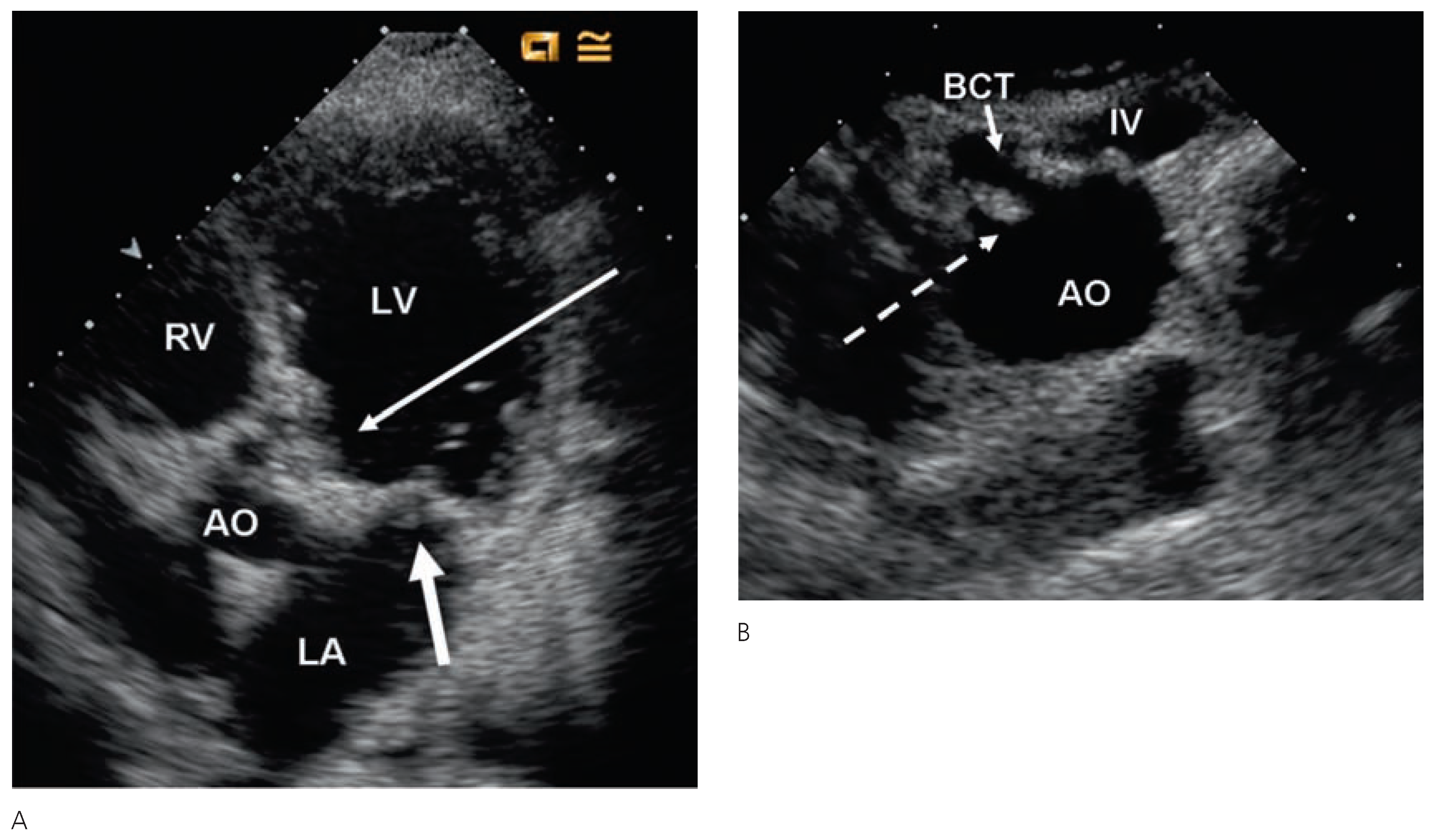
Figure 4 is the example of a 41 year old patient with extensive aortic and mitral valve calcifications including mitral and aortic annulus. He also had impressive calcifications involving the aortic arch.
ECG findings can be summarised as follows: in 18 patients (72%) the ECG was abnormal with complete right bundle branch block in 6 patients, complete left bundle branch block in 3 patients, left anterior hemiblock in 3 patients, and complete AV block necessitating pacemaker implantation in 3 patients. First degree AV block was present in 3 patients. In 6 patients, a history of atrial fibrillation or flutter was present.

Cardiac Interventions 

Cardiac interventions were performed in 17 patients (63%) and included 13 open heart surgeries, percutaneous coronary interventions in 5 patients and 3 pacemaker implantations.
Twelve patients underwent 13 cardiac surgeries (12 at our center). Two of 12 patients (17%) died in the early postoperative period due to heart failure. Cardiac surgery involved 11 valve replacements or repair in 10 patients, anterior pericardectomy and right ventricular epicardiotomy in 1 patient, and coronary artery bypass grafting due to subtotal left main stenosis and occlusion of the proximal right coronary artery in 1 patient. In this last patient, intraoperatively, the tissue was described as friable and not compatible with normal coronary artery disease. This patient developed as a complication aneurysmatic dilatation of the left main coronary artery due to the friable tissue of the stenotic coronary artery after pericardial patch repair. Another patient had previous mitral valve replacement and underwent reoperation two years later for severe constriction and severe tricuspid regurgitation with a very prolonged hospital course. The average stay for heart surgery at our hospital was 9.5 ± 2.8 days; however, some patients were referred to another hospital for subsequent postoperative care.
Table 4. Patient history, cardiac findings and causes of death in the 6 patients who died.
Cardiovascmed 10 00387 i005
Successful percutaneous transluminal angioplasty was performed in 5 patients. There were no unusual complications.
One patient had a pacemaker implantation as the only intervention.

Follow-up and causes of death 

Follow-up after the echocardiographic examination was performed by the end of March 2000 by a clinical visit or notice of death in all patients; follow-up time was 21 ± 8 months. Six patients died. In at least five of these patients, the cause of death was related to heart disease. The findings, presumed causes of death and findings at autopsy (if available) in these 6 patients are summarised in Table 4. Endocarditis occurred in 2 patients (14%). In one patient, staphylococcal endocarditis developed 2 years after aortic valve replacement and was the cause of death. This patient had additional severe RT-induced changes including bilateral carotid artery stenosis, pericarditis and substituted hypothyroidism 15 years after RT. In another patient, streptococcal endocarditis developed in combined aortic valve disease of a tricuspid aortic valve, she underwent aortic valve replacement.

Discussion

Our data show what happens if symptomatic heart disease develops in the long-term follow-up after RT to the chest – especially with the old RT technology used up to the early 1980s. Complex combined disease of valves, coronary arteries, conduction system, myocardium and pericardium may be present with many complications and a poor prognosis. Cardiac interventions in this patient group are frequently necessary. Careful long-term follow-up is needed in this patient group.
Significant heart disease following RT to the chest is rare; in an analysis done at the University Hospital in Zurich including 352 patients treated for Hodgkin’s lymphoma, a group of 112 patients underwent cardiac check-up including echocardiography. Nearly all these patients had been treated with <2.0 Gy per fraction to the anterior cardiac region. Sclerosis of mitral and/or aortal valves were observed in 36 of them (32%), but most were not clinically significant: only 1 patient had moderate aortic stenosis and one patient moderate mitral regurgitation [12].
  • Spectrum of radiation induced heart disease

Pericardial disease 

Acute or delayed pericarditis is the earliest described and most widely recognised form of radiation induced cardiac disease [20]. Time to onset of pericarditis ranges from 4 months to several years after RT and may present with pericardial effusion or manifest as constrictive pericarditis. Most patients with severe pericardial disease have received >40 Gray with little or no subcarinal shielding. Greenwood reported a 7% incidence of constrictive pericarditis [21]. Hancock described a 4% risk of constrictive pericarditis requiring pericardectomy [3]. In our patients, the incidence of a restrictive and/or constrictive physiology was 28%; however, severe constrictive pericarditis requiring surgery was rare.

Valvular heart disease 

It has been controversial whether valvular abnormalities may be considered as a late effect of RT. In a necropsy report, Brosius described 12 instances of mural endocardial fibrosis and 13 cases of valvular endocardial fibrosis [5]. Hancock found that 12 of 635 patients treated with RT for Hodgkin’s disease died of cardiac disease (RR: 29.6; 95% confidence interval [CI]: 16.0 to 49.3) including seven deaths from acute myocardial infarction ([AMI] RR: 41.5; 95% CI: 18.1 to 82.1), three from valvular heart disease, and two from radiation pericarditis / pancarditis. Three patients underwent valve replacement, 29 other patients had new murmurs [3]. Fifty percent of these patients had received ≥44 Gray without subcarinal blocking. In our series, valvular abnormalities were common (at least moderate valvular heart disease in 76%), as we collected our patients from the echocardiography database which, because of the selection bias, overestimates the prevalence of valvular changes. However, we believe that valvular abnormalities do have an impact on overall mortality in this patient group due to endocarditis and need for valve surgery. In 3 of the 6 patients who died during short-term follow-up, valvular heart disease was the likely cause. In a recent paper, Heidenreich et al. reported that in 294 asymptomatic patients with previous mediastinal irradiation due to Hodgkin’s disease with a mean mantle irradiation dose of 43 ± 0.3 Gy, valvular heart disease was common and increased with time following irradiation [6]. The number needed to screen to detect one candidate for endocarditis prophylaxis was only 1.6 (95% CI: 1.3 to 1.9) for those treated at least 20 years ago. The most common findings included at least mild aortic regurgitation in 60%, at least moderate tricuspid regurgitation in 4% and aortic stenosis in 16% in these asymptomatic patients [6]. This is very similar to the findings in our paper with 56% of patients having at least mild aortic regurgitation, 48% at least mild aortic stenosis and 12% at least moderate tricuspid regurgitation.

Coronary artery disease after radiation therapy 

Coronary artery disease and acute myocardial infarction seem to be increased in patients late after irradiation with a relative risk of 41.5 for acute myocardial infarction in the Stanford cohort [22]. Left main and ostial right coronary stenosis seem to be typical [7]. In one study, thallium-201 scintigraphy in patients after mediastinal RT was abnormal in 85% of these patients [23]. Mean time at diagnosis of coronary artery disease was 16 years after RT.
In animal studies, RT alone produced only rare changes in the media of larger vessels but animals also given high-cholesterol diet were noted to have marked atherosclerosis [24]. Other coronary risk factors seem to potentiate the risk of development of typical epicardial stenosis in these patients [13]. Coronary artery disease was present in 48% of our patients. Many of our patients have other coronary risk factors. RT is an important risk factor for coronary artery disease so that patients with a history of prior thoracic RT should control other cardiovascular risk factors rigidly. An additional cardiac risk factor in this patient group is hypothyroidism which can occur after mediastinal radiation [25]. This should be aggressively treated. Additionally, if hypothyroidism develops after prior mediastinal radiation, concomitant cardiac disease has to be sought.

Conduction system disease 

Conduction abnormalities have been observed as very late effects especially in patients who did not receive subcarinal blocking. ECG abnormalities have been described in 46–48% of patients [2,26,27]. In 1996, in a letter it was noted that advanced degrees of heart block are unusual in heart disease due to RT [28]. In our patients, conduction abnormalities were frequent, but pacemaker implantation was rarely required. Still, careful follow-up and evaluation of syncope or dizziness with Holter monitoring in this patient group is essential.

Surgery 

The risk of cardiac surgery in patients after mediastinal RT is increased [29,30]. Mortality in our small group was 17%. Surgery is often more difficult due to the radiation changes produced on the pericardium and epicardium of the heart. The thickened valves are more difficult to repair and frequently lead to valve replacement [31]. The risk of valve replacement with previous mediastinal radiation therapy is increased in the presence of constrictive pericarditis, reduced preoperative ejection fraction, and larger cardiopulmonary bypass times [29]. In a series of the Mayo Clinic, early mortality in patients undergoing valve replacement was 40% [29].
The plane between pericardium and great vessels is at times totally blurred with no clear separation between the pericardium and the epicardium of the right ventricle or the adventitia of the aorta. This fusion of tissue significantly increases the risk of iatrogenic injury while preparing the heart and great vessels. The coronary lesions are located predominantly in the ostial or proximal regions of the epicardial vessels [30]. The mammary arteries, after previous mediastinal radiation, are frequently unsuitable vessels for bypasses. The stiffness of the tissues, including the heart itself, makes the exposure of the valves and coronary arteries more difficult.
Finally, the risk of mediastinitis is increased due to the decreased microvascularisation of the sternum and the increased mechanical stress applied on the sternum to obtain correct exposure.

Limitations 

There is no way to definitely diagnose or exclude radiation-induced heart disease. RT should be considered as a risk factor in addition to the common coronary risk factors of coronary artery disease or degenerative disease of valves or conduction system. However, looking at the patient histories of these symptomatic patients, the cardiac changes in clinically symptomatic patients are definitively more severe than in a comparable population of the same age.
This is an observational study. Thus outcome of patients can not be linked with certainty to the individual exposure.
Radiotherapy to the chest is still commonly used in Hodgkin’s disease, breast cancer and other malignancies. Nowadays, total doses are less than in earlier regimens and fractionated over a 4- to 5-week period which reduces the incidence of radiation induced damage. Most of our patients (84%) had their radiotherapy before 1985. Besides, respiratory gating and intensity-modulated radiation therapy are among the techniques being investigated to reduce cardiac exposure [11]. However, even modern techniques might not eliminate cardiac toxicity completely.
Tissue Doppler imaging or strain rate imaging was not performed in any of these patients, thus systolic and diastolic dysfunction might have been underestimated.

Conclusions

Patients with a history of RT to the chest have an increased incidence of cardiac disease. If symptomatic heart disease develops in the long-term follow-up after high dose mediastinal irradiation, complex, combined disease of heart valves, coronary arteries, myocardium, and conduction system as well as diastolic dysfunction is frequent. Cardiac interventions in patients after chest irradiation with symptomatic heart disease are more complex and difficult and require prolonged and special care. We recommend regular cardiac follow-up with history, physical examination, rigorous control of cardiovascular risk factors, ECG recordings, exercise testing, echocardiography, Holter monitoring and/or computed tomography of the chest in the presence of symptoms or a heart murmur in patients with a previous history of mediastinal RT.

Conflicts of Interest

There is no conflict of interest.

References

  1. Cohn, K.E.; Stewart, J.R.; Fajardo, L.F.; Hancock, E.W. Heart disease following radiation. Medicine (Baltimore). 1967, 46, 281–98. [Google Scholar] [CrossRef]
  2. Benoff, L.J.; Schweitzer, P. Radiation therapy-induced cardiac injury. Am Heart J. 1995, 129, 1193–6. [Google Scholar] [CrossRef]
  3. Hancock, S.L.; Donaldson, S.S.; Hoppe, R.T. Cardiac disease following treatment of Hodgkin’s disease in children and adolescents. J Clin Oncol. 1993, 11, 1208–15. [Google Scholar] [CrossRef] [PubMed]
  4. Applefeld, M.M. Radiation-induced cardiac disease. Am Heart J. 1996, 131, 1235–6, author reply 1237–8. [Google Scholar] [CrossRef]
  5. Brosius, F.C., 3rd; Waller, B.F.; Roberts, W.C. Radiation heart disease. Analysis of 16 young (aged 15 to 33 years) necropsy patients who received over 3,500 rads to the heart. Am J Med. 1981, 70, 519–30. [Google Scholar] [CrossRef]
  6. Heidenreich, P.A.; Hancock, S.L.; Lee, B.K.; Mariscal, C.S.; Schnittger, I. Asymptomatic cardiac disease following mediastinal irradiation. J Am Coll Cardiol. 2003, 42, 743–9. [Google Scholar] [CrossRef]
  7. McEniery, P.T.; Dorosti, K.; Schiavone, W.A.; Pedrick, T.J.; Sheldon, W.C. Clinical and angiographic features of coronary artery disease after chest irradiation. Am J Cardiol. 1987, 60, 1020–4. [Google Scholar] [CrossRef] [PubMed]
  8. Dautzenberg, B.; Arriagada, R.; Chammard, A.B.; Jarema, A.; Mezzetti, M.; Mattson, K.; et al. A controlled study of postoperative radiotherapy for patients with completely resected nonsmall cell lung carcinoma. Groupe d’Etude et de Traitement des Cancers Bronchiques. Cancer. 1999, 86, 265–73. [Google Scholar] [CrossRef]
  9. Hooning, M.J.; Aleman, B.M.; van Rosmalen, A.J.; Kuenen, M.A.; Klijn, J.G.; van Leeuwen, F.E. Cause-specific mortality in long-term survivors of breast cancer: A 25-year follow-up study. Int J Radiat Oncol Biol Phys. 2006, 64, 1081–91. [Google Scholar] [CrossRef] [PubMed]
  10. Jones, J.M.; Ribeiro, G.G. Mortality patterns over 34 years of breast cancer patients in a clinical trial of post-operative radiotherapy. Clin Radiol. 1989, 40, 204–8. [Google Scholar] [CrossRef]
  11. Prosnitz, R.G.; Chen, Y.H.; Marks, L.B. Cardiac toxicity following thoracic radiation. Semin Oncol. 2005, 32 (Suppl. 3), S71–80. [Google Scholar] [CrossRef]
  12. Glanzmann, C.; Huguenin, P.; Lutolf, U.M.; Maire, R.; Jenni, R.; Gumppenberg, V. Cardiac lesions after mediastinal irradiation for Hodgkin’s disease. Radiother Oncol. 1994, 30, 43–54. [Google Scholar] [CrossRef]
  13. Glanzmann, C.; Kaufmann, P.; Jenni, R.; Hess, O.M.; Huguenin, P. Cardiac risk after mediastinal irradiation for Hodgkin’s disease. Radiother Oncol. 1998, 46, 51–62. [Google Scholar] [CrossRef]
  14. Schiller, N.B.; Shah, P.M.; Crawford, M.; DeMaria, A.; Devereux, R.; Feigenbaum, H.; et al. Recommendations for quantitation of the left ventricle by two-dimensional echocardiography. American Society of Echocardiography Committee on Standards, Subcommittee on Quantitation of Two-Dimensional Echocardiograms. J Am Soc Echocardiogr. 1989, 2, 358–67. [Google Scholar] [CrossRef]
  15. Appleton, C.P.; Firstenberg, M.S.; Garcia, M.J.; Thomas, J.D. The echo-Doppler evaluation of left ventricular diastolic function. A current perspective. Cardiol Clin. 2000, 18, 513–46, ix. [Google Scholar] [CrossRef]
  16. Hatle, L.K.; Appleton, C.P.; Popp, R.L. Differentiation of constrictive pericarditis and restrictive cardiomyopathy by Doppler echocardiography. Circulation. 1989, 79, 357–70. [Google Scholar] [CrossRef]
  17. Bonow, R.O.; Carabello, B.A.; Chatterjee, K.; de Leon, A.C., Jr.; Faxon, D.P.; Freed, M.D.; et al. ACC/AHA 2006 guidelines for the management of patients with valvular heart disease: a report of the American College of Cardiology/American Heart Association Task Force on Practice Guidelines (writing Committee to Revise the 1998 guidelines for the management of patients with valvular heart disease) developed in collaboration with the Society of Cardiovascular Anesthesiologists endorsed by the Society for Cardiovascular Angiography and Interventions and the Society of Thoracic Surgeons. J Am Coll Cardiol. 2006, 48, e1–148. [Google Scholar] [CrossRef] [PubMed]
  18. Connolly, H.M.; Crary, J.L.; McGoon, M.D.; Hensrud, D.D.; Edwards, B.S.; Edwards, W.D.; et al. Valvular heart disease associated with fenfluramine-phentermine. N Engl J Med. 1997, 337, 581–8. [Google Scholar] [CrossRef] [PubMed]
  19. Weissman, N.J.; Tighe, J.F., Jr.; Gottdiener, J.S.; Gwynne, J.T. An assessment of heart-valve abnormalities in obese patients taking dexfenfluramine, sustained-release dexfenfluramine, or placebo. Sustained-Release Dexfenfluramine Study Group. N Engl J Med. 1998, 339, 725–32. [Google Scholar] [CrossRef]
  20. Hancock, E.W. Subacute effusive-constrictive pericarditis. Circulation. 1971, 43, 183–92. [Google Scholar] [CrossRef] [PubMed]
  21. Greenwood, R.D.; Rosenthal, A.; Cassady, R.; Jaffe, N.; Nadas, A.S. Constrictive pericarditis in childhood due to mediastinal irradiation. Circulation. 1974, 50, 1033–9. [Google Scholar] [CrossRef]
  22. Hancock, S.L.; Tucker, M.A.; Hoppe, R.T. Factors affecting late mortality from heart disease after treatment of Hodgkin’s disease. JAMA. 1993, 270, 1949–55. [Google Scholar] [CrossRef]
  23. Pierga, J.Y.; Maunoury, C.; Valette, H.; Socie, G.; Girinski, T.; Tchernia, G.; et al. Follow-up thallium-201 scintigraphy after mantle field radiotherapy for Hodgkin’s disease. Int J Radiat Oncol Biol Phys. 1993, 25, 871–6. [Google Scholar] [CrossRef]
  24. Amromin, G.D.; Gildenhorn, H.L.; Solomon, R.D.; Nadkarni, B.B. The Synergism Of X-Irradiation And Cholesterol-Fat Feeding On The Development Of Coronary Artery Lesions. J Atheroscler Res. 1964, 4, 325–34. [Google Scholar] [CrossRef]
  25. Hancock, S.L.; Cox, R.S.; McDougall, I.R. Thyroid diseases after treatment of Hodgkin’s disease. N Engl J Med. 1991, 325, 599–605. [Google Scholar] [CrossRef] [PubMed]
  26. Gottdiener, J.S.; Katin, M.J.; Borer, J.S.; Bacharach, S.L.; Green, M.V. Late cardiac effects of therapeutic mediastinal irradiation. Assessment by echocardiography and radionuclide angiography. N Engl J Med. 1983, 308, 569–72. [Google Scholar] [CrossRef] [PubMed]
  27. Pohjola-Sintonen, S.; Totterman, K.J.; Salmo, M.; Siltanen, P. Late cardiac effects of mediastinal radiotherapy in patients with Hodgkin’s disease. Cancer. 1987, 60, 31–7. [Google Scholar] [CrossRef]
  28. Applefeld, M.M.; Cole, J.F.; Pollock, S.H.; Sutton, F.J.; Slawson, R.G.; Singleton, R.T.; et al. The late appearance of chronic pericardial disease in patients treated by radiotherapy for Hodgkin’s disease. Ann Intern Med. 1981, 94, 338–41. [Google Scholar] [CrossRef] [PubMed]
  29. Handa, N.; McGregor, C.G.; Danielson, G.K.; Daly, R.C.; Dearani, J.A.; Mullany, C.J.; et al. Valvular heart operation in patients with previous mediastinal radiation therapy. Ann Thorac Surg. 2001, 71, 1880–4. [Google Scholar] [CrossRef]
  30. Veeragandham, R.S.; Goldin, M.D. Surgical management of radiation-induced heart disease. Ann Thorac Surg. 1998, 65, 1014–9. [Google Scholar] [CrossRef]
  31. Crestanello, J.A.; McGregor, C.G.; Danielson, G.K.; Daly, R.C.; Dearani, J.A.; Orszulak, T.A.; et al. Mitral and tricuspid valve repair in patients with previous mediastinal radiation therapy. Ann Thorac Surg. 2004, 78, 826–31, discussion 826–31. [Google Scholar] [CrossRef] [PubMed]
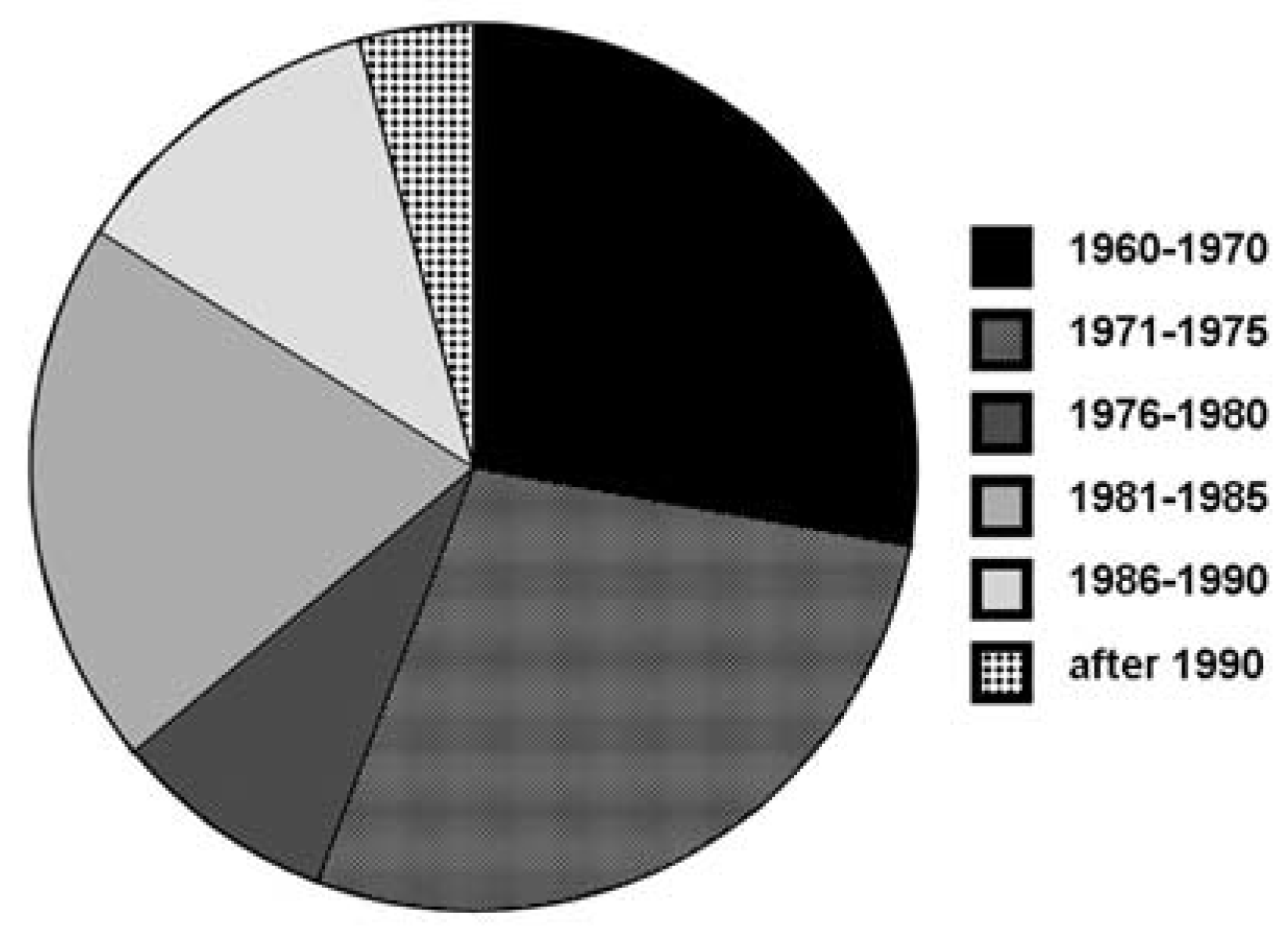
Figure 1. Distribution of year when radiotherapy was performed.
Cardiovascmed 10 00387 g001
Figure 2. Echocardiographic image in a 37 year old female (patient #10) 15 years after previous radiotherapy with extensive calcifications of mitral and aortic valve. There is the parasternal long axis view of the young patient #10 who died after mitral and aortic valve replacement. It shows the calcific mitral and aortic valve. Typically, these patients may have normally sized (shrunk?) atria despite atrioventricular regurgitation. LV = left ventricle; AO = aorta; LA = left atrium.
Cardiovascmed 10 00387 g002
Figure 3. This is the image of the 35 year old woman (patient #17) with postactinic changes on both sides of the heart with mitral annulus calcification, mitral and aortic valve sclerosis as well as pronounced tricuspid annulus calcification and shrunk, thickened tricuspid valve leaflets. RV = right ventricle; RA = right atrium; LA = left atrium; LV = left ventricle. Small arrow: tricuspid annulus calcification. Large arrow: tricuspid valve leaflets.
Cardiovascmed 10 00387 g003
Figure 4. Aortic and mitral valve disease in a 41 year old man (patient # 9) 18 years after RT for Hodgkin’s disease. A Extensive calcifications involving the aortic and mitral valve including mitral and aortic annulus. This is the apical long-axis view showing the severely calcified aortic valve (large arrow). Also the mitral valve is moderately calcified (small arrow). RV = right ventricle; LV = left ventricle; LA = left atrium; AO = aorta. B Pronounced calcifiactions of the aortic arch especially at the take-off of the brachiocephalic trunk. This is the aortic arch where calcific plaques are shown even at the origin of the cerebral arteries (broken arrow). BCT = brachiocephalic trunk; IV = innominate vein; AO = aorta.
Cardiovascmed 10 00387 g004