Next Article in Journal
Viewpoint: «Ist die Medizin ein Business?»
Previous Article in Journal
Long-Term Follow-Up After Thoracic Radiotherapy: Symptomatic Heart Disease Is an Ominous Sign
 
 
Cardiovascular Medicine is published by MDPI from Volume 28 Issue 1 (2025). Previous articles were published by another publisher in Open Access under a CC-BY (or CC-BY-NC-ND) licence, and they are hosted by MDPI on mdpi.com as a courtesy and upon agreement with Editores Medicorum Helveticorum (EMH).
Case Report

Variation on the Anchor Balloon Technique for Difficult Stent Delivery

by
Jean-François Surmely
1,2,* and
Stéphane Cook
1
1
Swiss Cardiovascular Center, Bern, Switzerland
2
Bern University Hospital, CH-3010 Bern, Switzerland
*
Author to whom correspondence should be addressed.
Cardiovasc. Med. 2007, 10(12), 397; https://doi.org/10.4414/cvm.2007.01284
Submission received: 28 September 2007 / Revised: 28 October 2007 / Accepted: 28 November 2007 / Published: 28 December 2007

Summary

Stent delivery failure happens in about five percent of coronary stent implantation. In this case report, we describe a new technique, which improves the support of guiding catheter and allowed the adequate delivery of a second stent after using an anchor balloon technique in a stent implanted distally. This technique is relatively easy to perform, and has a low risk of complications, as the anchor balloon is inflated inside a stent.

Introduction

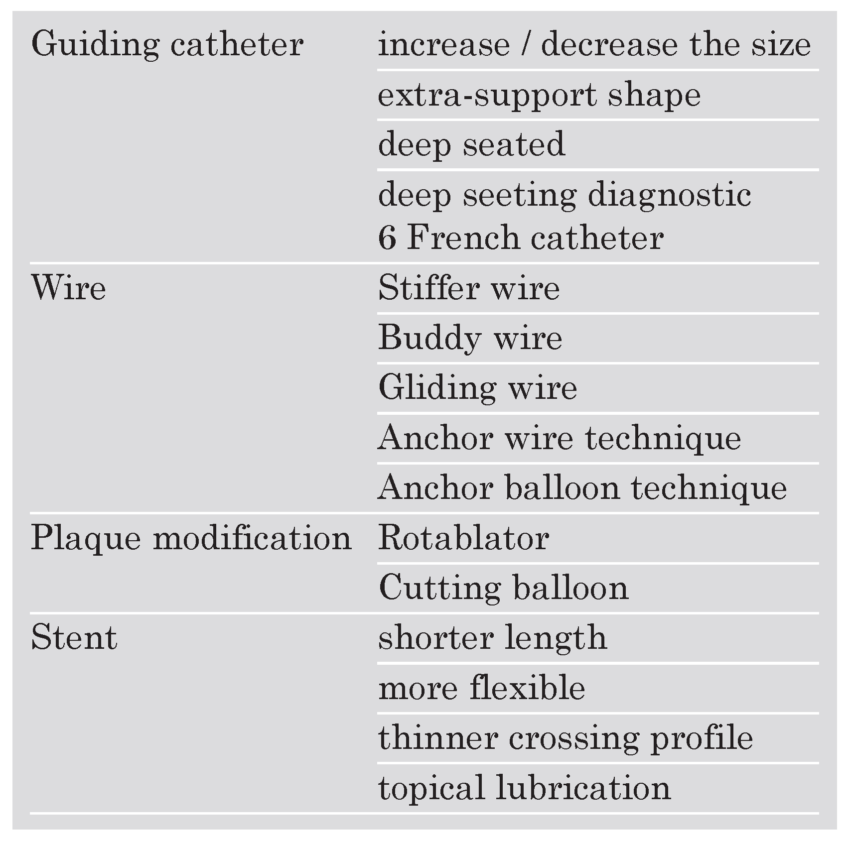
Stent delivery failure happens in five percent of coronary stent implantation, which can result in suboptimal revascularisation and has been associated with increased complications rates [1,2]. Successful stent delivery is influenced by lesion and/or stent characteristics. Several tips and tricks have been developed in order to improve the success rate of stent delivery in difficult coronary morphology or anatomy (Table 1). From these, the Anchor balloon technique was first introduced for the percutaneous treatment of chronic total occlusions [3,4]. It is achieved by inflating a balloon in a non-target vessel in order to obtain an adequate guiding catheter support. The following case report illustrates a variation of this technique which allowed successful stent delivery in a difficult percutaneous coronary intervention (PCI) of a right coronary artery (RCA) lesion, with a Shepherd’s Crook take-off.
Table 1. Tips and tricks to overcome difficult stent delivery.
Cardiovascmed 10 00397 i001

Case report

A 74-year-old woman was referred to us for primary PCI in a setting of acute inferior myocardial infarction. The ECG showed typical ST-elevation in leads II, III and aVF. Physical examination showed an unstable haemodynamic status with hypotension and bradycardia. A left ventricular angiography revealed a moderate reduced systolic function (EF 50%) due to inferoposterior hypokinesia. The coronary angiogram showed no significant disease of the left coronary tree, but an occlusion of the proximal part of the RCA with a Shepherd’s Crook shape take-off (Figure 1A). A Judkins right 4 (6 French) guiding catheter was chosen, and prompt successful recanalisation was achieved with a Whisper® guidewire (Guidant Corp, USA). After pre-dilatation with a Maverick® balloon (3.0 x 9 mm, 24 atm, Boston Sci., USA, Figure 1B,C), a first sirolimus-eluting stent (Cypher® 3.5 x 23 mm; Cordis, USA) was implanted at 24 atm in the mid-RCA (Figure 1D,E). Because of a suboptimal result and in order to cover the proximal part of the lesion (Figure 1F, arrow), we decided to implant a second stent (Cypher® 3.5 x 13 mm, Cordis, USA) proximally to the first stent. The delivery of this stent was impossible even after changing for 5 French guiding catheter and deep intubation, use of stiffer buddy-guidewire (Magnum®, Biotronik, Switzerland) and multiple balloon dilatation of the segment. This was probably due to the special angulation of the vessel, which forced the second stent meshes to hang against the first stent struts. We finally decided to anchor the guiding catheter by inflating a 3.0 x 9 mm balloon (Maverick®, Boston Sci., USA, 14 bars) inside the distally implanted stent (Figure 2A,B). This allowed placing the stent correctly over the second guidewire. Then the balloon was deflated and retrieved as well as the first Magnum® wire (Figure 2C). Finally, the stent (Cypher® 3.5 x 8 mm, 16 bars) could be inflated with a good final result (Figure 2D).

Discussion

With the advent of drug-eluting stents, an increasing number of difficult lesions are treated by PCI. Full lesion coverage is desirable and has been shown to improve long-term outcome. Despite improvements in stents profile, stent delivery continues to be problematic in complex coronary anatomy, and occurs in about 5% of cases. Unsuccessful stent placement is associated with a poorer short- (suboptimal angiographic result, acute or subacute vessel closure, need for urgent CABG) and long-term (death, myocardial infarction, repeat vascularisation) prognosis [1,2]. Characteristics associated with stent delivery failure include vessel tortuosity, lesion severity, lesion length, calcifications, stent length and structure, or poor guiding catheter support in dilated aortic root, or in unadjusted take-off of the proximal segment of the target coronary artery [5].
Over the years, an increasing number of tips and tricks have been developed to overcome this problem (Table 1). Increasing the guiding catheter back-up support can be achieved by either exchanging for an extra-support shape guiding catheter (eg AL, Voda left, XB left), or by exchanging for a larger one (7 or 8 French), or by deep-seating the guiding catheter, or by exchanging for a diagnostic 6 French catheter which due to its flexibility allows more profound cannulation [6,7]. Increased back up can also be obtained by using a stiffer wire, or by inserting a second wire (buddy wire technique) in order to straighten the vessel. A variant of the buddy wire technique is the gliding wire technique, where a second hydrophilic wire is inserted in order to allow the stent to glide over the hydrophilic coating of the second wire. A second wire can also be inserted in a non-target vessel (Anchor wire technique). Stabilisation of the guiding catheter can be obtained by inflating a balloon in a non-target vessel (Anchor balloon technique). Plaque modification, in particular for calcified lesions, can be obtained with Rotablator treatment before stent delivery. Shorter or more flexible stents with favourable crossing profiles can also be a solution, and should probably be selected as first choice in cases with difficult coronary anatomy or morphology. Recently, topical stent lubrification with Rotaglide has been shown to be feasible and effective [8].
In the present case, several of these techniques were tried unsuccessfully. In this RCA lesion without any significant non-target side-branch, it was not possible to use the Anchor wire/balloon technique. We performed therefore a variation on the Anchor balloon technique, which is depicted in Figure 2 and consists of inflating a balloon inside the distal vessel. The guiding catheter was consequently anchored which allowed the adequate delivery of a second stent. This technique is relatively easy to perform, and has a low risk of complications when the Anchor balloon is inflated inside a stent. This technique could be more easily performed with fixed wire balloons, such as ACE® (Boston Sci., USA) or FIX® (Schwager Medica, Winterthur, Switzerland) catheters. This Anchor balloon technique should be done into a drug eluting stent only shortly after its implantation to be sure to benefit from the properties of the drug to prevent restenosis. Indeed the drug coated around the stent is completely eluted during the first two weeks following the implantation of the stent and restenosis is probably no longer prevented if a balloon inflation is performed thereafter with possible subsequent arterial wall lesions.

Conflicts of Interest

There is no conflict of interest.

References

  1. Cantor, W.J.; Lazzam, C.; Cohen, E.A.; Bowman, K.A.; Dolman, S.; Mackie, K.; et al. Failed coronary stent deployment. Am Heart J. 1998, 136, 1088–95. [Google Scholar] [CrossRef] [PubMed]
  2. Lohavanichbutr, K.; Webb, J.G.; Carere, R.G.; Solankhi, N.; Jarochowski, M.; D’Yachkova, Y.; et al. Mechanisms, management, and outcome of failure of delivery of coronary stents. Am J Cardiol. 1999, 83, 779–81, A779. [Google Scholar] [CrossRef] [PubMed]
  3. Fujita, S.; Tamai, H.; Kyo, E.; Kosuga, K.; Hata, T.; Okada, M.; et al. New technique for superior guiding catheter support during advancement of a balloon in coronary angioplasty: the anchor technique. Catheter Cardiovasc Interv. 2003, 59, 482–8. [Google Scholar] [CrossRef] [PubMed]
  4. Hirokami, M.; Saito, S.; Muto, H. Anchoring technique to improve guiding catheter support in coronary angioplasty of chronic total occlusions. Catheter Cardiovasc Interv. 2006, 67, 366–71. [Google Scholar] [CrossRef] [PubMed]
  5. Tan, K.; Sulke, N.; Taub, N.; Sowton, E. Clinical and lesion morphologic determinants of coronary angioplasty success and complications: current experience. J Am Coll Cardiol. 1995, 25, 855–65. [Google Scholar] [CrossRef] [PubMed]
  6. Bartorelli, A.L.; Lavarra, F.; Trabattoni, D.; Fabbiocchi, F.; Loaldi, A.; Galli, S.; et al. Successful stent delivery with deep seating of 6 French guiding catheters in difficult coronary anatomy. Catheter Cardiovasc Interv. 1999, 48, 279–84. [Google Scholar] [CrossRef]
  7. Keller, P.F.; Gosselin, G.; Gregoire, J.; Guedes, A.; Verin, V. Feasibility of the PCI through 6F diagnostic catheters. Catheter Cardiovasc Interv. 2007, 69, 410–5. [Google Scholar] [CrossRef] [PubMed]
  8. Chan, A.W.; Ramee, S.R.; Collins, T.; Quintana, H.; White, C.J. Rotaglide-facilitated stent delivery: mission accomplished. Catheter Cardiovasc Interv. 2003, 59, 477–81. [Google Scholar] [CrossRef] [PubMed]
Figure 1. A Coronary angiogram demonstrating an occlusion of the proximal part of the RCA with a Shepherd’s Crook shape take-off. B,C Successful recanalisation. D,E Implantation of one sirolimus-eluting stent (Cypher® 3.5 x 23 mm; Cordis, USA). F Suboptimal result proximally.
Cardiovascmed 10 00397 g001
Figure 2. Anchor-technique. A,B A 3.0 × 9 mm balloon (Maverick®, Boston Sci., USA) was inflated inside the formerly implanted stent. This allowed placing the second stent correctly over the second guidewire. C The balloon was deflated and retrieved as well as the first Magnum® wire and the second stent (Cypher® 3.5 x 8 mm. Cordis, USA) could be implanted. D Final result.
Cardiovascmed 10 00397 g002