Systematic Review

Antioxidant Potential of Opuntia dillenii: A Systematic Review of Influencing Factors and Biological Efficacy

by
Ruymán Santana-Farré
1,2,†,
Nisa Buset-Ríos
1,† and
Mussa Makran
2,*
1
Department of Basic Health Sciences, Faculty of Health Sciences, Universidad Fernando Pessoa-Canarias, 35450 Santa María de Guía, Las Palmas, Spain
2
Department of Nutrition, Faculty of Health Sciences, Universidad Fernando Pessoa-Canarias, 35450 Santa María de Guía, Las Palmas, Spain
*
Author to whom correspondence should be addressed.
These authors contributed equally to this work.
Nutraceuticals 2025, 5(3), 22; https://doi.org/10.3390/nutraceuticals5030022
Submission received: 17 July 2025 / Revised: 4 August 2025 / Accepted: 8 August 2025 / Published: 13 August 2025

Abstract

Opuntia dillenii has gained considerable scientific attention as a potential natural source of antioxidants. This systematic review compiles and evaluates current evidence regarding its antioxidant activity. A PRISMA-guided literature search was conducted using PubMed, Scopus, and Web of Science, identifying 37 eligible studies. These studies employed two main methodological approaches: chemical-based assays and biological models. Chemical assays, including radical scavenging and reducing power assays, demonstrated a broad range of antioxidant activity influenced by factors such as the extraction method, plant part, plant maturity, and geographic origin. Polysaccharides, betalains, and polyphenols were consistently identified as primary contributors to these effects. Biological models further supported the antioxidant properties of O. dillenii extracts. In animal studies, administration of the extracts significantly improved oxidative stress biomarkers, increasing glutathione levels, reducing malondialdehyde concentrations, and enhancing the activity of antioxidant enzymes, particularly in the liver and other digestive tissues like the colon, stomach, and pancreas. Cellular studies using hepatocyte, macrophage, enterocyte, and neuronal cell lines produced comparable results, confirming the antioxidant effects. In conclusion, O. dillenii exhibits promising antioxidant potential across various experimental models. However, the absence of human clinical trials highlights the need for further research to establish its efficacy and safety as a nutraceutical product.

1. Introduction

The balanced generation of reactive oxygen species (ROS) is essential for maintaining numerous physiological processes, a phenomenon known as oxidative eustress. However, excessive ROS production can disrupt redox homeostasis, leading to oxidative stress that damages vital biomolecules, including DNA, proteins, and lipids [1]. This oxidative imbalance also promotes chronic low-grade inflammation, impairing mitochondrial function and triggering cellular apoptosis [2]. These mechanisms collectively contribute to tissue damage and organ dysfunction, representing a common pathological feature underlying numerous chronic diseases such as cardiovascular disorders, diabetes mellitus, neurodegenerative diseases, and cancer [3]. In response to this challenge, biomedical research has increasingly concentrated on identifying natural compounds with antioxidant properties, particularly those derived from dietary sources. Plant-based antioxidants play a critical role in neutralizing free radicals, mitigating oxidative damage, and potentially preventing the progression of diseases associated with oxidative stress [4]. Consequently, the study of dietary compounds with antioxidant activity is fundamental for developing complementary strategies for disease prevention and health promotion. In this sense, numerous naturally occurring bioactive compounds, particularly those from plant-based sources such as carotenoids and polyphenols, have demonstrated antioxidant properties through both direct and indirect mechanisms. They can directly scavenge reactive oxygen and nitrogen species, prevent cellular damage, and also modulate endogenous defenses [5]. Notably, many phytochemicals activate the Keap1/Nrf2/ARE pathway, promoting the expression of antioxidant and detoxifying enzymes like superoxide dismutase (SOD), catalase (CAT), and glutathione peroxidase (GPx). Through these complementary actions, natural compounds help to keep a redox balance and hold therapeutic potential against chronic diseases associated with oxidative stress.
Within this context, Opuntia dillenii has garnered particular interest among the over 200 species from the Opuntia genus, owing to its high nutritional value and its diverse array of secondary metabolites, including betanin, phenolic compounds, flavonoids, tocopherols, and sugars, that exhibit a wide range of biological activities [6]. This cactus, widely distributed across arid and semi-arid regions, has been traditionally used in various cultures both as food and in folk medicine, which has spurred a growing body of research aimed at characterizing its biological properties [7]. From an ethnopharmacological perspective, O. dillenii has been attributed to several health-promoting effects, such as hypoglycemic, hypolipidemic, and nephroprotective activities, although its antioxidant capacity remains the most extensively studied.
Published results to date reveal considerable variability in the antioxidant potency across different organs of O. dillenii, such as seeds, peel, pulp, flowers, and cladodes (as shown in Figure 1), as well as differences attributable to extraction methods, solvent polarity, and the geographical origin of the plant material. Furthermore, advanced extraction techniques, including ultrasound-assisted extraction and pressurized liquid extraction, have shown potential to significantly enhance the yield and antioxidant capacity, adding additional variables that influence comparability across studies.
It is important to acknowledge that the interpretation of in vitro antioxidant assays remains subject to debate. Concerns have been raised regarding the physiological relevance of chemical assays like DPPH (2,2-diphenyl-1-picrylhydrazyl). This is especially true when compared to assays that evaluate the capacity of extracts to neutralize biologically relevant ROS such as hydroxyl radical (OH), hydrogen peroxide (H2O2), superoxide anion radical (O2−•), and nitric oxide (NO). Additionally, extrapolating in vitro findings to human physiology requires caution, as the bioavailability and metabolism of phenolic compounds can substantially alter their in vivo efficacy. Furthermore, methodological heterogeneity among studies, including differences in experimental designs and result expression, complicates direct comparisons and the formulation of definitive conclusions regarding O. dillenii antioxidant potential.
To date, existing reviews addressing the antioxidant properties of O. dillenii have predominantly adopted a narrative approach, focusing on describing its potential antioxidant capacity based on in vitro chemical assays and the presence of phytochemical constituents [7,9]. However, these reviews lack a systematic methodology to critically assess the available evidence and identify the key factors influencing the antioxidant activity of this species. Most importantly, no comprehensive review has examined the biological models used to evaluate the antioxidant effects of O. dillenii, which are crucial for determining its real efficacy in biological systems beyond simple chemical-based tests. The characterization of these biological models, including cell culture systems and animal studies, is essential for translating antioxidant potential into meaningful therapeutic applications for chronic disease management. Specifically, by applying oxidative stress to these models and analyzing biomarkers (e.g., antioxidant enzyme activity), it is possible to assess whether the antioxidant activity of O. dillenii translates into real biological effects, and this will provide new insight into its potential for preventing diseases related to oxidative stress and its use as a nutraceutical for diet-based therapeutic interventions.
It is also important to emphasize that O. dillenii has received comparatively little attention in scientific literature, especially when contrasted with O. ficus-indica, the most widely consumed and researched species worldwide [10]. This significant gap in the literature highlights the urgent need for a comprehensive review that not only consolidates the existing research but also identifies critical shortcomings in the current understanding. Such a review would not only guide future research efforts but also foster the promotion of O. dillenii as a valuable resource for both food and pharmaceutical uses.
In light of these gaps, the present systematic review aims to provide a comprehensive and critical analysis and to synthesize the existing evidence on the antioxidant activity of O. dillenii, considering the evaluation methods employed and the factors contributing to result variability. Ultimately, it emphasizes the potential of this cactus as a natural source of bioactive compounds with nutraceutical applications and highlights the key avenues for future research necessary to solidify its role as an antioxidant agent.

2. Methods

2.1. Data Sources and Search Strategy

This systematic review was performed following the PRISMA 2020 guidelines [11,12]. The search was carried out on April 2025 using three different databases: PubMed, Web of Science (WOS), and Scopus. The search strategy was built for each database combining the different terms creating the query: (“Opuntia dillenii” or “O. dillenii” or “Opuntia stricta var. dillenii”) and “antioxidant”. No date, type of publication, or language limits were applied as a filter.

2.2. Inclusion and Exclusion Criteria

Eligibility criteria for this review included the following: (i) In vitro or in vivo original research studies, (ii) studies that referred to O. dillenii, and (iii) studies reporting data on antioxidant capacity. We excluded the following: (i) reviews and systematic reviews, (ii) studies based only on chemical characterization, (iii) studies that combine O. dillenii with other products, (iv) studies related to food technology, (v) studies on ethnobotanics, (vi) studies that referred to species different from O. dillenii, (vii) studies that do not report data on antioxidant capacity, and (viii) studies not related to food applications (Supplementary Table S1).

2.3. Data Extraction

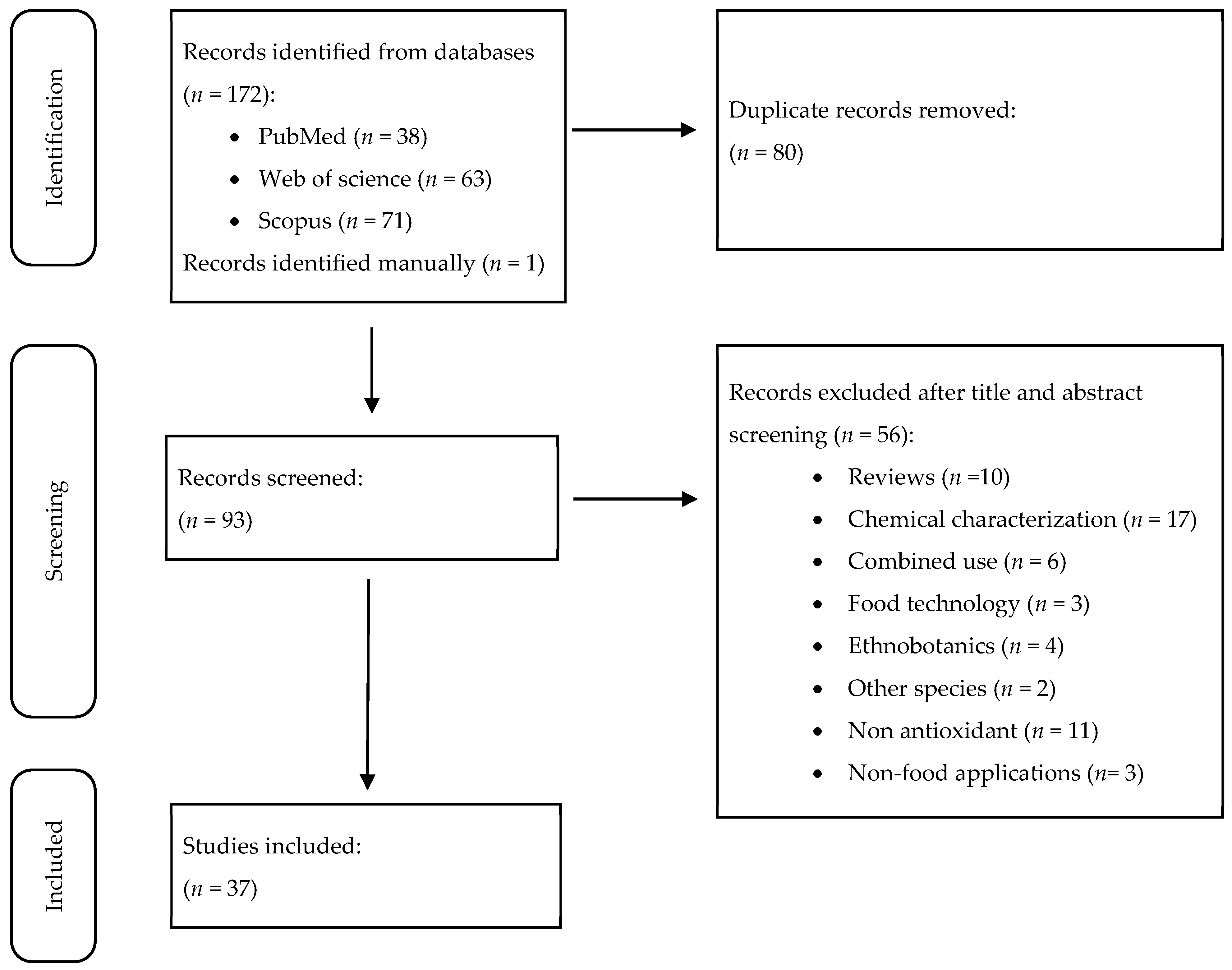
Titles and abstracts of the retrieved manuscripts were examined for their eligibility. Data extraction was carried out by three different researchers (R.S., N.B., and M.M.). After excluding 80 duplicated studies, 56 articles were excluded based on the exclusion criteria. Also, one additional article was added manually. This screening led to the identification of 37 studies suitable for review (Figure 2). Disagreements during each stage of the process were resolved through fair discussion.

2.4. Risk of Bias

For in vitro chemical-based antioxidant assays, no risk of bias was assessed due to the lack of a proper tool for this type of experiment. For cell-based and animal studies, the three reviewers (R.S., N.B., and M.M.) independently assessed the risk of bias by using the SYRCLE tool for in vivo research works [13] and also by adapting it to the in vitro assays. The items analyzed were as follows: (i) selection bias, (ii) performance bias, (iii) detection bias, (iv) attrition bias, (v) reporting bias, and (vi) other sources of bias. Once discussed together, the bias information for each study was summarized in the Supplementary Tables S2 and S3 where “Yes” indicates a low risk of bias, “No” indicates a high risk of bias, and “Unclear” indicates that the information reported was not enough.

2.5. Data Synthesis and Qualitative Analysis

Once the 37 studies were selected, the full texts were analyzed by the three researchers (R.S., N.B., and M.M.) and two different data extraction sheets were elaborated on. For the in vitro chemical-based antioxidant assays the sheet compiled the following aspects: (i) part of the plant employed, (ii) method for the extract preparation, (iii) assays performed, and (iv) results obtained. For the animal, cellular, and in silico approaches, the sheet compiled the following aspects: (i) biological model employed, (ii) treatments applied, (iii) assays performed, and (iv) results obtained. Once again, any disagreement that arose during this phase was resolved through fair discussion.

3. Results and Discussion

Amid the growing scientific interest in natural products with antioxidant properties, O. dillenii has emerged as a species of particular relevance, as evidenced by the 37 studies examined in this review. Its antioxidant activity has been explored using a wide array of experimental approaches, which can be broadly classified into two main categories. The first comprises in vitro chemical-based assays aimed at evaluating the radical-scavenging capacity or reducing power of plant extracts. The second includes investigations conducted in biological systems, such as cell cultures and animal models, designed to assess antioxidant effects under conditions that more closely mimic physiological environments. In this review, these two methodological approaches are analyzed separately to offer a clearer and more comprehensive understanding of the antioxidant potential of O. dillenii.

3.1. Antioxidant Activity Assessed Using in Vitro Chemical-Based Methods

3.1.1. Overview of in Vitro Assays

In vitro chemical-based assays are widely used as preliminary tools to assess the antioxidant potential of plant extracts. These methods evaluate the ability of antioxidants to neutralize free radicals or reactive species, reduce oxidants, or chelate prooxidant metal ions (see Table 1). The most common are radical-scavenging assays, which measure the capacity of antioxidants to neutralize free radicals by donating electrons or hydrogen atoms, thereby stabilizing reactive species [14]. Well-established examples include the DPPH and ABTS•+ (2,2′-azino-bis-3-ethylbenzothiazoline-6-sulfonic acid) assays, which quantify the decolorization of stable radicals. Additionally, more targeted assays assess the scavenging of biologically relevant ROS, such as OH, H2O2, peroxyl radicals (evaluated via the oxygen radical absorbance capacity, ORAC assay), O2•−, and NO [14]. On the other hand, reducing power assays evaluate the electron-donating ability of antioxidants, reflecting their capacity to reduce oxidized intermediates [15]. The ferric reducing antioxidant power (FRAP) assay, for example, measures the conversion of ferric ions (Fe3+) to ferrous ions (Fe2+). Complementarily, total antioxidant capacity (TAC) assays provide an overall estimation of plant antioxidant activity by integrating both radical-scavenging and reducing capacities. Additionally, metal-chelating assays assess the ability of antioxidants to bind prooxidant metal ions, such as Fe2+, thereby inhibiting radical-generating reactions like the Fenton reaction [16]. The ferrous ion-chelating (FIC) activity assay is the most commonly applied method in this category. Finally, lipid-oxidation-inhibition assays determine the ability of antioxidants to protect lipid substrates from oxidative degradation. In this sense, the β-carotene bleaching assay evaluates the prevention of β-carotene discoloration in the presence of linoleic acid-derived radicals, while lipid peroxidation assays, often based on malondialdehyde (MDA) quantification or by monitoring the formation of conjugated dienes, measure the inhibition of lipid peroxidation in model systems [17].
Once the main in vitro chemical-based methods have been described, the following section presents and analyzes the 24 studies identified in this category (Table 2), organized according to their specific research approaches related to the antioxidant activity of O. dillenii.

3.1.2. Antioxidant Potential

This section summarizes studies that have evaluated the antioxidant capacity of O. dillenii extracts, with a focus on comparisons to standard antioxidants and other Opuntia species. In this context, several studies have reported that the essential oil of O. dillenii generally exhibits modest antioxidant activity in the DPPH assay when compared to ascorbic acid, a widely recognized reference compound. For example, higher half-maximal inhibitory concentration (IC50) values have been observed for the essential oil than for ascorbic acid, indicating lower antioxidant efficacy. This trend has been reported for oils extracted using supercritical CO2 fluid (11.4% (v/v) vs. 0.24 mg/mL) [20], as well as through petroleum ether extraction (0.38 vs. 0.23 µg/mL) [29]. Furthermore, the essential oil demonstrated a lower percentage inhibition in the concentration range of 10–100 µg/mL (36.5–78.1 vs. 46.5–81.4%), except at higher concentrations (500 and 1000 µg/mL), where the inhibition levels were comparable [27]. The results from the DPPH assay are further corroborated by the β-carotene bleaching method, in which the essential oil exhibited an IC50 of 20.7% (v/v), whereas the positive control, butylated hydroxytoluene (BHT), demonstrated a significantly stronger effect with an IC50 of 0.44 mg/mL [20]. Taken together, these findings suggest that while O. dillenii essential oil does exhibit measurable antioxidant activity, its potency is generally modest and highly variable when compared to well-known standard antioxidants.
Although less studied, the flower extract showed lower DPPH-scavenging activity than ascorbic acid (IC50: 58.7 vs. 1.2 µg/mL) [23]. However, it was more effective in OH- and H2O2-scavenging assays (IC50: 159.7 and 131.1 µg/mL, respectively), whereas ascorbic acid showed no measurable activity under the same conditions. These findings are particularly relevant given that the scavenging of ROS, such as OH and H2O2, is considered more physiologically meaningful than DPPH-based assays, as ROS play a direct role in oxidative-stress-related cellular damage. Similarly, another study that evaluate cladode extracts reported lower DPPH (IC50: 43.0 vs. 9.7 µg/mL) and FIC activities (10.8 vs. 16.1 mg EDTA/g) compared to BHT, but comparable ABTS•+ (7290.0 vs. 10,165.4 µmol Trolox equivalents (TE)/g) and FRAP values (998.9 vs. 1342.2 mmol Fe2+/g) [21]. Notably, the same extract exhibited higher β-carotene-bleaching inhibition than BHT (19.6 vs. 8.8% inhibition). This suggests potential efficacy in lipid-peroxidation protection despite moderate radical-scavenging capacity in certain assays.
The peel extract of O. dillenii has shown notable antioxidant activity compared to other Opuntia species [39]. In the DPPH assay, its radical-scavenging capacity was comparable to that of O. ficus-indica and O. robusta (1.19–1.20 mg TE/g dry weight (DW)), as was its TAC (18.90–19.16 mg TE/g DW). However, O. dillenii outperformed O. ficus-indica in the ABTS•+ assay (0.38 vs. 0.30 mg TE/g DW) and exhibited higher FRAP than O. robusta (0.83 vs. 0.49 mg TE/g DW).

3.1.3. Antioxidant Activity According to Plant Part

Notably, considerable research has focused on determining which part of O. dillenii exhibits the highest antioxidant capacity, with particular attention given to the peel and pulp. The peel consistently showed greater DPPH-radical-scavenging activity than the pulp, both in terms of the percentage inhibition (20.7–71.8 vs. 24.9–75.0%) [30] and antioxidant activity expressed as TE (53.1 vs. 26.7 µmol TE/g DW) [34]. It also exhibited a non-significant trend toward higher activity in the ABTS•+ radical cation decolorization assay (1.24 vs. 1.01 µM TE), significantly higher antioxidant capacity in the oxygen radical absorbance capacity assay (0.14 vs. 0.10 µM TE) [19], and markedly higher FRAP (17.4 vs. 2.3 µmol TE/g DW) [34]. Additionally, the peel showed greater inhibition of lipid peroxidation (76.4 vs. 73.2%) [19] and stronger antioxidant activity in the β-carotene bleaching assay (53.8 vs. 51.9%) [34]. These differences have been attributed to the peel’s higher content of total polyphenols (133.4 vs. 91.5 mg gallic acid equivalents/100 g) and flavonoids (32.5 vs. 29.2 mg quercetin equivalents/100 g) [19], as well as to its elevated levels of specific phenolic compounds, including hydroxybenzoic acids (2.2–7.0 vs. 0.8–2.2 mg/g extract), hydroxycinnamic acids (0.2–3.6 vs. 0.2 mg/g extract), and flavonoids (0.4–44 vs. 0.2–13.4 mg/g extract) [34]. However, one study contradicted these findings by reporting no significant differences between the peel and pulp in the ABTS assay (102.6 vs. 99.5 µmol TE/g DW) and even observing a higher ORAC value for the pulp (160.0 vs. 115.4 µmol TE/g DW) [33]. These results suggest that other factors, such as plant maturation or extraction methods, may also influence the observed antioxidant activity, beyond the anatomical part of the plant.
The seed is another plant part that has been extensively evaluated. One study reported that the seed exhibited the highest antioxidant activity compared to the peel and pulp fruit, based on ABTS•+ (2.15 vs. 1.01–1.24 µM TE), ORAC (0.18 vs. 0.10–0.14 µM TE) and lipid peroxidation inhibition (92.3 vs. 73.2–76.4%) assays [19]. These results were associated with its elevated total polyphenol (212.8 vs. 91.5–133.4 mg gallic acid equivalents/100 g) and flavonoid (144.1 vs. 29.2–32.5 mg quercetin equivalents/100 g) contents. However, these findings were not corroborated by another study, which reported greater DPPH-radical-scavenging activity in the pulp juice compared to the seed oil (IC50, 8.2 vs. 27.2 µg/mL) [24]. In contrast to the relatively consistent findings favoring the peel over the pulp in terms of antioxidant activity, comparisons between the seed and fruit pulp or peel have shown greater variability across studies. These discrepancies are likely due to differences in extraction methods, particularly considering that the latter study evaluated pulp juice [24], while the former used methanolic extracts [19]. Additionally, it is important to note that in one study, the seed was extracted using a methanolic solvent [19], whereas in another, oil was extracted from the seed [34], potentially influencing the outcomes. On the other hand, only one study has evaluated the antioxidant activity of seed oil in comparison to flower extracts, reporting that the differences observed were influenced by both the solvent polarity and the type of antioxidant assay employed [38]. In the DPPH assay, seed oil exhibited higher antioxidant activity than flower extracts obtained with more polar solvents such as butanol, ethanol, and ethyl acetate. Similarly, in the FRAP assay, seed oil outperformed all flower extracts (dichloromethane, butanol, ethanol, and ethyl acetate), with the sole exception of the hexane extract. The observed variations in antioxidant activity depending on the solvent type may be related to the solubility of the bioactive compounds within the extracts. Polar solvents, such as ethanol and butanol, tend to dissolve a broader range of hydrophilic compounds, which could lead to higher antioxidant activity in seed oil. In contrast, non-polar solvents like hexane may extract a different set of compounds, potentially explaining the exceptions observed in the FRAP assay.
Another study reported divergent antioxidant profiles among different plant parts. In this regard, cladodes exhibited the stronger inhibition of lipid peroxidation compared to the fruit (IC50: 87 vs. 353 µg/mL) and greater ABTS•+-radical-scavenging activity (IC50: 27 vs. 46 µg/mL) [18]. Conversely, the fruit showed superior DPPH-radical-scavenging capacity (IC50: 317 vs. >1000 µg/mL) and higher TAC (1.4 vs. 0.4 µM ascorbic acid equivalents (AAE)/g). These contrasting results suggest that antioxidant activity can differ substantially between plant tissues, depending on the mechanism of action targeted by each assay (e.g., hydrogen atom transfer vs. electron transfer). Therefore, the use of multiple complementary antioxidant assays is crucial for accurately characterizing and comparing the antioxidant potential of various plant parts.

3.1.4. Extraction Methods and Antioxidant Yield

Several studies have focused on optimizing extraction methods to maximize the antioxidant potential of plant-derived extracts. These efforts have included the evaluation of various solvents and the implementation of innovative techniques such as ultrasound-assisted extraction or pressurized liquid extraction.
In general, polar solvents like ethanol have shown greater DPPH-radical-scavenging activity compared to less polar solvents such as hexane, particularly in peel (34.8–75.0 vs. 24.9–62.7%) and pulp (32.9–71.8 vs. 20.7–56.0%) extracts [30]. However, these findings have not been consistently replicated in other studies. For instance, peels from the Nador region exhibited higher antioxidant activity, measured via DPPH and ABTS•+ assays, when extracted with low-polarity solvents such as diethyl ether (IC50: 190 and 550 µg/mL), outperforming polar alternatives like ethanol (IC50: 530 and 736 µg/mL), ethyl acetate (IC50: 720 and 536 µg/mL), and water (IC50: >2000 and 970 µg/mL) [28]. In contrast, peels from the Essaouira region showed optimal antioxidant activity (DPPH and ABTS•+) with solvents of intermediate polarity such as ethyl acetate (IC50: 340 and 307 µg/mL) and ethanol (IC50: 500 and 460 µg/mL), while exhibiting reduced activity with both water (IC50: 880 and 820 µg/mL) and diethyl ether (IC50: >2000 and 520 µg/mL) [28]. These findings indicate that the antioxidant extraction profile is strongly influenced by the geographical origin of the fruit, which in turn determines the predominant polarity of its bioactive compounds. As a result, extracts from different regions may respond better to polar [30], intermediate, or non-polar solvents [28].
Data on juice is comparatively limited, and observed extraction patterns vary with the fruit variety [28]. For example, in the Nador variety, aqueous extraction yielded the highest antioxidant activity (vs. organic solvents) in terms of DPPH (IC50, 690 vs. 1340 to >2000 µg/mL), ABTS•+ (IC50: 750 vs. 1660 to >2000 µg/mL), and TAC (84 vs. 53–73 mg AAE/g DW). In contrast, the Essaouira variety showed more variable responses, lacking a consistent extraction pattern [28]. In this same study, seed extracts obtained from solvents with intermediate polarity (i.e., ethanol) have demonstrated greater DPPH (IC50: 45–53 µg/mL) and ABTS•+ (IC50: 88–130 µg/mL) scavenging activity, as well as TAC (281–410 mg ascorbic acid/g DW), outperforming both low-polarity solvents (IC50: DPPH, 118–380 µg/mL; ABTS•+, 857 to >2000 µg/mL; TAC, 71–110 AAE/g DW) and water (IC50: DPPH > 2000 µg/mL; ABTS•+ 380–700 µg/mL; 53–230 AAE/g DW) [28].
Other parts of the plant, such as flowers or cladodes, have been less investigated regarding the effect of different solvents on the extraction yield and antioxidant activity. For example, the antioxidant activity of flower extracts measured via DPPH and FRAP assays declined progressively with increasing solvent polarity, although exact quantitative data could not be retrieved from the referenced study [18]. Regarding cladodes, it has been observed that ethanol generally provides better results than water, yielding extracts with higher NO-scavenging activity (IC50: 0.06 vs. 0.15 µg/mL), FRAP (Abs 700 nm: 1.97 vs. 1.39), TEAC (0.59 vs. 0.46 mM TE/g), and TAC values (62.99 vs. 60.44 mm AAE/g), with the exception of DPPH activity, which was lower in ethanolic extracts (IC50: 0.60 vs. 0.54 µg/mL) [31]. These differences can be attributed to the solvent’s ability to extract a broader range of bioactive compounds. Ethanol, being both polar and slightly non-polar, extracts both hydrophilic and lipophilic antioxidants, leading to higher overall activity. Water, being more polar, primarily extracts hydrophilic compounds, which may explain the lower activity in some assays. Regarding DPPH, the higher activity observed in water extracts could be due to specific compounds that interact more effectively with DPPH, suggesting a different mechanism of action in this assay.
Beyond solvent selection, recent studies have highlighted the potential of alternative extraction techniques to further enhance the recovery of antioxidant compounds. Notably, ultrasound-assisted extraction [32] and pressurized liquid extraction [35] have demonstrated significantly greater efficiency compared to conventional methods. These techniques markedly increase antioxidant capacity as measured based on the ORAC assay (330.7–618.9 vs. 151.9 and 321.4–470.7 vs. 151.8 µmol TE/g, respectively). These results reinforce the notion that advanced extraction technologies can substantially improve both the yield and bioactivity of antioxidant compounds, whether applied independently or in combination with optimized solvent systems.

3.1.5. Identification of Antioxidant Bioactive Compounds

The identification and isolation of bioactive compounds from plants are essential for pinpointing the specific components responsible for their antioxidant activity. This process involves extracting various plant parts to isolate these compounds, enabling a direct link between the observed antioxidant effects and the bioactive components. Numerous studies, compiled in this review, have focused on identifying the key compounds responsible for the antioxidant activity of O. dillenii. In one such study, a hydromethanolic extraction of the fruit yielded an ABTS•+ value of 35.2 µmol TE/g fresh fruit [26]. Following the removal of hydrocolloids and proteins, this value markedly decreased to 15.7 µmol TE/g fresh fruit, a reduction attributed to the loss of non-pigmented compounds such as organic acids, known contributors to antioxidant activity. Subsequent fractionation of the extract produced two pigment-rich fractions: a yellow fraction, containing primarily betaxanthins, and a red fraction, composed of betacyanins and polyphenols. The antioxidant capacity of these fractions, as measured based on ABTS•+, was 1.7 µmol TE/g for the yellow extract and 5.4 µmol TE/g for the red extract. These data indicate that betacyanins and polyphenols contribute more substantially to the overall antioxidant potential of the fruit [26]. Corroborating these findings, another study reported that a betacyanin-rich extract from the fruit pulp exhibited markedly higher antioxidant activity than the synthetic antioxidant BHT, used as a positive control [41]. This was evidenced by a lower DPPH IC50 value (2.4 vs. 7.3 mg/mL) and a higher total antioxidant capacity (273.3 vs. 168.3 mg AAE/g DW). This highlights the significant contribution of betacyanins to the antioxidant potential of O. dillenii, particularly given their high concentration in the fruit pulp (59.9 mg/100 g fresh fruit).
In addition to pigments, polysaccharides extracted from O. dillenii have also been identified as important contributors to its antioxidant activity. One study demonstrated a positive correlation between the purity of cladode-derived polysaccharides (27.4–90.1%) and their antioxidant capacity, as determined based on DPPH-radical-scavenging activity (IC50: 3105–408 µg/mL) [22]. Moreover, an inverse relationship between the molecular weight and antioxidant activity has been reported [25]. Specifically, the lower-molecular-weight fraction (ODP-Ia, 339 kDa) exhibited markedly higher radical-scavenging activity than the higher-molecular-weight fraction (ODP-IIa, 943 kDa). This is demonstrated in various assays, including DPPH (58.4 vs. 45.6%), OH (45.7 vs. 41.4%), and O2−• (43.7 vs. 37.2%). Collectively, these findings highlight the significant role of polysaccharides in the antioxidant potential of O. dillenii and underscore the importance of molecular weight as a key factor modulating their antioxidant activity.

3.1.6. Effect of Maturity Stage on Antioxidant Activity

The effect of plant maturity on antioxidant activity has been examined in several studies, particularly in relation to the cladode age. One study compared polysaccharide extracts from tender (2–4 months old) and mature (5–10 months old) cladodes, reporting similar DPPH-radical-scavenging activity between both groups (60.5 vs. 58.1%) [25]. However, tender cladodes exhibited higher O2−•-scavenging activity (37.4 vs. 29.1%) but lower OH-scavenging activity (39.6 vs. 46.2%) than older ones. These results point to complex age-related changes in antioxidant activity, although the underlying mechanisms remain largely uncharacterized. Further evidence of age-related variability was provided by another study comparing young (<1-year-old) and mature (>2-year-old) cladodes [40]. In this case, although TAC remained relatively constant between age groups (15.56 vs. 15.12 mg TE/g DW), the antioxidant profile changed noticeably. Specifically, DPPH-scavenging capacity decreased with age (0.58 vs. 0.82 mg TE/g DW), while antioxidant activity increased in both FRAP (0.92 vs. 0.42 mg TE/g DW) and ABTS•+ assays (0.43 vs. 0.30 mg TE/g DW). These results indicate that, despite the stable overall antioxidant potential, the contribution of individual antioxidant mechanisms shifts with age. In this study, phytochemical analyses revealed that, although the total polyphenol content remained relatively stable between young and mature cladodes (4629.3 vs. 5061.7 mg gallic acid equivalents/100 g), there were significant age-related increases in total flavonoid (1903 vs. 4253 µg/g) and total phenolic acids (2100 vs. 3050 µg/g) [40]. These compositional changes indicate a qualitative shift in the phenolic profile with plant age, likely altering the antioxidant response in specific assays (e.g., FRAP and ABTS•+) without markedly changing TAC.

3.1.7. Geographical Influence on Antioxidant Activity

Varietal and geographical differences have been explored in relation to the antioxidant capacity of O. dillenii. In one study, the essential oils of three Moroccan varieties (Imtchan, Aknari, and Harmoucha) were evaluated using DPPH, FRAP, and FIC assays. The Imtchan variety exhibited the highest antioxidant activity (IC50 15.2, 23.3, and 35.3 µg/mL, respectively), followed by Aknari (38.4, 30.2, and 42.8 µg/mL, respectively), and Harmoucha (42.2, 55.9, and 39.5 µg/mL, respectively) [37]. These inter-varietal differences are likely attributable to the compositional variation in bioactive compounds, particularly polyphenols. For instance, 4-hydroxybenzoic acid, a compound with recognized antioxidant properties, was found in the highest concentrations in the Imtchan variety, followed by Aknari and Harmoucha (10.5, 5.1, and 1.5 mg/100 g, respectively. This suggests a possible link between this compound and antioxidant activity. Further evidence of the geographical influence was provided by a study comparing two varieties from distinct Moroccan regions: Nador and Essaouira [28]. Among the different matrices analyzed, juice extracts showed the most marked geographical differentiation. TAC was substantially higher in samples from Essaouira compared to those from Nador (220–830 vs. 53–82 mg AAE/g DW). Similar trends were observed in DPPH (IC50: 280–850 vs. 1340 to >2000 µg/mL) and ABTS•+ assays (IC50: 95–213 vs. 1660 to >2000 µg/mL). The marked geographical differences in antioxidant activity suggest that environmental factors, such as climate, soil composition, and cultivation practices, influence the bioactive compound content in the juice extracts. These factors may lead to variations in antioxidant capacity, with certain regions promoting higher levels of compounds beneficial for health. Such findings underscore the importance of considering the geographic origin when assessing the antioxidant potential of plant-based extracts.

3.1.8. Impact of Colonic Fermentation on Antioxidant Activity

Although less frequently explored in the literature, the impact of colonic fermentation on the antioxidant activity of O. dillenii offers critical insights into the metabolic fate and functional potential of its bioactive compounds post-ingestion. One study conducted the in vitro colonic fermentation of O. dillenii, monitoring the changes in antioxidant capacity over time under conditions simulating the human gut microbiota [36]. Results revealed a gradual decline in DPPH-radical-scavenging activity after 24 and 48 of the fermentation process (from 0.30 to 0.26 and 0.20 µmol TE/g). In contrast, antioxidant capacity measured based on ABTS•+ showed a progressive increase over the same time intervals (0.22 to 0.27 and 0.28 µmol TE/g), as did FRAP values (2.84 to 3.49 and 4.13 µmol FeSO4/g). These divergent trends suggest that microbial metabolism may degrade certain native antioxidants while simultaneously generating new metabolites or liberating previously bound compounds with distinct redox characteristics. These findings highlight the importance of considering not only the initial (native) antioxidant profile of plant-derived foods, but also the biochemical modifications that these compounds undergo during gastrointestinal passage.

3.2. Antioxidant Activity in Biological Systems: Cell-Based and Animal Models

After summarizing the chemical-based methods used to assess the antioxidant properties of O. dillenii, the review now turns to studies employing biological systems. These include cellular, animal, and in silico models, each offering complementary insights into antioxidant mechanisms in more physiologically relevant contexts. A total of 14 studies employing such biological models have been identified and are summarized in Table 3. In cellular and animal models, oxidative stress is typically induced by chemical agents such as H2O2, lead acetate, or AAPH, which promote lipid peroxidation and deplete endogenous antioxidant defenses. This allows the protective effects of O. dillenii extracts to be evaluated under oxidative conditions. The parameters measured across these studies encompass both direct markers of oxidative stress and components of the antioxidant defense system. Primary ROS and NO are commonly quantified, along with enzymes involved in their generation, including myeloperoxidase (MPO) and inducible nitric oxide synthase (iNOS) [42]. Notably, ROS levels can be determined via intracellular fluorescent staining or dynamically monitored using the cellular antioxidant activity (CAA) assay, which allows real-time, physiologically relevant assessments of oxidative stress in live cells [43]. In addition to these direct markers, intermediate by-products of oxidative damage, such as MDA, a hallmark of lipid peroxidation, and reduced glutathione (GSH), a key non-enzymatic antioxidant, are frequently measured [44]. Furthermore, the activity of endogenous antioxidant enzymes, including SOD, catalase, GPx, and NAD(P)H:quinone oxidoreductase 1 (NQO1), is often assessed to determine the extent to which O. dillenii extracts modulate the cellular antioxidant defense response. Beyond cellular and animal approaches, in silico methods have recently emerged as valuable tools for evaluating antioxidant activity. These computational models allow for the simulation of molecular interactions, such as the binding affinity between bioactive compounds from O. dillenii and key antioxidant-related proteins or enzymes [45]. By predicting how well a molecule can interact with targets involved in the oxidative stress pathway (i.e., those that regulate ROS production or antioxidant defense), these studies offer mechanistic insights into the antioxidant activity of dietary bioactive compounds.
Once these methodological approaches have been introduced, the following sections will describe the findings of the reviewed studies in detail, organized into three categories: cellular, animal, and in silico models.

3.2.1. Antioxidant Activity in Animal Models

Among the various biological systems used to assess the antioxidant potential of O. dillenii, numerous in vivo pre-clinical studies have utilized murine models to investigate the protective capacity of this plant against oxidative stress in different tissues. Most of these studies used Wistar rats [49,50,51,52,53,54], while only two employed Sprague–Dawley rats [46,48], and another used Chinese Kunming mice [47]. To induce oxidative stress, researchers have employed a variety of toxic agents, including streptozotocin, CCl4, acetic acid, paracetamol, lead acetate, ethanol, and high-fructose/high-fat diets (see Table 2). In addition, oxidative stress biomarkers were primarily analyzed in the liver, which is the most extensively studied tissue in these models. Other matrices assessed include serum, kidney, pancreas, colon, stomach, or brain. While chemical-based antioxidant assays have examined a wide array of plant parts, in vivo studies have primarily focused on the fruit of O. dillenii. These studies have used fruit juice, pulp or peel extracts, and essential oils from the seeds.
According to the SYRCLE tool, the nine animal studies presented similar patterns of risk of bias. The initial selection of animals and the composition of the groups were adequate in all studies. However, insufficient information was found on sequence concealment, so selection bias could not be ruled out. The housing and handling of the animals was controlled and consistent across all studies. However, in three of the studies, the caretakers or researchers were not blinded to the treatment each animal received during the experiments. Regarding the assessment of results by the evaluators, most studies did not provide sufficient information, and only two studies specified the lack of blinding of those who evaluated the results. Regarding attrition bias, only one article stated that there was no bias in losses, but the rest did not provide information on this issue. Finally, none of the articles showed evidence of publication bias.
In the context of experimental results, two studies reported that a 7-day treatment with fruit juice (2.5–5 mL/kg/day) led to significant reductions in oxidative stress markers in both the colon [50] and liver [51]. Specifically, a reduction in MDA levels accompanied by an increase in GSH content was reported in both tissues. Interestingly, the increase in hepatic GSH was observed only at the higher dose, whereas in the colon, GSH levels increased at both doses. This may be explained by the fact that the colon, being in direct contact with ingested substances, receives greater exposure to bioactive compounds, while the liver exposure depends on intestinal absorption, potentially limiting its antioxidant benefit. On the other hand, CAT activity was upregulated in the liver [51], while the colon showed a marked reduction in the pro-oxidant enzyme MPO, further supporting the antioxidant potential of O. dillenii juice [50].
Besides juices, other extracts derived from both pulp and peel have also been investigated. Pulp extracts administered at 100 and 200 mg/kg for 10 days showed hepatic improvements in oxidative stress parameters such as MDA levels and CAT activity [52]. In contrast, peel extracts were tested in another study at lower doses (25 and 100 mg/kg) over a much longer period (8 weeks), yielding improvements in SOD activity, MDA levels, and the GSH/GSSG ratio at higher doses and enhanced GPx activity at both doses [54]. However, no significant changes were observed in CAT activity, total antioxidant capacity (ORAC), or carbonyl protein content. Direct comparisons between the study evaluating the pulp and peel extract are complicated by differences in the study design, including the dosing regimens and duration. Nonetheless, one comparative study evaluated the antioxidant activity of both extracts in a murine model of ethanol-induced gastric ulcer [53]. The results clearly showed that peel extracts had significantly greater antioxidant effects than pulp, with increases in SOD (102 vs. 55%), CAT (100 vs. 75%), and GPx activities (104 vs. 58%), as well as a greater reduction in MDA levels (23 vs. 17%). These findings align well with data from the chemical-based assays presented earlier, which also demonstrated superior antioxidant potential in the peel compared to the pulp.
Another of the most prominent objectives pursued across the compiled studies is the identification of the specific compounds responsible for the antioxidant properties of O. dillenii. Three particular studies aimed to investigate whether the antioxidant activity of O. dillenii fruit was attributable to its polysaccharide content [46,47,48]. Accordingly, polysaccharides extracted from the fruit pulp were administered to rats and mice subjected to models of cerebral ischemia (200 mg/kg) [46] or streptozotocin-induced diabetes (50–200 mg/kg) [47,48]. The results demonstrated antioxidant effects, evidenced by a reduction in MDA levels (25–44%) and iNOS protein expression, along with increased activities of SOD (17–99%), CAT (62–283%), and GPx (20–82%) in the serum, liver, kidney, and pancreas [46,47,48]. These findings suggest that the polysaccharide fraction of the pulp plays a key role in mediating the antioxidant properties of O. dillenii, as previously described in the chemical-based assays in the preceding section.
As for seed essential oil, only one study to date has evaluated its antioxidant effect in vivo. This study reported a reduction in hepatic MDA levels following a 7-day intervention at 2 mL/kg [49]. However, the range of oxidative stress biomarkers assessed was limited, and further research is needed to draw robust conclusions about the antioxidant efficacy of O. dillenii seed oil.

3.2.2. Antioxidant Activity in Cell Culture Studies

Compared to animal studies, research on the antioxidant properties of O. dillenii in cell culture systems is more limited. To assess the quality and risk of bias of the six selected articles, the SYRCLE tool was adapted and found to be informative for cell models. None of the selected articles described a randomization method for assigning treatments, as homogeneity is assumed in cell lines and, therefore, in the established groups. The items of allocation concealment, random accommodation, and random evaluation of results are not applicable to cell experiments. All selected studies agreed on the absence of information on attrition bias and the lack of selection in the presentation of results.
Most in vitro studies have focused on hepatic cell lines, specifically HepG2, Huh-7, and Hepa 1c1c7, as well as on RAW 264.7 macrophages, each used in two studies (see Table 3). In contrast, only one study has evaluated intestinal Caco-2 cells, and another has employed PC12 neuronal cells. Importantly, all cell-based assays used extracts or fractions derived exclusively from the fruit.
In this sense, in one study using HepG2 cells exposed to lead acetate to induce oxidative stress, treatment with pulp extract led to increased intracellular levels of GSH and decreased MDA levels, demonstrating protective antioxidant effects [52]. In addition to reducing oxidative stress biomarkers in liver cell lines, O. dillenii fruit extracts have also shown the capacity to modulate NO production in immune cells [57]. Specifically, in a study using LPS-stimulated murine macrophages (RAW 264.7), treatment with whole fruit extract for 4 h resulted in a significant 34.1% reduction in NO secretion [57]. However, this effect was not confirmed in another study using the same RAW 264.7 cell line, where fruit extract treatment for only 15 min did not modify iNOS protein expression [55]. This discrepancy may be related to differences in the treatment duration, as the shorter incubation time may have been insufficient to affect iNOS levels. Alternatively, the reduction in NO production could result from direct inhibition of the iNOS enzyme activity by the extract, rather than downregulation of its expression. These possibilities highlight the complexity of the mechanisms of action of bioactive compounds and underscore the importance of experimental conditions when evaluating their antioxidant effects.
Similarly to studies conducted in murine models, several investigations using cellular antioxidant assays also focus on identifying the specific compounds responsible for the observed effects. In this context, one study extracted polysaccharides from the pulp, fractionated them (451, 555, and 804 kDa), and applied the resulting fractions to Huh-7 liver cells subjected to oxidative stress induced by H2O2 [56]. All fractions reduced MDA levels, but lower-molecular-weight fractions (451 and 555 kDa) tended to be more effective in enhancing SOD and CAT activity and GSH levels. This relationship between molecular weight and antioxidant activity had already been observed in previous chemical-based assays [25], further supporting the relevance of polysaccharide size in determining bioactivity. However, the lowest weight fraction did not significantly affect GPx activity, suggesting that the relationship between molecular weight and antioxidant activity may not be strictly linear [56]. Beyond hepatic models, the antioxidant properties of O. dillenii polysaccharides have also been demonstrated in neuronal cells. In a study using H2O2-induced oxidative stress in a rat pheochromocytoma cell line (PC12), treatment with polysaccharide extract significantly reduced intracellular ROS levels [46]. Following the growing interest in identifying the specific compounds responsible for the antioxidant activity of O. dillenii, another study fractionated the fruit extract into 12 distinct fractions to evaluate their individual contributions [58]. Using HepG2 cells under oxidative stress induced by AAPH, several fractions reduced intracellular levels of ROS and lipid peroxidation, with the most active ones being richer in phenolic acids (e.g., piscidic, quinic, and eucomic acids). Altogether, these results highlight that both phenolic compounds and polysaccharides contribute to the antioxidant activity of O. dillenii.
Another study directly compared the CAA of a whole fruit extract between hepatic (HepG2) and intestinal cells (Caco-2) [57]. The CAA was markedly higher in liver cells (26.6–40.9%) compared to intestinal cells (9.1–29.5%), suggesting greater bioactivity in hepatic environments. However, a key limitation of this study is that the extract was applied directly to liver cells, bypassing the intestinal barrier. Therefore, while the results are interesting, they may not fully reflect the in vivo bioavailability and metabolic fate of the bioactive compounds. In fact, as mentioned earlier in this review, animal studies have shown that the antioxidant activity of O. dillenii is more pronounced in the intestine than in the liver, suggesting that bioavailability may differ across tissues. This observation likely explains why the study in cells, which found higher activity in liver cells, may not accurately reflect in vivo conditions. The direct application of the extract to liver cells in this study bypassed absorption and digestion processes, which could limit the relevance of these findings to the actual bioavailability and distribution of bioactive compounds.
Figure 3 provides an overview of the biological models used, the oxidative stress markers assessed, and the antioxidant effects reported following treatment with O. dillenii extracts.

3.2.3. Antioxidant Activity in in Silico Study

Molecular docking is an emerging in silico technique that has gained relevance in antioxidant research. This is due to its ability to predict the binding affinity between bioactive compounds and molecular targets involved in oxidative stress [45]. This computational method simulates the interaction between small molecules (ligands) and specific proteins (receptors), providing insight into potential mechanisms of action at a molecular level. In the case of O. dillenii, only one study to date has employed molecular docking to explore its antioxidant potential [37]. Specifically, the analysis focused on the interaction between polyphenols identified in the seed essential oil and NADPH oxidase, a key enzyme in the generation of ROS. The results revealed that several of these polyphenols exhibited greater binding affinity for NADPH oxidase than Trolox, a well-established reference antioxidant. Notably, rutin (−6.99 kcal/mol), vanillic acid (−6.62 kcal/mol), catechin (−6.65 kcal/mol), and p-coumaric acid (−6.07 kcal/mol) all demonstrated stronger predicted interactions compared to Trolox (−6.36 kcal/mol). These findings suggest that one possible mechanism underlying the antioxidant activity of these compounds is their direct binding to NADPH oxidase, potentially leading to its inhibition. However, while these results are promising, further studies are needed to explore other molecular targets involved in the antioxidant activity of O. dillenii. In addition to polyphenols, other compounds such as polysaccharides, which have demonstrated antioxidant potential, should be investigated. A broader understanding of the molecular mechanisms behind the antioxidant effects of O. dillenii will provide more comprehensive insights into its potential therapeutic applications.

4. Conclusions and Future Perspectives

This systematic review provides a comprehensive and integrative assessment of the antioxidant activity of O. dillenii, drawing upon findings from both chemical-based assays and biological models. Among the most consistent findings across the literature is the superior antioxidant capacity exhibited by the fruit peel compared to the pulp. This observation highlights the significant potential of the fruit peel as a valuable plant-based by-product, traditionally regarded as waste. This suggests its promising role in the development of functional ingredients and sustainable nutraceutical applications. In contrast, the antioxidant potential of other O. dillenii components, including seeds, flowers, and cladodes, remains poorly characterized, with available data demonstrating high variability and a lack of comparative studies across plant tissues.
A central theme emerging from this review is the critical role of solvent polarity in modulating antioxidant extraction efficiency. Numerous studies report that polar solvents tend to extract higher antioxidant yields, particularly from peels and pulps. Nevertheless, exceptions have been documented, especially in studies comparing specimens from diverse geographical origins. In these cases, solvents of intermediate or low polarity have occasionally demonstrated superior extraction performance. This seems to indicate that both solvent characteristics and the underlying phytochemical composition of O. dillenii interact to influence antioxidant outcomes. These findings underscore the pressing need to establish standardized and optimized extraction protocols, which would enhance reproducibility and facilitate inter-study comparability.
Although still limited in scope, the identification of the specific bioactive compounds responsible for antioxidant effects is another relevant aspect addressed in this review. Such identification is essential for elucidating cause–effect relationships and ultimately informing evidence-based dietary recommendations. The compounds most consistently implicated include betalains, particularly betacyanins, and polysaccharides. Their antioxidant activity appears to be modulated by structural features such as molecular weight, with lower-molecular-weight polysaccharide fractions generally exerting more potent effects.
Crucially, data from biological models support the chemical assay findings, confirming the antioxidant effects of O. dillenii at both cellular and tissue levels. However, the current body of evidence remains limited, with the majority of studies focusing on the digestive system, primarily on the liver. A notable gap in the literature is the lack of studies targeting other pathophysiologically relevant tissues, such as those associated with cardiovascular and neurodegenerative diseases, in which oxidative stress plays a central etiological role. Given the rising global burden of these conditions, this omission represents a critical research shortcoming. Furthermore, despite promising preclinical evidence, no human clinical trials have been conducted to date. This constitutes a significant barrier to the translational application of O. dillenii-derived compounds within the nutraceutical sector, particularly in the context of oxidative-stress-related disease prevention.
Based on the collective findings of this review, several future research directions are strongly recommended. Firstly, researchers should prioritize the use of standardized, validated extraction protocols to reduce methodological heterogeneity. Secondly, expanding in vitro and in vivo experimentation to include tissues such as the heart, brain, and vascular endothelium is essential to better characterize the therapeutic scope of O. dillenii. Thirdly, investigations should aim to explore synergistic interactions between key phytochemical classes, such as betalains, polyphenols, and polysaccharides, to uncover potential additive or synergistic mechanisms contributing to antioxidant capacity. Lastly, well-designed, placebo-controlled clinical trials are urgently needed to assess the bioavailability and efficacy of O. dillenii extracts in humans, thus paving the way for their inclusion in evidence-based nutraceutical interventions.
In conclusion, O. dillenii demonstrates substantial antioxidant potential across multiple experimental models. However, the current evidence base is still insufficient to support its widespread application in human health. Bridging the identified research gaps through rigorous, multidisciplinary studies is essential to fully realize the nutraceutical promise of this underutilized plant species.

Supplementary Materials

The following supporting information can be downloaded at: https://www.mdpi.com/article/10.3390/nutraceuticals5030022/s1, Table S1: Excluded records; Table S2: Risk of bias assessment in animal studies based on the SYRCLE’s tool; Table S3: Risk of bias assessment in cell culture studies based on the SYRCLE’s tool.

Author Contributions

Conceptualization, methodology, writing—original draft preparation, writing—review and editing, and funding acquisition, R.S.-F., N.B.-R., and M.M. All authors have read and agreed to the published version of the manuscript.

Funding

This research was supported by the Internal Research Grant Program of the Universidad Fernando Pessoa Canarias (project code 025-2025).

Data Availability Statement

No new data were created or analyzed in this study. Data sharing is not applicable to this article.

Acknowledgments

Figure 3 was partly generated using Servier Medical Art (https://smart.servier.com/), licensed under CC BY 4.0 (https://creativecommons.org/licenses/by/4.0/), accessed on 7 July 2025.

Conflicts of Interest

The authors declare no conflicts of interest.

Abbreviations

The following abbreviations are used in this manuscript:
AAEAscorbic acid equivalents
AAPH2,2′-Azodiisobutyramidine dihydrochloride
ABTS•+2,2′-Azino-bis (3-ethylbenzothiazoline-6-sulfonic acid)
BHTButylated hydroxytoluene
BuOHButanol
CAACellular antioxidant activity
CATCatalase
DEAEDiethylaminoethyl
DCMDichloromethane
DPPH2,2-Diphenyl-1-picrylhydrazyl
DWDry weight
EDTAEthylenediaminetetraacetic acid
EtOAcEthyl acetate
EtOHEthanol
FICFerrous ion-chelating
FRAPFerric reducing antioxidant power
GPxGlutathione peroxidase
GSHReduced glutathione
GSSGOxidized glutathione
H2O2Hydrogen peroxide
HxHexane
IC50Half-maximal inhibitory concentration
i.g.Intragastric administration
iNOSInducible nitric oxide synthase
i.p.Intraperitoneal administration
LPSLipopolysaccharide
MDAMalondialdehyde
MPOMyeloperoxidase
NADPHNicotinamide adenine dinucleotide phosphate
NONitric oxide
NQO1NAD(P)H: quinone oxidoreductase 1
O2−•Superoxide anion
OHHydroxyl radical
ODPOpuntia dillenii polysaccharide
ORACOxygen radical absorbance capacity
p.o.Per Os (oral administration)
ROSReactive oxygen species
SODSuperoxide dismutase
TACTotal antioxidant capacity
TETrolox equivalents

References

  1. Sies, H.; Berndt, C.; Jones, D.P. Oxidative Stress. Annu. Rev. Biochem. 2017, 86, 715–748. [Google Scholar] [CrossRef] [PubMed]
  2. Sharifi-Rad, M.; Anil Kumar, N.V.; Zucca, P.; Varoni, E.M.; Dini, L.; Panzarini, E.; Rajkovic, J.; Fokou, P.V.T.; Azzini, E.; Peluso, I.; et al. Lifestyle, Oxidative Stress, and Antioxidants: Back and Forth in the Pathophysiology of Chronic Diseases. Front. Physiol. 2020, 11, 694. [Google Scholar] [CrossRef] [PubMed]
  3. Jomova, K.; Raptova, R.; Alomar, S.Y.; Alwasel, S.H.; Nepovimova, E.; Kuca, K.; Valko, M. Reactive oxygen species, toxicity, oxidative stress, and antioxidants: Chronic diseases and aging. Arch. Toxicol. 2023, 97, 2499–2574. [Google Scholar] [CrossRef] [PubMed]
  4. Muscolo, A.; Mariateresa, O.; Giulio, T.; Mariateresa, R. Oxidative Stress: The Role of Antioxidant Phytochemicals in the Prevention and Treatment of Diseases. Int. J. Mol. Sci. 2024, 25, 3264. [Google Scholar] [CrossRef]
  5. Hunyadi, A. The Mechanism(s) of action of antioxidants: From scavenging reactive oxygen/nitrogen species to redox signaling and the generation of bioactive secondary metabolites. Med. Res. Rev. 2019, 39, 2505–2533. [Google Scholar] [CrossRef]
  6. Rodrigues, C.; de Paula, C.D.; Lahbouki, S.; Meddich, A.; Outzourhit, A.; Rashad, M.; Pari, L.; Coelhoso, I.; Fernando, A.L.; Souza, V.G.L. Opuntia spp.: An Overview of the Bioactive Profile and Food Applications of This Versatile Crop Adapted to Arid Lands. Foods 2023, 12, 1465. [Google Scholar] [CrossRef]
  7. Shirazinia, R.; Rahimi, V.B.; Kehkhaie, A.R.; Sahebkar, A.; Rakhshandeh, H.; Askari, V.R. Opuntia dillenii: A forgotten plant with promising pharmacological properties. J. Pharmacopunct. 2019, 22, 16–27. [Google Scholar] [CrossRef]
  8. Land, E.O.; Casanova, G.G. Opuntia tuna (L.) Mill. In the Canary Islands Biodiversity Database (Banco de Datos de Biodiversidad de Canarias), Government of the Canary Islands. Available online: https://www.biodiversidadcanarias.es/biota/especie/F00241 (accessed on 7 July 2025).
  9. Loukili, E.H.; Merzouki, M.; Taibi, M.; Elbouzidi, A.; Hammouti, B.; Yadav, K.K.; Khalid, M.; Addi, M.; Ramdani, M.; Kumar, P.; et al. Phytochemical, Biological, and Nutritional Properties of the Prickly Pear, Opuntia dillenii: A Review. Saudi Pharm. J. 2024, 32, 102167. [Google Scholar] [CrossRef]
  10. Ardisana, E.F.H. The Future of Opuntia spp.: Sustainability and Global Impact. In Opuntia spp.: Superfood of the Future and its Biotechnological Potential, 1st ed.; Pérez-Álvarez, S., Ardisana, E.F.H., Eds.; Deep Science Publishing: London, UK, 2025; pp. 60–75. [Google Scholar]
  11. Page, M.J.; McKenzie, J.E.; Bossuyt, P.M.; Boutron, I.; Hoffmann, T.; Mulrow, C.D.; Shamseer, L.; Tetzlaff, J.; Akl, E.A.; Brennan, S.; et al. The PRISMA 2020 Statement: An Updated Guideline for Reporting Systematic Reviews. BMJ 2021, 372, n71. [Google Scholar] [CrossRef]
  12. Page, M.J.; Moher, D.; Bossuyt, P.M.; Boutron, I.; Hoffmann, T.C.; Mulrow, C.D.; Shamseer, L.; Tetzlaff, J.M.; Akl, E.A.; Brennan, S.E.; et al. PRISMA 2020 Explanation and Elaboration: Updated Guidance and Exemplars for Reporting Systematic Reviews. BMJ 2021, 371, n160. [Google Scholar] [CrossRef]
  13. Hooijmans, C.R.; Rovers, M.M.; de Vries, R.B.M.; Leenaars, M.; Ritskes-Hoitinga, M.; Langendam, M.W. SYRCLE’s risk of bias tool for animal studies. BMC Med. Res. Methodol. 2014, 14, 43. [Google Scholar] [CrossRef]
  14. Sanchez-Moreno, C. Review: Methods Used to Evaluate the Free Radical Scavenging Activity in Foods and Biological Systems. Food Sci. Technol. Int. 2002, 8, 121–137. [Google Scholar] [CrossRef]
  15. Gulcin, İ.; Alwasel, S.H. Fe3+ Reducing Power as the Most Common Assay for Understanding the Biological Functions of Antioxidants. Processes 2025, 13, 1296. [Google Scholar] [CrossRef]
  16. Gulcin, İ.; Alwasel, S.H. Metal Ions, Metal Chelators and Metal Chelating Assay as Antioxidant Method. Processes 2022, 10, 132. [Google Scholar] [CrossRef]
  17. Laguerre, M.; Lecomte, J.; Villeneuve, P. Evaluation of the Ability of Antioxidants to Counteract Lipid Oxidation: Existing Methods, New Trends and Challenges. Prog. Lipid Res. 2007, 46, 244–282. [Google Scholar] [CrossRef]
  18. Badami, S.; Channabasavaraj, K.P. In Vitro Antioxidant Activity of Thirteen Medicinal Plants of India’s Western Ghats. Pharm. Biol. 2007, 45, 392–396. [Google Scholar] [CrossRef]
  19. Chang, S.-F.; Hsieh, C.-L.; Yen, G.-C. The protective effect of Opuntia dillenii Haw fruit against low-density lipoprotein peroxidation and its active compounds. Food Chem. 2008, 106, 569–575. [Google Scholar] [CrossRef]
  20. Liu, W.; Fu, Y.-J.; Zu, Y.-G.; Tong, M.-H.; Wu, N.; Liu, X.-L.; Zhang, S. Supercritical carbon dioxide extraction of seed oil from Opuntia dillenii Haw. and its antioxidant activity. Food Chem. 2009, 114, 334–339. [Google Scholar] [CrossRef]
  21. Loganayaki, N.; Manian, S. In Vitro antioxidant properties of indigenous underutilized fruits. Food Sci. Biotechnol. 2010, 19, 725–734. [Google Scholar] [CrossRef]
  22. Yang, Q.; Chen, H.; Zhou, X.; Zhang, J. Optimum extraction of polysaccharides from Opuntia dillenii and evaluation of its antioxidant activities. Carbohydr. Polym. 2013, 97, 736–742. [Google Scholar] [CrossRef]
  23. Kumar, A.S.; Ganesh, M.; Peng, M.M.; Jang, H.T. Phytochemical, antioxidant, antiviral and cytotoxic evaluation of Opuntia dillenii flowers. Bangladesh J. Pharmacol. 2014, 9, 351–355. [Google Scholar] [CrossRef]
  24. Ghazi, Z.; Ramdani, M.; Tahri, M.; Rmili, R.; Elmsellem, H.; El Mahi, B.; Fauconnier, M.-L. Chemical Composition and Antioxidant Activity of seeds oils and fruit juice of Opuntia ficus indica and Opuntia dillenii from Morocco. J. Mater. Environ. Sci. 2015, 6, 2338–2345. [Google Scholar]
  25. Li, H.; Yuan, Q.; Zhou, X.; Zeng, F.; Lu, X. Extraction of Opuntia dillenii Haw. Polysaccharides and Their Antioxidant Activities. Molecules 2016, 21, 1612. [Google Scholar] [CrossRef]
  26. Betancourt, C.; Cejudo-Bastante, M.J.; Heredia, F.J.; Hurtado, N. Pigment composition and antioxidant capacity of betacyanins and betaxanthins fractions of Opuntia dillenii (Ker Gawl) haw cactus fruit. Food Res. Int. 2017, 101, 173–179. [Google Scholar] [CrossRef] [PubMed]
  27. Alsaad, A.J.A.; Altemimi, A.B.; Aziz, S.N.; Lakhssassi, N. Extraction and identification of cactus Opuntia dillenii seed oil and its added value for human health benefits. Pharmacogn. J. 2019, 11, 579–587. [Google Scholar] [CrossRef]
  28. Katanić, J.; Yousfi, F.; Caruso, M.C.; Matić, S.; Monti, D.M.; Loukili, E.H.; Boroja, T.; Mihailović, V.; Galgano, F.; Imbimbo, P.; et al. Characterization of bioactivity and phytochemical composition with toxicity studies of different Opuntia dillenii extracts from Morocco. Food Biosci. 2019, 30, 100410. [Google Scholar] [CrossRef]
  29. Bouhrim, M.; Daoudi, N.E.; Ouassou, H.; Benoutman, A.; Loukili, E.H.; Ziyyat, A.; Mekhfi, H.; Legssyer, A.; Aziz, M.; Bnouham, M. Phenolic Content and Antioxidant, Antihyperlipidemic, and Antidiabetogenic Effects of Opuntia dillenii Seed Oil. Sci. World J. 2020, 2020, 5717052. [Google Scholar] [CrossRef]
  30. Alsaad, A.J.A.; Mohammed, L.S. Study of the Antioxidant Activity of Cactus (Opuntia dellienii) Fruits (Pulp and Peels) and Characterisation of Their Bioactive Compounds by GC-MS. Basrah J. Agric. Sci. 2021, 34, 204–219. [Google Scholar] [CrossRef]
  31. Ben Lataief, S.; Zourgui, M.-N.; Rahmani, R.; Najjaa, H.; Gharsallah, N.; Zourgui, L. Chemical composition, antioxidant, antimicrobial and cytotoxic activities of bioactive compounds extracted from Opuntia dillenii cladodes. J. Food Meas. Charact. 2021, 15, 782–794. [Google Scholar] [CrossRef]
  32. Gómez-López, I.; Lobo-Rodrigo, G.; Portillo, M.P.; Cano, M.P. Ultrasound-Assisted “Green” Extraction (UAE) of Antioxidant Compounds (Betalains and Phenolics) from Opuntia stricta Var. dilenii’s Fruits: Optimization and Biological Activities. Antioxidants 2021, 10, 1786. [Google Scholar] [CrossRef] [PubMed]
  33. Gomez-Maqueo, A.; Soccio, M.; Cano, M.P. In Vitro Antioxidant Capacity of Opuntia Spp. Fruits Measured by the LOX-FL Method and Its High Sensitivity Towards Betalains. Plant Foods Hum. Nutr. 2021, 76, 354–362. [Google Scholar] [CrossRef]
  34. Vieira, E.A.; Coêlho, J.G.S.; Grisi, C.V.B.; Santos, B.S.; Silva Junior, J.C.; Alcântara, M.A.; Meireles, B.R.L.A.; Santos, N.A.; Cordeiro, A.M.R.T.M. Correlation and influence of antioxidant compounds of peels and pulps of different species of cacti from Brazilian caatinga biome using principal component analysis. S. Afr. J. Bot. 2022, 147, 434–442. [Google Scholar] [CrossRef]
  35. Gómez-López, I.; Mendiola, J.A.; Portillo, M.P.; Cano, M.P. Pressurized green liquid extraction of betalains and phenolic compounds from Opuntia stricta Var. dillenii whole fruit: Process optimization and biological activities of green extracts. Innov. Food Sci. Emerg. Technol. 2022, 80, 103066. [Google Scholar] [CrossRef]
  36. de Sousa Rocha, S.; da Silva, S.R.F.; da Silva, J.Y.P.; de Medeiros, V.P.B.; Aburjaile, F.F.; de Oliveira Carvalho, R.D.; da Silva, M.S.; Tavares, J.F.; do Nascimento, Y.M.; dos Santos Lima, M.; et al. Exploring the potential prebiotic effects of Opuntia dillenii (Ker Gawl). Haw (Cactaceae) cladodes on human intestinal microbiota. J. Funct. Foods 2024, 118, 106259. [Google Scholar] [CrossRef]
  37. El Hassania, L.; Mounime, K.; Elbouzidi, A.; Taibi, M.; Mohamed, C.; Abdelkhaleq, L.; Mohammed, R.; Naceiri Mrabti, H.; Zengin, G.; Addi, M.; et al. Analyzing the Bioactive Properties and Volatile Profiles Characteristics of Opuntia dillenii (Ker Gawl.) Haw: Exploring Its Potential for Pharmacological Applications. Chem. Biodivers. 2024, 21, e202301890. [Google Scholar] [CrossRef]
  38. Elouazkiti, M.; Zefzoufi, M.; Elyacoubi, H.; Gadhi, C.; Bouamama, H.; Rochdi, A. Phytochemical Analysis and Bioactive Properties of Opuntia dillenii Flower Extracts, Compound, and Essential Oil. Int. J. Food Sci. 2024, 2024, 6131664. [Google Scholar] [CrossRef] [PubMed]
  39. Marhri, A.; Rbah, Y.; Allay, A.; Boumediene, M.; Tikent, A.; Benmoumen, A.; Melhaoui, R.; Elamrani, A.; Abid, M.; Addi, M. Comparative Analysis of Antioxidant Potency and Phenolic Compounds in Fruit Peel of Opuntia robusta, Opuntia dillenii, and Opuntia ficus-indica Using HPLC-DAD Profiling. J. Food Qual. 2024, 2024, 2742606. [Google Scholar] [CrossRef]
  40. Marhri, A.; Rbah, Y.; Allay, A.; Boumediene, M.; Tikent, A.; Benmoumen, A.; Melhaoui, R.; Elamrani, A.; Abid, M.; Addi, M. HPLC-DAD Profiling of Phenolic Components and Comparative Assessment of Antioxidant Potency in Opuntia robusta, Opuntia dillenii, and Opuntia ficus-indica Cladodes at Diverse Stages of Ripening. Oleo Sci. 2024, 73, 1529–1540. [Google Scholar] [CrossRef] [PubMed]
  41. Rehioui, M.; Benzidia, B.; Abbout, S.; Barbouchi, M.; Erramli, H.; Hajjaji, N. Physicochemical properties of Moroccan Opuntia dillenii fruit, extraction of betacyanins and study of its stability and antioxidant activity. J. Microbiol. Biotechnol. Food Sci. 2024, 14, e9098. [Google Scholar] [CrossRef]
  42. Anavi, S.; Tirosh, O. iNOS as a metabolic enzyme under stress conditions. Free Radic. Biol. Med. 2020, 146, 16–35. [Google Scholar] [CrossRef]
  43. Wolfe, K.L.; Liu, R.H. Cellular antioxidant activity (CAA) assay for assessing antioxidants, foods, and dietary supplements. J. Agric. Food Chem. 2007, 55, 8896–8907. [Google Scholar] [CrossRef]
  44. Cipak Gasparovic, A.; Zarkovic, N.; Zarkovic, K.; Semen, K.; Kaminskyy, D.; Yelisyeyeva, O.; Bottari, S.P. Biomarkers of oxidative and nitro-oxidative stress: Conventional and novel approaches. Br. J. Pharmacol. 2017, 174, 1771–1783. [Google Scholar] [CrossRef]
  45. Mhya, D.H.; Jakwa, A.G.; Agbo, J. In Silico Analysis of Antioxidant Phytochemicals with Potential NADPH Oxidase Inhibitory Effect. J. Health Sci. Med. Res. 2023, 41, e2022912. [Google Scholar] [CrossRef]
  46. Huang, X.; Li, Q.; Li, H.; Guo, L. Neuroprotective and antioxidative effect of cactus polysaccharides In Vivo and In Vitro. Cell Mol. Neurobiol. 2009, 29, 1211–1221. [Google Scholar] [CrossRef]
  47. Zhao, L.Y.; Lan, Q.J.; Huang, Z.C.; Ouyang, L.J.; Zeng, F.H. antidiabetic effect of a newly identified component of Opuntia dillenii polysaccharides. Phytomedicine 2011, 18, 661–668. [Google Scholar] [CrossRef]
  48. Gao, J.; Han, Y.-L.; Jin, Z.-Y.; Xu, X.-M.; Zha, X.-Q.; Chen, H.-Q.; Yin, Y.-Y. Protective effect of polysaccharides from Opuntia dillenii Haw. fruits on streptozotocin-induced diabetic rats. Carbohydr. Polym. 2015, 124, 25–34. [Google Scholar] [CrossRef]
  49. Bouhrim, M.; Ouassou, H.; Choukri, M.; Mekhfi, H.; Ziyyat, A.; Legssyer, A.; Aziz, M.; Bnouham, M. Hepatoprotective effect of Opuntia dillenii seed oil on CCl4 induced acute liver damage in rat. Asian Pac. J. Trop. Biomed. 2018, 8, 254–260. [Google Scholar] [CrossRef]
  50. Babitha, S.; Bindu, K.; Nageena, T.; Veerapur, V.P. Fresh Fruit Juice of Opuntia dillenii Haw. Attenuates Acetic Acid–Induced Ulcerative Colitis in Rats. J. Diet. Suppl. 2019, 16, 431–442. [Google Scholar] [CrossRef] [PubMed]
  51. Mahmad, M.A.; Babitha, S.; Tejaskumar, H.; Pooja, T.; Veerapur, V.P. Fresh fruit juice of Opuntia dillenii Haw attenuates paracetamol induced hepatotoxicity in rats. Res. J. Pharm. Technol. 2020, 13, 3317–3322. [Google Scholar] [CrossRef]
  52. Shirazinia, R.; Golabchifar, A.A.; Rahimi, V.B.; Jamshidian, A.; Samzadeh-Kermani, A.; Hasanein, P.; Hajinezhad, M.; Askari, V.R. Protective Effect of Opuntia dillenii Haw Fruit against Lead Acetate-Induced Hepatotoxicity: In Vitro and In Vivo Studies. Evid. Based Complement. Altern. Med. 2021, 2021, 6698345. [Google Scholar] [CrossRef]
  53. Abdelkader Saidi, S.; Al-Shaikh, T.M.; Hamden, K. Evaluation of Gastroprotective Effect of Betalain-Rich Ethanol Extract from Opuntia stricta Var. dillenii Employing an In Vivo Rat Model. J. Food Qual. 2023, 2023, 2215454. [Google Scholar] [CrossRef]
  54. Besné-Eseverri, I.; Martín, M.Á.; Lobo, G.; Cano, M.P.; Portillo, M.P.; Trepiana, J. Antioxidant and Anti-Inflammatory Effects of Opuntia Extracts on a Model of Diet-Induced Steatosis. Antioxidants 2024, 13, 1416. [Google Scholar] [CrossRef] [PubMed]
  55. Haq, I.U.; Mirza, B.; Kondratyuk, T.P.; Park, E.-J.; Burns, B.E.; Marler, L.E.; Pezzuto, J.M. Preliminary evaluation for cancer chemopreventive and cytotoxic potential of naturally growing ethnobotanically selected plants of Pakistan. Pharm. Biol. 2013, 51, 316–328. [Google Scholar] [CrossRef] [PubMed]
  56. Liu, R.; Chu, F.; Yan, Z.; Chen, H. Structural characterization of three acidic polysaccharides from Opuntia dillenii Haw. fruits and their protective effect against hydrogen peroxide-induced oxidative stress in Huh-7 cells. Food Sci. Hum. Wellness 2024, 13, 1929–1942. [Google Scholar] [CrossRef]
  57. Parralejo-Sanz, S.; Antunes-Ricardo, M.; Lobo, M.G.; Requena, T.; Cano, M.P. Assessment of the Immunomodulatory Potential of Betalain- and Phenolic-Rich Extracts from Opuntia Cactus Fruits. Food Biosci. 2025, 65, 106093. [Google Scholar] [CrossRef]
  58. Serrano-Sandoval, S.N.; Parralejo-Sanz, S.; Lobo, M.G.; Cano, M.P.; Antunes-Ricardo, M. A bio-Guided search of anti-steatotic compounds in Opuntia stricta var. dillenii by fast centrifugal partition chromatography. Food Chem. 2025, 464, 141682. [Google Scholar] [CrossRef]
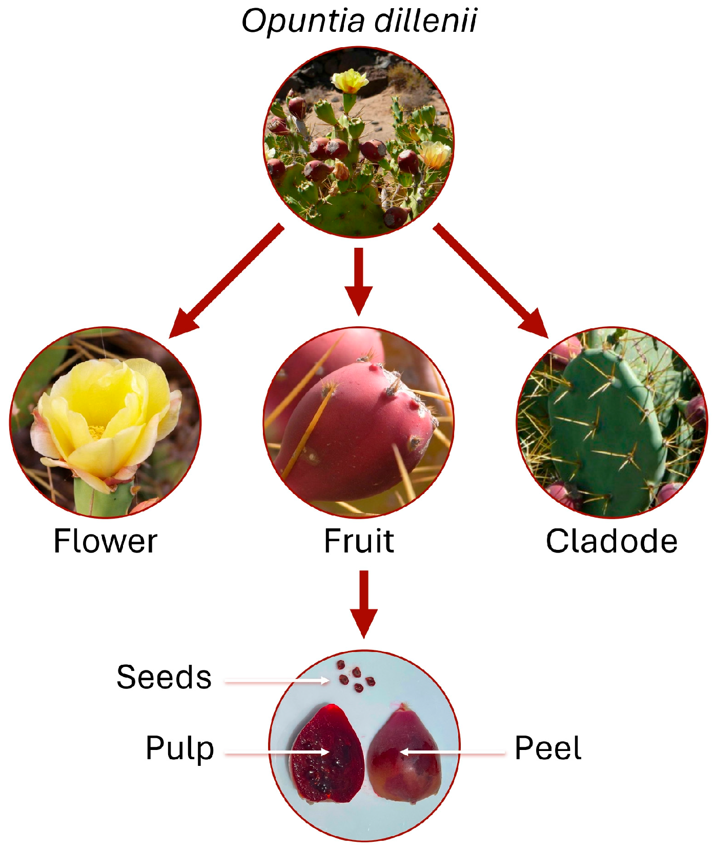
Figure 1. Opuntia dillenii on Canary Island, Spain. Image created using photographs from the Banco de Datos de Biodiversidad de Canarias [8].
Nutraceuticals 05 00022 g001
Figure 2. Search strategy for primary studies.
Nutraceuticals 05 00022 g002
Figure 3. Improvement in oxidative stress parameters mediated by different parts of Opuntia dillenii fruit in animal and cellular models. CAT: catalase; GPx: glutathione peroxidase; GSH: reduced glutathione; iNOS: inducible nitric oxide synthase; MDA: malondialdehyde; MPO: myeloperoxidase; ROS: reactive oxygen species SOD: superoxide dismutase.
Nutraceuticals 05 00022 g003
Table 1. Comparison of in vitro chemical-based antioxidant assays.
AssayMechanism of ActionTargeted Reactive SpeciesApplicability
DPPH and ABTS•+Radical scavenging
(electron/hydrogen donation)
DPPH and ABTS•+ radicalsMeasures the ability to neutralize free radicals
FRAPReduction of Fe3+
(single electron transfer)
N/A (measure of reducing power)Measures the ability to donate electrons
TACReduction of molybdate
(single electron transfer)
ORACRadical scavenging
(hydrogen atom transfer)
ROO radicalMeasures the capacity to neutralize ROO radicals and inhibit lipid oxidation
β-Carotene bleachingRadical scavenging
(single electron transfer)
Lipid peroxidationRadical scavenging
(hydrogen atom transfer)
ROS scavenging assaysRadical scavenging
(hydrogen atom transfer)
OH, H2O2, O2•−, and NOEvaluates antioxidant activity against biologically relevant ROS
FICMetal ion chelationFe2+Assesses the ability to chelate iron ions, preventing radical formation
ABTS•+: 2,2′-azino-bis(3-ethylbenzothiazoline-6-sulfonic acid); DPPH: 2,2-diphenyl-1-picrylhydrazyl; FIC: ferrous iron chelating; FRAC: ferric reducing antioxidant capacity; H2O2: hydrogen peroxide; N/A: not applicable; NO: nitric oxide; OH: hydroxyl radical; O2−•: superoxide anion; ORAC: oxygen radical absorbance capacity; ROO: peroxyl radicals; ROS: reactive oxygen species; TAC: total antioxidant capacity.
Table 2. Compilation of in vitro chemical-based antioxidant assays applied to Opuntia dillenii extracts.
Plant PartExtraction MethodAssayResultsReference
Fruit or cladodesMethanolic extraction (1:5, w/v) for 7 daysLipid peroxidation
DPPH
ABTS•+•OH
H2O2
NO
TAC
[µg/mL]: 0.45–1000
IC50 (µg/mL): fruit vs. cladodes
Lipid peroxidation: 353 vs. 87
DPPH: 317 vs. >1000
ABTS•+: 46 vs. 27
OH: >1000 in both
H2O2: >1000 in both
NO: >700 in both
TAC (µM AAE/g): fruit vs. cladodes
1.4 vs. 0.4
[18]
Peel, pulp, and seeds from fruit Two successive methanolic extractions (1:1, w/v) for 1 h at 4 °C, followed by centrifugation (14,000× g, 10 min)ABTS•+ and ORAC
(1 mg/mL)
Lipid peroxidation
(10 µg/mL)
Peel vs. pulp vs. seeds
ABTS•+ (μM TE)
1.24 vs. 1.01 vs. 2.15
ORAC (μM TE)
0.14 vs. 0.10 vs. 0.18
Lipid peroxidation (% inhibition)
76.4 vs. 73.2 vs. 92.3
[19]
SeedsSupercritical CO2 fluid extraction of seed oil (46.96 MPa, 46.51 °C, 2.79 h, and CO2 flow rate of 10 kg/h) DPPH
β-carotene bleaching
[%, v/v]: 10–100
IC50 (%, v/v)
DPPH: 11.4
β-Carotene bleaching: 20.7
[20]
Whole fruitTwo successive methanolic extractions (1:5, w/v) using mechanical shaker for 24 h at 25 °C, followed by centrifugation (6000× g, 10 min) DPPH
ABTS•+
FIC
FRAP
β-carotene bleaching
DPPH (IC50, µg/mL): 43.0
ABTS•+ (μmol TE/g): 7290.0
FIC (mg EDTA/g): 10.8
FRAP (mmol Fe2+/g): 998.9
β-Carotene bleaching (% inhibition): 19.6
[21]
Not specifiedTwo successive aqueous extractions (1:8–1:12, w/v) in a shaking water bath (100 rpm, 80–90 °C, 50–70 min), followed by centrifugation (6000 rpm, 15 min) and isolation of polysaccharides via 80% ethanol precipitation (24 h) and washing with ethanol and acetone DPPHHigher polysaccharide purity (27.4–90.1%) was associated with stronger antioxidant activity (IC50, 3105–408 µg/mL)[22]
FlowersBolling methanolic extraction (1:10, w/v), followed by centrifugation (150× g, 15 min)DPPH
OH
H2O2
[µg/mL]: 10–500
IC50 (µg/mL)
DPPH: 58.7
OH: 159.7
H2O2: 131.1
[23]
Seeds and fruit pulpSeed: organic solvent extraction of oil (1:2.5, w/v) for 2 h
Pulp juice: mixing of seedless pulp for 5 min
DPPH
[µg/mL]: 5–20
IC50 (µg/mL)
Oil: 27.2
Pulp juice 8.2
[24]
Cladodes from tender (2–4-month-old) and old (5–10-month-old) plantsPolysaccharide extract: aqueous extraction (1:60, w/v), for 3 h at 95 °C, followed by centrifugation (1810× g, 15 min) and protein removal
Polysaccharide fractions from tender plant (I, Ia, Ib, II, IIa, and IIb): DEAE-cellulose anion exchange chromatography and Sephacryl S-400 gel filtration
DPPH
OH
O2−•
[mg/mL]: 6.4
Radical-scavenging activity (%)
DPPH: tender (60.5), old (58.1), I (46.2), Ia (58.4), Ib (15.3), II (33.9), IIa (45.6), and IIb (8.8)
OH: tender (39.6), old (46.2), I (39.8), Ia (45.7), Ib (12.5), II (39.1), IIa (41.4), and IIb (0.8)
O2−•: tender (37.4), old (29.1), I (36.5), Ia (43.7), Ib (13.4), II (30.0), IIa (37.2), and IIb (5.2)
[25]
FruitCrude extract: 60% methanol extraction at 10 °C, until discoloration
Purified extract: removal of hydrocolloids and proteins from the crude extract
Yellow/red fraction: chromatographic separation (column C18) of the purified extract
ABTS•+Radical-scavenging activity
(μmol TE/g fresh fruit)
Crude extract: 35.2
Purified extract: 15.7
Yellow fraction: 1.7
Red fraction: 5.4
[26]
SeedsHydro distillation extraction of essential oil (1:20, w/v), for 4.5 h, followed by centrifugation (5000 rpm, 2 min) DPPH
[µg/mL]: 10–1000
Radical-scavenging activity
36.5–78.1%
[27]
Seeds, fruit peel, and fruit pulp juice from two Moroccan regions (Nador and Essaouira)Organic solvent extraction: diethyl ether maceration (1:2.2, w/v) for 24 h, followed by extraction with diethyl ether (Et2O), ethyl acetate (EtOAc), or ethanol (EtOH)
Water extraction of the powder recovered after organic extraction
DPPH
ABTS•+
TAC
Nador vs. Essaouira
DPPH (IC50, µg/mL)
Peel: Et2O: 190 vs. >2000; EtOAc: 720 vs. 340; EtOH: 530 vs. 500; water: >2000 vs. 880
Seed: Et2O: 119 vs. >2000; EtOAc: 220 vs. 380; EtOH: 63 vs. 45; water: >2000 in both
Juice: Et2O: 1340 vs. 370; EtOAc: >2000 vs. 850; EtOH: 1450 vs. 280; water: 690 vs. 370
ABTS•+ (IC50, µg/mL)
Peel: Et2O: 550 vs. 570; EtOAc: 536 vs. 307; EtOH: 736 vs. 460; Water: 970 vs. 820
Seed: Et2O: >2000 in both; EtOAc: 920 vs. 857; EtOH: 130 vs. 88; water: 700 vs. 380
Juice: Et2O: >2000 vs. 190; EtOAc: >2000 vs. 95; EtOH: 1660 vs. 100; water: 750 vs. 213
TAC (mg AAE/g DW)
Peel: Et2O: 190 vs. 140; EtOAc: 130 vs. 190; EtOH: 150 vs. 240; water: 82 vs. 77
Seed: Et2O: 95 vs. 92; EtOAc: 130 vs. 129; EtOH: 281 vs. 410; water: 71 vs. 110
Juice: Et2O: 73 vs. 230; EtOAc: 53 vs. 220; EtOH: 64 vs. 420; water: 84 vs. 830
[28]
SeedsExtraction of essential oil with petroleum ether (1:5, w/v) for 24 hDPPH
[mg/mL]: 0.1–6
IC50
0.38 mg/mL
[29]
Fruit peel and pulpPolar (ethanol) and non-polar (hexane) solvent extractions (1:30, w/v) for 48 hDPPH
[µg/mL]: 20–80
Radical-scavenging activity (%)
Ethanol + peel: 34.8–75.0
Ethanol + pulp: 32.9–71.8
Hexane + peel: 24.9–62.7
Hexane + pulp: 20.7–56.0
[30]
CladodesAqueous or ethanolic extraction (1:4, w/v) for 24 hDPPH
ABTS•+
TAC
FRAP
NO
Water vs. ethanol
DPPH (IC50, mg/mL): 0.54 vs. 0.60
ABTS•+ (mM TE/g): 0.46 vs. 0.59
TAC (mg AAE/g): 60.44 vs. 62.99
FRAP (Abs 700 nm): 1.39 vs. 1.97
NO (IC50, mg/mL): 0.15 vs. 0.06
[31]
Whole seedless fruitUltrasound-assisted extraction with ethanol (1:50, w/v) for 5 min, at different temperatures (20–50 °C), amplitudes (20–50%), and ethanol concentration (15–80%), followed by centrifugation (10,000× g)
Standard extraction: 50% methanolic extraction (1:30, w/v) with Ultraturax (25,000 rpm, 2 min), followed by centrifugation (10,000 rpm, 10 min) and two successive re-extractions with 50% and 100% methanol
ORACRadical-scavenging activity
(µmol TE/g DW)
Ultrasound-assisted extraction: 330.7–618.9
Standard extraction: 151.8
[32]
Peels and pulp from fruitThree successive 50% methanol extractions, followed by pure methanol extractionABTS•+
ORAC
Peel vs. pulps (µmol TE/g DW)
ABTS•+: 102.6 vs. 99.5
ORAC: 115.4 vs. 160.0
[33]
Peels and pulp from fruitEthanolic extraction (1:10, w/v) at 30 °C, for 4 h with stirring (200 rpm), followed by centrifugation (2800× g, 15 min)DPPH
FRAP
β-carotene bleaching
Peel vs. pulps
DPPH (µmol TE/g DW): 53.1 vs. 26.7
FRAP (µmol TE/g DW): 17.4 vs. 2.3
β-Carotene (% inhibition): 53.8 vs. 51.9
[34]
Whole seedless fruitPressurized liquid extraction with ethanol for 10 min with constant pressure (10.34 MPa), at different temperatures (25–65 °C) and ethanol concentration in water (0–100%, v/v)
Standard extraction: 50% methanolic extraction (1:30, w/v) with Ultraturax (25,000 rpm, 2 min), followed by centrifugation (10,000 rpm, 10 min) and two successive re-extractions with 50% methanol and 100% methanol
ORACRadical-scavenging activity
(µmol TE/g DW)
Pressurized liquid extraction: 321.4–470.7
Standard extraction: 151.8
[35]
Cladodes pulp48 h anaerobic in vitro colonic fermentation (40:40:20 v/v/w medium/inoculum/cladodes)DPPH
ABTS•+
FRAP
0 vs. 24 vs. 48 h of colonic fermentation
DPPH (µmol TE/g): 0.30 vs. 0.26 vs. 0.20
ABTS•+ (µmol TE/g): 0.22 vs. 0.27 vs. 0.28
FRAP (µmol FeSO4/g): 2.84 vs. 3.49 vs. 4.13
[36]
Seeds of three variants: Aknari (AK), Harmoucha (HA), and Imtchan (IM)Cold pressing extraction of essential oil for 45 minDPPH
FRAP
FIC
IC50 (µg/mL): AK vs. HA vs. IM
DPPH: 38.4 vs. 42.2 vs. 15.2
FRAP: 30.2 vs. 55.9 vs. 23.4
FIC: 42.8 vs. 39.5 vs. 35.3
[37]
Flowers and seedsFlowers: 70% ethanolic (EtOH) extraction (4 days), followed or not by liquid–liquid extraction with hexane (Hx), dichloromethane (DCM), ethyl acetate (EtOAc), or butanol (BuOH)
Seeds: steam-distillation followed purification of essential oil with Hx
DPPH
FRAP
DPPH (IC50)
Hx < DCM < oils < BuOH = EtOAc < EtOH
FRAP (IC50)
Hx < oils < DCM < BuOH = EtOAc < EtOH
[38]
Fruit peel50% acetone extraction (1:50, w/v) with vortexing for 10 min, followed by centrifugation (5000 rpm, 15 min)DPPH
ABTS•+
FRAP
TAC
mg TE/g DW
DPPH: 1.2
ABTS•+: 0.4
FRAP: 0.8
TAC: 19.2
[39]
Tender (<1 year) and old (>2 years) cladodes50% acetone extraction (1:50, w/v) with vortexing for 10 min, followed by centrifugation (5000 rpm, 15 min)DPPH
ABTS•+
FRAP
TAC
mg TE/g DW: tender vs. old
DPPH: 0.82 vs. 0.58
ABTS•+: 0.30 vs. 0.43
FRAP: 0.42 vs. 0.92
TAC: 15.56 vs. 15.12
[40]
Fruit pulpBetacyanin extraction with 50% ethanol (1:2, w/v) for 24 h, followed centrifugation (5000 rpm, 10 min)DPPH
TAC
DPPH (IC50, mg/mL): 2.4
TAC (mg AA/g): 273.3
[41]
AAE: ascorbic acid equivalent; ABTS•+: 2,2′-azino-bis(3-ethylbenzothiazoline-6-sulfonic acid); DEAE: diethylaminoethyl; DPPH: 2,2-diphenyl-1-picrylhydrazyl; DW: dry weight; EDTA: ethylenediaminetetraacetic acid; FIC: ferrous iron chelating; FRAC: ferric reducing antioxidant capacity; H2O2: hydrogen peroxide; IC50: half-maximal inhibitory concentration; NO: nitric oxide; OH: hydroxyl radical; O2−•: superoxide anion; ORAC: oxygen radical absorbance capacity; TAC: total antioxidant capacity; TE: Trolox equivalent.
Table 3. Studies evaluating antioxidant properties of Opuntia dillenii using animal, cellular, and in silico approaches.
Biological ModelTreatmentAssayResultsReference
Animal model studies
MCAO-induced cerebral ischemia in male Sprague–Dawley ratsSingle dose (200 mg/kg) of polysaccharide extract (i.p.)iNOS protein expression in cortex↓ iNOS[46]
Streptozotocin-induced diabetes in male Chinese Kunming mice50, 100, or 200 mg/kg/day of polysaccharide extracted from cladodes for 21 days (i.g.)Liver levels of MDA and antioxidant enzyme activity (SOD and GPx)↓ MDA (27, 39, and 40%) at all doses
↑ SOD (18 and 22%) at 100 and 200 mg/kg
↑ GPx (54, 58, and 82%) at all doses
[47]
Streptozotocin-induced diabetes in male Sprague–Dawley rats50, 100, or 200 mg/kg/day of polysaccharide extracted from pulp fruit for 28 days (p.o.)Serum, liver, kidney, and pancreatic levels of MDA and antioxidant enzyme activity (SOD, CAT, and GPx)Effect with 200 mg/kg
↓ MDA (44, 25, 31, and 26%)
↑ SOD (30, 17, 17, and 99%)
↑ CAT (283, 108, 66, and 62%)
↑ GPx (58, 20, 55, and 64%)
[48]
CCl4-induced liver injury in Wistar rats2 mL/kg/day of seed essential oil for 7 days (p.o.)MDA level in liver↓ MDA[49]
Acetic-acid-induced colitis in male albino Wistar rats2.5 or 5 mL/kg/day of juice for 7 days (p.o.) MPO activity, levels of MDA, and GSH content in colonEffect with both doses
↓ MPO and MDA
↑ GSH
[50]
Paracetamol-induced liver injury in albino Wistar rats2.5 or 5 mL/kg/day of juice for 7 days (p.o.) Levels of MDA and GSH, and CAT activity in liver↓ MDA (both doses)
↑ GSH (5 mL/kg)
↑ CAT (both doses)
[51]
Lead-acetate-induced liver injury in male Wistar rats100 or 200 mg/kg/day of pulp fruit extract for 10 days (p.o.)Levels of MDA and CAT activity in liverEffect with both doses
↓ MDA
↑ CAT
[52]
Ethanol-induced gastric ulcer in male Wistar ratsSingle dose (200, 400, or 800 mg/kg) of pulp or peel fruit extract (p.o.)MDA levels and antioxidant enzyme activity (SOD, CAT, and GPx) in gastric tissueEffect with 800 mg/kg of pulp vs. peel
↓ MDA (17 vs. 23%)
↑ SOD (55 vs. 102%)
↑ CAT (75 vs. 100%)
↑ GPx (58 vs. 104%)
[53]
High-fat/high-fructose diet-induced liver steatosis in male Wistar rats25 or 100 mg/kg/day of peel fruit extract for 8 weeks (p.o.)Levels of MDA and protein carbonyl, antioxidant capacity (ORAC), GSSG/GSH ratio, and antioxidant enzyme activity (SOD, CAT, and GPx) in liver↔ CAT, ORAC, and carbonyl protein
↑ GPx at 25 and 100 mg/kg
↑ SOD (87%) at 100 mg/kg
↓ MDA (29%) at 100 mg/kg
↓ GSSG/GSH at 100 mg/kg
[54]
Cell culture studies
H2O2-induced oxidative stress in rat pheochromocytoma cells (PC12)0.1, 0.25, or 0.5 mg/mL of polysaccharide extract for 30 minIntracellular levels of ROS↓ ROS at 0.5 mg/mL[46]
LPS-stimulated murine macrophage (RAW 264.7)
Murine liver cells (Hepa 1c1c7)
RAW 264.7: fruit extract for 15 min (concentrations not indicated)
Hepa 1c1c7: 20 μg/mL of fruit extract for 48 h
iNOS protein expression
(RAW 264.7)
NQO1 enzyme activity
(Hepa 1c1c7)
↔ iNOS and NQO1 [55]
Lead-acetate-induced oxidative stress in human liver cells (HepG2)20, 40, or 80 μg/mL of pulp fruit extract for 24 hIntracellular levels of GSH and MDA↑ GSH (40 and 80 μg/mL)
↓ MDA (all concentrations)
[52]
H2O2-induced oxidative stress in human liver cells (Huh-7)50, 100, or 200 μM of polysaccharide fraction with different molecular weights (F1, 804; F2, 555; F3, 415 kDa) from pulp fruit, for 1 hIntracellular levels of GSH and MDA and antioxidant enzyme activity (SOD, CAT, and GPx)↑ GSH (F2; 200 μM of F3)
↓ MDA (all fractions)
↑ SOD (200 μM of F1; F2 and F3)
↑ CAT (200 μM of F1 and F2; 100 and 200 μM of F3)
↑ GPx (200 μM of F1 and F2)
[56]
Human intestinal (Caco-2) and liver (HepG2) cells, and LPS-stimulated murine macrophage (RAW 264.7)Caco-2 and HepG2: 3.1, 6.3, 12.5, 25, and 50 μg/mL of whole fruit extract for 2 h
RAW 264.7: 25 μg/mL of whole fruit extract for 4 h
CAA (Caco-2 and HepG2)
NO secretion (RAW 264.7)
CAA (Caco-2 vs. HepG2)
9.1–29.5 vs. 26.6–40.9%
↓ NO (34.1%)
[57]
AAPH-induced oxidative stress in human liver cells (HepG2)25 μg/mL of seedless fruit extract or its 12 fractions (individually) for 20 minIntracellular levels of ROS and inhibition of lipid peroxidation↓ ROS (4–27%)
↓ Lipid peroxidation (74.6–82.2%)
[58]
In silico study
Molecular dockingNADPH oxidase
(protein target)
Polyphenols identified in seed essential oil (ligand)
Binding affinity ligand and target proteinPolyphenol with grater affinity (kcal/mol) than Trolox (–6.36): rutin (–6.99), vanillic acid (–6.62), catechin (–6.65), and p-coumaric acid (–6.07)[37]
AAPH: 2,2′-azodiisobutyramidine dihydrochloride; CAA: cellular antioxidant activity; CAT: catalase; CCl4: carbon tetrachloride; GPx: glutathione peroxidase; GSH: reduced glutathione; GSSG: oxidized glutathione; H2O2: hydrogen peroxide; i.g.: intragastric administration; i.p.: intraperitoneal administration; iNOS: inducible nitric oxide synthase; LPS: lipopolysaccharide; MCAO: middle cerebral artery occlusion; MDA: malondialdehyde; MPO: myeloperoxidase; NADPH: nicotinamide adenine dinucleotide phosphate; NO: nitric oxide; NQO1: NAD(P)H:quinone oxidoreductase 1; ORAC: oxygen radical absorbance capacity; p.o.: oral administration; ROS: reactive oxygen species; SOD: superoxide dismutase. Changes in the oxidative stress parameter compared to the control group (animal or cells exposed to the oxidative-stress-inducing agent) are indicated as follows: ↑ for increase, ↓ for decrease, and ↔ for no change.
Disclaimer/Publisher’s Note: The statements, opinions and data contained in all publications are solely those of the individual author(s) and contributor(s) and not of MDPI and/or the editor(s). MDPI and/or the editor(s) disclaim responsibility for any injury to people or property resulting from any ideas, methods, instructions or products referred to in the content.