Communication

Exploring the Efficacy of Extracts for Cosmetic Creams: In Vivo and In Vitro Assessments

1
Faculty of Biotechnology, University of Agronomic Science and Veterinary Medicine, 011464 Bucharest, Romania
2
Hofigal Export Import SA, 042124 Bucharest, Romania
3
Eurofins Evic Product Testing Romania SRL, 040256 Bucharest, Romania
4
National Institute for Chemical Pharmaceutical Research and Development-ICCF, 031299 Bucharest, Romania
5
Indian Scientific Education and Technology Foundation, Lucknow 226002, India
*
Author to whom correspondence should be addressed.
Nutraceuticals 2023, 3(3), 306-314; https://doi.org/10.3390/nutraceuticals3030024
Submission received: 16 May 2023 / Revised: 26 June 2023 / Accepted: 27 June 2023 / Published: 30 June 2023
(This article belongs to the Special Issue Natural Nutraceuticals in Actual Therapeutic Strategies)

Abstract

:
Plants with therapeutic potential are a rich resource for the inhabitants of a country, with Romania among them, having a diverse flora not only from plants but also from fungi. New trends in biopharmaceuticals are also addressed in studies related to topical administration, representing a great advantage; research has determined biological activities in vivo and in vitro. This study aimed to present the main active compounds from extracts of Lactarius piperatus, Centaurea cyanus, and Ribes rubrum. In vitro (DPPH scavenging activity and reducing power) and in vivo activities were determined. A survey of hydrating cosmetic products and the completion of a patch test (in vivo test) under dermatological control were realized to confirm the skin compatibility of the product in a panel of human subjects after a single application. The cosmetic product did not induce allergic reactions, erythema, irritation, or itching. This study showed that the extracts incorporated in the formulation of the face cream had a nutricosmetic effect. The research demonstrated a result of increasing skin elasticity and an anti-inflammatory effect following direct application to the skin of volunteers. The product offered in vitro/in vivo protection against the action of free radicals; it was effective in reducing the side effects of exposure to the action of blue light.

Graphical Abstract

1. Introduction

Romania has a diverse flora from plants and fungi to wild fruits [1]. Plants with therapeutic potential are a valuable resource for obtaining functional products in the biotechnological industry. New trends in biopharmaceuticals are also addressed in studies related to topical administration, representing a great advantage; research has determined biological activities in vivo and in vitro [2].
Skincare cosmetics to improve skin appearance and treat dermatological conditions have increased in recent years [3,4]. Plant-based bioactive compounds are becoming more popular as cosmetic ingredients because they protect and heal the skin [3]. Compared to synthetic cosmetics, plant-based products are easier to tolerate, biodegradable, and have biological and therapeutic activities. The potential side effects that can occur in the case of synthetic skin care products are skin irritation, dryness, different allergic reactions, and skin barrier perturbation.
Using a carrier vehicle to deliver phenolic compounds increases the in vivo bioavailability and reduces the yield of biotransformations that limit the expression of bioactivity [5]. For example, gallic acid has no stability in the fermentative action of the microbiota, but resistance to the action of oxidative stress is mediated by this compound [6]. Its action is limited by the amount absorbed in the small intestine because the remaining amount is inaccessible in vivo [7].
Centaurea cyanus has been used in traditional medicine to protect and heal the skin. It has a calming effect, which can be assimilated to the one caused by chamomile products [8]. Ribes rubrum has a high content of vitamins and minerals that help reparative processes and has a known antimicrobial effect. It is a fruit rich in polyphenols with combined antioxidant and anti-inflammatory effects, which help in regeneration processes and reduce possible local infections [9]. Lactarius piperatus, a little-known product in the cosmetic industry, needs to be more exploited. It is conditionally edible but with numerous biopharmaceutical effects due to a complex pattern of bioactive molecules. Antibiofilm effects have been demonstrated, leading us to use it to reduce the inflammatory processes associated with skin lesions, but also for a cicatrizing result due to polysaccharides [10]. This study examines the in vivo and in vitro biological activity of Lactarius piperatus, Centaurea cyanus, and Ribes rubrum. Their anti-inflammatory effects were studied, and each plant’s anthocyanin, proanthocyanidin, and phenol contents were determined. A hydrating cosmetic product was also manufactured and a patch test was conducted under dermatological control to confirm the skin compatibility of the investigated product in a panel of healthy human subjects after a single application under maximizing and controlled experimental conditions.

2. Materials and Methods

2.1. Biological Material

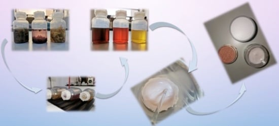
For the research, we used three plant extracts but also mushrooms: peppery milkcap—Lactarius piperatus (Lp), cornflour—Centaurea cynanus (A), and redcurrants—Ribes rubrum (Cr). The substrates were selected based on the possibility of valorizing the local resources from Parâng Mountain, Romania. Ethanol and acetic acid were used as solvents to prepare the extracts of the three samples. Extraction was performed in Duran bottles (24 h, at room temperature under stirring, overnight) at 1% plant/mushroom concentration with the following solvent mixture (v/v): ethanol/water/acetic acid = 50/49.5/0.5. After process extraction, the mixture was filtered under vacuum using Whatman filter paper no. 1. The extract was concentrated under vacuum with a Buchi rotavapor [11].

2.2. Determination of Bioactive Compounds

2.2.1. Determination of Phenol Quantity

The determination of total phenol compounds was realized spectrophometrically. The reaction mixture was kept at room temperature for 15 min; then, the absorbance was read using a Thermo spectrophotometer at 765 nm [12].

2.2.2. Determination of Flavonoid Quantity

The determination of total flavonoid compounds was realized spectrophometrically. The reaction mixture was kept for one hour at room temperature, after which the absorbance was read with a Thermo spectrophotometer at a wavelength of 450 nm [13].
Polysaccharidic compounds were determined by precipitation with cold ethanol in accordance with a previous method [14].

2.2.3. HPLC Assay

The determinations were conducted according to previous methods in published studies [12,13,15]. According to a previous method, a 3 mL aliquot of the sample was weighed and then diluted with 3 mL of methanol. Before injection into the high-pressure liquid chromatography (HPLC) system, the solution underwent filtration using a 0.2 μm polypropylene filter. Simultaneously, approximately 0.8 g of extract was ultrasonicated for 30 min in 20 mL of methanol. This solution also underwent filtration using a 0.2 μm polypropylene filter before being injected into the HPLC system. For the analysis, a Hitachi Chromaster HPLC system was utilized, which consisted of a 5160 pump, a 5310 column oven, 5260 thermostated autosamplers, and a 5430 DAD detector. A ZORBAX SB-C18, 4.6 × 150 mm, 3.5 μm column was used for separation. An adapted, reverse-phase HPLC (RP-HPLC) method was developed, employing a mobile phase composed of acetonitrile, methanol, and water with gradient elution. The analytical wavelengths for detection were set at 320 nm, 369 nm, 285 nm, and 267 nm. To generate calibration curves, stock solutions of each reference standard were diluted to five different concentration levels. The calibration curves’ determination coefficient (R2) values ranged between 0.996 and 0.999.

2.3. Cosmetic Cream Formulation

The protective face cream was obtained by mixing a 300 mg face cream base (Oleya) with 250 μL Lp, 250 μL A, 250 μL Cr, and 50 μL cosmetic dye [16]. The mixture was combined manually in a heat-resistant container made from borosilicate glass. The container was maintained in a water bath for 3–5 min at 50 °C and constant stirring until the mixture was fully homogeneous. After that, the mixture was split into different specific containers made from plastic and stored at room temperature [17,18]. The cream base did not contain sulfates, parabens, or perfume.

2.4. Determination of the In Vivo Antioxidant Potential under Blue Light

This method was based on a protocol published in a previous study [19,20]. It used a strain of Saccharomyces boulardii from the collection of the Laboratory of Pharmaceutical Biotechnology, Faculty of Biotechnology, USAMV (University of Agronomic Sciences and Veterinary Medicine), Bucharest. The viability analysis involved determining the critical time using the reaction mixture: 0.2 mL sample (extract) and 0.2 mL culture medium [16]. The intersection between the lines of viability and mortality (at time intervals of 15, 30, 45, and 60 min) was defined as the critical exposure time at ʎ = 320 nm. It was expressed as the critical point (%). The untreated sample was the control. The antioxidant potential in vivo was determined minutes after the bioactive compound’s assimilation [21].

2.5. Determination of the In Vitro Antioxidant Capacity of the Extracts

The DPPH scavenging activity was used to evaluate the extracts’ antioxidant capacity. The values were expressed as scavenging activity (%) and TBHQ (tert-butylhydroquinone) was used as a control. The determination of the reducing power was the second method used for in vitro evaluation. The higher the absorbance, the stronger the reducing power of the samples. TBHQ was used as a control [11,21].

2.6. Human Patch Test under Dermatological Control

The tests were realized at Eurofins Romania. Dermatological control was used to confirm the skin compatibility of the investigationed product (cosmetic cream) in a panel of healthy human subjects after a single application under maximizing and controlled experimental conditions. The simple blind performed a monocentric clinical study. The study’s product application modality was the occlusive patch Finn Chamber standard®: an aluminum cupula was kept in position by a hypoallergenic adhesive (Scanpor® (inner diameter: 8 mm, surface: 50 mm2)) and the quantity applied was 20 μL.
The number of test subjects was five valid cases (two men and three women). The specific inclusion criteria for the test subjects were 18 to 70 years old, female and male with a phototype (Fitzpatrick): II to IV, with all skin types. The specific non-inclusion criteria were test subjects with a history of adverse reactions to ethanol, colophony, rubber, nickel, aluminum, patch materials, and adhesive plasters and a family or personal history of atopy. All subjects agreed to wear the equipment provided by the investigating center and follow the staff’s instructions during the study.
The tests were realized by the application of the investigational product to the upper back of the test subject by the technician in charge of the study at the investigating center under the following maximizing conditions of exposure: once (on D1), occlusive patch Finn Chamber standard®: aluminum cupula kept in position by a hypoallergenic adhesive: Scanpor® (inner diameter: 8 mm, surface: 50 mm2–quantity applied = 20 μL during a defined time (48 +/− 4 h). Distilled water was applied to a skin area on the upper back under a semi-occlusive patch for a specified time (48 +/− 4 h) as a control area to take into account the possible effects not directly related to the investigational product but due to the patch material.
Checking of skin compatibility was based on a skin examination of the treated and control areas by the same investigator or technician, supervised by the investigator, at the investigating center on D1, before patching, and on D3, 15 to 30 min after patch removal. Sensations of discomfort were reported directly by the test subjects to the investigator or technician during the study. Descriptive analysis was based on the percentage of reactive test subjects (erythema and other visible signs of reactivity).
Skin reactions (Supplementary Table S1) were recorded by a visual skin examination of the treated and control areas performed at the investigating center by the same investigator or technician, supervised by the investigator, under a standard “daylight” source. This was performed on D1, before patching, and on D3, 15 to 30 min after patch removal (or more, if redness appeared after the removal of the adhesive).
In case of clinical signs, the dermatologist would identify, check, grade, and follow the reaction until complete resolution. Concurrently with the clinical examinations performed after patch removal, the test subjects were questioned about their possible discomfort. In case of strong discomfort felt during patch wearing at home, the test subjects must inform the investigator by phone. If necessary, the patch would be removed, and the investigator would quickly perform a skin examination (before the next visit to the investigating center). An area where erythema was graded as 2 or more (with or without infiltration) was evaluated on subsequent days to note whether the reaction diminished or increased to differentiate between an allergic reaction and an irritation. Digital photographs of the skin were systematically taken when justified (adverse effects) [20].

2.7. Microbial Contamination Testing

An evaluation of the microbial load that the product may have was carried out by the method of testing the total number of viable aerobic microorganisms under the conditions provided by FE 5. The technique used was based on the basic principles of the deep inoculation method. The following culture media were used (Merck Romania): Casein soya-bean digest agar (CaSoA), Casein soya-bean digest broth (CaSoB), Sabouraud with chloramphenicol (SDA), Medium H, Medium G, Cetrimide agar (N), Baird–Parker agar (O), and peptone water buffer solution with sodium chloride, pH = 7. After the expiration of the incubation period, the results were interpreted based on the selection of Petri plates showing microbial growth for each tested dilution. The sample was tested for the total number of aerobic microorganisms, and the identification of pathogenic or conditionally pathogenic microorganisms was compliant from the point of view of the microbial load, presenting values below the maximum imposed by the European Pharmacopoeia [22].

2.8. Statistical Analysis

All parameters investigated were evaluated in a minimum of three independent determinations, and the results were expressed as the means ± standard deviation (SD). The mean and SD values were calculated using the IBM SPSS Statistics 23 software package (IBM Corporation, Armonk, NY, USA). The significance level for the calculations was set as follows using the normal distribution of the variables: significant, p ≤ 0.05; very significant, p ≤ 0.01; and highly significant, p ≤ 0.001. The differences were analyzed by an ANOVA followed by a Tukey post hoc analysis. The experimental data were analyzed and correlated with the IBM SPSS Statistics software package (IBM Corporation, Armonk, NY, USA).

3. Results

3.1. Determination of Major Bioactive Compounds

Important differences were noticed for Cr and Lp samples. According to Figure 1 (right), the highest concentrations of phenolic compounds were found in the Centaurea cyanus extract, at 5114.2167 ± 88.72 (p ≤ 0.001). The values were 60% and 71% lower than A. This ratio was also maintained in the case of flavonoid levels (Figure 1), where the differences exceeded 80%. Lp showed a 69% higher amount of flavonoids than Cr, inversely to the total amount of phenols.
Cr exhibited the highest concentration of flavonoids, specifically quercetin-3-glucoside (510.78 μg/mL) and hyperoside (361.68 μg/mL), surpassing A by 40% (Figures S1 and S2). An interesting observation was the similarity in the pattern of phenolic acid compounds between A and Cr. Notably, chlorogenic and caffeic acids were present in twice the amount compared to other acids in both extracts. On the other hand, Lp only displayed the presence of catechin at a concentration of 12.86 μg/mL (Figure S3; Table S2—Supplementary Materials). These quantifications were obtained through an extraction method specifically designed to isolate flavonoids, which are the predominant components with protective effects against oxidative stress.

3.2. Determination of the Antioxidant Effect In Vitro

Evaluating the antioxidant potential in vitro by scavenging DPPH activity demonstrated a balance between A and Lp, with values over 65%. A value of 83.07 ± 0.05 was recorded for Lp, equivalent to 1% TBHQ (used as a standard). These results resulted from other bioactive compounds detailed in Section 3.1, such as anthocyanins and proanthocyanidins. This pattern was absent in sample A, corresponding to the data calculated by an in vitro analysis of antiradical activity.
As standard, the reduction power showed close values, between 0.79 and 0.96, at least 50% lower than those of TBHQ (Figure 2). These data respected the paternal structure of bioactive compounds, demonstrating specificity with the method used in the analysis. In addition, the extracts tested did not show chelating activity (data not shown), which means that the products in which they will be integrated cannot act on the cause of oxidative stress.

3.3. Determination of the Antioxidant Effect In Vivo

The critical point corresponding to the in vivo evaluation of the antioxidant effect is presented in Figure 3. The data showed that the content of phenolic compounds was correlated with the value of the critical point, which corresponds with previous studies [17]. In addition, the Lp extract had a similar critical point of 1.90 ± 0.01% (p < 0.01 vs. the untreated sample), and these low values were interpreted by the presence of other categories of functional compounds (e.g., polysaccharides or tocopherol residues). These components were not a target of the extracts made, but their presence is common in these mushroom extracts [23]. Although protection was exercised through several mechanisms [24], the presence of polysaccharides provides exoprotection to the action of hydroxyl radicals generated in the reaction medium, as demonstrated by the low value of the critical point.

3.4. Human Patch Test under Dermatological Control

Upon careful examination of the cutaneous skin tissue of treated and control areas, it was observed that the skin area had a higher degree of hydration (Table S3). Subjects did not report any discomfort throughout the study. Subjects 1 and 4 had more reactive skin, as can be noticed in Table S3, and they showed redness in the patch area but not in the treated and control area. The subjects were asked about the possible discomfort they felt during the study, and they stated that they felt comfortable. The human patch test had significant results; the tested cosmetic product did not induce allergic reactions, erythema, irritation, or itching.

4. Discussion

This study presents the in vitro/in vivo effects of a nutricosmetic cream based on three functional extracts from natural sources [25,26]. Through the support provided by the three extracts, the tested cream supports the skin, acting as a barrier to the aggression of UV-Vis radiation that causes an accelerated aging effect by inducing oxidative stress [21]. In addition to published studies on Grifola frondosa, the current study also demonstrated the effect of improving the epithelial function due to L. piperatus. The beneficial effects were also determined by the functional composition (hyperoside, quercetin-3-glucoside, and chlorogenic and caffeic acids) that A and Cr bring into the product formulation.
The presence of components with antioxidant effects demonstrated the effectiveness of the three extracts in reducing oxidative stress. The correlation of in vitro data with in vivo data was essential in demonstrating the efficiency of the formula used in reducing the processes leading to the induction of oxidative stress caused by the action of UV-Vis rays. Increased skin elasticity is a new effect cause by the new species of fungi (L. piperatus), which is generally considered to be of no economic importance [23]. Although it is used in small quantities, it contains compounds with various biological functions. The same situation occurs in case A, a plant less used by local communities [27]. This study demonstrated the high biological value of introducing multifunctional extracts in cosmetic formulas, with side effects not recorded. Reducing oxidative stress is a direction the cosmetics industry is considering to eliminate the early signs of aging. Manufacturers also aim to promote new natural components that can be easily integrated into classic formulas [28].
The microbiological analysis of the cream regarding microbial contamination, following the total number of aerobic microorganisms, but also that of pathogenic or conditionally pathogenic microorganisms, is compliant from the point of view of the microbial load. The determined values were significantly below the maximum imposed by the European Pharmacopoeia (Table S4).
It can be considered that the presence of phenolic compounds (mainly the flavonoid fraction) acted as a protector in the presence of free radicals, with anti-inflammatory and antimicrobial effects on the skin [29,30]. The Lp extract’s polysaccharides and other bioactive components supported the elasticity and led to a UV-radiation-repellent effect [30,31]. The re-image amount of these polysaccharides varied at around 1.50 ± 0.13 g/100 mL. The amount of vitamin C, which was 1 mg/mL extract, also contributed to the nutricosmetic action of the product formula tested in vivo. These data confirm previous studies that support different therapeutic properties of Lp species [30,32], adding a nutraceutical value to these data. In addition, the results agreed with studies that have shown these products’ cytotoxic effects, which is essential in reducing side effects resulting from prolonged exposure to UV-Vis radiation [32,33,34,35].

5. Conclusions

This preliminary study revealed the nutricosmetic effect of the three extracts incorporated into the formulation of a face cream. These extracts demonstrated in vitro and in vivo protection against free radicals, effectively mitigating the adverse effects caused by prolonged exposure to blue light emitted by commonly used electronic devices. Furthermore, an in vivo study supported these findings by demonstrating that direct application of the face cream to the skin of volunteers led to increased skin elasticity and induced anti-inflammatory effects. Additionally, the enhanced bioavailability of phenolic compounds and the antimicrobial properties of the cream suggest its potential for protective and reparative use in cases of sunburn.

Supplementary Materials

The following supporting information can be downloaded at: https://www.mdpi.com/article/10.3390/nutraceuticals3030024/s1, Table S1: Operations at the investigating center for recording the skin reactions; Table S2: The medium level of bioactive compounds in the extracts; Table S3: Skin examination after in vivo product tests; Table S4: Microbiological analysis of the functional cream; Figure S1: Chromatographic data for bioactive compounds in the extracts of Centaurea cynanus; Figure S2: Chromatographic data for bioactive compounds in the extracts of Ribes rubrum; Figure S3: Chromatographic data for bioactive compounds in the extracts of Lactarius piperatus.

Author Contributions

E.V. and C.M.P. designed the experiments, analyzed the data, and wrote the paper; A.S. performed the chromatographic analysis; N.E. contributed to biological analyses; and S.K.S. and I.D. analyzed the samples/results. The authors discussed and made comments on the results. All authors have read and agreed to the published version of the manuscript.

Funding

This research received no external funding.

Institutional Review Board Statement

Not applicable.

Informed Consent Statement

Informed consent was obtained from all subjects involved in the study.

Data Availability Statement

Not applicable.

Conflicts of Interest

The authors declare no conflict of interest.

References

  1. Petran, M.; Dragos, D.; Gilca, M. Historical ethnobotanical review of medicinal plants used to treat children diseases in Romania (1860s–1970s). J. Ethnobiol. Ethnomed. 2020, 16, 15. [Google Scholar] [CrossRef] [PubMed] [Green Version]
  2. Patra, J.K.; Das, G.; Fraceto, L.F.; Campos, E.; Rodriguez-Torres, M.; Acosta-Torres, L.S.; Diaz-Torres, L.A.; Grillo, R.; Swamy, M.K.; Sharma, S.; et al. Nano based drug delivery systems: Recent developments and future prospects. J. Nanobiotechnol. 2018, 16, 71. [Google Scholar] [CrossRef] [PubMed] [Green Version]
  3. Goyal, A.; Sharma, A.; Kaur, J.; Kumari, S.; Garg, M.; Sindhu, R.K.; Rahman, M.H.; Akhtar, M.F.; Tagde, P.; Najda, A.; et al. Bioactive-Based Cosmeceuticals: An Update on Emerging Trends. Molecules 2022, 27, 828. [Google Scholar] [CrossRef]
  4. Sator, P.G. Skin treatments and dermatological procedures to promote youthful skin. Clin. Interv. Aging 2006, 1, 51–56. [Google Scholar] [CrossRef] [PubMed]
  5. Vulić, J.; Šeregelj, V.; Kalušević, A.; Lević, S.; Nedović, V.; Tumbas Šaponjac, V.; Čanadanović-Brunet, J.; Ćetković, G. Biodisponibilitatea și bioactivitatea fenolicelor încapsulate și a carotenoizilor izolați din deșeurile de ardei roșu. Molecules 2019, 24, 2837. [Google Scholar] [CrossRef] [PubMed] [Green Version]
  6. Vamanu, E.; Gatea, F.; Sârbu, I. In Vitro Ecological Response of the Human Gut Microbiome to Bioactive Extracts from Edible Wild Mushrooms. Molecules 2018, 23, 2128. [Google Scholar] [CrossRef] [Green Version]
  7. Hussain, M.B.; Hassan, S.; Waheed, M.; Javed, A.; Farooq, M.A.; Tahir, A. Biodisponibilitatea și Calea Metabolică a Compușilor Fenolici, Aspecte Fiziologice ale Plantelor ale Compușilor Fenolici; IntechOpen: London, UK, 2019. [Google Scholar]
  8. Garbossa, W.A.C.; Berardo, P.M.; Campos, G.M. Euterpe oleracea, Matricaria chamomilla, and Camellia sinensis as promising ingredients for development of skin care formulations. Ind. Crops Prod. 2016, 83, 1–10. [Google Scholar] [CrossRef]
  9. Kostic, M.; Ivanov, M.; Fernandes, Â.; Calhelha, R.C.; Glamoclija, J.; Barros, L.; Sokovi’c, M.; Ciric, A. A Comparative Study of Lactarius Mushrooms: Chemical Characterization, Antibacterial, Antibiofilm, Antioxidant and Cytotoxic Activity. J. Fungi 2023, 9, 70. [Google Scholar] [CrossRef]
  10. Choi, E.J.; Park, B.; Lee, J.; Kim, J. Anti-atopic dermatitis properties of Cordyceps militaris on TNFα/IFNγ-stimulated HaCaT cells and experimentally induced atopic dermatitis in mice. Phys. Act. Nutr. 2020, 4, 7–14. [Google Scholar] [CrossRef]
  11. Vamanu, E.; Gatea, F.; Sârbu, I.; Pelinescu, D. An In Vitro Study of the Influence of Curcuma longa Extracts on the Microbiota Modulation Process, In Patients with Hypertension. Pharmaceutics 2019, 11, 191. [Google Scholar] [CrossRef] [Green Version]
  12. Uddin, R.; Saha, M.R.; Subhan, N.; Hossain, H.; Jahan, I.A.; Akter, R.; Alam, A. HPLC-Analysis of Polyphenolic Compounds in Gardenia jasminoides and Determination of Antioxidant Activity by Using Free Radical Scavenging Assays. Adv. Pharm. Bul. 2014, 4, 273–281. [Google Scholar] [CrossRef]
  13. Saidi, A.; Hambaba, L.; Bensaad, M.S.; Melakhessou, M.A.; Bensouici, C.; Ferhat, N.; Kahoul, M.A.; Helal, M.; Sami, R.; Alharthy, S.A.; et al. Phenolic Characterization Using cLC-DAD Analysis and Evaluation of In Vitro and In Vivo Pharmacological Activities of Ruta tuberculata Forssk. Antioxidants 2022, 11, 1351. [Google Scholar] [CrossRef]
  14. Vamanu, E. Biological activities of the polysaccharides produced in submerged culture of two edible Pleurotus ostreatus mushrooms. J Biomed. Biotechnol. 2012, 2012, 565974. [Google Scholar] [CrossRef] [PubMed] [Green Version]
  15. Vamanu, E.; Dinu, L.D.; Luntraru, C.M.; Suciu, A. In Vitro Coliform Resistance to Bioactive Compounds in Urinary Infection, Assessed in a Lab Catheterization Model. Appl. Sci. 2021, 11, 4315. [Google Scholar] [CrossRef]
  16. Smaoui, S.; Hlima, H.B.; Chobba, I.B.; Kadri, A. Development and stability studies of sunscreen cream formulations containing three photo-protective filters. Arab. J. Chem. 2017, 10, S1216–S1222. [Google Scholar] [CrossRef] [Green Version]
  17. Soto, M.L.; Parada, M.; Falqué, E.; Domínguez, H. Personal-Care Products Formulated with Natural Antioxidant Extracts. Cosmetics 2018, 5, 13. [Google Scholar] [CrossRef] [Green Version]
  18. Butkeviciute, A.; Ramanauskiene, K.; Janulis, V. Formulation of Gels and Emulgels with Malus domestica Borkh: Apple Extracts and Their Biopharmaceutical Evaluation In Vitro. Antioxidants 2022, 11, 373. [Google Scholar] [CrossRef]
  19. Blue Light Facts: Is Light Blue Bad for Your Eyes? Available online: https://www.allaboutvision.com/cvs/blue-light.htm (accessed on 6 March 2023).
  20. Zlabiene, U.; Baranauskaite, J.; Kopustinskiene, D.M.; Bernatoniene, J. In Vitro and Clinical Safety Assessment of the Multiple W/O/W Emulsion Based on the Active Ingredients from Rosmarinus officinalis L., Avena sativa L. and Linum usitatissimum L. Pharmaceutics 2021, 13, 732. [Google Scholar] [CrossRef] [PubMed]
  21. Dabulici, C.M.; Sârbu, I.; Vamanu, E. The Bioactive Potential of Functional Products and Bioavailability of Phenolic Compounds. Foods 2020, 9, 953. [Google Scholar] [CrossRef]
  22. Bashir, A.; Lambert, P. Microbiological study of used cosmetic products: Highlighting possible impact on consumer health. J Appl. Microbiol. 2020, 128, 598–605. [Google Scholar] [CrossRef] [PubMed]
  23. Vamanu, E.; Nita, S. Antioxidant capacity and the correlation with major phenolic compounds, anthocyanin, and tocopherol content in various extracts from the wild edible Boletus edulis mushroom. Biomed. Res. Int. 2013, 2013, 313905. [Google Scholar] [CrossRef] [Green Version]
  24. Vreeburg, R.A.; Airianah, O.B.; Fry, S.C. Fingerprinting of hydroxyl radical-attacked polysaccharides by N-isopropyl-2-aminoacridone labelling. Biochem. J. 2014, 463, 225–237. [Google Scholar] [CrossRef] [Green Version]
  25. Roncea, F.; Cazacincu, R.; Mireşan, H.; Roșca, R.A. Nutricosmetics. Updates and perspectives. Farmacist 2019, 6, 191. [Google Scholar] [CrossRef]
  26. Laneri, S.; Di Lorenzo, R.M.; Bernardi, A.; Sacchi, A.; Dini, I. Aloe barbadensis: A Plant of Nutricosmetic Interest. Nat. Prod. Commun. 2020, 15, 7. [Google Scholar] [CrossRef]
  27. Wu, Y.; Choi, M.-H.; Li, J.; Yang, H.; Shin, H.-J. Mushroom Cosmetics: The Present and Future. Cosmetics 2016, 3, 22. [Google Scholar] [CrossRef]
  28. Gigliobianco, M.R.; Cortese, M.; Nannini, S.; Di Nicolantonio, L.; Peregrina, D.V.; Lupidi, G.; Vitali, L.A.; Bocchietto, E.; Di Martino, P.; Censi, R. Chemical, Antioxidant, and Antimicrobial Properties of the Peel and Male Flower By-Products of Four Varieties of Punica granatum L. Cultivated in the Marche Region for Their Use in Cosmetic Products. Antioxidants 2022, 11, 768. [Google Scholar] [CrossRef] [PubMed]
  29. Tungmunnithum, D.; Thongboonyou, A.; Pholboon, A.; Yangsabai, A. Flavonoids and Other Phenolic Compounds from Medicinal Plants for Pharmaceutical and Medical Aspects: An Overview. Medicines 2018, 5, 93. [Google Scholar] [CrossRef]
  30. de Andrade Arruda Fernandes, I.; Maciel, G.M.; Ribeiro, V.R.; Rossetto, R.; Pedro, A.C.; Haminiuk, C.W.I. The role of bacterial cellulose loaded with plant phenolics in prevention of UV-induced skin damage. Carbohyd. Pol. Technol. Appl. 2021, 2, 100122. [Google Scholar] [CrossRef]
  31. Alharbi, K.L.; Raman, J.; Shin, H.-J. Date Fruit and Seed in Nutricosmetics. Cosmetics 2021, 8, 59. [Google Scholar] [CrossRef]
  32. Ma, X.; Yang, M.; He, Y.; Zhai, C.; Li, C. A review on the production, structure, bioactivities and applications of Tremella polysaccharides. Int. J. Immunopathol. Pharmacol. 2021, 35, 20587384211000541. [Google Scholar] [CrossRef]
  33. Kosanic, M.; Petrović, N.; Milosevic-Djordjevic, O.; Grujičić, D.; Tubic, J.; Marković, A.; Stanojkovic, T.P. The health promoting effects of the fruiting bodies extract of the peppery milk cap mushroom Lactarius piperatus (Agaricomycetes) from Serbia. Int. J. Med. Mushrooms 2020, 22, 4. [Google Scholar] [CrossRef]
  34. Kozarski, M.; Klaus, A.; Jakovljevic, D.; Todorovic, N.; Vunduk, J.; Petrović, P.; Niksic, M.; Vrvic, M.M.; Van Griensven, L. Antioxidants of Edible Mushrooms. Molecules 2015, 20, 19489–19525. [Google Scholar] [CrossRef] [Green Version]
  35. El-Wahed, A.A.A.; Khalifa, S.A.M.; Elashal, M.H.; Musharraf, S.G.; Saeed, A.; Khatib, A.; Tahir, H.E.; Zou, X.; Naggar, Y.A.; Mehmood, A.; et al. Cosmetic Applications of Bee Venom. Toxins 2021, 13, 810. [Google Scholar] [CrossRef]
Figure 1. Total phenolic compounds (right) and flavonoid compounds (left) values in the functional extracts. Different letters represent significant statistical differences (p ≤ 0.05), n = 3.
Nutraceuticals 03 00024 g001
Figure 2. DPPH scavenging activity (right) and reducing power (left) of the functional extracts. Different letters represent significant statistical differences (p ≤ 0.05), n = 3.
Nutraceuticals 03 00024 g002
Figure 3. The critical time of the functional extracts after in vivo analysis. Different letters represent significant statistical differences (p ≤ 0.05), n = 3.
Nutraceuticals 03 00024 g003
Disclaimer/Publisher’s Note: The statements, opinions and data contained in all publications are solely those of the individual author(s) and contributor(s) and not of MDPI and/or the editor(s). MDPI and/or the editor(s) disclaim responsibility for any injury to people or property resulting from any ideas, methods, instructions or products referred to in the content.