Review

Environmental Fate, Biological Interactions, and Toxicity Mechanisms of Engineered Nanoparticles (ENPs)

by
Christina M. Brenckman
1,
Ashish D. Borgaonkar
2,*,
William H. Pennock III
1,
Genoa R. Warner
3 and
Jay N. Meegoda
1,*
1
Department of Civil and Environmental Engineering, New Jersey Institute of Technology, 323 MLK Blvd., Newark, NJ 07102, USA
2
School of Applied Engineering and Technology, New Jersey Institute of Technology, 323 MLK Blvd., Newark, NJ 07102, USA
3
Department of Chemistry and Environmental Science, New Jersey Institute of Technology, 323 MLK Blvd., Newark, NJ 07102, USA
*
Authors to whom correspondence should be addressed.
Int. J. Environ. Res. Public Health 2026, 23(9), 1103; https://doi.org/10.3390/ijerph23091103
Submission received: 3 July 2026 / Revised: 19 August 2026 / Accepted: 20 August 2026 / Published: 25 August 2026

Highlights

Public health relevance—How does this work relate to a public health issue?
  • Health effects from nanoparticles occur when particles are breathed into the lungs. Ultrafine particles can travel deep into the lungs and cause inflammation and oxidative stress. Hence children, pregnant women, older adults, and those with respiratory conditions and heart disease may face increased risk from exposure to nanoparticles.
  • Nanoparticles can enter the bloodstream after inhalation and lodge in the liver, spleen, kidneys and brain. There is concern that they could build up in the body and cause adverse health effects. Hence, damage at the cellular/genetic level from oxidative stress, inflammation, cell membrane damage, and DNA damage can lead to health effects like heart disease, neurological disorders and cancer.
Public health significance—Why is this work of significance to public health?
  • To better understand the impact of engineered nanoparticles (ENPs) on human health and toxicological changes in response to engineered nanoparticle exposure, including oxidative stress, inflammation, cell damage, and genotoxicity.
  • To benchmark the risk assessment process by identifying how nanoparticle toxicity is influenced by the properties of the particles themselves, as well as transformations that occur in the environment and within biological systems, instead of just the chemical composition.
Public health implications—What are the key implications or messages for practitioners, policy makers and/or researchers in public health?
  • It is important to understand ENPs as an emerging public health issue and begin surveillance efforts, considering nanoparticle-specific characteristics (size, surface chemistry, shape, and environmental transformations) during risk assessments instead of extrapolating from traditional toxicological methods used for chemicals.
  • There is a need to standardize the methods used for nanoparticle characterization, toxicity testing, exposure assessments, and environmental sampling and create evidence-based regulations and occupational health exposure limits to safeguard workers, consumers, and susceptible populations.

Abstract

Engineered nanoparticles (ENPs) are being used with increasing frequency in industrial and consumer products across applications in industry, agriculture, ecology, and biomedicine. The unique physicochemical characteristics that make nanoparticles desirable in product design also affect their fate in the environment, interactions with living systems, and mechanisms of toxicity. Traditional testing methods and toxicological paradigms based on dissolved chemicals are poorly suited to understand ENP risks, primarily due to their small size, large SA:Vs, increased reactivity, and a surface chemistry that can be tuned during synthesis. Nanoparticle toxicity is dependent on complex relationships between particle characteristics, transformations in the environment, resulting exposure scenarios, and biological effects. Here we review ENP toxicity across a property → transformation → exposure → toxicity continuum, with a focus on how particle properties affect environmental and biological transformations relevant to toxicity. Properties such as size, shape, surface chemistry, dissolution, redox activity, and aggregation propensity are reviewed with respect to effects on transport and bioavailability, cellular uptake, biodistribution, and toxicity mechanisms. Transformations including aggregation, oxidation, dissolution/sulfidation, aging and eco-corona formation are discussed with regard to impacts on exposure and risk. Finally, major mechanisms of toxicity including oxidative stress, ion toxicity, membrane damage, inflammation, and genotoxicity are discussed with regard to nano–bio interactions. Analytical challenges associated with studying ENPs, shortcomings of the current risk assessment methods, and emerging Safe-by-Design approaches are also reviewed. Furthermore, connections between the fields of engineered nanoparticle toxicology and microplastics/nanoplastics are discussed, with particular focus on overlapping physicochemical properties, transformations, exposures, and biological mechanisms.

1. Introduction

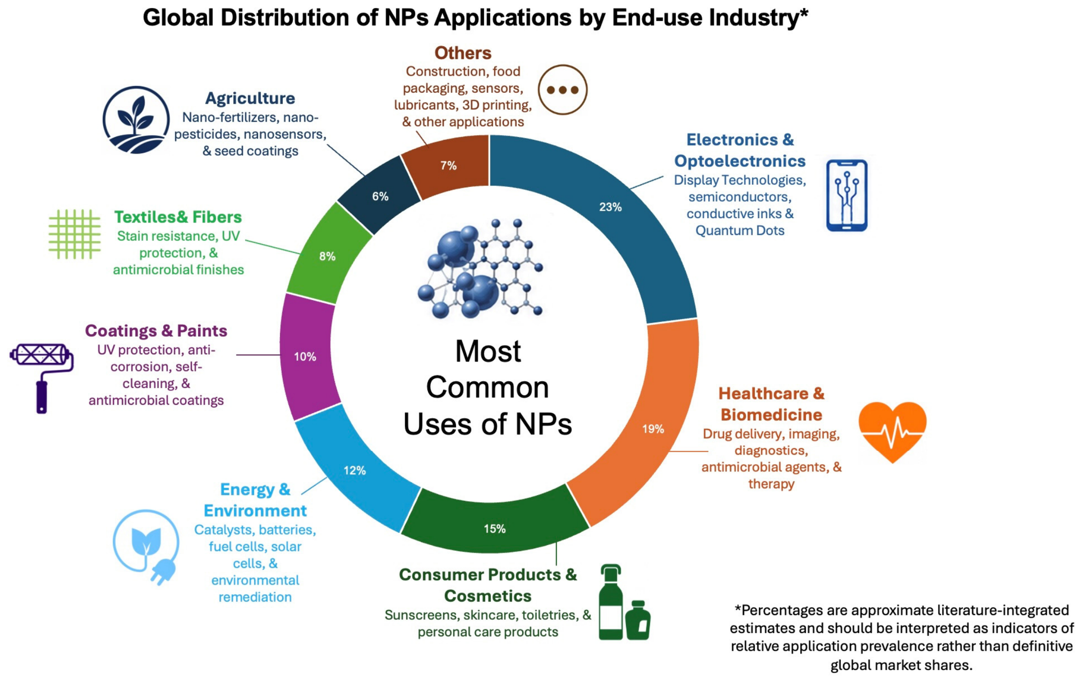
Nanomaterials (NMs) are defined as materials with at least one external dimension or internal structure feature in the size range of approximately 1–100 nm. Nanoparticles (NPs) are categorized as a subset of NMs where all three external dimensions fall within the nano-size range. Engineered nanomaterials (ENMs) are those nanomaterials that are deliberately produced, while engineered nanoparticles (ENPs) are nanoparticles that are intentionally synthesized for industrial, environmental, agricultural or biomedical use. Since engineered nanoparticles are the focus of this review, we will refer to them as ENPs throughout unless referring to nanomaterials in general [1,2]. Over the years, numerous engineered nanomaterials (ENMs) have been synthesized, including metallic ENPs, carbon-based nanomaterials, quantum dots (QDs), polymeric nanomaterials, and nanotubes [3]. In particular, metal-based ENPs have garnered significant research interest, as their diminutive size imbues them with unique physicochemical properties that differ from their bulk material counterparts [1]. These physicochemical properties include changes in surface reactivity, optical properties, and catalytic activity. Consequently, metal-based NPs have found widespread industrial application across biomedical fields [4], coatings [5], cosmetics [6], and more. Engineered Nanoparticles (ENPs) have also found utility in a wide variety of technologies and applications, ranging from antimicrobial surfaces to photocatalysts, electronics, drug delivery, and medical imaging devices [7]. These technologies lead to environmental release during production, processing, and consumer use of ENP-containing products. Intentional releases of NPs into the environment are also on the rise with the increased use of nano-enabled pesticides, fertilizers, and nanosensors in agricultural settings [8,9]. ENPs are used in a wide variety of products such as electronics, biomedicine, cosmetics, energy production/storage, coating surfaces, agriculture, and environmental cleanup. Percentages vary by market sector, but typically, electronics, medical/hygiene, consumer goods, and advanced material applications account for the largest market share of nanoparticles (Figure 1) [10,11,12,13]. Percentages are calculated by combining market share sizes reported across various industry publications from 2022–2024. Totals have been normalized to equal 100% and should be used as rough estimates of comparative category volumes, not precise market splits (modified from Keller et al. (2023), Malik et al. (2023), Mekuye and Abera (2023) and Eker et al. (2024) [10,11,12,13]).
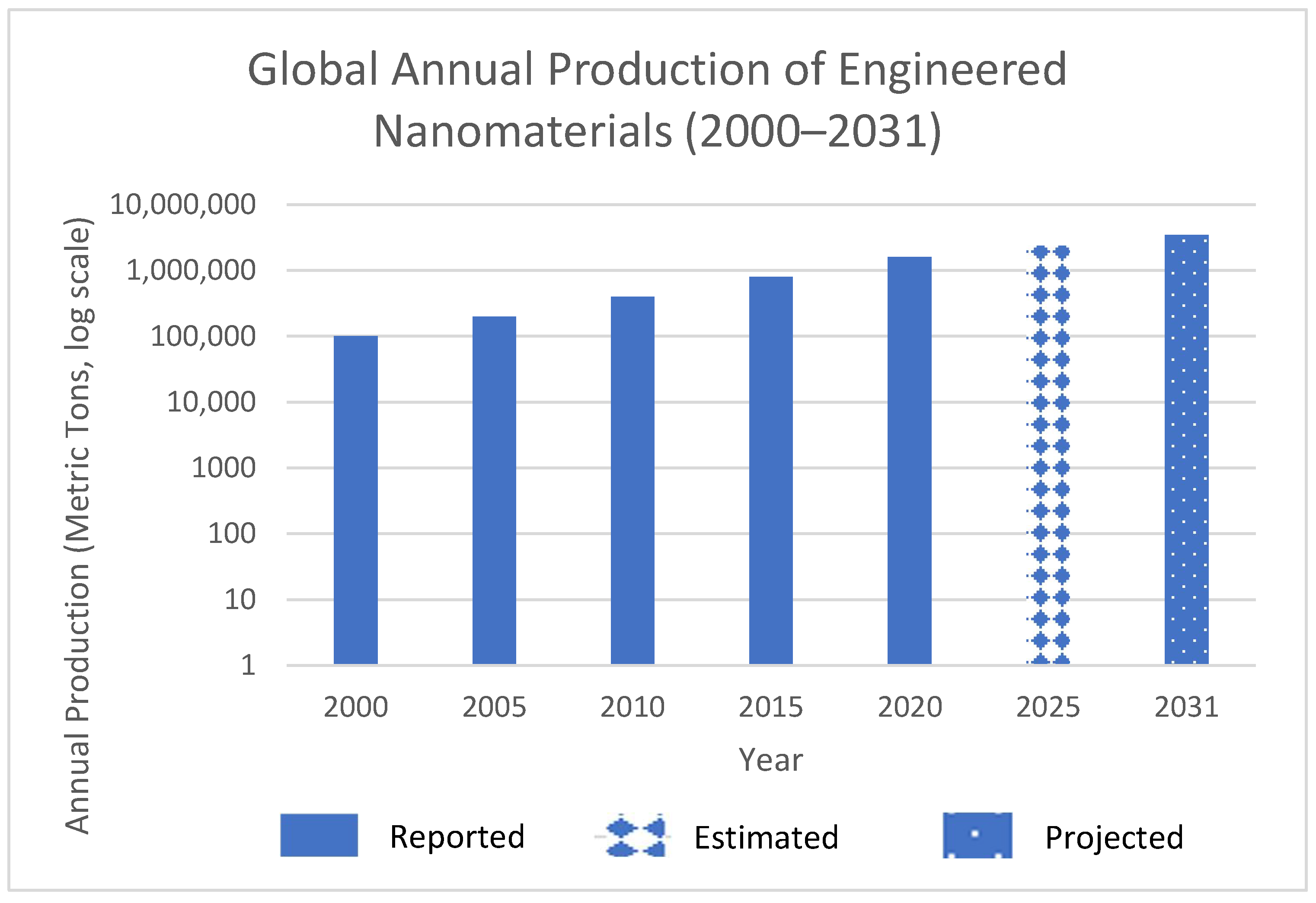
Published estimates of cumulative worldwide production of engineered nanomaterials (ENMs) have grown from approximately 100,000 metric tons (MT) in 2000 to values over 1.6 million MT in 2020 (Figure 2). The newer estimates reflect values that are either estimated or projected, rather than reported historical production, which is indicated in Figure 2. While these production values are shown for this reason alone, to demonstrate growth in the scale of ENP use, production is important to consider when evaluating exposure and risk, because increased production and use create more opportunities for release to occur during manufacturing, product application, and disposal [11,14].
Over 1800 nano-enabled consumer and industrial products containing ENPs are currently on the market globally [15,16]. Furthermore, the nanoparticle industry is expected to continue growing, with projected revenues to exceed 1 trillion U.S. dollars in the next 10–15 years [16,17]. Coupled with estimates that the world population will exceed 9 billion people by the year 2050 [16,18], pressure to increase food production will likely heighten concerns over long-term food security. Nanotechnology has been proposed as one mechanism through which agricultural production can be enhanced. Nano-fertilizers, nano-pesticides, smart delivery systems, and nanosensors are just a few examples of nanotechnology applications that have been suggested to increase crop yields while maintaining food safety.
Figure 2. Global annual production of engineered nanomaterials (2000–2031). Production values for 2000, 2005, 2010, 2015, and 2020 were compiled from the published literature and market sources (e.g., [11,14]). The 2025 value represents an estimate synthesized from recent market analyses, while the 2031 value represents a projected global production based on published market forecasts and the solid line values represent reported annual production [14,17]. Reported production data, estimates, and projections are distinguished in the figure using different bar patterns.
Ijerph 23 01103 g002
Engineered Nanomaterials (ENMs) used today include metals and metal-oxides such as silver (AgNPs), titanium dioxide (TiO2 NPs), and zinc oxide (ZnO NPs), carbon-based materials such as graphene and carbon nanotubes (CNTs), as well as other hybrid or composite nanostructures [19]. With continued production and integration into commercial goods, unintentional environmental exposures to ENMs are inevitable. Therefore, recent attention has been focused on determining how ENMs may affect microbial physiology and ecology [20,21].
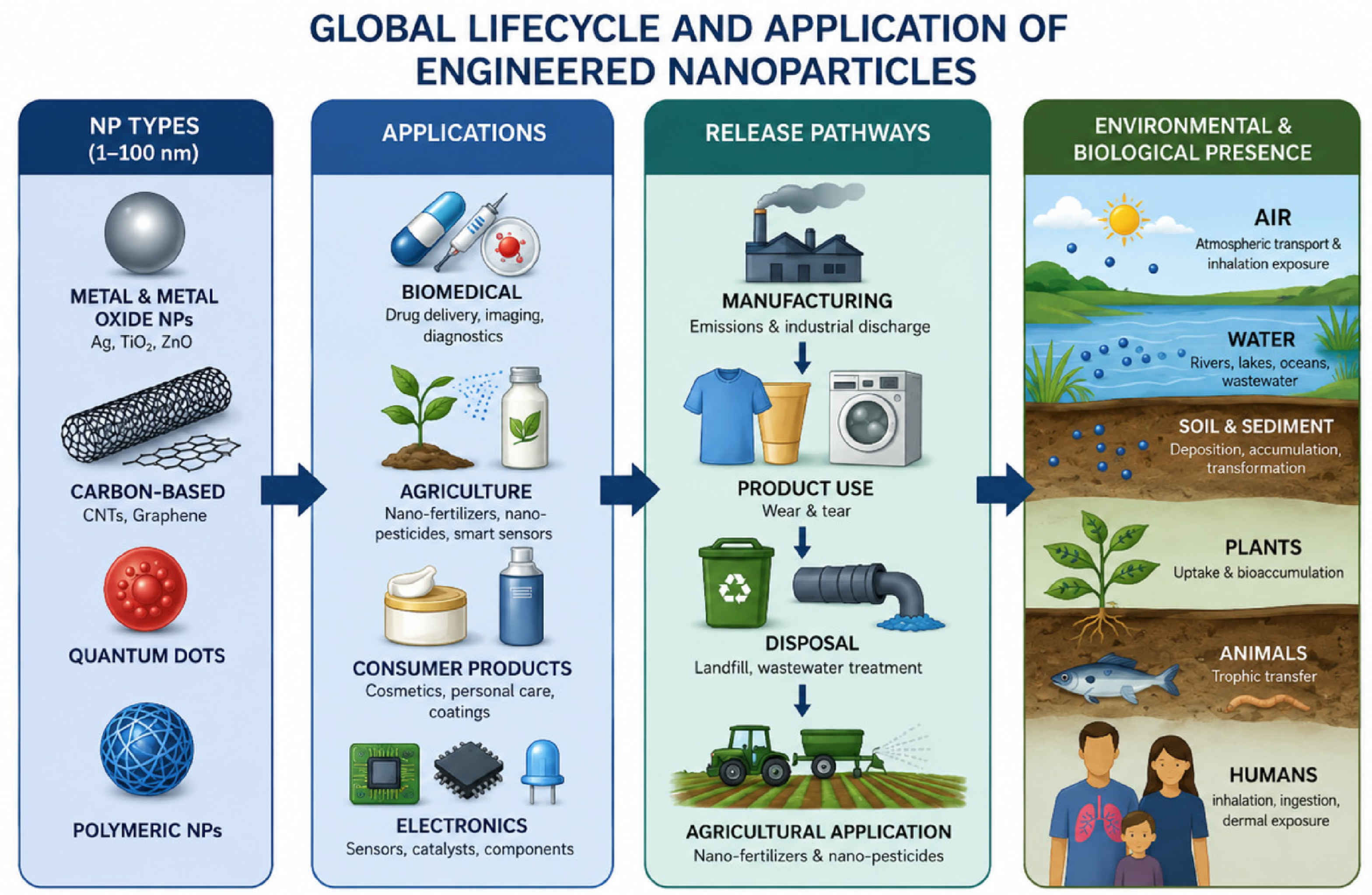
Unintentional releases of ENPs into the environment have increased with expanding NP-enabled technologies. These releases occur during manufacturing processes, consumer use, and disposal [22,23]. As a result, NPs have been detected in numerous environmental matrices, including air, surface water, soil and sediment, and within organisms such as microorganisms, plants, and higher-order species [24]. Rapidly increasing production volumes and widespread distribution have led to calls for better understanding of NP fate and transport as well as their resultant effects in the environment [25]. An overview of major ENPs types, application sectors, and pathways of environmental release is presented in Figure 3.
Due to their small size, NPs exhibit unique behaviors that are distinct from those of conventional chemicals. The cellular activity of NPs is largely dictated by their intrinsic physicochemical properties. Properties such as surface charge, shape, aggregation state, and surface chemistry influence cellular uptake, biodistribution, and biomolecule absorption onto the NP surface [26,27]. Once in biological media, NPs are rapidly coated with adsorbed proteins and biomolecules, which is often referred to as the protein corona. This protein corona can alter NP interactions with cell receptors and lead to modified cellular responses [28]. Because of these dynamic properties, NPs do not follow some of the same principles used to predict traditional toxicant behavior. Classic toxicology has been based on bulk mass and chemical stability.
Frameworks for chemical risk assessment based on chemical hazard and exposure characterization are still widely used as a starting point for ENPs. However, these frameworks often fail to incorporate nanoform-specific behavior. Recent OECD guidance addresses many of these shortcomings by recommending detailed characterization of nanomaterial physical and chemical properties (the use of well-characterized reference materials when testing is required but information is limited), nanoparticle-specific sample preparation and dosimetry, and dose descriptors that account for particle number or surface area in addition to mass concentration [29,30,31]. European guidance documents and research projects have been developed with a similar emphasis on nanoform-specific assessment and Safe- and Sustainable-by-Design frameworks that include consideration of safety throughout material design. A limitation of both OECD and European resources is that environmental and biological transformations, including dissolution and ion release, aggregation, oxidation, and corona formation, have not been fully incorporated into standardized testing or regulatory decision frameworks. Harmonization and implementation of OECD and European nanomaterial-specific guidance will be critical to ensure ENP hazard and exposure assessments account for changes in particle properties, exposure, and toxicity throughout the life cycle of a material [22,32].
It should also be noted that intrinsic nanoscale properties are unlikely to solely dictate toxicity. Instead, they drive a series of transformations that ultimately dictate exposure conditions and subsequent biological effects. Environmental and biological transformations such as sulfidation, oxidation, aggregation, eco-corona formation, and biomolecule absorption can alter NP reactivity. These transformations can affect reactive oxygen species (ROS) generation, DNA damage, inflammatory response, and adverse effects [14,23]. For this reason, ENP hazards should be considered through a property → transformation → exposure → toxicity continuum, where properties drive transformations that dictate exposure conditions and relevant mechanisms.
It is anticipated that this review will help summarize knowledge gaps on engineered nanoparticles (ENPs) across the property → transformation → exposure → toxicity continuum. To this end, this review focuses on identifying how key physicochemical properties of ENPs control their environmental and biological transformations, exposure, and toxicological effects in humans and ecosystems. This review will briefly highlight what is currently known about ENP physicochemical properties, environmental fate and transformations, biological transformations, toxicity mechanisms, analytical methods for detection and characterization, current approaches to risk assessment, and Safe-by-Design approaches. In addition, this review highlights the current overlap between ENP research and that of microplastics/nanoplastics by comparatively discussing physicochemical properties, environmental transformations, exposures, and toxicological mechanisms. Lastly, this effort serves as an extension of the authors’ previous reviews on plastic pollution [33], harmful algal blooms [34], and PFAS—microplastic interactions [35], with a focus on engineered nanoparticles.

2. Literature Review and Methodology

A focused review of the engineered nanoparticle (ENP) toxicity literature is provided, with the discussion following a continuum from particle properties to environmental and biological transformations, exposure and subsequent toxicity. Literature search dates spanned from January 2026 to June 2026, with the most recent search conducted in June 2026. Emphasis was placed on the peer-reviewed literature published within the last 6 years (July 2020–June 2026) to provide an overview of the most recent and relevant findings in nanotoxicology. However, older publications (>6 years) were included when they were considered landmark studies within the field or necessary for the introduction of key concepts.
Web of Science, Scopus, PubMed, Google Scholar, and ScienceDirect were used to collect the literature. Search terms used combinations of the following: “engineered nanoparticles”, “engineered nanomaterials”, “nanoparticle toxicity”, “nanotoxicology”, “environmental transformation”, “aggregation”, “protein corona”, “eco-corona”, “oxidative stress”, “biodistribution”, “cellular uptake”, “risk assessment”, and “Safe-by-Design”. Boolean operators (AND/OR) were used where appropriate.
Peer-reviewed journal articles published in English that investigated ENP physicochemical properties, environmental transformations or fate, biological transformations or fate, exposure routes, toxicological mechanisms, characterization methods, or risk assessments were given highest priority. Review articles were included when original research articles were not available. Articles discussing naturally occurring nanoparticles were included if they added mechanistic relevance to the review, but articles specific to ENPs took precedence. Guidance documents, regulatory resources, technical reports, and other gray literature were included to support discussion on regulatory, methodological, market, or technical issues as needed. Conference abstracts, editorials, and obscure sources without adequate information to determine relevance were excluded.
Titles and abstracts of collected articles were screened to determine relevance. Articles that were deemed possibly eligible were retrieved and assessed in full-text for eligibility. Records collected from multiple databases were removed if they duplicated another article. Following screening, full-text assessment, and removal of duplicate records, a total of 155 sources were retained and incorporated into the final review.
The studies selected for inclusion were then grouped together and summarized based on the property → transformation → exposure → toxicity framework outlined in this review. Articles were initially grouped based on the engineered nanoparticle property being studied (i.e., particle size, morphology/surface structure, surface chemistry, aggregation/agglomeration state, dissolution, and redox potential/reactivity). Articles elucidating environmental and biological changes to these nanoparticle properties were integrated into the transformation stage of the framework (e.g., aggregation/agglomeration, oxidation, sulfidation, dissolution, protein/eco-corona formation). Studies focused on human and environmental exposure routes, biodistribution, and bioavailability were included in the exposure stage, and articles reporting oxidative stress, inflammation, membrane damage, ion-mediated toxicity, genotoxicity, and tissue-specific effects were compiled in the toxicity stage. Using this framework allowed us to take key findings from studies on a variety of nanoparticles and group them into one framework linking nanoparticle properties to their environmental fate, exposure, and toxicity.
NP properties such as size, shape, surface chemistry, aggregation, dissolution, and redox potential are some of the primary factors that control reactivity. Here, environmental and biological transformations are reviewed with a focus on how they impact bioavailability and the resultant internal dose. Aggregation, oxidation, sulfidation, dissolution, and protein corona or eco-corona formation are some of the important transformation processes that ENPs undergo in media and biotic fluids. Exposure routes are varied and can include inhalation, ingestion, dermal exposure, occupational exposure, and trophic transfer up the food chain. Toxicological effects focus on mechanisms related to oxidative stress, inflammation, membrane disruption, ion-related toxicity, and genotoxicity. Particle number and particle surface area are alternative metrics used for dosing and are discussed where relevant. Often, mass concentration does not adequately describe exposures to nanoparticles. Knowledge gaps, including the need for chronic exposure data and standardized testing, are mentioned throughout. Table 1 lists some representative biological endpoints that have been measured in nanotoxicology studies along with their major targets, method of measurement, representative biomarkers/endpoints, and references. Please note that this table is intended to serve as a sampling of commonly utilized experimental endpoints and is not meant to be an all-inclusive classification of mechanisms of toxicity induced by nanoparticles. Representative mechanistic toxicity endpoints, cellular targets, measurement methods, and biomarkers used in nanotoxicology are summarized in Table 1.

3. Toxicological Behavior of NPs Determined by Nanoscale Properties

For ENPs, a combination of inherent physicochemical properties controls environmental transformation, biological fate, and internal dose. Due to continuous exposures of ENPs to environmental and biological media during their life span, they are prone to physicochemical changes that affect their behavior [7,22]. Such environmental changes may include aggregation, oxidation, sulfidation, dissolution and eco-corona formation, which occur prior to and/or during exposure and will impact the fate and bioavailability of nanoparticles in the environment. Intrinsic nanoparticle properties, including particle size, the resulting high surface area-to-volume ratio (SA:V), morphology, surface chemistry, and surface charge, govern the extent to which these transformations occur and ultimately influence nanoparticle reactivity and fate [9,26]. Within a biological system, toxicokinetic properties including absorption, biodistribution, cellular uptake, biotransformation and elimination pathways will affect how much of a dose reaches the target tissue [9,26,27]. Toxicodynamics will be used to describe the biological responses occurring due to exposure to the internal dose; these include oxidative stress, inflammation, membrane damage, genotoxicity, and other forms of cytotoxicity [14]. The relationships among key nanoparticle physicochemical properties, transformation processes, exposure-related effects, and toxicological outcomes are summarized in Table 2.

3.1. Size and Morphology

Nanomaterial properties such as size and morphology primarily dictate NP performance and behavior. Particle size and shape directly impact surface reactivity, optical properties, and the ability to cross cellular membranes. Nanomaterial size and shape are critically important for the biomedical applications of NPs and have been widely exploited for successful cellular uptake and tunable optical properties for use in medical imaging agents and cancer therapeutics [10,41].
Overall NP size has also been shown to influence biological fate, including circulation time, cellular uptake, and targeting ability. Smaller NPs are typically internalized by cells more efficiently, which can lead to enhanced cellular uptake [42,43]. Moreover, NP shape can affect cellular uptake, with certain geometries participating more readily in cellular transport than others. This can be due to shape-dependent interactions with the cell membrane and endocytic machinery [42,43].
As particle size decreases, the SA:V of NPs increases. This means a greater proportion of the total atoms make up the surface of the particle instead of the core. A higher SA:V results in more reactive sites on the NP. Many of the adverse effects associated with NPs, such as ROS generation, lipid peroxidation, redox reactions, and protein corona formation, are related to the NP surface. Therefore, smaller NPs will typically induce a larger response at equal mass compared to larger particles of the same material [44,45]. This concept is one of the reasons why mass concentration is not always a good dose metric for NPs. Two NP exposures that contain the same mass concentration can have very different surface areas and numbers of particles. Smaller particles will typically have a higher surface area and more particles at the same mass concentration compared to larger particles. Because of this, surface-area- or particle-number-based exposure metrics have been gaining more attention in the field of nanotoxicology [45].
Particle size also affects the mechanism of cellular uptake. Smaller particles (<50 nm) are more likely to be internalized by cells via endocytosis, leading to a higher concentration of nanoparticles inside the cell and greater potential to reach sensitive target sites [44,45]. Additionally, NP shape affects how easily the particles can be internalized by cells. Different shapes can affect the mechanism of uptake and cause physiologically distinct responses once inside the cell. For example, NP shape has been shown to impact lysosomal stability, mitochondrial integrity, and oxidative and inflammatory stress responses [44,45]. Once engulfed by the cell, membrane wrapping energetics will differ based on the particle shape. Sphere-shaped NPs will deform the membrane around them evenly, while rod-shaped NPs (or NPs with a high aspect ratio) will affect how the membrane interacts with the NP, which can alter uptake kinetics and subsequent intracellular routing [46]. This can cause variation in toxicological responses by altering intracellular localization and which cellular pathways are affected, such as oxidative stress, genotoxicity, and apoptotic pathways.
Size continues to play a role even after NP uptake. Once in the circulation, small NPs can more easily transverse biological barriers such as epithelial and endothelial cells. This includes cells in the lungs and gastrointestinal (GI) tract, allowing for deeper tissue penetration and potential secondary organ exposure. Under the right physicochemical conditions, some NPs can travel across the blood–brain barrier, which has led to concerns over potential NP neurotoxicity [47]. Biodistribution studies have found that size affects organ distribution, with many particles accumulating in organs responsible for clearance, including the liver and spleen [48].
Clearance represents the final stage in this size-dependent cascade. Particles approaching or below renal filtration thresholds are more likely to be eliminated through glomerular filtration, whereas larger NPs are preferentially sequestered by the mononuclear phagocyte system, particularly macrophages in the liver and spleen [49]. Hydrodynamic diameter and surface chemistry also play roles in blood circulation time and organ distribution [50]. Slowly cleared NPs will continuously expose tissues to their reactive surfaces prolonging oxidative stress and inflammation. In situations with occupational or repeated exposures, slow clearance rates may contribute to higher incidences of tissue damage over time.
Ultimately NP size influences cellular uptake kinetics, cellular trafficking, organ distribution, and clearance mechanisms. Understanding how these processes are connected and dictated by NP size is critical to understanding the mechanisms behind NP toxicity and should be carefully considered when designing safer NPs.

3.2. Surface Chemistry and Coatings

Particle size and morphology also have effects on NP behavior in biological systems. Surface charge, functional groups, and coatings on NPs also play a role in determining how particles interact with cellular membranes, which proteins adhere to their surfaces, how they are recognized by immune cells, and how stable the particles are under biological conditions. These properties matter because NPs will first interact with cells at the nano–bio interface, so even minor changes in surface properties can tip the balance of toxicological responses.
Surface properties are intrinsically linked to NP function because they determine how NPs will interact with their surroundings. Surface chemistry and morphology can be exploited to tune NP performance because NPs have a high SA:V. For instance, changes to surface chemistry by adding different functional groups can alter the reactivity and selectivity of NPs. This has been applied in gas sensors, where surface wettability and gas adsorption alter sensing performance [42,51]. Furthermore, NPs can have physicochemical properties that are distinct from bulk materials of the same composition; this includes electronic and mechanical properties. These properties can differ significantly at sizes ≲ 10 nm [42,52].
One property that plays a large role is electrostatics. Cell membranes are typically negatively charged due to the presence of charged phospholipid head groups and membrane-bound glycoproteins. As a result, NPs with positive surface charges will have stronger electrostatic attractions to the membrane, which can affect how well they adsorb to the surface or are internalized by cells. On the other hand, positive NP surfaces have been shown to cause membrane damage and ROS generation more than other surface charges [53]. Surface charge can be partially hidden by protein corona formation, but charge effects can still influence how well NPs are able to penetrate cells and the signals that they send to cells [54].
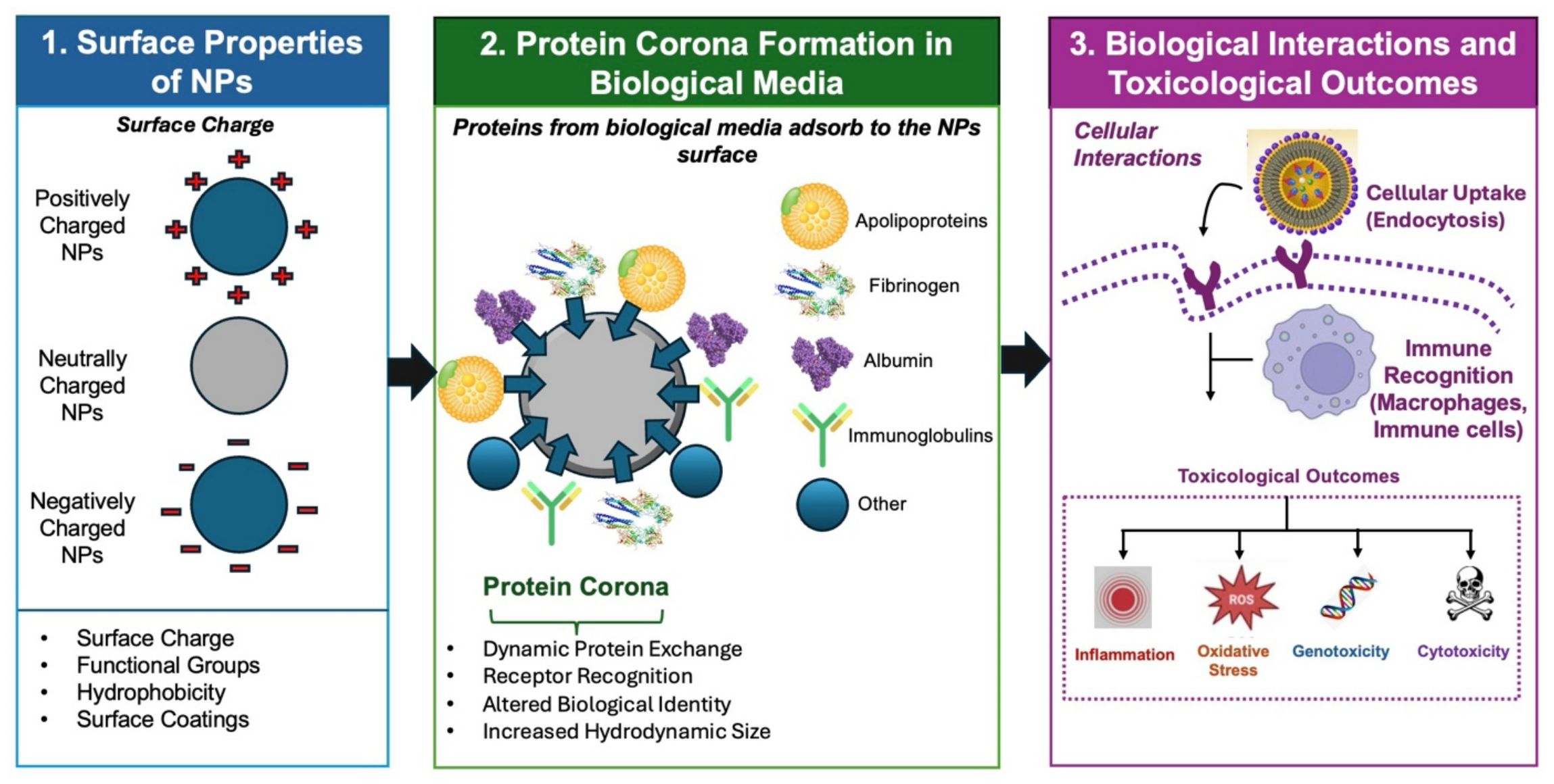
After exposure to biological media, NPs will become coated with a layer of adsorbed proteins. This protein corona can dictate how the particle will interact with cells. The surface chemistry of the NP, including functional groups, hydrophobicity, and charge, will influence what proteins are present in the corona. Because the protein corona can mediate receptor recognition, immune cell uptake, and intracellular trafficking, surface properties have an indirect effect on the biomolecular interactions of NPs [55]. Protein coronas can mitigate NP toxicity, such as by promoting macrophage uptake, but can also alter the fate of NPs in the body to enhance accumulation in certain tissues [56]. The protein corona is dynamic and can change over time with protein exchange, so NPs characterized in vitro may behave differently in vivo [57]. In some studies, surface charge had a greater effect on uptake and cytotoxicity than protein corona composition [58]. This presents a tradeoff where surface properties that favor cellular uptake can also cause membrane damage and inflammation. Figure 4 illustrates how nanoparticle surface properties influence protein corona composition and subsequently affect cellular recognition, uptake, and toxicological responses. Environmental conditions such as pH, ionic strength, and media composition further influence these processes.
Surface coatings can also influence NP behavior in biological media. Surface coatings can be used to improve colloidal stability and prevent aggregation as well as provide biocompatibility. However, these coatings can break down in vivo. Surface chemistry has been shown to alter the stability and degradation of NP coatings [59]. If coatings degrade or desorb, this could increase the availability of ions that leak from the NPs or increase catalytic activity on the surface of NPs, causing ROS generation. Toxicity differences have been seen with different NP coatings, leading to ferroptosis in endothelial cells [60]. Protein corona formation can also change how stable coatings are on NPs [61]. Hydrodynamic size, which takes into account any surface coating on the NPs, also affects circulation time and elimination, linking surface properties to the biological half-life of NPs [50]. Overall, surface charge, functional groups, and the stability of surface coatings all influence NP toxicity. By mediating how NPs interact with cell membranes and protein molecules, as well as the uptake and activation of immune cells, the surface of NPs can determine if they will provoke oxidative stress, inflammation, and cytotoxicity. Surface chemistry is an important consideration for assessing NP hazards to human health.

3.3. Dissolution, Ion Release, and Redox Activity

Size, surface charge, and surface chemistry do not tell the whole story for many metal and metal oxide NPs. Another important factor that influences the toxicity of metal and metal oxide NPs is dissolution. Dissolution rates are affected by both the properties of the NP itself, such as composition and crystallinity, and environmental conditions such as pH, salt concentration, and the media the NPs are placed in. Silver (Ag), zinc oxide (ZnO), copper oxide (CuO), and certain types of iron oxides can release metal ions into intracellular or extracellular environments that can damage cellular membranes, inhibit mitochondrial activity, and disrupt cellular signaling. Specifically for ZnO and Ag NPs, which have been shown to be partially soluble, ion release has been identified as the main cause of cytotoxicity rather than the NP itself [45].
The release of ions from metal and metal oxide NPs is only one part of the equation. Transition metals can enhance ROS generation through surface-associated redox cycling. Redox-active metals can catalyze the conversion of H2O2 to •OH through Fenton or Fenton-like reactions. Surface defects, exposed catalytic residues, and dissolved metal ions from NPs can contribute to ROS production [62]. The electrons required for these reactions to take place can come from glutathione or ascorbic acid. Once ROS levels are increased above the physiological regulatory capacity of the cell, oxidative damage can occur. Transition metals can also enhance redox cycling. Transition metals like iron and copper can exist in multiple oxidation states (i.e., Fe2+ and Fe3+). This means that they can undergo repeated cycles of reduction and oxidation in the presence of hydrogen peroxide or other cellular oxidants. Redox cycling can continue ROS production after an initial oxidative stress event. Continued production of ROS has been linked to DNA damage, activation of NF-κB pro-inflammatory signaling, and apoptosis [45,62]. Dissolution and redox activity can potentiate each other, because as more ions are released, there are more catalytically active metals to participate in redox cycling. As metals are oxidized at the NP surface, more metal ions are released.
Dissolution rates, ion toxicity, and metal redox activity can work in tandem to cause NP toxicity. All of these properties are linked to the composition of the NP and can lead to oxidative stress independent of particle contact with cells. Understanding how these processes are affected by NP properties is important for proper risk assessment.

3.4. Aggregation

Aggregation is one of several physicochemical changes that ENPs can undergo in environmental media [14,63]. When NPs are released into aquatic environments or soils, they often do not exist as single nanometer-sized particles. Aggregation may occur between NPs themselves (homo-aggregation) or between NPs and natural colloids/suspended material forming mixed aggregates (hetero-aggregation) [14,64]. Due to their small size, NPs are flocculated extremely quickly, particularly by Brownian motion, but also by fluid shear and differential sedimentation, provided that the absolute surface potential is less than 20 mV [65].
Homo-aggregation may become relevant at high NP concentrations or environmental conditions (pH) near the NP’s point of zero charge (PZC) when electrostatic repulsion is minimized [14,66]. However, ENPs are rarely present alone in environmental systems. In natural waters and soils, there are various dissolved and suspended minerals, organic molecules, and biogenic particles. Consequently, hetero-aggregation of ENPs is typically viewed as the most relevant aggregation mechanism in environmental settings [14,67].
Alteration of the aggregation state can affect bioavailability. For instance, as particles aggregate into larger clusters, diffusion rates decrease, and modes of contact with cell membranes can change. Metal-based NPs are expected to aggregate in freshwater environments in the presence of NOM. The degree to which NOM stabilizes particles depends on NOM characteristics: NOM can impart steric and electrostatic stabilization to particles but may also promote aggregation depending on NOM concentration and identity [68]. Similarly, the degree and chemistry of NOM surface coverage may be used to predict hetero-aggregation between engineered NPs and natural colloids and ultimately predict the bioavailability of particles to biological receptors [69]. Aggregation could decrease uptake directly from water by increasing particle size but may enhance exposure through exposure to sediment-dwelling organisms.
Aggregation status also determines whether NPs remain in suspension or become part of the sediment. Clusters of ENPs that aggregate into larger sizes will have an increased hydrodynamic diameter and increased sedimentation rate. This alters how and where exposure occurs (water column vs. sediment). Results from multiple studies suggest that surface chemistry, NOM, and ionic strength all influence this balance of colloidal stability versus sedimentation [70]. Sedimentation potentially decreases exposure to organisms that interact with surface waters but could increase NP exposure to organisms that interact with sediments. As a result, aggregation influences not only the extent of exposure but how it is partitioned in the environment.
Aggregate structures can shift between media. Once NPs aggregate in environmental water they may disaggregate partially or change structure when exposed to biological media such as cell culture media or blood plasma. The interaction of NPs with various lipids and proteins can destabilize previously formed aggregates or promote clustering in biological fluids [61]. Single particle analytical studies have shown that aggregation and corona formation occur simultaneously when NPs are placed in complex media such as cell culture media, and the composition of the corona can shift particles from being aggregated to disaggregated [71]. Aggregation behavior may therefore be different in natural water systems than in blood plasma or within cells.
Homo-aggregation and hetero-aggregation affect ENP transport, exposure, and toxicity. Shifting size distribution through aggregation can influence sedimentation, and restructuring within various media determines how NPs released into the environment may affect human health. Consequently, understanding the underlying mechanisms of this process is crucial for forecasting potential risks within environmental health contexts. Major environmental and biological transformation processes, their primary drivers, environmental consequences, biological effects, and representative nanoparticle materials are summarized in Table 3.

4. Toxicity Modification of NPs Due to Environmental Transformations

4.1. Behavior in Aqueous Systems

NP behavior in water columns is dictated by numerous fluctuating physicochemical factors that influence their fate, transport, transformation, and toxicity potential. Ionic strength and pH are two primary factors, influencing NP bioavailability through the modulation of surface charge potentials, colloidal stability, and aggregation. NPs are stabilized by electrical double layers, causing them to repel other particles in solution via electrostatic forces. High ionic strength environments can cause this layer to collapse due to compression by salts (found in marine and estuarine environments), which decreases repulsion forces and enhances NP aggregation potential. Aggregates decrease particle mobility, causing them to settle faster than their unbound counterparts and redistribute NPs to the sediment. While aggregation could conceivably decrease NP dispersion, it does not necessarily decrease toxicity and could aid in bioaccumulation while also prolonging exposure time [73].
Low-ionic-strength environments would favor the dispersion and protracted stability of NPs in suspension, potentially causing more exposure to pelagic and limnic life and increasing bioavailability to higher trophic levels. NOM can adsorb onto particle surfaces, causing steric stabilization. Steric forces provided by NOM could potentially combat aggregation at higher ionic strengths; however, surface chemistry will also change with NOM association, which could lead to decreased or increased biological response, depending on particle type. Therefore, the fate of NPs in an aquatic environment is dependent on both dispersion and environmental transformations [74]. The major environmental factors influencing NP behavior and toxicity in water systems are shown in Table 4.
The pH of the surrounding media is another equally important control factor that can impact both the surface charge of NPs and speciation (chemical form). Colloidal stability decreases and aggregation increases as pH approaches the PZC, because electrostatic repulsion is lost. However, moving further away from the PZC, surface charge increases, which can increase interactions with biological membranes. Positively charged NPs often have increased affinity for surfaces and can increase cellular uptake and membrane damage. Changes in speciation with pH can also impact redox behavior and ROS generation, linking aqueous chemistry with oxidative stress and toxicity mechanisms [75].
While colloidal chemistry and transformations are important, dissolution is another important process controlling NP toxicity. Many metal and metal oxide NPs, including silver, zinc oxide, and copper oxide, are toxic due to dissolved ions. Dissolution of these NPs is surface-controlled and time-dependent, often increasing as pH decreases or in the presence of complexing ligands such as chloride or natural organic matter. Environmental parameters including pH, ionic strength, temperature, and ligand concentration can impact the rate of dissolution. Because dissolution is time-dependent, it has been argued that dissolution should not be thought of as a material property but rather that dissolution rates can change over the lifespan or aging of an NP [76]. This can be especially important if dissolved ions are the primary cause of toxicity, as transformation from a particle-dominated to dissolved-ion-dominated form can occur over short environmental time scales. An NP may not show high levels of toxicity if evaluated shortly after introduction into the environment but could show delayed toxicity as it dissolves over time [77].
Aggregation and dissolution kinetics are tightly interlinked. As NPs aggregate, the surface area available for dissolution decreases, which can slow down ion release. Environmental transformations such as oxidative reactions, sulfidation, dissolution itself, or surface modification by natural organic matter could enhance dissolution by altering surface reactivity. At the same time, hetero-aggregation with naturally occurring colloids can form reactive interfaces that increase dissolution [73,74]. This results in a spectrum of NP states, from completely dissolved ions to suspended NPs to suspended NP aggregates.
In summary, NP behavior in water is controlled by transformation processes in which ionic strength and pH influence colloidal behavior, while dissolution rates can control whether NPs or dissolved ions are the dominant exposure form. Understanding these relationships is critical to linking exposure with toxicity because both can be influenced by NP transformations and ultimately determine whether toxicity is from particle or ion effects. Transformation should therefore be considered when linking environmental conditions to toxicological responses in order to create a predictive, transformation-aware framework for assessing NP toxicity in aquatic environments.

4.2. Soil and Sediment Dynamics

When NPs enter soils and sediments, they experience a completely different environment than that of the water column. Sorption, longer retention times, and continued physicochemical transformations are all dominant processes that affect NP behavior in these matrices. Soil properties, including mineralogy, organic matter content, porewater chemistry, and redox conditions, control NP mobility, whether NPs sorb to solids or dissolve, and what type of transformations they may undergo once introduced. Recent reviews have summarized that the primary transformations found in soils include physical changes like aggregation and sorption to soils, dissolution and redox transformations, and biologically mediated transformations from microbial and plant-associated activities [78,79].
Aging is a key transformation that NPs undergo in both soils and sediments. Aging of NPs in soils and sediments refers to any change that an NP undergoes after its original introduction into the environment. This can include aggregation, heteroaggregation with natural colloids, adsorption onto clay minerals or natural organic matter, or the formation of an eco-corona. Together, these changes typically result in NPs becoming less mobile and transitioning from freely dissolved or suspended NP forms to NPs that are strongly associated with soil or sediment particles. This would be expected to decrease the bioavailability of NPs; however, it may simply change how organisms are exposed, from a waterborne exposure to exposure through consumption of contaminated particles/sediments. Studies focused on soil highlight the importance of NP aging in changes in how NPs interact with microbes, plants, and soil invertebrates. Ultimately, toxicity could be changed as a result of aging [78,79].
Surface passivation is another transformation that limits NP reactivity. NP surface passivation occurs when a coating forms on the NP surface and decreases its reactivity. In many cases, this can occur through sulfidation, although oxide coatings, natural organic matter coatings, and other types of transformation can also passivate NPs. Passivation often leads to decreased toxicity by preventing dissolution, limiting membrane interactions, or decreasing ROS generation. Reviews of engineered NP transformations have shown that many environmentally relevant processes actually decrease the acute toxicity of NPs by limiting both dissolution rates and particle–cell interactions. However, passivation can also increase the environmental persistence of NPs [14,80].
Even though passivation, sulfidation, and aging typically decrease NP toxicity, there are scenarios where these transformations can have the opposite effect. For example, if transformation is incomplete, there can be leftover reactive material that continues to release ions. Also, transformation products can have mixed solubility, where some fraction of the material remains mobile enough to still be transported or taken up by organisms [81].
Metreveli et al. (2025) conducted a long-term floodplain mesocosm study that found sulfurized silver NPs had strong accumulation in sediments and biofilms but were also enriched in several biological compartments (amphipods, mussels, plants), suggesting they still had the potential for trophic transfer [82]. This study is unique because it shows that even after NPs have undergone transformations, they can still be important to ecological systems if they still have the ability to accumulate and/or move through food webs [82].
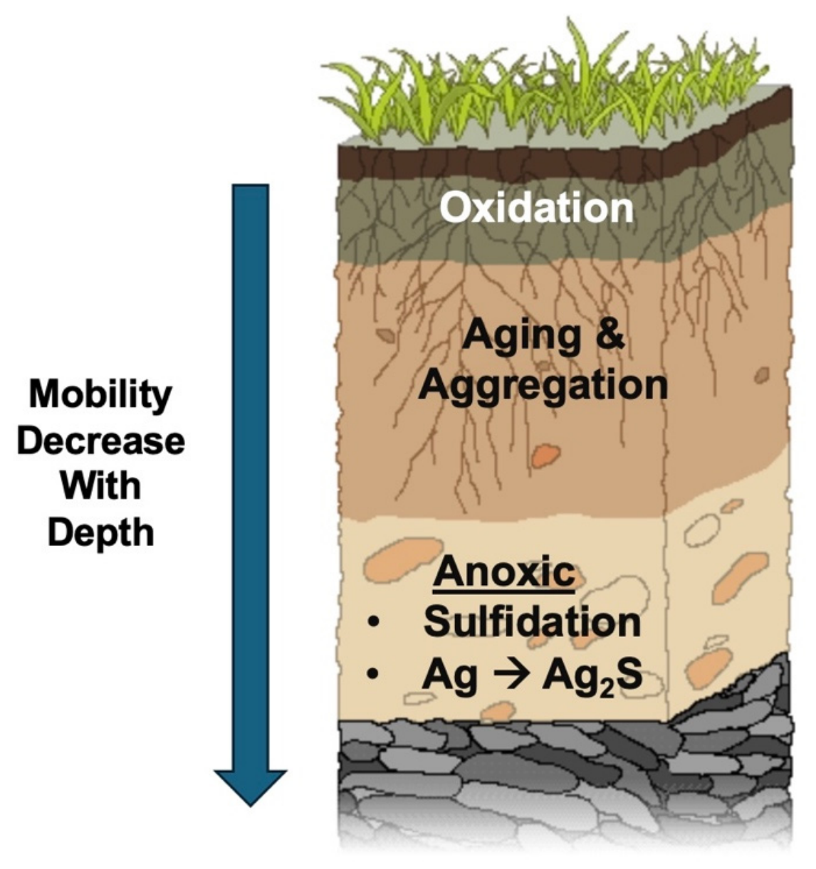
In conclusion, soils and sediments act as reactive sinks where NP transformations can continue to occur long after their initial release. Aging will typically cause NPs to associate more strongly with soil particles; sulfidation can occur rapidly under sulfide-rich conditions and typically decreases ion release (for metal NPs). In addition, passivation can decrease NP reactivity. Depending on the specifics, these transformations can lead to decreased acute toxicity by reducing dissolution or shifting exposure from acute waterborne exposure to chronic exposure via contact with or ingestion of contaminated sediments. Therefore, understanding how environmental conditions affect NP transformations is key to understanding their ultimate toxicological impact in soils and sediments [14,78,79]. Figure 5 illustrates examples of transformation pathways in soils/sediments, including oxidation, aging, and sulfidation.

4.3. Atmospheric Pathways and Inhalation-Relevant Transformations

Airborne NPs can be directly emitted (primary) or formed from gas-phase precursors (secondary). Secondary particle formation involves chemical reactions of gas-phase species prior to partitioning into the particulate phase. Volatile organic compounds (VOCs), sulfur oxides (SOx), and nitrogen oxides (NOx) readily oxidize in the atmosphere to form lower volatility products that nucleate particles or condense onto preexisting particles [83]. Secondary particle formation has been increasingly recognized as the predominant source of atmospheric ultrafine particles, contributing significantly to ambient particle number concentrations both outdoors and indoors [84,85]. Freshly nucleated particles are often in the nanometer size range and grow rapidly via condensation and coagulation to form a broad spectrum of particles that can evolve on short time scales. Chemical transformation of airborne NPs continues during transport via oxidative processes. Atmospheric oxidants (ozone, OH radicals, NOx) can react with particle surfaces to initiate multiphase reactions that change surface chemistry and composition. Oxidation can increase particle polarity and introduce functional groups that promote interactions with water and biological surfaces. Oxidation may also drive particle growth and alter hygroscopic properties that affect deposition mechanisms following inhalation exposure. Multiphase oxidation reactions have now been shown to play a central role in the formation and atmospheric aging of anthropogenic aerosols, dramatically impacting both physicochemical properties and potential biological impacts [85].
Particle size greatly impacts doses from inhalation exposures. Ultrafine particles (<100 nm) have high diffusivity and are small enough to deposit deeply into the lung alveolar region. Recent observations have shown even higher concentrations of nm-sized particles between 1 and 3 nm being formed from routine indoor sources such as during cooking activities. Termed nanocluster aerosols, these particles are not commonly measured by routine air quality monitors such as PM2.5, yet they can dominate particle number concentrations indoors and may contribute significantly to inhalation exposure [84]. Rapid changes in size from condensation and coagulation do not preclude efficient lung deposition, however, due to their small size. Additionally, high surface area and chemical reactivity resulting from particle transformation processes can promote biological interactions such as oxidative stress responses.
In summary, secondary atmospheric processes result in a dynamic environment in which NPs are formed and chemically transformed prior to exposure. Secondary formation processes dominate particle number concentrations in the atmosphere, while oxidative aging alters chemical composition. Both factors can influence how particles deposit within the respiratory tract and interact with biological surfaces. Atmospheric transformation should therefore be carefully considered when characterizing NP exposure.

4.4. Eco-Corona Formation and Aging

Following release to environmental media, NPs rarely exist in their original state for any length of time. Instead, NPs rapidly adsorb the natural and biological components present in the environment to form what is commonly known as an eco-corona. The eco-corona consists of a mixture of naturally occurring organic matter, microbial byproducts, protein biomolecules, and other dissolved organic matter that becomes attached to NP surfaces. Due to the rapid formation of this layer upon exposure to environmental conditions, the eco-corona essentially becomes the particle’s surface, dictating how the particle will behave and interact with its surroundings [86,87].
The adsorption of environmental molecules has been shown to impact the biological uptake of NPs. In certain cases, formation of the eco-corona will effectively mask the NP surface and prevent cellular interactions, leading to reduced uptake. In other cases, adsorbed molecules may contain functional groups that promote binding to cellular receptors, leading to enhanced uptake. Composition of the eco-corona is strongly influenced by environmental conditions and surrounding media, including pH, ionic strength, and type of natural organic matter present, all of which can alter corona structure and composition [39].
Eco-corona formation can also impact NP stability and transport. Formation of a natural organic matter coating can increase NP stability by imparting charge repulsion or steric stabilization, leading to longer suspension times. Increased stability can prolong particle transport and the potential for exposure across environmental media. At the same time, natural colloids and suspended solids can cause adsorbed NPs to aggregate and settle, redirecting them toward soil and sediment compartments. Overall, formation of the eco-corona can impact both the dispersion and ultimate fate of NPs in the environment [39].
The toxicity of NPs has also been shown to correlate with eco-corona formation in many cases. Immediately following release, the eco-corona can act as a barrier, to reduce reactivity and slow the dissolution of toxic ions, particularly for metal-containing NPs. By sequestering reactive surfaces, the eco-corona can effectively decrease oxidative stress and other toxicological responses. However, a decrease in short-term reactivity does not necessarily equate to a decrease in risk, as prolonged environmental or biological exposure times can still occur due to enhanced NP stability [87].
At times, eco-corona formation may even enhance NP toxicity rather than reduce it. The eco-corona can mediate transport across biological membranes or alter how NPs are distributed following uptake into organisms. Additionally, because eco-coronas can facilitate the transport of NOM, they may also contribute to the transport of other environmental contaminants. Last, eco-coronas are dynamic and can evolve with time as NPs age or undergo biological interactions. Continual restructuring of the eco-corona can expose new particle surfaces or alter behavior over an extended period of time, causing delayed effects that differ from the initial toxicity observed upon release [39].
Due to this complexity, eco-corona formation should be considered one step in a larger aging process. As NPs migrate from one environmental compartment to another, they will continually adsorb and shed organic materials to develop a surface coating reflective of their current chemical surroundings. These changes in surface composition will affect NP stability, transport, and biological interactions. Therefore, toxicity is not static but will evolve as the NP ages across different environments. In some instances, this may mean early transformation events reduce toxicity, while later transformations facilitate persistence or alter exposure routes [39,88].
In conclusion, the eco-corona serves as an important interface between NPs and their environment. While it can decrease initial NP toxicity by reducing reactivity and dissolution rates, the eco-corona can also increase particle stability, prolong residence time, and alter biological interactions. Ultimately, how eco-corona formation affects NP fate and toxicity is context-dependent, underscoring the importance of studying eco-corona formation and dynamics during NP risk assessment.

4.5. Transformation Products and Secondary Phases

ENPs typically undergo chemical transformations once released into environmental systems. The chemistry of the dissolved oxygen, reduced sulfur species, and NOM produced are most relevant to this process, yielding secondary phases such as metal oxides, metal sulfides, and surface bound complexes. Such transformations can alter NP composition, size, shape, dissolution rate, and other properties that influence behavior and toxicity. Because the properties of these transformation products often differ from the as-released NP, they are an important factor in determining ENP fate and toxicity [14].
Under oxidizing conditions, ENPs will likely undergo oxidation reactions that produce oxide coatings or completely oxidized particles. Oxide layers passivate particle surfaces, decreasing dissolution rate and ion release. Oxidation can also alter particle surface charge and redox potential, which may affect aggregation and interactions with biological surfaces. Oxide formation is generally thought to decrease acute toxicity, though exceptions are observed due to the ability of oxidized particles to participate in redox chemistry [14].
In contrast, ENPs will undergo sulfidation reactions under reducing conditions. Environments rich in dissolved sulfide include sediments, wastewater treatment systems, and localized microzones lacking oxygen. The sulfidation process forms metal-sulfide compounds (e.g., Ag2S), which are often much less soluble than their parent ENPs. Dissolution has been linked to ENP toxicity for many materials, making sulfidation an important pathway for decreasing short-term ENP toxicity. For instance, researchers have shown that the transformation of metal oxide ENPs into metal sulfides greatly diminishes observed toxicity, which is attributed to the lower dissolution rates of the sulfide [89]. Contrary to expectations, several studies have also demonstrated transient increases in biological effects with sulfidation. Partial transformation or dissolved ion concentrations that do not decrease with sulfidation have been suggested as possible explanations [90].
In addition to oxide and sulfide formation, dissolved NOM, biomolecules, and inorganic ligands can bind to ENP surfaces and form surface-associated complexes. Binding of NOM can impart colloidal stability, affect aggregation kinetics, and influence ENP transport through media. Surface complexes can also alter NP interactions with biological systems by changing how cells recognize and take up ENPs, partially through modifications to the biomolecular layer that adsorbs to NP surfaces. Depending on the system, surface complexation can either increase or decrease toxicity through these mechanisms [91].
In summary, the toxicity of ENP transformation products will largely depend on environmental conditions and the mechanisms of transformation. Oxidation and sulfidation often reduce acute toxicity by decreasing dissolution and subsequent ion release. However, these transformation processes can enhance ENP persistence and shift effects to chronic time scales. Transformation can also introduce new mechanisms of action by altering ENP surface chemistry and reactivity, promoting stability, or affecting ENP partitioning to regions of the environment. In some cases, transformation products may even increase toxicity, such as when NPs transform into more toxic compounds. Recent evidence indicates that environmental transformations often do not decrease risk but shift it from an acute, ion-mediated mechanism to a chronic, particle-mediated effect [14].
Transformation into secondary phases such as metal oxides, metal sulfides, and surface complexes is an important process controlling ENP fate and toxicity. Environmental transformations change the chemical identity of ENPs, making it critical to consider transformed ENPs during risk assessment. Rather than treating ENPs as static materials, it is necessary to view them as dynamic and evolving with changing environmental conditions. An overview of dominant transformation pathways and the associated general trends in toxicity across environmental compartments is shown in Table 5.

5. NP Exposure Pathways and Ecological Transfer

NPs will not remain in the same environmental compartment into which they are released. They are transported through physical and biological processes to other environmental compartments based on where exposure happens and how effects will transfer. These processes include both transport (advection, sedimentation, diffusion) and transformations that can change NP size, surface chemistry, or association to natural particles. They may cause NPs to remain suspended or to become incorporated into sediment or biofilms on organisms. Exposure often happens over time and during various processes, as particles are transported between media such as water, soil and organisms. This concept is important to consider when linking exposure to effects, because environmental fate processes link directly to uptake, accumulation, and transfer processes at the organism level [78].

5.1. Microbial Interactions and Functional Disruption

Species-dependent responses of microbes to ENPs have been observed. Microbial response to nanoparticles can also depend on properties of the nanoparticles themselves (i.e., composition, size, surface chemistry, concentration), environmental parameters and differences in cell envelope among microorganisms. In soils, sediments and surface waters, microorganisms can be one of the first organisms to come into contact with ENPs. Exposure to ENPs has been associated with changes in microbial community composition, although the magnitude and direction of these responses differ among studies and microbial taxa. Reported effects include reduced microbial diversity in some systems and shifts in community structure or dominance patterns in others, depending on the nanoparticle characteristics and exposure conditions. It is important to consider these changes, as microorganisms are responsible for driving many ecosystem services, such as nutrient cycling, organic matter decomposition and redox transformations [78].
Microorganisms can be affected by NPs through a variety of routes. These NPs can interact directly with the membrane or generate ROS that can damage proteins, lipids, and DNA. In metal systems, dissolved ions can act directly once released from the NP, which can contribute to overall toxicity. The damages that occur through ROS formation or ion dissociation can inhibit/enhance enzyme activity and overall microbial metabolism, leading to decreased microbial efficiency. Microorganisms can also alter quorum sensing and biofilm formation efficiencies. Alterations in community-level properties can affect how microorganisms function within ecosystems [92].
Microbial processes drive a variety of ecosystem functions. Because of this, any changes to microbial activity could alter nutrient availability, ecosystem cycling, and overall ecosystem health. For example, the inhibition of nitrifying bacteria can impact overall nitrogen cycling efficiency. Shifts in decomposer community composition can also impact rates of organic matter breakdown. In this way, NP exposures can create cascading effects from the micro to ecosystem scale [78].

5.2. Primary Producers and Food Web Transfer

Primary producers act as a gateway for NP exposure into many ecosystems. Algae and plant cells are consistently exposed to NPs in aquatic environments through exposure from water, sediment and soil. They also can take up NPs from these environmental compartments through surface adsorption followed by internalization. NP uptake into algae generally occurs through attachment to the cell surface. From there, particles can either be taken up by the cell or remain on the surface. Root uptake of NPs by plants generally introduces NPs into the root, and some translocation to the stem and leafy sections of plants can occur depending on the surface properties and size of the NPs [39].
Under controlled experimental exposure conditions, surface adsorption has been shown to alter photosynthesis, nutrient uptake, and plant growth. At environmentally relevant concentrations, these effects are generally more variable and depend on nanoparticle properties and environmental conditions. Oxidative stress is commonly seen at higher NP concentrations and can lead to decreases in growth and overall productivity. Environmental conditions can play a large role in these responses, as pH, ionic strength, and organic matter can all influence NP behavior prior to exposure [39,87].
Primary producers are key players when discussing the trophic transfer of NPs. Many species directly ingest algae or plant tissues, allowing for NPs associated with these organisms to be transferred through the food web. The transfer of NPs from algae to Daphnia magna and onward to goldfish has been shown through experimental studies. The question of whether NPs biomagnify or biodilute through trophic transfer is still unanswered. Factors such as transformation, metabolism, and feeding behaviors all play a role in the transfer of NPs. As mentioned earlier, some NPs can become associated with natural organic matter or taken up into cells, which may allow for easier movement through the food web [39].

5.3. Invertebrates and Vertebrates

Invertebrates and fish/vertebrates can be exposed to NPs through contact with the surrounding environment as well as consumption of other prey organisms that have accumulated or ingested NPs. Aquatic invertebrates such as crustaceans, worms, and mollusks are of specific interest because they are found at lower levels of the food web and are more likely to interact with sediments and suspended particles. These organisms can ingest NPs from water columns or surrounding sediments, which can lead to accumulation in the digestive system. Experimental studies have reported alterations in growth, reproduction, and behavior following NP exposure, although these effects are frequently observed under controlled laboratory conditions using concentrations that may exceed typical environmental levels [93].
Some species of invertebrates live within the sediment, where they are exposed to NPs in porewater, adsorbed to sediment particles, or associated with organic matter. This makes ingestion and accumulation of these NPs very likely. Studies have found that this combination of exposure pathways and environmental transformation lead to the bioavailability and accumulation of NPs in benthic organisms [78]. Environmental transformations such as aggregation and bio-corona formation can alter the ability of an NP to interact with biological tissues, which can decrease reactivity but may increase persistence.
Fish and terrestrial invertebrates/vertebrates experience exposure routes similar to previously mentioned organisms. Exposure can happen through ingestion, contact with skin or gills, and the uptake of particles through diets. Some of these NPs can accumulate in different tissues, such as the liver, kidneys, and brain. Experimental studies have demonstrated oxidative stress and inflammatory responses following NP exposure. While environmentally relevant exposures may also pose ecological risks, the magnitude of these effects under field conditions remains less well characterized. As stated before, these organisms are likely not exposed to pristine NPs but ones that have been altered in the environment [39,78].

5.4. Mixtures and Co-Stressors

Complete exposure scenarios will often involve mixtures of chemicals and/or stressful conditions. ENPs will not exist in the environment on their own. There will likely be other contaminants, biotic factors, and abiotic stressors present that could affect the overall toxicity of NPs. One common example is the binding ability of NPs. Many organic contaminants and metals have been shown to bind to the surface of NPs. This could allow for enhanced transport of these chemicals, as they can be introduced to organisms on NP surfaces. However, binding can also decrease contaminant bioavailability, leading to less toxicity. The strength of the bond and environmental conditions play a large role in determining which way the interaction goes [86].
It is critical to separate discussions of contaminant transport from combined toxicity. While examining responses to ENP/co-contaminant exposures, some published studies report the response to alteration of contaminant transport properties (such as increased transport or delivery to an organism) by ENPs rather than an increased toxicity response. In these cases, ENPs function primarily as carriers through adsorption of the contaminant to the ENP surface. Other studies legitimately show combined biological interactions of ENPs and contaminants that lead to altered toxicological responses. Such effects can be additive, synergistic, or antagonistic, depending on the characteristics of the ENPs and co-contaminant under specific environmental conditions and the biological system examined. Below, we try to differentiate between these mechanisms where the literature allows.
Environmental conditions can also impact NP transformations. Variations in pH, temperature, and even ionic strength can impact NP dissolution or surface charge, altering how they will interact in the environment with co-contaminants and organisms. As mentioned before, organic matter can passivate NPs, which can decrease NP toxicity but can also enhance transport of these particles. Temperature and redox conditions can also influence transformations of both ENPs and organism sensitivity to ENPs [78].
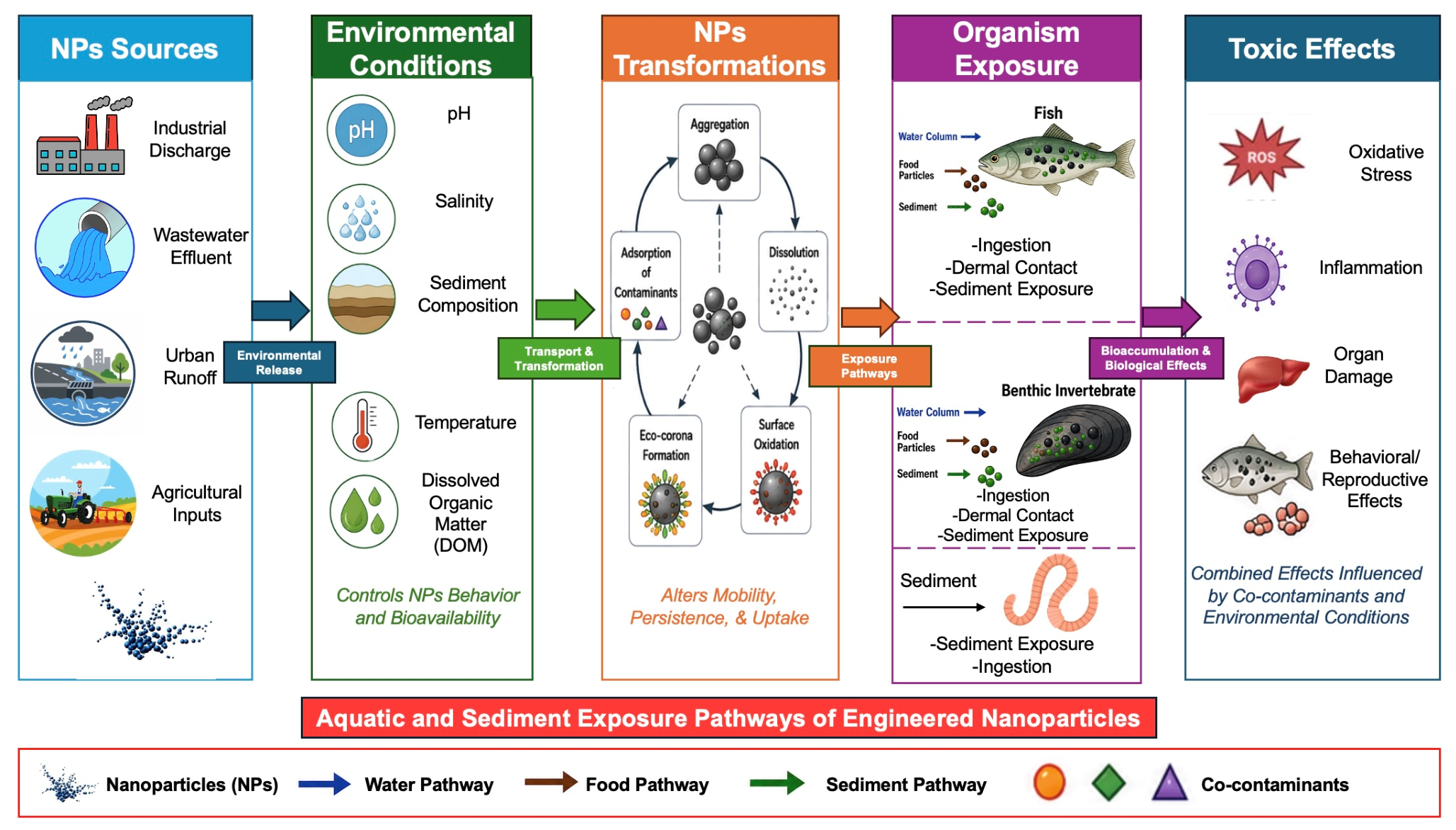
Due to these many factors, it is hard to determine how ENPs will behave in the real world. For this reason, it is important to evaluate the effects of ENPs under a variety of conditions and take into consideration how the environment can alter their toxicity. In some cases, mixture exposures can have additive or synergistic effects, while others can cancel each other out. It is important to recognize these possibilities when understanding how ENPs will affect biological organisms and the environment [78]. Figure 6 summarizes the major environmental factors influencing nanoparticle transport, transformation, exposure pathways, bioaccumulation, and toxicity under environmentally realistic conditions. Environmental parameters influence nanoparticle fate and bioavailability. Once in the environment, NPs can undergo various transformation processes, including agglomeration/aggregation, dissolution, oxidation, eco-corona formation, and contaminant desorption, which can affect how organisms are exposed (i.e., exposure routes). Exposure can lead to oxidative stress, inflammation, damage to organs, and reproductive and behavior effects. Toxicity can be altered by the presence of other contaminants. This figure illustrates aquatic and sediment exposure pathways. Airborne transport and inhalation exposure are discussed separately in the text.

6. NP-Bio Interactions Relevant to Human Health

After release into the environment and subsequent physicochemical modification, ENPs will undergo nano–bio interactions that lead to adverse outcomes. ENPs interact with biological fluids and form a protein corona that changes their physiological identity and impacts cellular recognition and internalization, biodistribution, and clearance [56,94]. These biological behaviors regulate the toxicokinetic behavior of these materials (i.e., absorption, distribution, cellular uptake, biotransformation, and excretion), which controls what dose reaches target tissues [95,96,97]. Toxicodynamic outcomes include oxidative stress, inflammation, immune modulation, membrane disruption, and genotoxicity, as described below.

6.1. Human Exposure Pathways

Humans can be exposed to ENPs through inhalation, ingestion, and dermal contact. Inhalation is considered the most efficient route, as NPs that are suspended in the air can be inhaled into the respiratory system. Airborne nanoscale particles originate from engineered sources, naturally occurring processes, and incidental combustion-derived ultrafine particles (UFPs). Because this review focuses on ENPs, the subsequent discussion refers specifically to ENPs unless general respiratory deposition mechanisms derived from UFP studies are being described. Inhaled NPs can travel deep into the lungs, where they interact with surfactants, epithelial cells, and alveolar macrophages. Experimental studies using ENPs have demonstrated that inhaled particles can translocate across the air–blood barrier and distribute to distal organs such as the liver, spleen, kidneys, and brain [30,98].
Deposition and distribution throughout the body is strongly related to the surface properties and size of the NPs. Smaller particles have been shown to diffuse more rapidly through biological media and have been shown to have increased surface reactivity. As NPs reach the air–liquid interface, many environmental transformations can occur. Factors such as protein corona formation and aggregation can change the behavior of NPs throughout the body [30].
Ingestion of NPs can occur through food, drinking water, and materials that encounter food. Studies have shown that engineered NPs can be introduced into foods through direct addition, migration from food packaging materials, and the environmental contamination of raw products [98]. Once consumed, NPs interact with a dynamic gastrointestinal environment. The gastrointestinal milieu is made of changing pH conditions, digestive enzymes, bile salts, and gut microbiota. These conditions will impact the stability, aggregation, and dissolution of NPs, which determine the bioaccessibility of NPs [32].
Therefore, the ingested or nominal concentration must be differentiated from the gastrointestinal bioaccessible dose, since what is ingested may not be representative of the dose available to interact with and/or be taken up across the gastrointestinal epithelium. Nanoparticles may experience physicochemical transformations during transit through the gastrointestinal tract that modify their aggregation, dissolution, and subsequent biological effects [26,98]. Alterations can impact the form and magnitude of material available for intestinal uptake and ultimately impact the dose received [26,68]. As a result, gastrointestinal transformations should be accounted for when extrapolating an ingested nanoparticle concentration to a potential adverse outcome.
Unlike inhalation, the uptake of NPs across the intestinal epithelium is generally low. However, NPs that are less than ~100 nm have been shown to translocate across biological membranes through transcellular pathways or to be taken up by specialized cells called M cells. After systemic uptake, NPs can be distributed to other organs and tissues through the body.
Skin also serves as a route of exposure for NPs. Minimal penetration has generally been observed through intact skin. However, NP uptake through the skin may increase following the wear or degradation of nano-enabled materials, which releases NPs, or when the skin barrier is compromised (e.g., damaged or abraded skin). Regulatory agencies have concluded that the penetration of intact skin is dependent on NP physicochemical properties, including size distribution, surface chemistry, and exposure duration [32]. Despite inefficient uptake through healthy skin surfaces, NPs have shown detectable penetration through skin under certain circumstances. ENP exposure cannot be easily quantitatively characterized due to variable concentrations across occupational, consumer, and general environmental settings and because particle mass concentration alone is not sufficient to characterize nanoparticle dose. High mass concentrations of airborne particles have been reported in occupational settings related to ENP production, handling, and spraying, as well as the processing of powders. Consumer exposures are extremely variable and are highly dependent on the product used, route of application, and extent to which particles are released. In general, environmental settings, concentrations are orders of magnitude lower than occupational settings and are difficult to measure, as ENPs must be differentiated from the natural and incidental nanoparticles present within complex matrices. For these reasons, exposure characterization should include particle size distributions, and if available, exposure assessments should report particle number concentration, mass concentration, and surface area rather than using one measure based on mass. These measures are particularly important to differentiate because the same mass concentration of differently sized nanoparticles could translate to different particle numbers and reactive surface areas, resulting in different biologically effective doses [45,99].
A further distinction should be made between nominal, delivered and internal dose. Nominal dose is the concentration applied or present initially in an exposure medium. Delivered dose is that amount which actually contacts or reaches the biological interface or target surface. For nanoparticles, nominal and delivered doses may be quite different, since diffusion, sedimentation, aggregation, dissolution, and other modifications will change the delivered dose form and amount reaching cells or tissues [30]. Internal dose is that fraction of the delivered dose that is then absorbed or internalized and enters systemic circulation, cells, or target organs. As such, toxicity occurring at the nominal concentration may not be representative of the dose that the biology actually received. Dosimetry for nanoparticles should consider particle size, particle number, surface area, aggregation/agglomeration state, dissolution, and transformation during exposure when quantifying the delivered concentration to biological response [30].
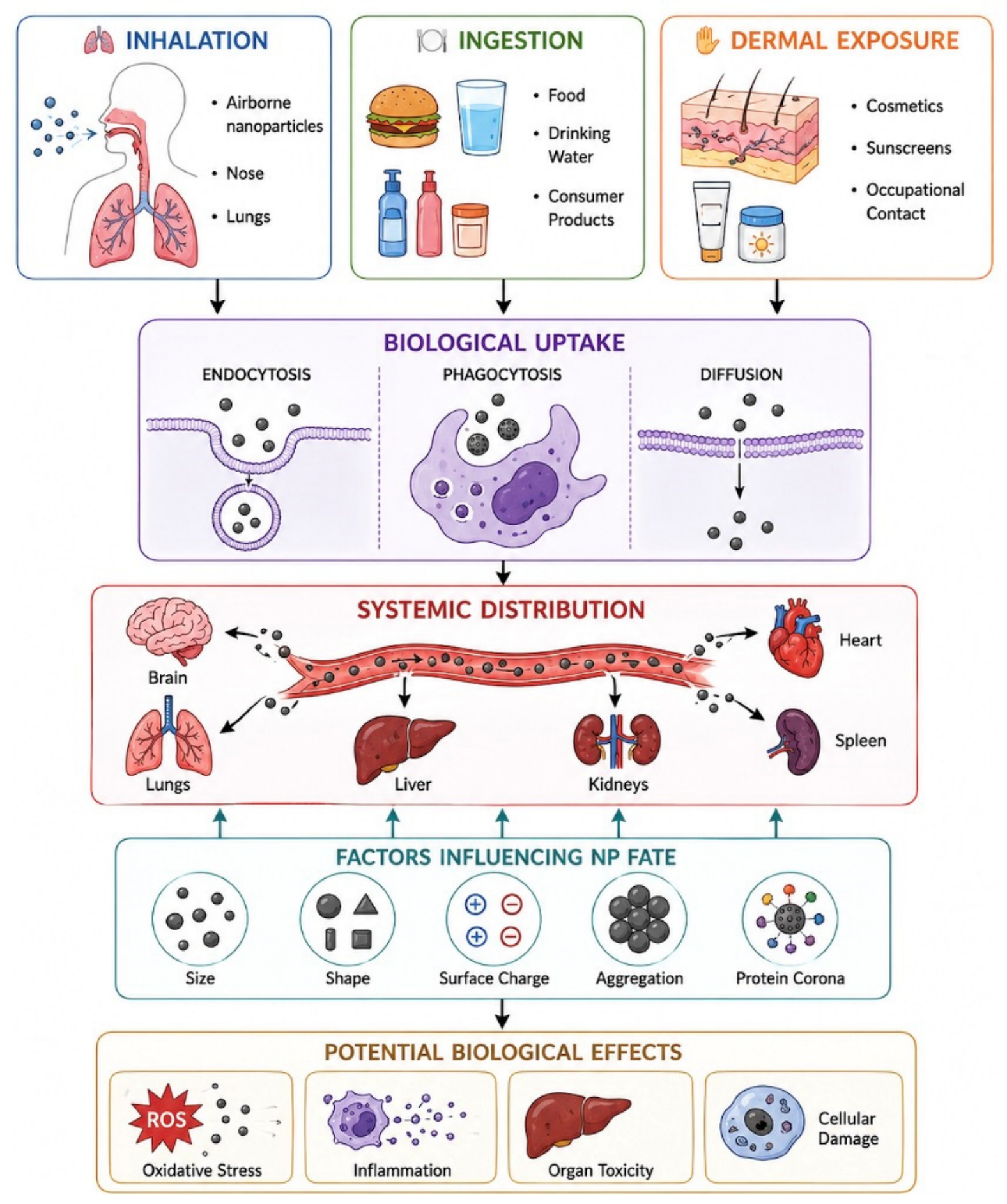
Environmental transformations play a key role in all routes of exposure. External exposure does not define toxicological outcomes. Interaction with biological fluids, cellular uptake, and distribution throughout the body after exposure is what determines NP fate within the body [98]. Figure 7 summarizes the progression from nanoparticle exposure to biological effects, highlighting the critical roles of cellular uptake mechanisms, systemic transport, and physicochemical properties in determining nanoparticle fate and toxicity. Inhaled NPs: exposure to airborne NPs in the respiratory tract. Ingested NPs: exposure through food, drinking water, consumer products, cosmetics or sunscreens into the gastrointestinal tract. Dermal exposure: exposure through skin contact during work hours; transdermal penetration/uptake. Biological uptake: endocytosis, phagocytosis, or passive diffusion into cells. Distributed to: central nervous system (brain), respiratory system (lungs), hepatic system (liver), renal system (kidneys), cardiovascular system (heart), lymphatic system (spleen). Physicochemical properties of NPs: particle size, shape, surface charge, aggregation/agglomerated state, protein corona. Toxicity: oxidative stress, inflammation, organ toxicity, and cell damage.

6.2. Protein Corona and Physiological Identity of Nanomaterials

Upon entering the body, NPs will immediately be exposed to soluble biomolecules that comprise complex media such as blood. A biomolecular corona will form on the NP surface within seconds to minutes of exposure and will define the NP’s biological identity. How the NP interacts with biological media will depend on the protein corona that forms on its surface, not its engineered surface. The corona is determined by both NP properties (size, charge, hydrophobicity/hydrophilicity) and the environmental conditions (protein and lipid concentrations) to which it is exposed. The corona is continuously evolving as NPs travel through various biological compartments and become exposed to different proteins and environmental conditions [94,100].
Protein corona formation is initiated by rapid adsorption of high abundance plasma proteins such as albumin, immunoglobulins, fibrinogen, and apolipoproteins. This interaction is facilitated by electrostatic forces, hydrophobic forces, and van der Waals interactions. Over time, low-affinity proteins will be replaced by proteins with a higher affinity to the NP’s surface, generating a “hard corona” that is surrounded by a “soft corona” of proteins that dynamically exchange with the environment [26,28]. Recent reviews have shown that corona formation is not a passive process. When proteins bind to the surface of NPs they can undergo conformational changes. Structural changes can affect protein function and can alter how the NPs will interact with biological systems such as cell membranes and biological barriers [101].
Cellular uptake and distribution throughout the body will be affected by the protein corona on NP surfaces. Since the corona acts as the NP’s new identity, how it will distribute throughout the body will be defined by the protein corona characteristics. For example, a protein corona rich in opsonin will target the NPs to the liver and spleen through macrophage recognition. A different protein corona could allow the NPs to evade the immune system, leading to prolonged circulation time and distribution throughout the body [100]. Recent studies have shown that even slight changes in corona composition can alter NP diffusion throughout biological fluids. Since biological fluids are crowded environments, protein–NP interactions can play a large role in NP transport [102].
Immune recognition of NPs is also driven by the protein corona. The identity of the protein corona (what proteins are bound to the NP surface) and the conformation of these proteins (if the protein changes structurally after binding to the NP surface) can determine if the NPs will elicit an immune response or not. Depending on how the protein conformation changes can determine if the protein will activate the complement system, causing downstream inflammatory responses. Structural changes can also hide reactive surfaces on the NPs, decreasing recognition and allowing the NPs to go unrecognized by the immune system [94]. These findings suggest that nanoparticle-induced immunotoxicological effects can be blamed not on the physicochemical properties of the NP itself but rather on the interactions between the NP surface and the proteins within the biological media it enters [100].

6.3. Cellular Uptake and Organ Distribution

Regardless of the route of entry into the body, most NP–cellular interactions are via active transport processes. Endocytosis is the general term used to describe the cellular uptake of NPs and includes clathrin-mediated endocytosis, caveolae-mediated uptake, macropinocytosis, and phagocytosis. These pathways differ across cell types and NP formulations, therefore making it difficult to generalize which pathway is preferred. Cellular uptake is toxicologically relevant because it can determine where particles localize within the cell and whether or not they have access to sensitive cellular compartments [103,104].
Experimental studies of engineered nanoparticles have shown that inhaled ENPs deposited in the lung do not always remain localized. Similar pulmonary deposition patterns have also been described for combustion-derived ultrafine particles, although these particles differ from ENPs in their source, composition, and physicochemical properties. Studies have shown that inhaled NPs can be taken up by airway or alveolar epithelial cells and pulmonary alveolar macrophages. From the lung, NPs can enter the bloodstream and distribute to other organs and tissues. This could help explain why we see effects of inhaled NPs distal to the lung, including the accumulation of particles in organs that serve as filtration and detoxification organs as well as other sensitive tissues [105,106].
NP movement across biological barriers is of considerable interest because it influences systemic distribution and potential toxicity. Evidence describing translocation across biological barriers includes both ENP-specific studies and investigations of combustion-derived ultrafine particles. These particle categories are discussed separately because they differ in origin, composition, and exposure scenarios. While selective, many studies done in vitro and experimentally have shown that NPs are able to penetrate through the blood–brain barrier through receptor- and adsorptive-mediated transcytosis when functionalized or engineered with the appropriate ligands for targeted drug delivery. Ligand-modified lipid and polymeric NPs have been shown to cross the BBB via receptor-mediated transcytosis, and surface charge and size have also been shown to aid in transcytosis across the BBB [107]. Also, the breakdown of the BBB due to inflammation, disease, or injury can lead to greater nanoparticle accumulation into the brain.
Another biological barrier that NPs have been shown to cross is the placenta. Studies performed using human placental perfusion models, trophoblast cell models, and ex vivo human placental tissue have shown that certain NPs (polystyrene, silica, gold, and lipid NPs) are able to cross the placenta, but translocation depends on nanoparticle size, composition, surface chemistry, and stage of gestation [108].
However, these studies do not suggest that all NPs are able to freely cross these biological barriers. What they show is that certain types of NPs have the ability to cross the BBB or placenta when specific physicochemical properties and biological conditions are present. These findings should be taken into consideration when assessing human health risks [107,108]. Table 6 lists the main routes of NP uptake, biological barriers, distribution into organs, and the resulting toxicological effects, as studied in experimental models.

6.4. Immune System and Inflammatory Responses

Immune responses can be affected by NP exposure. Once NPs are exposed to biological fluids, they will acquire a protein corona. This protein corona will determine how the immune system recognizes the NPs, which receptors will be activated upon NP interaction, and if the response will be muted or if it will continue to activate downstream immune responses. Therefore, immune responses to NPs can range from toleration to releasing large amounts of cytokines and driving inflammation. The disease state of an individual can also alter protein corona composition, which would alter how immune cells interact with the NP and could modulate cytokine production [109].
One of the first indications of inflammation is cytokine activation. Studies have shown that accidental NP–immune system interactions can lead to inflammation through cytokine release and activation of the complement system. Severe cases have shown responses similar to cytokine release syndrome. Inflammation becomes a concern when NPs begin to accumulate in the body either through an inability to clear them or continuous exposure. Inflammatory responses are also elevated when NPs are introduced into an already inflamed environment [83,109,110].
Inflammation can become a chronic issue if exposure continues over a long period of time and the body is unable to clear NPs. Continuous retention of NPs has been linked to prolonged macrophage activation, oxidative stress, and prolonged signaling within the affected tissue or organ. Inflammation is not always captured in traditional toxicity tests because they use short-term cytotoxicity assays. Factors such as how long the NP remains in the body, repeated exposures, and if the exposed individual was already in an inflammatory state all play large roles in the development of chronic effects [106,109,111].
Table 6. Biological uptake pathways, barrier crossing, and toxicological consequences of nanoparticle exposure.
ProcessMechanismBiological ConsequenceExample FindingsReferences
EndocytosisCellular uptake through membrane invaginationIntracellular accumulation and altered cell functionCellular uptake efficiency depends on NP size, charge, and surface chemistry[103]
PhagocytosisEngulfment of particles by macrophages and immune cellsParticle clearance, inflammatory signalingMajor uptake pathway for larger NPs and aggregates[112]
Pulmonary TranslocationMovement from lung alveoli into systemic circulationDistribution to secondary organsInhaled NPs detected in liver, spleen, kidney, and brain[108]
Blood–Brain Barrier (BBB) CrossingTransport through endothelial cells or tight-junction disruptionNeuroinflammation and oxidative stressSmaller NPs show enhanced BBB penetration[107]
Placental TransferMaternal–fetal translocation across placental tissuesPotential developmental toxicityMultiple engineered NPs have been detected in fetal tissues[107,108]
Protein Corona FormationAdsorption of proteins onto NP surfacesAlters cellular recognition and biodistributionCorona composition influences uptake and toxicity profiles[56,94]
Cytokine ActivationInduction of inflammatory mediators (TNF-α, IL-6, IL-1β)Acute and chronic inflammationCommon response following NP exposure in immune cells[27]
Oxidative StressExcess generation of ROSDNA damage, lipid peroxidation, apoptosisFrequently reported mechanism of NP toxicity[26]
Although associations between NP exposure and autoimmune disease are still being explored, there is currently no indication that NP exposure can induce autoimmune disease. It is important to note that NP-mediated immune modulation has been identified when exploring NP interactions with the immune system. It has been shown that NPs are able to modulate antigen presentation, cytokine production, and tolerance pathways. This property is currently being applied to create NP therapies for autoimmune diseases. Immune modulation caused by NP exposure has the potential to impact immune homeostasis in predisposed individuals. Instead of causing autoimmune disease, NPs have been shown to either potentiate or suppress the immune response based on NP physicochemical properties, exposure circumstances, and host genetics, immune health, and underlying disease. Therefore, we can conclude that NP-mediated immunological effects are likely dependent on exposure and individual parameters, and further research is needed to determine any associations with autoimmune disease [113,114].

7. Toxicity Mechanisms and Human Health Endpoints

7.1. Oxidative Stress and Reactive Oxygen Species (ROS)

A commonality between these findings is that oxidative stress is involved in NP-induced toxicity. This is due to an imbalance between ROS generation and antioxidant capacity. The bulk of evidence for oxidative stress comes from in vitro cell culture and animal studies, where ROS generation has been one of the most commonly reported mechanisms of action. Evidence in humans is mostly limited to occupational exposure studies, which have reported measures of oxidative stress. Little data exist from the general population. In normal physiology, ROS are produced continuously as byproducts of cellular metabolism and are kept in check by enzymes and antioxidant defenses. However, when ROS production exceeds capacity for clearance and scavenging, ROS begin to damage sensitive molecules, initiating downstream consequences, which result in tissue damage and disease [115,116].
The mitochondria are one of the first targets of ROS-mediated damage. Exposure to NPs has been shown to disrupt the electron transport chain, resulting in electron leakage and generation of ROS. As ROS attack mitochondrial membranes, cellular respiration decreases, decreasing membrane potential and further exacerbating oxidative stress in a positive feedback loop that can ultimately lead to apoptosis [117,118].
ROS can also attack membrane lipids, starting lipid peroxidation reactions. Lipid peroxidation changes the physical properties of membranes, including fluidity and permeability, which compromises membrane structural integrity and normal cell signaling. In addition to damaging the membrane, byproducts of peroxidation such as malondialdehyde can initiate additional oxidative stress responses and inflammation [115].

7.2. Ion-Mediated Toxicity

Concurrently with oxidative stress responses, several metal and metal oxide NPs exert toxicity mediated by ion release. Metallic and metal oxide NPs often derive their toxicity from dissolved ions once they enter solution-based biological environments. This mechanism has been demonstrated primarily in in vitro studies and experimental animal models, particularly for metal-based ENPs such as silver and zinc oxide nanoparticles. Human evidence remains limited. Physiological fluids can dissolve metal NPs, releasing highly reactive ions like Ag+, Zn2+, and Cu2+ into cells and tissues [116,119]. Once inside cells, free ions can directly interfere with protein function. For example, cations like Ag+ and Hg2+ have high affinities for sulfhydryl groups on proteins, particularly enzymes. Binding can alter protein conformation and disrupt catalytic activity. The perturbation of antioxidant enzymes or other metabolic proteins further disturbs cellular homeostasis and promotes oxidative stress [116].
Ion flux can also disrupt Ca2+ signaling pathways. Calcium signaling must be tightly regulated, as fluctuations in calcium homeostasis are known to mediate everything from contraction to cell death. Exposure to NPs or dissolved ions can cause the release of calcium from lysosomes or mitochondria or interfere with calcium channels. Dysregulated Ca2+ can also trigger mitochondrial permeability transitions, leading to apoptosis or necrosis [118].
It should be noted that NPs can induce oxidative stress responses and ion-mediated effects simultaneously. As mentioned previously, oxidative stress is a predominant mechanism of NP toxicity, but many metals can trigger oxidative stress while also disrupting ion signaling and cellular metabolism. These processes can affect any organ with high energy demand or ion turnover disproportionately, which includes the liver, kidney, lungs, and brain. However, this correlation does not necessarily indicate causation, as multiple lines of evidence have found that NPs can accumulate in several organs after exposure and induce oxidative stress, inflammation, and cell death, leading to organ toxicity [97,120].

7.3. Membrane Disruption and Physical Stress

Physical disruption to cellular membranes is another established mode of NP toxicity. Evidence for membrane disruption is derived largely from mechanistic in vitro studies, with animal studies supporting tissue-level injury following ENP exposure. Direct evidence in exposed human populations is limited. After uptake by endocytosis, many NPs become sequestered in lysosomes, where the acidic pH can cause dissolution or surface reactivity. In some instances, this can cause destabilization of the lysosomal membrane, allowing proteolytic enzymes such as cathepsins to leak into the cytoplasm. Activation of cathepsin-mediated cell death has been linked to inflammation and cell death [116,117]. Mechanical interference is another physically mediated stress that can be caused by NPs. Nanoparticle characteristics such as size, shape, surface charge, and rigidity can affect their interactions with biological membranes and the cytoskeleton. For example, high aspect ratio nanomaterials (HARNs) or those with significant rigidity can have different cellular impacts than spherical NPs of similar size [121,122].
While not all NPs have been shown to exhibit asbestos-like properties, this is an area of concern for high aspect ratio and persistent nanomaterials. Certain HARNs, such as carbon nanotubes, have been shown to exhibit persistent inflammation and fibrosis, along with biomarkers of frustrated phagocytosis, when they are longer, thinner, and more bio-persistent [123,124].

7.4. Genotoxicity and Chronic Endpoints

Exposure to NPs has recently been associated with genotoxic effects, either due to direct interactions between particles and DNA or indirectly through oxidative stress and the production of ROS. Evidence for genotoxicity is supported primarily by in vitro assays and animal studies demonstrating DNA damage and chromosomal abnormalities. Human studies are comparatively limited and are largely restricted to occupationally exposed populations, making causal links to disease risk less certain. Damage to DNA can take many forms, but one of the most common consequences of NP exposure is stranding breaks. Both single- and double-strand breaks in DNA lead to mutations and changes in the genetic code if not repaired properly [116]. Epigenetic changes are another consequence of NP exposure and can lead to changes in cell regulation. NPs have been shown to alter DNA methylation, histone modification, and noncoding RNA expression. Unlike genetic changes, epigenetic modifications do not change the DNA sequence but can have large impacts on which genes are expressed [125].
The combination of genotoxic and epigenetic impacts has led to concern over NP carcinogenicity. Chronic inflammation, cellular stress, and genotoxicity can lead to tumor formation. The carcinogenic potential of NPs varies based on the type of NP and conditions, but there is some evidence that long-term exposures to certain NPs may increase cancer risk under certain exposure scenarios [117]. Additional chronic effects of NP exposure include neurotoxicity and endocrine disruption. Studies have shown that NPs can readily cross the blood–brain barrier and cause neuroinflammation oxidative stress, as well as neurotransmitter disruption. Similarly, NP exposure has been linked to endocrine-disrupting activity [118].

7.5. Sub-Lethal, Chronic, and Combined Effects

Cumulative evidence from the existing literature indicates that repeated or chronic exposure to nanoparticles can cause oxidative stress-related sublethal effects, metabolic changes, and immune response changes [115]. The available literature is still very diverse in terms of nanoparticle type, model used, concentration applied, and duration of exposure. The exposure levels used in many studies are experimental exposures that do not equate to real world or general population human exposures. For this reason, there is a lack of quantitative data exploring the chronic effects at low doses that would be seen environmentally. This is an important gap in knowledge, because human exposures will more realistically occur at lower concentrations over repeated or long-term exposures instead of the high doses seen in acute studies [115].
It should also be noted that combined or synergistic effects under co-exposure scenarios should be considered, particularly in complex environments. Field exposures to NPs will often involve co-exposure to metals, organic contaminants, or persistent pollutants such as PFAS. Interactions between NPs and other chemicals can affect particle bioavailability, transport, trophic transfer, and toxicodynamics. Combined stresses could conceivably be weaker, stronger, or qualitatively different from each stressor alone [35,126].
Developmental/reproductive effects also are a relatively understudied adverse outcome following exposure to nanoparticles. Developmental anomalies, decreased fertility, changes in embryonic development, and transgenerational effects have been observed in laboratory animal and fish models after exposure to nanoparticles [116,118]. However, variations in nanoparticle dose and composition, dosing concentration, length of exposure, and test model make it difficult to compare these studies to environmentally relevant and/or occupational human exposures. Data supporting similar developmental/reproductive effects due to exposure to nanoparticles in humans are limited.
Overall, the mechanistic evidence for ENP toxicity is strongest from controlled in vitro and animal studies. Occupational studies provide supportive evidence for biological effects in highly exposed populations, whereas evidence from the general population remains comparatively limited because of lower exposure levels and challenges associated with exposure assessment.

8. Detection and Characterization of NPs for Toxicity Assessment

8.1. Essential Properties for Toxicological Interpretation

The characterization of NPs should extend beyond bulk mass concentrations and should account for properties relevant to toxicological interpretation. Research from the past several years has shown repeatedly that NP toxicity is driven by multiple properties in combination. Size and size distributions are critical properties that control the surface area and chemical reactivity of particles. Surface state, including charge, coatings, oxidation state, and weathering (discussed below), also directly controls aggregation behavior and biomolecular interactions (corona formation, see Section 6.2). For metal NPs, dissolution must also be characterized due to the toxic effects of metal ions [127].
Particle number concentration is also an important property for exposure and toxicological consideration. Because a gram of NPs can contain vastly more particles than a gram of bulk metal or polymer, number concentration is often more relevant than mass when discussing nanomaterial exposures [128].
Surface-related properties further influence toxicity. The surface state, including charge, coatings, oxidation, and weathering, governs aggregation, biomolecular interactions, and corona formation. In addition, dissolution behavior must be considered, particularly for metal-based NPs, where ion release can significantly contribute to observed toxicity [127].

8.2. Analytical Techniques in Environmental and Biological Matrices

Many different analytical tools have been used to measure and characterize NPs in environmental and biological samples. Inductively coupled plasma mass spectrometry (ICP-MS) is among the most widely used techniques for measuring metal NPs but cannot detect individual NPs. Single particle techniques such as spICP-MS enable detection and measurement of nanoparticle size, number concentration, and elemental composition at very low concentrations and have become some of the most widely used environmental nanoscience techniques as a result [128].
Field-flow fractionation (FFF) separates NPs by size using a variety of methods and can often be coupled with ICP-MS or light scattering techniques. Fractionation can be useful in highly heterogeneous samples to separate particles before detection [127]. Microscopy and imaging tools such as transmission electron microscopy (TEM) and scanning electron microscopy (SEM) are also important tools for NP detection and characterization. Both of these methods allow users to visualize nanoparticles and identify morphology and aggregate structure, but they are limited to relatively small sample volumes and require significant preparation [129].
There are also a variety of spectroscopic and hybrid techniques that can be used to identify chemical composition and achieve fuller characterization. These include, but are not limited to, Raman spectroscopy, Fourier transform infrared spectroscopy (FTIR), and scanning electron microscopy coupled with energy dispersive X-ray spectroscopy (SEM-EDS). Frequently, studies will use a combination of techniques (such as spICP-MS with electron microscopy) to provide both bulk and nanoparticle-specific information. While no method can currently detect and quantify all properties of interest on its own, multi-method approaches are considered the gold standard for nanoparticle analysis [129]. Table 7 summarizes the principal analytical techniques currently used for nanoparticle detection and characterization, together with their major strengths, limitations, and recent applications.

8.3. Challenges in Measuring Realistic Exposure

As reviewed above, there are still major obstacles in our ability to detect and quantify NPs under environmentally realistic conditions. Matrix effects from environmental or biological media (e.g., wastewater, soil, sediment, biofluids) can interfere with nearly all of the aforementioned detection techniques. Ionic strength, dissolved organic carbon, and other dissolved constituents can affect nanoparticle aggregation and interfere with signals [130].
Detection limits also remain a challenge, as NPs are often found at very low concentrations that approach or fall below the limits of many instruments. Limits of detection are worse for smaller particles, which also tend to be more biologically relevant than larger particles [130]. Adding to this complexity is the formation of what is known as an eco- or protein corona. Once NPs are introduced into an environmental or biological medium, proteins and other biomolecules found in that media will begin to adsorb onto the NP surface. A dynamic corona forms around the particle, and its composition will change with time and local environmental conditions. The formation of a corona can alter the apparent surface chemistry, hydrodynamic size, aggregation state, and interactions of NPs in different media. This makes it difficult to tell when a particle has been transformed from its original state during characterization [128].
A second complication is often the differentiation of ENPs from naturally occurring nanoparticles and nanoparticles produced unintentionally through combustion, weathering, or other human activities. Environmental particles of similar size, morphology, elemental makeup, or surface chemistry can originate from multiple sources and cannot necessarily be identified using one analytical method. Dissolved metal ions and molecular species may also be present with particles of the same species in both environmental and biological samples. The presence of dissolved species can complicate interpretation of total nanoparticle concentrations as well as the environmental transformations that the particles may undergo. As a result, accurately characterizing many samples will require complimentary analytical methods that can discriminate between particulate and dissolved species and characterize particle composition, morphology, and surface chemistry [128].
Finally, there is a general lack of standardization between sampling, analysis, and data reporting between studies. Sampling protocols, method detection limits, and reported units can vary widely, making it difficult to compare datasets. Several recent reviews on the subject have concluded that no best method exists for every scenario and that we need better standards to reproduce and quantify realistic NP exposures [127].

9. Risk Assessment and Safe-by-Design Strategies

9.1. Limitations of Current Risk Frameworks

While traditional chemical risk assessment provides an important starting point, two major limitations have been identified with directly applying these frameworks to NPs. Nanomaterials are dynamic substances whose hazard profile may change upon release into the environment. For example, as they travel through media, NPs can dissolve, release ions, adsorb biomolecules, and change surface chemistry. The OECD’s nanomaterial guidance recognizes these concerns and states that well-characterized reference materials (RMs) should be used when data are limited; several reviews have also reinforced the idea that exposure and risk are dependent on the nanoform rather than chemical composition alone [29,99].
Existing hazard and risk assessment strongly relies on traditional dose descriptors (usually mass concentration) and short-term testing. However, as described in Section 3, particle number, size distribution, surface area, and surface transformation all affect cellular uptake and toxicity potential. Next-generation risk assessment has therefore called for incorporation of these data streams and new approach methodologies beyond traditional testing [22,30,31,124].

9.2. Dose Metrics and Mechanism-Based Modeling

Engineered NPs pose a unique challenge to our understanding and interpretation of dose. The OECD guidelines explicitly state that number concentration or particle surface area can function as a dose metric for nanomaterials, rather than (or in addition to) mass concentration. OECD also states that nanomaterial characterization may be reported as any number-, area-, volume- or mass-based distributions, depending on technique [30,31].
Mechanistic modeling is crucial to closing gaps between exposure and risk, especially since NPs will rarely stay in their original form after manufacture. Recent reviews on exposure modeling have highlighted the importance of including homo- and heteroaggregation, dissolution, physicochemical transformations, and corona formation. Meanwhile, newer nanomaterial risk-assessment frameworks are shifting toward mechanism-based models that link transformation to biological outcomes [22,99].
There are several modeling efforts that have tried to include transformation in their fate, exposure, dosimetry, and risk assessment predictions. These models include environmental fate models, which consider transformation processes like aggregation, dissolution, sedimentation, corona formation, etc., to predict the transport, persistence, and resulting environmental compartment concentrations of NPs. Exposure models use outputs from these environmental models to consider transformation-dependent properties like particle size or agglomeration state to predict realistic exposure scenarios through inhalation, ingestion, or dermal contact. Dosimetry models use information about particle size distribution, particle number, surface area and their deposition behavior in different media to predict the dose delivered or particle mass concentration that reaches target tissues instead of just administered mass concentration. Finally, next-generation frameworks for risk assessment use transformation-dependent exposures along with mechanistic toxicity information to begin to better characterize nanoparticle risks and hazards. All of these types of models/frameworks consider how nanoparticle transformation directly affects model inputs, exposure estimates, and dose metrics [22,30,31,99].

Core Components of Nanomaterial Risk Assessment

The process for conducting nanomaterial risk assessments closely resembles traditional chemical risk assessment; however, important nanoparticle-specific issues should be considered during each step of the process. Important issues to consider during hazard identification include identifying adverse biological responses and mechanisms of action. Studies have shown oxidative stress, inflammation, disruption of biological membranes, and genotoxicity to be key mechanisms of nanomaterial toxicity [14,36,95]. Alternatives to mass-based dose metrics should be considered during dose-response assessment. Amount of substance (particle number), particle surface area, and surface reactivity have all been suggested as better descriptors of nanomaterial activity in a biological system than mass concentration [22,45]. Relevant environmental and occupational exposure routes should be identified and characterized during exposure assessment. Potential transformations that can occur during exposure that impact bioavailability and delivered dose include aggregation, dissolution, oxidation, and corona formation [14,61,70]. Toxicity and bioavailability should be taken into account during risk characterization, since they can be affected by both physicochemical properties and environmental transformations [22,32]. Uncertainty analysis is also an important step in nanomaterial risk assessment. Limitations in current analytical methodologies, exposure assessment, environmental conditions, and lack of chronic toxicity data are among the factors that influence the level of uncertainty in risk estimates [127,130].

9.3. Safe-by-Design (SbD) Frameworks for Toxicity Mitigation

Safe-by-Design (SbD) refers to strategies aimed at reducing potential risks from ENPs during the design and manufacturing stages. SbD frameworks incorporate mitigation strategies into the design process to complement, rather than replace, post-production hazard testing, toxicity testing, and risk assessment throughout a nanomaterial’s life cycle. By selectively altering the physicochemical properties that influence toxicity, scientists and manufacturers can greatly reduce hazards while preserving desired material functions. This strategy has been embraced by emerging regulations such as the safe and sustainable by design (SSbD) principles put forth by the European Commission, which emphasizes a life-cycle approach to safety and sustainability starting from synthesis [134].
It should be noted that, within SbD, hazard reduction needs to be distinguished from risk reduction. Hazard reduction changes an intrinsic property of an ENP that affects its potential to adversely affect. Risk reduction, however, also relies on the dose, exposure duration and route of exposure. The latter therefore implies that a design modification that results in the reduction of one toxicity mechanism may not result in an overall risk reduction. Surface reactivity, dissolution, aggregation and coating stability can all impact environmental persistence, transport, bioavailability, biodistribution and transformation behavior. Changes to one of these parameters can therefore improve or exacerbate one or more of these behaviors. Evaluation of SbD approaches should therefore look at such trade-offs across the life cycle of a material instead of focusing on the reduction of toxicity as a single endpoint [22,99].
Surface reactivity tuning is one of the main tactics of SbD. Because the surface is the primary site of nano–bio interactions, altering surface properties can have outsized effects on biological responses. For instance, NPs can be modified to have lower surface charge or be coated with polymers that resist unspecific protein binding. By limiting interactions with biological molecules, cellular uptake can be reduced, thereby mitigating oxidative stress and inflammation [135]. However, reducing surface reactivity or increasing coating stability may also alter particle persistence, transport, cellular uptake, and biodistribution. Therefore, a reduction in a specific hazard endpoint should not be assumed to represent a proportional reduction in overall risk.
Controlled dissolution is another method of mitigation that is particularly applicable to metal-based NPs. Ion dissolution and release is often the primary mode of metal NP toxicity, and regulating dissolution can have large impacts on biocidal effectiveness and toxicity. Several strategies exist to tune dissolution, such as alloying, encapsulation, or surface coatings that slow ion release. Reduced dissolution rates may limit acute exposure to ion-mediated toxicity but can prolong particulate persistence or alter transport, biodistribution, or chronic exposure potential. Faster dissolution rates may limit particle persistence but enhance dissolved species exposures that may be toxic. Dissolution kinetics should therefore be optimized based on both particle and transformation-product hazard rather than simply minimized [30].
Biodegradability-/transformation-aware design are further options within SbD. The degradation/transformation of materials after completion of the desired function can be engineered into a material. Transformation can be induced via pH, redox, or biologically mediated routes. Importantly, biodegradability does not inherently lead to innocuous end products. Dissolved species, smaller particles, and other transformation products could occur with higher/lower mobility, bioavailability, persistence, or hazard characteristics than the parent material. The identity, fate, exposure potential and toxicity of pertinent transformation products should be considered along with persistence and functionality of the parent nanomaterial during the design stage [136].

9.4. Transformation-Aware Safe-by-Design Strategies

Several transformation-aware Safe-by-Design (SbD) strategies have been proposed to reduce nanoparticle hazards while maintaining desired functionality. As described above, surface coatings can be used to improve the colloidal and environmental stability of NPs. Biologically inert or “stealth” coatings like polymers and biomimetic coatings can improve colloidal stability, decrease protein corona formation, and decrease nonspecific cellular uptake reducing toxicity [135].
Dissolution-controlled design represents another important SbD strategy for metal-based nanoparticles. As one of the more well-established modes of NP toxicity, regulated dissolution has been used to reduce ion release from metal NPs. Coated or core-shell nanoparticle designs allow for significantly slower dissolution rates, which decreases acute toxicity. Interestingly, while coated silver NPs exhibit significantly lower toxicity than uncoated particles, they can retain antimicrobial activity under realistic settings [30].
Triggered transformation strategies are another avenue of SbD that has shown promise. These materials are designed to remain stable during production and usage but undergo a transformation event once released into the environment. pH-sensitive polymer coatings, redox-responsive transformations, and biodegradable envelopes can all improve the long-term safety profile of nanomaterials [136].
Material substitution is another general SbD strategy that can reduce nanoparticle hazards without relying solely on surface modification. Rather than altering the surface or other properties of a material, lower-risk materials can be substituted in place of higher-risk NPs. Selecting nanomaterials with lower surface reactivity or increased biodegradation rates are two of the many ways nanoparticle risk can be reduced through safer material design (e.g., tuning composition, coating, dissolution behavior, etc.) [137,138].
Taken together, these examples highlight that NP toxicity is inherently dependent on the material’s surroundings and transformation state. Both the behavior and hazard posed by nanomaterials change as they interact with biological and environmental systems. For this reason, the toxicity of nanomaterials is not fixed but dynamically depends on their immediate environment and transformation history [138,139]. Accordingly, SbD decisions should balance functionality with hazard, persistence, transformation behavior, and exposure, recognizing that improvement in one parameter may introduce trade-offs in another. Integrating these considerations into early-stage development enables more effective risk mitigation and supports the advancement of safer, more sustainable nanotechnologies.

9.5. Limitations of Using Engineered NPs as Proxies for Naturally Occurring NPs

Synthetic NPs intentionally produced in the laboratory or through industrial manufacturing are referred to as ENPs, whereas NPs generated through natural geological, biological, or atmospheric processes are classified as naturally occurring NPs (NONPs). A third category, incidental anthropogenic NPs (INPs), consists of NPs unintentionally generated through human activities such as combustion processes, vehicle emissions, welding fumes, tire and brake wear, and the environmental degradation of manufactured materials, including plastics. Consequently, nanoplastics produced through the fragmentation of plastic products or pipes are considered incidental anthropogenic NPs rather than naturally occurring NPs. Despite their differences in origin, ENPs have often been used in experimental models to understand nanotoxicology, because it is nearly impossible to isolate, harvest, characterize, and replicate NONPs in sufficient quantity for experimentation. ENPs, however, serve as a useful analogue to study NONP transformations, exposure routes, environmental fate and distribution, and underlying mechanisms of toxicity [140,141]. Because ENPs can be synthesized and manufactured to have very specific physicochemical properties, researchers can control variables to determine how NP size, shape, surface chemistry, and surface charge impact biological responses [140]. Despite some differences in their physicochemical properties, NONPs and ENPs have several properties in common. NPs of both types have nanoscale dimensions and share a high SA:V. Furthermore, both NONPs and ENPs have the ability to adsorb biomolecules and environmental contaminants on their surface. Such surface adsorption can cause alterations in bio-transport and eco-transport, environmental fate, cell uptake, and subsequent biological responses by forming what is known as a bio-corona or eco-corona [142,143]. Additionally, both NONPs and ENPs can have surface charges that can impact NP agglomeration, membrane interactions, and protein binding, which can alter toxicity [142].
Extrapolation from ENPs to NONPs can be justified only when the characteristics and behavior of ENPs closely resemble those of NONPs of interest with respect to particle size, morphology, surface chemistry, and transformation pathways. One should be careful, since NONPs are subject to long-term environmental aging/weathering processes and heterogeneous surface conditioning that cannot be readily duplicated in the laboratory. Thus, ENPs should be used primarily for mechanistic studies, whereas environmental fate/risk assessments of NONPs should be based upon NONPs themselves.

9.5.1. Morphological Characteristics and Surface Topography

Particle morphology is one of three major properties that affect NP fate and transport as well as how NPs interact with biological systems. NONPs are formed by weathering, erosion, precipitation, volcanoes and biogeochemical cycling. After extended aging processes in the environment and exposure to numerous physicochemical changes, NONPs tend to have smoother surface morphologies. ENPs, on the other hand, are created synthetically and can have sharp edges, atomic-level crystal defects, and irregular surfaces, based on how they are produced [144].
Surface morphology can affect how particles are taken up by cells, how they interact with membranes, how they agglomerate and how they are transported through the environment. High aspect ratio and/or irregularly-shaped NPs may cause higher degrees of membrane stress and have altered cellular uptake kinetics relative to spherical NPs [142]. Recent advancements in NP synthesis techniques can produce ENPs that have highly uniform size distributions and well-defined shape and surface morphologies, including almost spherical nanoparticles with smooth surfaces. While many naturally occurring NPs have heterogeneous, irregular, or weathered morphologies, some naturally occurring NPs, such as extracellular vesicles, ferritin nanocages, some biomineral NPs and virus particles, have relatively smooth or highly regular surfaces. Engineered NPs therefore can typically be synthesized to approximate the morphology of carefully chosen naturally occurring NPs and can serve as experimental surrogates for mechanistic laboratory investigations [140,144].

9.5.2. Surface Charge and Colloidal Behavior

Surface charge can also play a major role in nanoparticle stability/aggregation tendencies, protein binding, and interaction with cell membranes. NONPs commonly have surface charges developed by weathering processes, the adsorption of dissolved ions, the surface chemistry of minerals, and interactions with NOM. Surface charges can play a role in colloidal stability as well as interaction with biota and environmental matrices [143].
ENPs can also be modified to have positive, negative or neutral surface charges through surface functionalization and modification chemistry. Modification of the surface charge has also been shown to alter protein corona formation, cellular uptake, biodistribution, and immune recognition. Positively charged NPs have been shown to interact with negatively charged cell membranes more readily than ENPs with different surface charges, which can lead to greater uptake but also cytotoxicity and oxidative stress [142].
Since both NONPs and ENPs can have surface charges that are environmentally relevant, many of the mechanisms that control aggregation, transport, bioavailability, and nano–bio interactions should be similar. Thus, studies using surface-modified ENPs can help draw conclusions about naturally occurring NPs [142,143].

9.5.3. Surface Adsorbates, Corona Formation, and Environmental Conditioning

Environmentally released NPs and formed NPs often do not have a chemically inert surface. Surface adsorbates such as organic matter, biomolecules, metal ions, clay minerals, and other components found in the environment often coat naturally occurring NPs. Surface coatings can influence nanoparticle transport, aggregation, dissolution kinetics, environmental fate, and biological interactions [143].
ENPs can also be intentionally coated with surface functional groups that resemble naturally occurring surface adsorbents. Surface coatings on ENPs are often applied to increase colloidal stability, increase biocompatibility, tune aggregation properties, and manipulate biological interactions. NONPs and ENPs rapidly become coated with additional adsorbed molecules after release into the environment or into biological systems. These adsorbed molecules can form what are known as eco-coronas and protein coronas [142,143].
Often, the interaction of NPs with cells, proteins, and environmental contaminants is dictated by these surface adsorbents. For example, surface adsorbents can conceal reactive surfaces on NPs and affect cell recognition, mediate uptake, and alter toxicity. Since both NONPs and ENPs experience adsorption and desorption events throughout their environmental lifetime, research using ENPs can be extremely useful for studying the behavior and toxicity of NONPs. On the other hand, environmental aging processes, heterogeneous surface coatings, and differences in origins and formation routes limit the extrapolation of laboratory ENP studies to environmental nanoparticles found in nature [140,143].

10. Microplastic/Nanoplastic Toxicology Relevance to Engineered NP Toxicology

ENP work has provided several clear principles that could also be reasonably applied to nanoplastics: particle size, surface area-to-volume ratio, surface charge, aggregation/aggregation potential, corona formation, cellular uptake, and oxidative stress production are all major factors contributing to nanoparticle biological activity and toxicity. These commonalities at the nanoscale can help to mechanistically inform nanoplastic effects when data are unavailable.
ENP microplastics and nanoplastics have inherently different origins but have overlapping physicochemical properties that are thought to drive similar behavior in the environment and similar toxicological impacts. Although microplastics and nanoplastics are similar, they cannot be used interchangeably because particle size can have a large influence on environmental fate, biological interactions, and toxicological interpretation. As such, nanoplastics have been found to have a higher surface area-to-volume ratio, colloidal stability, and cellular uptake when compared to microplastics, therefore exhibiting behavior more similar to ENPs. Microplastics on the other hand have been associated primarily with transportation, ingestion, and localized physical interactions due to larger particle sizes. While some NPs are purposefully produced polymeric nanomaterials, most nanoplastics in the environment are formed from the breakdown of plastic. Many findings about ENP toxicology can be extended to nanoplastics with little conflict, while extension of the data from nanoplastics to microplastics must take into account the effects of particle size on environmental fate and biological interactions [145,146].
The general property → transformation → exposure → toxicity framework discussed throughout this review applies to both microplastics and nanoplastics. However, particle size can affect each stage differently. While environmental aging, oxidation, aggregation, eco-corona formation, and biofouling all occur for microplastics and nanoplastics alike, fragmentation can break down microplastics into secondary nanoplastics with qualitatively different behaviors. In comparison to microplastics, nanoplastics are generally more mobile through environmental media, bioavailable, uptaken by cells, and able to translocate across biological barriers. For these reasons, nanoplastics often represent a closer mechanistic parallel to engineered nanoparticles than larger microplastics [146].
Mirroring physicochemical processes of microplastics, oxidative stress, inflammation, immune dysregulation, and genotoxic effects have also been observed after nanoplastic exposure. However, adverse effects are typically more severe after nanoplastic exposure due to its increased ability for cellular uptake, intracellular trafficking, and tissue distribution related to its smaller size. Alternatively, biological effects after microplastic exposure have been attributed to ingestion, particle retention, interactions with local tissues, and secondary nanoplastic formation following fragmentation. Therefore, while both microplastics and nanoplastics may undergo similar transformation mechanisms once in the environment and once internalized by organisms, particle size appears to be an important factor when considering environmental fate, cellular uptake, translocation across biological barriers, and toxicological effects [145].
Since plastic particles are everywhere in the environment and are becoming a prominent contaminant of emerging concern, it is important to understand how ENPs are similar to and different from microplastics and nanoplastics in order to move forward with predictive toxicology. Below are subsections that address topics discussed throughout this review paper and how they relate to microplastics and nanoplastics. Figure 8 illustrates the conceptual relationship between ENPs and microplastics/nanoplastics, highlighting shared physicochemical properties, environmental transformations, biological interactions, and mechanisms of toxicity that support the application of nanotoxicology principles to plastic particle risk assessment. Although these materials differ in composition and origin, they share many physicochemical properties, undergo similar environmental transformations (where applicable), interact with biological systems through common uptake and distribution pathways, and can induce overlapping mechanisms of toxicity and adverse biological outcomes. The figure highlights how shared behaviors influence environmental fate, bioavailability, and potential human and ecological health effects while acknowledging that the importance of individual processes depends on material composition, physicochemical properties, and environmental conditions.

10.1. Common Physicochemical Determinants of Toxicity

Despite the emphasis on ENPs throughout this review, much of what has been discussed regarding the mechanisms of ENP toxicity is also relevant to understanding the toxicity of microplastics and nanoplastics. Like ENPs, microplastics and nanoplastics share size-dependent physicochemical properties, which affect their environmental fate, biological interactions, and subsequent toxicity. Both NP shape and size contribute to their physicochemical properties. Nanoplastics, like engineered NPs, have high SA:Vs, greater chemical reactivity, and higher potential for uptake compared to larger particles [145,147].
Particle size is one of the most significant determinants of ENP toxicity. This concept can easily be applied to plastic particles, as smaller particles down to the nano-size range are more easily able to cross biological barriers and enter cells via endocytosis and passive diffusion. There have been reports of nanoplastics <100 nm in size passing through epithelial layers and localizing within intracellular vesicles and translocating to secondary organs, similar to engineered NPs [145,147].
Particle shape can also play a role in how microplastics and nanoplastics interact with biology. For example, fibrous-shaped plastics can cause physical damage and inflammatory responses similar to high-aspect-ratio ENPs such as carbon nanotubes. Nanoplastics that are spherical in shape have been shown to be taken up similarly to ENPs made of metals and polymers [145,147].

10.2. Environmental Transformations and Aging Processes

One major takeaway throughout this review is that environmental transformations are critical when evaluating nanoparticle toxicity. Microplastics and nanoplastics share many similar transformation pathways upon release into the environment. Oxidation, photodegradation, fragmentation, aggregation, and eco-corona formation all occur in plastic particles, changing their physicochemical properties and biological responses over time [146,148].
The surface roughness and oxidation of plastic particles are often increased with environmental aging. This alters the particle surface and creates additional reactive sites available for chemical interaction with biomolecules and contaminants of concern. This process is analogous to oxidation, dissolution, and surface transformations that ENPs experience. Fragmentation can also break down larger microplastics into secondary nanoplastics. This can change the number concentrations of particles and introduce more particles that are biologically available for uptake [146,148].
Environmental transformations should not be thought of as simply decreasing toxicity, as has been emphasized throughout this review for ENPs. Transformations can change exposure routes, bioavailability, and modes of action. Aged nanoplastics have been shown to often have increased cellular uptake and inflammatory potential compared to their pristine counterparts [146,148].

10.3. Corona Formation and Biological Identity

Formation of protein coronas and eco-coronas: A unique feature of ENPs described in this review are protein coronas and eco-coronas [35]. Microplastics and nanoplastics also readily form coronas upon release to environmental or biological media. The adsorption of proteins, lipids, natural organic matter, extracellular polymeric substances, and other biomolecules occurs rapidly upon exposure of plastic particles to environmental or biological media. These coronas are dynamic and will eventually determine the biological identity of that particle [39,149,150].
Coronas dictate how cells recognize particles, as well as uptake, immune responses, and biodistribution. Similar to what has been observed with ENPs, the corona can reduce or enhance toxicity depending on environmental conditions and the identity of adsorbed biomolecules. Due to similarities between ENPs and nanoplastics in corona-mediated biology, much of the knowledge gained from nanotoxicology can be extrapolated to plastic particle toxicity [39,149,150].

10.4. Shared Mechanisms of Toxicity

As previously discussed throughout this review, these modes of toxicity have also begun to be reported for microplastics and nanoplastics. Oxidative stress is the most prevalent biological outcome seen. Nanoplastics exposure has been linked to excessive ROS production, lipid peroxidation, mitochondrial damage, and pro-inflammatory signaling pathway activation (NF-κB and MAPK signaling cascades) [151].
Along the same vein, inflammation caused by exposure to ENPs is also seen with plastic particle exposure. Increased cytokine expression (TNF-α, IL-6, IL-1β) has been shown in various model organisms after nanoplastic exposure. These chronic inflammatory responses can then lead to tissue injury, fibrosis, immune dysfunction, and changes in organ physiology [152].
Similar to ENPs, genotoxicity has been shown with NP exposure as well. DNA strand breaks, chromosome damage, and changes in gene expression have been observed with both ENP and nanoplastics exposure [151]. These damages have been connected to the production of ROS and oxidative stress pathways more so than physical interactions with the DNA [151,152].

10.5. Microplastics and Nanoplastics as Carriers of Co-Contaminants

Similar to ENPs, microplastics and nanoplastics have the ability to transport environmental contaminants by adsorption of chemical pollutants. But unlike ENPs, the stability, ubiquitous presence in the environment, and hydrophobic surfaces of plastic particles have garnered additional concern as potential vectors for persistent organic contaminants, heavy metals, pharmaceuticals, and PFAS. Plastic particles have been shown to adsorb heavy metals, pesticides, pharmaceuticals, PFAS, persistent organic pollutants, and pathogenic microbes from the environment around them. The carrier effect discussed above is comparable to the mixture toxicity reviewed throughout this paper. Much like ENPs, which adsorb environmental contaminants, microplastics and nanoplastics have the potential to transform contaminant transport, bioavailability, and toxicity. As a result, observed toxicological responses could be attributed not only to particle exposure but to the cocktail of contaminants carried with the particles. This highlights the complexity of exposures and the need for transformation-aware and mixture-aware risk assessment [35].
With these caveats in mind, studies on ENPs still offer mechanistic insights into nanoplastic toxicity. However, they should not be viewed as analogous. First, unlike many ENPs, nanoplastics come in many polymer varieties and are often mixed with additives, including plasticizers, pigments, stabilizers, and flame retardants, which can affect toxicity independently. Nanoplastics are also often heterogeneous in size, shape, weathering state, and surface chemistry because most are not intentionally manufactured but rather formed through environmental weathering processes. These particles can also undergo changes after release through environmental aging, biofouling, and adsorption of other chemicals. These changes can impact their fate and behavior in ways that may alter exposure and toxicity profiles compared to engineered NPs.

11. Conclusions and Future Perspectives

Emerging findings from ENP research indicate that predicting and extrapolating ENP toxicity and fate in the environment cannot be assessed through the conventional endpoints used for dissolved chemicals. Multiple factors, including physicochemical properties, environmental transformations and interactions with biota, play a role in toxicity. Mechanisms of toxicity, such as oxidative stress, release of ions, membrane interaction, and genotoxicity, all show strong dependency on the physicochemical properties of the particles, especially particle size and surface chemistry, as well as transformation processes like agglomeration/aggregation, dissolution, protein corona formation, etc. It is now evident that ENPs must be considered as “dynamic systems”.
The evidence presented here suggests that several mechanistic concepts that have been established for ENPs, such as the role of particle size, surface chemistry, aggregation/agglomeration and corona formation, and environmental transformation affecting biological responses and toxicity are also applicable to microplastics and nanoplastics. Caution should be taken when attempting to directly extrapolate data between these particles, as there are distinct differences between nanoplastics and ENPs in terms of polymer type, additives present, particle heterogeneity, environmental aging, and source that can affect environmental fate and toxicity. Microplastics/nanoplastics have many of the same physicochemical properties that affect their fate in the environment and during biological interactions and ultimately determine their toxicological effects as ENPs. These properties include particle size and shape/morphology, surface chemistry, aggregation/agglomeration, and the subsequent protein corona that forms around them in biological media [145,146]. In addition, environmental aging processes, including oxidation, eco-corona formation and fragmentation, can change the bioavailability and toxicity of plastic particles similar to how environmental transformations affect engineered nanomaterials. Many of the same adverse biological effects that ENPs elicit, such as oxidative stress, inflammation, immune disruption, and genotoxicity, have also been observed with microplastic and nanoplastic exposures. Thus, concepts from nanotoxicology are key to understanding the impacts of plastics on the environment and human health.
Years of research have improved characterization tools, and our understanding of nano–bio interactions have aided in the predictive ability of nanoparticle behavior in the environment and during biological interactions. However, exposure-toxicokinetics/toxicodynamics relationships under environmentally relevant conditions are not yet fully understood. Relating changes in physicochemical properties to biological effects remains an important challenge, especially since these properties can change with environmental conditions. Toxicity has also been demonstrated to depend not only on intrinsic particle properties but also on the history of transformation and exposure conditions. Further development of characterization methods and the standardization of testing procedures will be important for correlating physicochemical changes to biological effects and for enabling Safe-By-Design strategies that alter particle characteristics to mitigate potential risk without sacrificing desired properties.
There are several obstacles in our current understanding of ENPs that need to be addressed. The first knowledge gap is understanding the effects of long-term, low-dose exposures. Studies typically performed look at the effects of NPs at high dose ranges, which would be considered acute exposures. However, in real-world scenarios, organisms are exposed to lower concentrations over longer periods of time. These longer exposures can have subtler effects that may not be realized until much later. The second gap is the lack of experimental validation under field conditions. Many studies and predictive models do not fully take into consideration the complexity of environmental conditions. There is still a need to study how ENPs behave in situ, where you can take into consideration natural variations and the presence of multiple stressors. The third gap addresses the need to standardize characterization and testing methods. Guidance documents and regulatory initiatives have been released by multiple international organizations, such as the OECD, European Union (EU), International Organization for Standardization (ISO), European Chemicals Agency (ECHA), European Food Safety Authority (EFSA), and the U.S. Food and Drug Administration (FDA), to increase the standardization of nanomaterial characterization, testing and risk assessment [134,153].
However, more collaboration between these organizations is needed. The last gap is related to future developments in computational models. Mechanistic models could be useful in predicting the risks associated with ENPs if they can be properly validated and standardized. Models that take into consideration ENP transformations, transport, and biological interactions could be used to predict effects over a larger range of variables than what can be tested experimentally. For this to be possible we need to improve our ability to gather high-quality data and standardize our inputs for these models.
Therefore, with the implementation of SbD principles in material development, along with understanding ENPs as “dynamic systems”, we can start to take steps in the right direction to bridge these gaps in our current knowledge and ensure the safe development and implementation of nanotechnology.


Author Contributions

Conceptualization, J.N.M.; methodology, J.N.M. and A.D.B.; validation, J.N.M. and C.M.B.; formal analysis, J.N.M. and C.M.B.; investigation, J.N.M., C.M.B. and A.D.B.; resources, A.D.B.; writing—original draft preparation, C.M.B., J.N.M., A.D.B., G.R.W. and W.H.P.III; writing—review and editing, C.M.B., A.D.B., J.N.M., W.H.P.III and G.R.W.; supervision, J.N.M. and A.D.B.; project administration J.N.M. and A.D.B.; funding acquisition, A.D.B. All authors have read and agreed to the published version of the manuscript.

Funding

This research received no external funding.

Institutional Review Board Statement

Not applicable.

Informed Consent Statement

Not applicable.

Data Availability Statement

The original contributions presented in this study are included in the article. Further inquiries can be directed to the corresponding authors.

Conflicts of Interest

The authors declare no conflicts of interest.

Abbreviations

The following abbreviations are used in this manuscript:
AgNPsSilver nanoparticles
CNTsCarbon nanotubes
BBBBlood–brain barrier
CuOCopper oxide
DLVODerjaguin–Landau–Verwey–Overbeek theory
DNADeoxyribonucleic acid
EC50Half maximal effective concentration
ENMsEngineered nanomaterials
ENPsEngineered nanoparticles
EPSExtracellular polymeric substances
FFFField-flow fractionation
FTIRFourier transform infrared spectroscopy
GIGastrointestinal
ICP-MSInductively coupled plasma mass spectrometry
HARNsHigh-aspect-ratio nanomaterials
IL-6Interleukin-6
LC50Median lethal concentration
LDHLactate dehydrogenase
NONPsNatural organic nanoparticles
NMsNanomaterials
NOMNatural organic matter
NOxNitrogen oxides
NPsNanoparticles
OECDOrganization for Economic Co-operation and Development
PZCPoint of zero charge
Py-GC/MSPyrolysis gas chromatography/mass spectrometry
QDQuantum dot
RMsWell-characterized reference materials
ROSReactive oxygen species
SA:VSurface area-to-volume ratio
SEMScanning electron microscopy
SEM-EDSScanning electron microscopy with energy-dispersive X-ray spectroscopy
SOxSulfur oxides
SbDSafe-by-Design
SSbDSafe and sustainable by design
TEMTransmission electron microscopy
TNF-αTumor necrosis factor alpha
TiO2Titanium dioxide
VOCsVolatile organic compounds
ZnOZinc oxide
spICP-MSSingle-particle inductively coupled plasma mass spectrometry

References

  1. Kim, S.H.; Bae, S.; Hwang, Y.S. Comparative bioaccumulation, translocation, and phytotoxicity of metal oxide nanoparticles and metal ions in soil-crop system. Sci. Total Environ. 2023, 856, 158938. [Google Scholar] [CrossRef] [Scilit]
  2. Tan, P.; Tang, Q.; Xu, S.; Zhang, Y.; Fu, H.; Ma, X. Designing Self-Assembling Chimeric Peptide Nanoparticles with High Stability for Combating Piglet Bacterial Infections. Adv. Sci. 2022, 9, 2105955. [Google Scholar] [CrossRef] [Scilit]
  3. Hatami, M.; Kariman, K.; Ghorbanpour, M. Engineered nanomaterial-mediated changes in the metabolism of terrestrial plants. Sci. Total Environ. 2016, 571, 275–291. [Google Scholar] [CrossRef] [Scilit]
  4. Alklaf, S.A.; Zhang, S.; Zhu, J.; Manirakiza, B.; Addo, F.G.; Guo, S.; Alnadari, F. Impacts of nano-titanium dioxide toward Vallisneria natans and epiphytic microbes. J. Hazard. Mater. 2022, 436, 129066. [Google Scholar] [CrossRef] [Scilit]
  5. Soran, M.-L.; Lung, I.; Opriș, O.; Culicov, O.; Ciorîță, A.; Stegarescu, A.; Zinicovscaia, I.; Yushin, N.; Vergel, K.; Kacso, I.; et al. The Effect of TiO2 Nanoparticles on the Composition and Ultrastructure of Wheat. Nanomaterials 2021, 11, 3413. [Google Scholar] [CrossRef] [Scilit]
  6. Jurkow, R.; Kalisz, A.; Húska, D.; Sękara, A.; Dastborhan, S. Sequential Changes in Antioxidant Potential of Oakleaf Lettuce Seedlings Caused by Nano-TiO2 Treatment. Nanomaterials 2021, 11, 1171. [Google Scholar] [CrossRef] [Scilit]
  7. Fadeel, B. Understanding the immunological interactions of engineered nanomaterials: Role of the bio-corona. Wiley Interdiscip. Rev. Nanomed. Nanobiotechnol. 2022, 14, e1798. [Google Scholar] [CrossRef] [Scilit]
  8. Usman, M.; Farooq, M.; Wakeel, A.; Nawaz, A.; Alam Cheema, S.A.; Rehman, H.U.; Ashraf, I.; Sanaullah, M. Nanotechnology in agriculture: Current status, challenges and future opportunities. Sci. Total Environ. 2020, 721, 137778. [Google Scholar] [CrossRef] [Scilit]
  9. Wang, D.; Saleh, N.B.; Byro, A.; Zepp, R.; Sahle-Demessie, E.; Luxton, T.P.; Ho, K.T.; Burgess, R.M.; Flury, M.; White, J.C.; et al. Nano-enabled pesticides for sustainable agriculture and global food security. Nat. Nanotechnol. 2022, 17, 347–360. [Google Scholar] [CrossRef] [Scilit]
  10. Eker, F.; Duman, H.; Akdaşçi, E.; Bolat, E.; Sarıtaş, S.; Karav, S.; Witkowska, A.M. A Comprehensive Review of Nanoparticles: From Classification to Application and Toxicity. Molecules 2024, 29, 3482. [Google Scholar] [CrossRef] [Scilit]
  11. Keller, A.A.; Ehrens, A.; Zheng, Y.; Nowack, B. Developing trends in nanomaterials and their environmental implications. Nat. Nanotechnol. 2023, 18, 834–837. [Google Scholar] [CrossRef] [Scilit]
  12. Malik, S.; Muhammad, K.; Waheed, Y. Nanotechnology: A Revolution in Modern Industry. Molecules 2023, 28, 661. [Google Scholar] [CrossRef] [Scilit]
  13. Mekuye, B.; Abera, B. Nanomaterials: An overview of synthesis, classification, characterization, and applications. Nano Sel. 2023, 4, 486–501. [Google Scholar] [CrossRef] [Scilit]
  14. Feculak, M.; Loureiro, S.; White, J.C.; Xing, B.; Wu, K.C.-W.; Sheteiwy, M.S.; Gao, Y.; Oleszczuk, P.; Jośko, I. Engineered nanoparticle transformations: Rethinking toxicity in water. Nano Today 2025, 65, 102804. [Google Scholar] [CrossRef] [Scilit]
  15. Abbas, Q.; Yousaf, B.; Ullah, H.; Ali, M.U.; Ok, Y.S.; Rinklebe, J. Environmental transformation and nano-toxicity of engineered nano-particles (ENPs) in aquatic and terrestrial organisms. Crit. Rev. Environ. Sci. Technol. 2020, 50, 2523–2581. [Google Scholar] [CrossRef] [Scilit]
  16. Islam, A.K.M.N.; Uddin, M.N.; Ridwan, A.; Neon, A.K.; Rab, M.F. Engineering nanoparticles (ENPs) in aquatic environments and soil-plant ecosystems: Transformation, toxicity, and environmental challenges. Front. Soil Sci. 2025, 5, 1705689. [Google Scholar] [CrossRef] [Scilit]
  17. Khan, W.; Asmatulu, E.; Asmatulu, R. Nanotechnology emerging trends, markets and concerns. In Nanotechnology Safety; Elsevier: Amsterdam, The Netherlands, 2025; pp. 1–21. [Google Scholar] [CrossRef] [Scilit]
  18. Pastrana-Pastrana, Á.J.; Rodríguez-Herrera, R.; Solanilla-Duque, J.F.; Flores-Gallegos, A.C. Plant proteins, insects, edible mushrooms and algae: More sustainable alternatives to conventional animal protein. J. Future Foods 2025, 5, 248–256. [Google Scholar] [CrossRef] [Scilit]
  19. Fritea, L.; Banica, F.; Costea, T.O.; Moldovan, L.; Dobjanschi, L.; Muresan, M.; Cavalu, S. Metal Nanoparticles and Carbon-Based Nanomaterials for Improved Performances of Electrochemical (Bio)Sensors with Biomedical Applications. Materials 2021, 14, 6319. [Google Scholar] [CrossRef] [Scilit]
  20. Chowdhury, A.; Ghosh, M. Engineered nanomaterials and the microbiome: Assessing disruptions in environmental and human microbial communities. Front. Nanotechnol. 2025, 7, 1666431. [Google Scholar] [CrossRef] [Scilit]
  21. Rajpal, V.R.; Nongthongbam, B.; Bhatia, M.; Singh, A.; Raina, S.N.; Minkina, T.; Rajput, V.D.; Zahra, N.; Husen, A. The Nano-Paradox: Addressing Nanotoxicity for Sustainable Agriculture, Circular Economy and SDGs. Sprinker Link. Available online: https://link.springer.com/article/10.1186/s12951-025-03371-5 (accessed on 11 February 2026).
  22. Hristozov, D.; Badetti, E.; Bigini, P.; Brunelli, A.; Dekkers, S.; Diomede, L.; Doak, S.H.; Fransman, W.; Gajewicz-Skretna, A.; Giubilato, E.; et al. Next Generation Risk Assessment approaches for advanced nanomaterials: Current status and future perspectives. NanoImpact 2024, 35, 100523. [Google Scholar] [CrossRef] [Scilit]
  23. Irumva, O.; Twagirayezu, G.; Xia, A.; Uwimpaye, F.; Nizeyimana, J.C.; Nizeyimana, I.; Uwimana, A.; Birame, C.S. Environmental fate, transport, impacts, and future perspectives of engineered nanoparticles in surface waters. Environ. Res. 2025, 285, 122267. [Google Scholar] [CrossRef] [Scilit]
  24. Rajput, V.; Minkina, T.; Mazarji, M.; Shende, S.; Sushkova, S.; Mandzhieva, S.; Burachevskaya, M.; Chaplygin, V.; Singh, A.; Jatav, H. Accumulation of nanoparticles in the soil-plant systems and their effects on human health. Ann. Agric. Sci. 2020, 65, 137–143. [Google Scholar] [CrossRef] [Scilit]
  25. Kang, M.; Weng, Y.; Liu, Y.; Wang, H.; Ye, L.; Gu, Y.; Bai, X. A Review on the Toxicity Mechanisms and Potential Risks of Engineered Nanoparticles to Plants. Rev. Environ. Contam. Toxicol. 2023, 261, 5. [Google Scholar] [CrossRef] [Scilit]
  26. Fu, F.; Crespy, D.; Landfester, K.; Jiang, S. In Situ Characterization Techniques of Protein Corona Around Nanomaterials. Chemical Society Reviews. Available online: https://pubs.rsc.org/en/content/articlelanding/2024/cs/d4cs00507d (accessed on 11 February 2026).
  27. Acet, B.Ö.; Gül, D.; Stauber, R.H.; Odabaşı, M.; Acet, Ö. A Review for Uncovering the ‘Protein-Nanoparticle Alliance’: Implications of the Protein Corona for Biomedical Applications. Nanomaterials 2024, 14, 823. [Google Scholar] [CrossRef] [Scilit]
  28. Sun, Y.; Zhou, Y.; Rehman, M.; Wang, Y.-F.; Guo, S. Protein Corona of Nanoparticles: Isolation and Analysis. Chem. Bio Eng. 2024, 1, 757–772. [Google Scholar] [CrossRef] [Scilit]
  29. OECD. Important Issues on Risk Assessment of Manufactured Nanomaterials. In OECD Series on the Safety of Manufactured Nanomaterials and Other Advanced Materials; OECD Publishing: Paris, France, 2022. [Google Scholar] [CrossRef] [Scilit]
  30. OECD. Guidance on Sample Preparation and Dosimetry for Manufactured Nanomaterials, 2025 Edition. In OECD Series on the Safety of Manufactured Nanomaterials and Other Advanced Materials; OECD Publishing: Paris, France, 2025. [Google Scholar] [CrossRef] [Scilit]
  31. OECD. Test No. 125: Nanomaterial Particle Size and Size Distribution of Nanomaterials. In OECD Guidelines for the Testing of Chemicals, Section 1; OECD Publishing: Paris, France, 2023. [Google Scholar] [CrossRef] [Scilit]
  32. Usmani, S.M.; Bremer-Hoffmann, S.; Cheyns, K.; Cubadda, F.; Dumit, V.I.; Escher, S.E.; Fessard, V.; Gutleb, A.C.; Léger, T.; Liu, Y.; et al. Review of New Approach Methodologies for Application in Risk Assessment of Nanoparticles in the Food and Feed Sector: Status and Challenges. EFSA Support. Publ. 2024, 21, 8826E. [Google Scholar] [CrossRef] [Scilit]
  33. Meegoda, J.N.; Pennock, W.H.; Brenckman, C.; Borgaonkar, A.D. Would the Oceans Become Toxic to Humanity Due to Use and Mismanagement of Plastics? Int. J. Environ. Res. Public Health 2024, 22, 17. [Google Scholar] [CrossRef] [Scilit]
  34. Brenckman, C.M.; Jayalakshmamma, M.P.; Pennock, W.H.; Ashraf, F.; Borgaonkar, A.D. A Review of Harmful Algal Blooms: Causes, Effects, Monitoring, and Prevention Methods. Water 2025, 17, 1980. [Google Scholar] [CrossRef] [Scilit]
  35. Brenckman, C.M.; Borgaonkar, A.D.; Pennock, W.H.; Meegoda, J.N. Combined Environmental Impacts and Toxicological Interactions of Per- and Polyfluoroalkyl Substances (PFAS) and Microplastics (MPs). Environments 2026, 13, 38. [Google Scholar] [CrossRef] [Scilit]
  36. Wei, L.; Liu, J.; Jiang, G. Nanoparticle-specific transformations dictate nanoparticle effects associated with plants and implications for nanotechnology use in agriculture. Nat. Commun. 2024, 15, 7389. [Google Scholar] [CrossRef] [Scilit]
  37. Déciga-Alcaraz, A.; Medina-Reyes, E.I.; Delgado-Buenrostro, N.L.; Rodríguez-Ibarra, C.; Ganem-Rondero, A.; Vázquez-Zapién, G.J.; Mata-Miranda, M.M.; Limón-Pacheco, J.H.; García-Cuéllar, C.M.; Sánchez-Pérez, Y.; et al. Toxicity of engineered nanomaterials with different physicochemical properties and the role of protein corona on cellular uptake and intrinsic ROS production. Toxicology 2020, 442, 152545. [Google Scholar] [CrossRef] [Scilit]
  38. Wang, J.; Xu, Y.; Zhou, Y.; Zhang, J.; Jia, J.; Jiao, P.; Liu, Y.; Su, G. Modulating the toxicity of engineered nanoparticles by controlling protein corona formation: Recent advances and future prospects. Sci. Total Environ. 2024, 914, 169590. [Google Scholar] [CrossRef] [Scilit]
  39. Yang, H.; Chen, Z.; Kong, L.; Xing, H.; Yang, Q.; Wu, J. A Review of Eco-Corona Formation on Micro/Nanoplastics and Its Effects on Stability, Bioavailability, and Toxicity. Water 2025, 17, 1124. [Google Scholar] [CrossRef] [Scilit]
  40. Oladipo, A.A. The double-edged nanoparticle: Remediation benefits vs. mechanistic toxicity risks in aquatic systems. Environ. Sci. Nano 2026, 13, 79–105. [Google Scholar] [CrossRef] [Scilit]
  41. Siddique, S.; Chow, J.C.L. Application of Nanomaterials in Biomedical Imaging and Cancer Therapy. Nanomaterials 2020, 10, 1700. [Google Scholar] [CrossRef] [Scilit]
  42. Abdullah, M.; Obayedullah, M.; Shuvo, M.S.I.; Khair, M.A.; Hossain, D.; Islam, M.N. A review on multifunctional applications of nanoparticles: Analyzing their multi-physical properties. Results Surf. Interfaces 2025, 21, 100635. [Google Scholar] [CrossRef] [Scilit]
  43. Öztürk, K.; Kaplan, M.; Çalış, S. Effects of nanoparticle size, shape, and zeta potential on drug delivery. Int. J. Pharm. 2024, 666, 124799. [Google Scholar] [CrossRef] [Scilit]
  44. Awashra, M.; Młynarz, P. The toxicity of nanoparticles and their interaction with cells: An in vitro metabolomic perspective. Nanoscale Adv. 2023, 5, 2674–2723. [Google Scholar] [CrossRef] [Scilit]
  45. Xuan, L.; Ju, Z.; Skonieczna, M.; Zhou, P.-K.; Huang, R. Nanoparticles-induced potential toxicity on human health: Applications, toxicity mechanisms, and evaluation models. MedComm 2023, 4, e327. [Google Scholar] [CrossRef] [Scilit] [PubMed]
  46. Zimmer, O.; Goepferich, A. How clathrin-coated pits control nanoparticle avidity for cells. Nanoscale Horiz. 2023, 8, 256–269. [Google Scholar] [CrossRef] [Scilit]
  47. Hersh, A.M.; Alomari, S.; Tyler, B.M. Crossing the Blood-Brain Barrier: Advances in Nanoparticle Technology for Drug Delivery in Neuro-Oncology. Int. J. Mol. Sci. 2022, 23, 4153. [Google Scholar] [CrossRef] [Scilit]
  48. Kumar, M.; Kulkarni, P.; Liu, S.; Chemuturi, N.; Shah, D. Nanoparticle biodistribution coefficients: A quantitative approach for understanding the tissue distribution of nanoparticles. Adv. Drug Deliv. Rev. 2023, 194, 114708. [Google Scholar] [CrossRef] [Scilit]
  49. Adhipandito, C.F.; Cheung, S.-H.; Lin, Y.-H.; Wu, S.-H. Atypical Renal Clearance of Nanoparticles Larger Than the Kidney Filtration Threshold. Int. J. Mol. Sci. 2021, 22, 11182. [Google Scholar] [CrossRef] [Scilit]
  50. Lu, B.; Wang, J.; Hendriks, A.J.; Nolte, T.M. Clearance of nanoparticles from blood: Effects of hydrodynamic size and surface coatings. Environ. Sci. Nano 2024, 11, 406–417. [Google Scholar] [CrossRef] [Scilit]
  51. Zhu, Z.-J.; Rotello, V.M.; Vachet, R.W. Engineered nanoparticle surfaces for improved mass spectrometric analyses. Analyst 2009, 134, 2183–2188. [Google Scholar] [CrossRef] [Scilit]
  52. Giesenberg, T.; Hayoz, P.; Vogel, T.; Muhlebach, A.; Frey, M.; Ilg, S.; Steck, R.K.; Michau, L.; Rime, F. Changing Surface Properties by Functionalized Nanoparticles. US20100178512A1, 15 July 2010. Available online: https://patents.google.com/patent/US20100178512A1/en (accessed on 27 January 2026).
  53. Balog, S.; de Almeida, M.S.; Taladriz-Blanco, P.; Rothen-Rutishauser, B.; Petri-Fink, A. Does the surface charge of the nanoparticles drive nanoparticle–cell membrane interactions? Curr. Opin. Biotechnol. 2024, 87, 103128. [Google Scholar] [CrossRef] [Scilit]
  54. Moscatiello, G.Y.; Natale, C.; Inserra, M.; Morelli, A.; Russo, L.; Battajini, N.; Sironi, L.; Panzeri, D.; Corbelli, A.; De Luigi, A.; et al. The surface charge both influences the penetration and safety of polystyrene nanoparticles despite the protein corona formation. Environ. Sci. Nano 2025, 12, 2857–2870. [Google Scholar] [CrossRef] [Scilit]
  55. Bilardo, R.; Traldi, F.; Vdovchenko, A.; Resmini, M. Influence of Surface Chemistry and Morphology of Nanoparticles on Protein Corona Formation. Wiley Interdiscip. Rev. Nanomed. Nanobiotechnology 2022, 14, e1788. Available online: https://wires.onlinelibrary.wiley.com/doi/10.1002/wnan.1788 (accessed on 11 February 2026).
  56. Bashiri, G.; Padilla, M.S.; Swingle, K.L.; Shepherd, S.J.; Mitchell, M.J.; Wang, K. Nanoparticle protein corona: From structure and function to therapeutic targeting. Lab. Chip 2023, 23, 1432–1466. [Google Scholar] [CrossRef] [Scilit]
  57. Guo, F.; Luo, S.; Wang, L.; Wang, M.; Wu, F.; Wang, Y.; Jiao, Y.; Du, Y.; Yang, Q.; Yang, X.; et al. Protein corona, influence on drug delivery system and its improvement strategy: A review. Int. J. Biol. Macromol. 2024, 256, 128513. [Google Scholar] [CrossRef] [Scilit]
  58. Barbalinardo, M.; Chiarini, F.; Teti, G.; Paganelli, F.; Mercadelli, E.; Bartoletti, A.; Migliori, A.; Piazzi, M.; Bertacchini, J.; Sena, P.; et al. Surface Charge Overrides Protein Corona Formation in Determining the Cytotoxicity, Cellular Uptake, and Biodistribution of Silver Nanoparticles. ACS Appl. Bio Mater. 2025, 8, 5032–5043. [Google Scholar] [CrossRef] [Scilit]
  59. Portilla, Y.; Fernández-Afonso, Y.; Pérez-Yagüe, S.; Mulens-Arias, V.; Morales, M.P.; Gutiérrez, L.; Barber, D.F. Different coatings on magnetic nanoparticles dictate their degradation kinetics in vivo for 15 months after intravenous administration in mice. J. Nanobiotechnol. 2022, 20, 543. [Google Scholar] [CrossRef] [Scilit]
  60. Zhang, X.; Kong, F.; Wang, T.; Huang, X.; Li, W.; Zhang, M.; Wen, T.; Liu, J.; Zhang, Y.; Meng, J.; et al. Iron oxide nanoparticles cause surface coating- and core chemistry-dependent endothelial cell ferroptosis. Nanotoxicology 2022, 16, 829–843. [Google Scholar] [CrossRef] [Scilit]
  61. Rabel, M.; Warncke, P.; Thürmer, M.; Grüttner, C.; Bergemann, C.; Kurland, H.-D.; Müller, F.A.; Koeberle, A.; Fischer, D. The differences of the impact of a lipid and protein corona on the colloidal stability, toxicity, and degradation behavior of iron oxide nanoparticles. Nanoscale 2021, 13, 9415–9435. [Google Scholar] [CrossRef] [Scilit]
  62. Kessler, A.; Hedberg, J.; Blomberg, E.; Odnevall, I. Reactive Oxygen Species Formed by Metal and Metal Oxide Nanoparticles in Physiological Media—A Review of Reactions of Importance to Nanotoxicity and Proposal for Categorization. Nanomaterials 2022, 12, 1922. [Google Scholar] [CrossRef] [Scilit]
  63. Amde, M.; Liu, J.; Tan, Z.-Q.; Bekana, D. Transformation and bioavailability of metal oxide nanoparticles in aquatic and terrestrial environments. A review. Environ. Pollut. 2017, 230, 250–267. [Google Scholar] [CrossRef] [Scilit]
  64. Hotze, E.M.; Phenrat, T.; Lowry, G.V. Nanoparticle Aggregation: Challenges to Understanding Transport and Reactivity in the Environment. J. Environ. Qual. 2010, 39, 1909–1924. [Google Scholar] [CrossRef] [Scilit]
  65. Youn, S.; Lawler, D.F. The (relative) insignificance of G revisited to include nanoparticles. AWWA Water Sci. 2019, 1, e1138. [Google Scholar] [CrossRef] [Scilit]
  66. Cornelis, G.; Hund-Rinke, K.; Kuhlbusch, T.; van den Brink, N.; Nickel, C. Fate and Bioavailability of Engineered Nanoparticles in Soils: A Review. Crit. Rev. Environ. Sci. Technol. 2014, 44, 2720–2764. [Google Scholar] [CrossRef] [Scilit]
  67. Schultz, C.; Powell, K.; Crossley, A.; Jurkschat, K.; Kille, P.; Morgan, A.J.; Read, D.; Tyne, W.; Lahive, E.; Svendsen, C.; et al. Analytical approaches to support current understanding of exposure, uptake and distributions of engineered nanoparticles by aquatic and terrestrial organisms. Ecotoxicology 2015, 24, 239–261. [Google Scholar] [CrossRef] [Scilit]
  68. Wang, T.; Liu, W. Emerging investigator series: Metal nanoparticles in freshwater: Transformation, bioavailability and effects on invertebrates. Environ. Sci. Nano 2022, 9, 2237–2263. [Google Scholar] [CrossRef] [Scilit]
  69. Oney, D.M.; Nason, J.A. Natural organic matter surface coverage as a predictor of heteroaggregation between nanoparticles and colloids. Environ. Sci. Nano 2021, 8, 687–697. [Google Scholar] [CrossRef] [Scilit]
  70. Wang, J.; Zhu, P.; Li, M.; Lu, R.; Yu, X.; Wang, L.; Zhao, L. The impact of natural organic matter on the aggregation, dissolution and sedimentation of cadmium telluride quantum dots and their interacting mechanism. Environ. Res. 2025, 274, 121356. [Google Scholar] [CrossRef] [Scilit]
  71. Silva, C.E.P.; Picco, A.S.; Galdino, F.E.; Azevedo, M.D.B.M.D.; Cathcarth, M.; Passos, A.R.; Cardoso, M.B. Distinguishing Protein Corona from Nanoparticle Aggregate Formation in Complex Biological Media Using X-ray Photon Correlation Spectroscopy. Nano Lett. 2024, 24, 13293–13299. [Google Scholar] [CrossRef] [Scilit]
  72. Wang, Y.; Tang, Z.; Tabusibieke, C.; Gao, H.; Lu, W. Nanopesticides by Design: A Review of Delivery Platforms, Environmental Fate, and Standards for Safe and Sustainable Crop Protection. Molecules 2026, 31, 453. [Google Scholar] [CrossRef] [Scilit]
  73. Amiri, P.; Behin, J. Fate of nanoparticles in aqueous environments: Stability, physicochemical interactions, and separation. Sci. Total Environ. 2025, 1007, 180882. [Google Scholar] [CrossRef] [Scilit]
  74. Nthunya, L.N.; Mosai, A.K.; López-Maldonado, E.A.; Bopape, M.; Dhibar, S.; Nuapia, Y.; Ajiboye, T.O.; Buledi, J.A.; Solangi, A.R.; Sherazi, S.T.H.; et al. Unseen threats in aquatic and terrestrial ecosystems: Nanoparticle persistence, transport and toxicity in natural environments. Chemosphere 2025, 382, 144470. [Google Scholar] [CrossRef] [Scilit]
  75. Ershov, V.A.; Ershov, B.G. Oxidative Dissolution and the Aggregation of Silver Nanoparticles in Drinking and Natural Waters: The Influence of the Medium on the Process Development. Toxics 2024, 12, 757. [Google Scholar] [CrossRef] [Scilit]
  76. Chakraborty, S.; Valsami-Jones, E.; Misra, S.K. Characterising Dissolution Dynamics of Engineered Nanomaterials: Advances in Analytical Techniques and Safety-by-Design. Small 2025, 21, 2500622. [Google Scholar] [CrossRef] [Scilit]
  77. Kalapus, M.; Gajewicz-Skretna, A.; Puzyn, T. How to describe the time-dependent dissolution of engineered nanomaterials? Comput. Struct. Biotechnol. J. 2024, 25, 75–80. [Google Scholar] [CrossRef] [Scilit]
  78. Wang, C.; Chen, L.; Xu, J.; Zhang, L.; Yang, X.; Zhang, X.; Zhang, C.; Gao, P.; Zhu, L. Environmental behaviors and toxic mechanisms of engineered nanomaterials in soil. Environ. Res. 2024, 242, 117820. [Google Scholar] [CrossRef] [Scilit]
  79. Haydar, M.S.; Zuluaga, M.Y.A.; Astolfi, S.; Cesco, S.; Pii, Y. From synthesis to soil: Transformations and impacts of nanoparticles in agroecosystems. Sci. Total Environ. 2025, 1000, 180374. [Google Scholar] [CrossRef] [Scilit]
  80. Sanjuan-Navarro, L.; Cortés-Bautista, S.; Vital, M.; von der Kammer, F. Dissolution and transformation of silver nanoparticles in flow systems: Sulfidation and effect of flow rate. Sci. Total Environ. 2025, 1006, 180939. [Google Scholar] [CrossRef] [Scilit]
  81. Stetten, L.; Kaegi, R.; Hofmann, T.; von der Kammer, F. Depth-dependent transformation of ZnO and Ag nanoparticles in sulfate-reducing sediments tracked using scanning transmission electron microscopy. Environ. Sci. Nano 2024, 11, 136–148. [Google Scholar] [CrossRef] [Scilit]
  82. Metreveli, G.; Kurtz, S.; Leuthold, K.; Kuehr, S.; Schlechtriem, C.; Lüderwald, S.; Bundschuh, M.; Schulz, R.; Schaumann, G.E. Distribution of sulfidized silver nanoparticles across abiotic and biotic environmental compartments: A long-term floodplain mesocosm study. Environ. Sci. Nano 2025, 12, 4629–4645. [Google Scholar] [CrossRef] [Scilit]
  83. Nguyen, H.-M.; Alexander, K.E.; Collinge, M.; Hickey, J.C.; Lanz, T.A.; Sheehan, M.J.; Newman, L.C.; Thorn, M. mRNA-LNPs Induce Immune Activation and Cytokine Release in Human Whole Blood Assays Across Diverse Health Conditions: Molecular Therapy. 2024. Available online: https://www.cell.com/molecular-therapy-family/molecular-therapy/fulltext/S1525-0016%2824%2900818-9 (accessed on 13 April 2026).
  84. Patra, S.S.; Jiang, J.; Ding, X.; Huang, C.; Reidy, E.K.; Kumar, V.; Price, P.; Keech, C.; Steiner, G.; Stevens, P.; et al. Dynamics of nanocluster aerosol in the indoor atmosphere during gas cooking. PNAS Nexus 2024, 3, pgae044. [Google Scholar] [CrossRef] [Scilit]
  85. Xiao, M.; Wang, M.; Mentler, B.; Garmash, O.; Lamkaddam, H.; Molteni, U.; Simon, M.; Ahonen, L.; Amorim, A.; Baccarini, A.; et al. Anthropogenic organic aerosol in Europe produced mainly through second-generation oxidation. Nat. Geosci. 2025, 18, 239–245. [Google Scholar] [CrossRef] [Scilit]
  86. Nielsen, M.B.; Skjolding, L.M.; Khort, A.; Hua, J.; Odnevall, I.; Baun, A.; Arvidsson, R.; Hansson, L.-A.; Cedervall, T.; Hansen, S.F. Inclusion of eco-corona formation and biotransformation in regulatory nanomaterial ecotoxicity and fate testing: Review and insights from the MISTRA environmental nanosafety project. Environ. Sci. Eur. 2025, 37, 193. [Google Scholar] [CrossRef] [Scilit]
  87. Liu, S.; Zhang, X.; Zeng, K.; He, C.; Huang, Y.; Xin, G.; Huang, X. Insights into eco-corona formation and its role in the biological effects of nanomaterials from a molecular mechanisms perspective. Sci. Total Environ. 2023, 858, 159867. [Google Scholar] [CrossRef] [Scilit]
  88. Reilly, K.; Davoudi, H.; Guo, Z.; Lynch, I. The Composition of the Eco-Corona Acquired by Micro- and Nanoscale Plastics Impacts on Their Ecotoxicity and Interactions with Co-Pollutants. 2022. Available online: https://books.rsc.org/books/edited-volume/1989/chapter/4621367/The-Composition-of-the-Eco-corona-Acquired-by (accessed on 30 March 2026).
  89. Feculak, M.; Loureiro, S.; Silva, P.V.; Chen, F.Y.; Oleszczuk, P.; Kończak, M.; Jośko, I. The multiple transformed ZnO ENPs in the aquatic environment: The mechanisms of formation and ecotoxicological impact. Environ. Sci. Nano 2026, 13, 1127–1140. [Google Scholar] [CrossRef] [Scilit]
  90. Wang, X.; Qi, M.; Wang, Y.; Wang, X.; Liu, Y.; Xu, M. Mild Sulfidation Aggravates the Dissolution and Cytotoxicity of Silver Nanoparticles in Mammalian Cells. Environ. Health 2026, 4, 1362–1372. [Google Scholar] [CrossRef] [Scilit]
  91. Du, T.; Wang, Z.; Chen, J.; Qu, G.; Zhang, T.; Chen, W. Sulfidation Affects Macrophage Uptake of Metal-Based Nanomaterials by Dictating Protein Corona Compositions and Aggregation States Collectively. Environ. Health 2025, 3, 1490–1500. [Google Scholar] [CrossRef] [Scilit]
  92. Islam, S. Toxicity and transport of nanoparticles in agriculture: Effects of size, coating, and aging. Front. Nanotechnol. 2025, 7, 1622228. [Google Scholar] [CrossRef] [Scilit]
  93. Gualtieri, M.; Melzi, G.; Costabile, F.; Stracquadanio, M.; La Torretta, T.; Di Iulio, G.; Petralia, E.; Rinaldi, M.; Paglione, M.; Decesari, S.; et al. On the dose-response association of fine and ultrafine particles in an urban atmosphere: Toxicological outcomes on bronchial cells at realistic doses of exposure at the Air Liquid Interface. Chemosphere 2024, 366, 143417. [Google Scholar] [CrossRef] [Scilit]
  94. Morbidelli, M.; Papini, E.; Tavano, R. Essential protocols for decoding the composition and the functional effects of the nanoparticle protein corona. Front. Nanotechnol. 2024, 6, 1500567. [Google Scholar] [CrossRef] [Scilit]
  95. Wang, F.; Zhou, L.; Mu, D.; Zhang, H.; Zhang, G.; Huang, X.; Xiong, P. Current research on ecotoxicity of metal-based nanoparticles: From exposure pathways, ecotoxicological effects to toxicity mechanisms. Front. Public Health 2024, 12, 1390099. [Google Scholar] [CrossRef] [Scilit]
  96. Havelikar, U.; Ghorpade, K.B.; Kumar, A.; Patel, A.; Singh, M.; Banjare, N.; Gupta, P.N. Comprehensive insights into mechanism of nanotoxicity, assessment methods and regulatory challenges of nanomedicines. Discov. Nano 2024, 19, 165. [Google Scholar] [CrossRef] [Scilit]
  97. Kumar, R.; Kumar, A.; Bhardwaj, S.; Sikarwar, M.; Sriwastaw, S.; Sharma, G.; Gupta, M. Nanotoxicity unveiled: Evaluating exposure risks and assessing the impact of nanoparticles on human health. J. Trace Elem. Miner. 2025, 13, 100252. [Google Scholar] [CrossRef] [Scilit]
  98. Meng, X.; Nag, R. Human health risk assessment of nanoparticles through food consumption—Occurrence, exposure, and toxicological implications. Sci. Total Environ. 2026, 1013, 181340. [Google Scholar] [CrossRef] [Scilit]
  99. Keller, A.A.; Zheng, Y.; Praetorius, A.; Quik, J.T.K.; Nowack, B. Predicting environmental concentrations of nanomaterials for exposure assessment—A review. NanoImpact 2024, 33, 100496. [Google Scholar] [CrossRef] [Scilit]
  100. He, W.; Wu, W.; Li, Y.; Liu, Q.; Ren, P.; Liu, H.; Chen, F. Next generation nanoparticle protein corona characterization methods. Nanomedicine 2025, 20, 769–771. [Google Scholar] [CrossRef] [Scilit]
  101. Rui, X.; Fu, K.; Wang, H.; Pan, T.; Wang, W. Formation Mechanisms of Protein Coronas on Food-Related Nanoparticles: Their Impact on Digestive System and Bioactive Compound Delivery. Foods 2025, 14, 512. [Google Scholar] [CrossRef] [Scilit]
  102. Cipriani, B.; Lopez, H. Impact of protein corona morphology on nanoparticle diffusion in biological fluids: Insights from a mesoscale approach. arXiv 2025, arXiv:2510.08340. [Google Scholar] [CrossRef] [Scilit]
  103. Peng, Y.; Yang, Z.; Sun, H.; Li, J.; Lan, X.; Liu, S. Nanomaterials in Medicine: Understanding Cellular Uptake, Localization, and Retention for Enhanced Disease Diagnosis and Therapy. Aging Dis. 2025, 16, 168–208. [Google Scholar] [CrossRef] [Scilit]
  104. Zhang, J.; Liu, R.; Yang, Z.; Luo, C.; Chen, J.; Guo, B.; Weitz, D.A.; Chen, D. Physicochemical and Surface Properties of Nanoparticles: Effects on Cellular Pathway and Uptake. ChemNanoMat 2025, 11, e202500043. [Google Scholar] [CrossRef] [Scilit]
  105. Zhang, S.; Zhang, H.; Li, Y.; Sun, Z.; Chen, Y. Recent advances on transport and transformation mechanism of nanoplastics in lung cells. Sci. Total Environ. 2024, 952, 175881. [Google Scholar] [CrossRef] [Scilit]
  106. Fathian-Nasab, M.-H.; Manavi, M.-A.; Gelivarisarshari, M.; Daghighi, S.-M.; Beyer, C.; Baeeri, M.; Sanadgol, N. Unveiling the hidden risks of human exposure to nanomedicine and nanopollutants: Nanoparticle-induced blood barrier disruption and tissue toxicity. Colloids Surf. B Biointerfaces 2025, 255, 114909. [Google Scholar] [CrossRef] [Scilit]
  107. Asimakidou, E.; Tan, J.K.S.; Zeng, J.; Lo, C.H. Blood–Brain Barrier-Targeting Nanoparticles: Biomaterial Properties and Biomedical Applications in Translational Neuroscience. Pharmaceuticals 2024, 17, 612. [Google Scholar] [CrossRef] [Scilit]
  108. Adams, S.; Stapleton, P.A. Nanoparticles at the maternal-fetal interface. Mol. Cell. Endocrinol. 2023, 578, 112067. [Google Scholar] [CrossRef] [Scilit]
  109. Shaw, J.R.; Caprio, N.; Truong, N.; Weldemariam, M.; Tran, A.; Pilli, N.; Pandey, S.; Jones, J.W.; Kane, M.A.; Pearson, R.M. Inflammatory Disease Progression Shapes Nanoparticle Biomolecular Corona-Mediated Immune Activation Profiles. Nat. Commun. 2025, 16, 924. [Google Scholar] [CrossRef] [Scilit]
  110. Yuan, F.; Li, Z.-D.; Li, Q.; Zeng, Y.; Zhang, G.; Li, Y. Designing nanoparticles to minimize unintended inflammatory responses: A step toward safer and more effective precision nanomedicine. Nanomedicine 2025, 20, 1213–1217. [Google Scholar] [CrossRef] [Scilit]
  111. Zhang, H.; Chen, H.; Hu, X.; Muhammad, W.; Liu, C.; Liu, W. Inflammation-modulating polymeric nanoparticles: Design strategies, mechanisms, and therapeutic applications. eBioMedicine 2025, 118, 105837. [Google Scholar] [CrossRef] [Scilit]
  112. Guo, Q.; Qian, Z.-M. Macrophage based drug delivery: Key challenges and strategies. Bioact. Mater. 2024, 38, 55–72. [Google Scholar] [CrossRef] [Scilit]
  113. Al-Thani, A.N.; Jan, A.G.; Hajialthakar, Z.; Awad, A.; Abbas, M. Next-generation nanoparticles for cancer and autoimmune therapy. Biochem. Pharmacol. 2025, 242, 117298. [Google Scholar] [CrossRef] [Scilit]
  114. Lee, B.; Yoo, J.; Lee, H.S.; Lee, Y. Targeted-Immunomodulatory Nanomedicines for the Treatment of Autoimmune Diseases via Multiple Administration Routes. Int. J. Nanomed. 2025, 20, 15493–15514. [Google Scholar] [CrossRef] [Scilit]
  115. Neogi, N.; Choudhury, K.P.; Hossain, I.; Hossain, S.; Sazid, M.G. Nanoparticle-Induced Oxidative Stress: Mechanisms and Implications for Human Health and Environmental Safety. Environ. Earth Sci. Proc. 2025, 37, 1. [Google Scholar] [CrossRef] [Scilit]
  116. Wang, Y.-L.; Lee, Y.-H.; Chou, C.-L.; Chang, Y.-S.; Liu, W.-C.; Chiu, H.-W. Oxidative stress and potential effects of metal nanoparticles: A review of biocompatibility and toxicity concerns. Environ. Pollut. 2024, 346, 123617. [Google Scholar] [CrossRef] [Scilit]
  117. Xu, X.; Pang, Y.; Fan, X. Mitochondria in oxidative stress, inflammation and aging: From mechanisms to therapeutic advances. Signal Transduct. Target. Ther. 2025, 10, 190. [Google Scholar] [CrossRef] [Scilit] [PubMed]
  118. Liu, H.; Li, H.; Yao, X.; Yan, X.; Peng, R. Environmental nanoplastics induce mitochondrial dysfunction: A review of cellular mechanisms and associated diseases. Environ. Pollut. 2025, 382, 126695. [Google Scholar] [CrossRef] [Scilit]
  119. Jarrar, B.; Almansour, M.; Al-Doaiss, A.; Lee, S.Y.; Melhem, W.; Jarrar, Q.; Sewelam, A. Metallic and metallic oxide nanoparticles toxicity primarily targets the mitochondria of hepatocytes and renal cells. Toxicol. Ind. Health 2024, 40, 667–678. [Google Scholar] [CrossRef] [Scilit]
  120. Vignesh, A.; Menaka, C.; Amal, T.C.; Selvakumar, S.; Vasanth, K. Exploring the dual impact of nanoparticles on human well-being: A comprehensive review of risks and benefits. Next Nanotechnol. 2025, 8, 100223. [Google Scholar] [CrossRef] [Scilit]
  121. Ji, Y.; Wang, Y.; Wang, X.; Lv, C.; Zhou, Q.; Jiang, G.; Yan, B.; Chen, L. Beyond the promise: Exploring the complex interactions of nanoparticles within biological systems. J. Hazard. Mater. 2024, 468, 133800. [Google Scholar] [CrossRef] [Scilit]
  122. Abegunde, S.M.; Alaka, M.O.; Awonyemi, O.I. Nanomaterial toxicity: A comprehensive review of mechanisms and mitigation strategies. Discov. Hazards 2025, 1, 3. [Google Scholar] [CrossRef] [Scilit]
  123. Murphy, F.; Dekkers, S.; Braakhuis, H.; Ma-Hock, L.; Johnston, H.; Janer, G.; di Cristo, L.; Sabella, S.; Jacobsen, N.R.; Oomen, A.G.; et al. An integrated approach to testing and assessment of high aspect ratio nanomaterials and its application for grouping based on a common mesothelioma hazard. NanoImpact 2021, 22, 100314. [Google Scholar] [CrossRef] [Scilit]
  124. Longhin, E.M.; Rios-Mondragon, I.; Mariussen, E.; Zheng, C.; Busquets, M.; Gajewicz-Skretna, A.; Hofshagen, O.-B.; Bastus, N.G.; Puntes, V.F.; Cimpan, M.R.; et al. Hazard assessment of nanomaterials: How to meet the requirements for (next generation) risk assessment. Part. Fibre Toxicol. 2024, 21, 54. [Google Scholar] [CrossRef] [Scilit]
  125. Zheng, M.; Chen, Z.; Xie, J.; Yang, Q.; Mo, M.; Liu, J.; Chen, L. The Genetic and Epigenetic Toxicity of Silica Nanoparticles: An Updated Review. Int. J. Nanomed. 2024, 19, 13901–13923. [Google Scholar] [CrossRef] [Scilit]
  126. Junaid, M.; Liu, S.; Yue, Q.; Wei, M.; Wang, J. Trophic transfer and interfacial impacts of micro(nano)plastics and per-and polyfluoroalkyl substances in the environment. J. Hazard. Mater. 2024, 465, 133243. [Google Scholar] [CrossRef] [Scilit]
  127. Blaimer, D.; Leopold, K. Analytical methods for identification, characterization, and quantification of metal-containing nanoparticles in biological and biomedical samples, food and personal care products. TrAC Trends Anal. Chem. 2024, 181, 118031. [Google Scholar] [CrossRef] [Scilit]
  128. Gregar, F.; Baron, D.; Pluháček, T. Advances in ICP-MS-Based Nanoparticle Characterization: Techniques and Challenges in Biological Sample Analysis. J. Sep. Sci. 2025, 48, e70259. [Google Scholar] [CrossRef] [Scilit]
  129. Olszok, V.; Rembe, P.; Grieb, T.; Wijeyeratnam, E.J.; Rosenauer, A.; Weber, A.P. Synergizing ICP-MS, STEM-EDXS, and SMPS single particle analytics exemplified by superlattice L1 0 Pt/Fe aerosol nanoparticles produced by spark ablation. Nanoscale Adv. 2024, 6, 3895–3903. [Google Scholar] [CrossRef] [Scilit]
  130. Gómez-Pertusa, C.; Grindlay, G.; García, M.J.V.; Yáñez, M.A.; Gras, L. Determination of nanoparticles in wastewaters by means spICP-MS after a separation/preconcentration treatment with nylon filters. Talanta 2026, 298, 128805. [Google Scholar] [CrossRef] [Scilit]
  131. Brits, M.; van Velzen, M.J.M.; Sefiloglu, F.Ö.; Scibetta, L.; Groenewoud, Q.; Garcia-Vallejo, J.J.; Vethaak, A.D.; Brandsma, S.H.; Lamoree, M.H. Quantitation of micro and nanoplastics in human blood by pyrolysis-gas chromatography–mass spectrometry. Microplast. Nanoplast. 2024, 4, 12. [Google Scholar] [CrossRef] [Scilit]
  132. Lee, S.-Y.; An, J.; Kwon, J.-H. Sequential quantification of number and mass of microplastics in municipal wastewater using Fourier-transform infrared spectroscopy and pyrolysis gas chromatography-mass spectrometry. Environ. Pollut. 2023, 336, 122452. [Google Scholar] [CrossRef] [Scilit]
  133. Seeley, M.E.; Lynch, J.M. Previous successes and untapped potential of pyrolysis–GC/MS for the analysis of plastic pollution. Anal. Bioanal. Chem. 2023, 415, 2873–2890. [Google Scholar] [CrossRef] [Scilit]
  134. European Commission. Chemicals and Advanced Materials. Available online: https://research-and-innovation.ec.europa.eu/research-area/industrial-research-and-innovation/chemicals-and-advanced-materials_en (accessed on 22 April 2026).
  135. Wohlleben, W.; Adam, V.; Lledó, P.C.; Dekkers, S.; Durand, C.; Haase, A.; Soeteman-Hernandez, L.G.; Livieri, A.; Martel-Martín, S.; Pizzol, L.; et al. A suite of tools for safe-and-sustainable-by-design advanced materials from the EU projects DIAGONAL, HARMLESS and SUNSHINE. RSC Sustain. 2025, 3, 5285–5302. [Google Scholar] [CrossRef] [Scilit]
  136. Chatzipanagiotou, K.-R.; Petrakli, F.; Steck, J.; Philippot, C.; Artous, S.; Koumoulos, E.P. Towards safe and sustainable by design nanomaterials: Risk and sustainability assessment on two nanomaterial case studies at early stages of development. Sustain. Futures 2025, 9, 100511. [Google Scholar] [CrossRef] [Scilit]
  137. Adewale, O.B.; Davids, H.; Cairncross, L.; Roux, S. Toxicological Behavior of Gold Nanoparticles on Various Models: Influence of Physicochemical Properties and Other Factors. Int. J. Toxicol. 2019, 38, 357–384. [Google Scholar] [CrossRef] [Scilit]
  138. Xia, T.; Zhang, H.; Wang, S.; Xin, W.; Liu, S. Environmental Health and Safety of Engineered Nanomaterials. In Nanomedicine; Springer: Singapore, 2023; pp. 801–846. [Google Scholar] [CrossRef] [Scilit]
  139. Naqvi, S.; Kumar, V.; Gopinath, P. Nanomaterial Toxicity: A Challenge to End Users. In Applications of Nanomaterials; Woodhead Publishing: Cambridge, UK, 2018; pp. 315–343. [Google Scholar] [CrossRef] [Scilit]
  140. Harrison, D.M.; Briffa, S.M.; Mazzonello, A.; Valsami-Jones, E. A Review of the Aquatic Environmental Transformations of Engineered Nanomaterials. Nanomaterials 2023, 13, 2098. [Google Scholar] [CrossRef] [Scilit]
  141. Raut, S.S.; Singh, R.; Lekhak, U.M. Naturally occurring nanoparticles (NONPs): A review. Next Sustain. 2024, 3, 100037. [Google Scholar] [CrossRef] [Scilit]
  142. Mahmoudi, M.; Landry, M.P.; Moore, A.; Coreas, R. The protein corona from nanomedicine to environmental science. Nat. Rev. Mater. 2023, 8, 422–438. [Google Scholar] [CrossRef] [Scilit]
  143. Ren, J.; Bao, Q.; Yang, Y.; Tang, Y.; Zhang, N.; Liu, G.; Zhang, S.; Gao, H.; Liu, S. Nano–eco interactions: A crucial principle for nanotoxicity evaluation. Environ. Sci. Nano 2023, 10, 3253–3270. [Google Scholar] [CrossRef] [Scilit]
  144. Srinivasan, L.V.; Rana, S.S. A critical review of various synthesis methods of nanoparticles and their applications in biomedical, regenerative medicine, food packaging, and environment. Discov. Appl. Sci. 2024, 6, 371. [Google Scholar] [CrossRef] [Scilit]
  145. Chen, Z.; Shi, X.; Zhang, J.; Wu, L.; Wei, W.; Ni, B.-J. Nanoplastics are significantly different from microplastics in urban waters. Water Res. X 2023, 19, 100169. [Google Scholar] [CrossRef] [Scilit]
  146. Pradel, A.; Catrouillet, C.; Gigault, J. The environmental fate of nanoplastics: What we know and what we need to know about aggregation. NanoImpact 2023, 29, 100453. [Google Scholar] [CrossRef] [Scilit]
  147. Arif, Y.; Mir, A.R.; Zieliński, P.; Hayat, S.; Bajguz, A. Microplastics and nanoplastics: Source, behavior, remediation, and multi-level environmental impact. J. Environ. Manag. 2024, 356, 120618. [Google Scholar] [CrossRef] [Scilit]
  148. Permana, R.; Chakraborty, S.; Valsami-Jones, E. Nanoplastics in aquatic environments: The hidden impact of aging on fate and toxicity. Environ. Chem. Ecotoxicol. 2025, 7, 429–444. [Google Scholar] [CrossRef] [Scilit]
  149. Dawson, A.L.; Bose, U.; Ni, D.; Nelis, J.L.D. Unravelling protein corona formation on pristine and leached microplastics. Microplast. Nanoplast. 2024, 4, 9. [Google Scholar] [CrossRef] [Scilit]
  150. Wang, J.; Cong, J.; Wu, J.; Chen, Y.; Fan, H.; Wang, X.; Duan, Z.; Wang, L. Nanoplastic-protein corona interactions and their biological effects: A review of recent advances and trends. TrAC Trends Anal. Chem. 2023, 166, 117206. [Google Scholar] [CrossRef] [Scilit]
  151. Tang, K.H.D. A review of the toxic effects of microplastics based on studies on mammals and mammalian cell lines. Environ. Sci. Adv. 2024, 3, 1669–1678. [Google Scholar] [CrossRef] [Scilit]
  152. Xiao, S.; Wang, J.; Digiacomo, L.; Amici, A.; De Lorenzi, V.; Pugliese, L.A.; Cardarelli, F.; Cerrato, A.; Laganà, A.; Cui, L.; et al. Protein corona alleviates adverse biological effects of nanoplastics in breast cancer cells. Nanoscale 2024, 16, 16671–16683. [Google Scholar] [CrossRef] [Scilit]
  153. Barhoum, A.; García-Betancourt, M.L.; Jeevanandam, J.; Hussien, E.A.; Mekkawy, S.A.; Mostafa, M.; Omran, M.M.; Abdalla, M.S.; Bechelany, M. Review on Natural, Incidental, Bioinspired, and Engineered Nanomaterials: History, Definitions, Classifications, Synthesis, Properties, Market, Toxicities, Risks, and Regulations. Nanomaterials 2022, 12, 177. [Google Scholar] [CrossRef] [Scilit] [PubMed]
Figure 1. Predominant end-use sectors for engineered nanoparticles (ENPs). Engineered nanoparticles find applications in numerous industrial sectors, such as electronics and optoelectronics, health care and biomedicine, consumer products and cosmetics, energy and environmental technology, coatings and paints, textiles, agriculture, etc. The percentages provide approximate literature-integrated estimates of the relative prevalence of these application sectors based on data reported in the cited sources. Because the underlying sources differ in reporting year, geographic coverage, nanomaterial classification, and market-sector definitions, the percentages should be interpreted as approximate indicators rather than definitive global market shares. No formal uncertainty intervals could be calculated from the available source data. Percentages were estimated and modified from Keller et al. (2023), Malik et al. (2023), Mekuye and Abera (2023) and Eker et al. (2024) [10,11,12,13].
Ijerph 23 01103 g001
Figure 3. Overview of engineered nanoparticle (ENP) types, major application sectors, and pathways of environmental release. NPs are incorporated into a wide range of industrial, agricultural, and consumer products, leading to emissions during manufacturing, product use, and disposal.
Ijerph 23 01103 g003
Figure 4. Influence of nanoparticle surface properties on protein corona formation and subsequent biological interactions. Surface charge, functional groups, hydrophobicity, and coatings determine protein adsorption patterns that alter cellular uptake, immune recognition, oxidative stress, inflammation, and cytotoxicity. Arrows indicate the direction of the influence among nanoparticle properties, protein corona formation, and subsequent biological responses.
Ijerph 23 01103 g004
Figure 5. Figure depicting fate and transformation of NPs in soil/sediment with depth. Oxidation occurs at nanoparticle surfaces at upper, oxygenated layers. Mobility decreases with depth due to aging/aggregation.
Ijerph 23 01103 g005
Figure 6. Schematic representation of how different environmental conditions (pH, salinity, temperature, DOM, sediment) influence nanoparticle transport and transformation, potential exposure routes (waterborne, sediment, diet), processes determining bioaccumulation, and the ultimate effects and toxicity of NPs in an aquatic system.
Ijerph 23 01103 g006
Figure 7. Routes of exposure once NPs enter the human body.
Ijerph 23 01103 g007
Figure 8. Conceptual framework comparing engineered nanomaterials (ENMs) and environmental microplastics and nanoplastics.
Ijerph 23 01103 g008
Table 1. Representative biological endpoints, measurement methods, and biomarkers used in nanotoxicology.
Biological EndpointCellular TargetMeasurement MethodBiomarkersReferences
Oxidative stressMitochondria, cellular membranes, proteins, and DNAReactive oxygen species (ROS) assaysROS (i.e., H2O2); antioxidant response (i.e., superoxide Dismutase (SOD))[36]
Membrane disruptionLipid bilayerLactate dehydrogenase (LDH) releaseMembrane leakage[37]
InflammationImmune signalingCytokine assaysIL-6, TNF-α[38]
GenotoxicityDNAComet/Micronucleus testDNA strand breaks[14]
Cellular UptakeEndocytosisTransmission electron microscopy (TEM)/flow cytometryInternal NP burden[39]
Table 2. Relationships among engineered nanoparticle properties, physicochemical transformations, exposure and toxicokinetic consequences, and toxicological outcomes.
Nanoscale PropertyControlling FactorsPhysicochemical Transformation Exposure/Toxicokinetic ConsequencePotential Toxicological ConsequencesReferences
Particle sizeDiameter, SA:VCellular internalization, biodistributionIncreased tissue penetrationMay influence oxidative stress and organ accumulation[36]
MorphologyShape, aspect ratioMembrane wrapping differencesUptake pathway variationMay contribute to lysosomal stress and apoptotic responses[37]
Surface chargeFunctional groupsProtein corona formationImmune interaction changesMay influence cytotoxicity and inflammatory responses[38]
Surface coatingsPolymer/ligand stabilityCorona evolutionCirculation persistenceMay alter bioavailability and subsequent biological responses[40]
Aggregation stateIonic strength, natural organic matter (NOM)Homo/hetero-aggregationSedimentation vs. suspensionMay alter exposure patterns and associated biological effects[39]
Table 3. Environmental and biological transformation processes.
Transformation ProcessPrimary DriversEnvironmental ConsequencesBiological EffectExample MaterialsReferences
Homo-aggregationSalinity, pH, electrolytesReduced mobilityLower pelagic exposureTiO2 NPs[72]
Hetero-aggregationNOM, biofilmsSediment accumulationBenthic exposureAgNPs[39]
DissolutionLow pH, intracellular fluidsMetal ion releaseIon-mediated toxicityZnO, AgNPs[40]
Bio/Eco-corona formationProteins, extracellular polymeric substances (EPSs)Stability modificationAltered uptakeMultiple ENPs[38]
Redox transformationSurface defects, lightROS productionOxidative stressMetal oxides[36]
BiotransformationOrganism microenvironmentReduced reactivityToxicity mitigationCuO, Ag[14]
Table 4. Environmental controls on NP behavior and toxicity in aqueous systems.
Environmental FactorMechanismEffect on NP BehaviorToxicity ImplicationReferences
Ionic strengthDouble-layer compressionIncreased aggregationReduced dispersion, increased localized exposure[73]
pHSurface charge modulationStability and membrane interaction changeIncreased uptake and ROS[75]
NOMSurface adsorption and steric stabilizationIncreased dispersion or decreased aggregationIncreased transport, toxicity variable[74]
TemperatureKinetic accelerationIncreased dissolutionIncreased ion-mediated toxicity[76]
Ligands (Cl, organics)ComplexationIon release modifiedIncreased/decreased toxicity (depending on speciation)[76]
Table 5. Cross-compartment comparison of NP transformation pathways and associated toxicity trends.
EnvironmentDominant ProcessesResulting FormsExposure PathwayToxicity TrendReferences
Water ColumnAggregation, dissolutionPositive ions, suspended particlesDirect exposure (pelagic/limnic)Mixed (positive particle ion)[73,77]
SoilAging, sorptionBound particlesRoot uptake, ingestionReduced acute, increased chronic[79]
SedimentSulfidation, passivationMetal sulfides (e.g., AG2S)Benthic ingestionReduced acute, persistent[14,81]
AtmosphereSecondary particle formation (nucleation), condensation/coagulation, oxidative agingUltrafine particles and chemically aged aerosolsInhalation (respiratory deposition)Enhanced pulmonary exposure due to efficient alveolar deposition and altered surface reactivity[84,85]
Table 7. Analytical techniques used for nanoparticle detection and characterization in environmental and biological samples.
TechniqueMain Information ObtainedMeasurement TypeStrengthsLimitationsReferences
Single-Particle ICP-MS (spICP-MS)Particle size, particle number concentration, elemental compositionDirect and calculatedHighly sensitive; detects individual nanoparticles; suitable for environmental concentrationsLimited to metal-containing nanoparticles; requires calibration[128]
Field-Flow Fractionation coupled with ICP-MS (FFF-ICP-MS)Particle size distribution, separation of heterogeneous particle mixturesDirect and inferredEffective separation before elemental analysis; useful for complex matricesTime-consuming; method optimization required[127]
Transmission Electron Microscopy (TEM)Morphology, particle size, shape, aggregation stateDirectHigh-resolution imaging at nanoscaleSmall sample volume; extensive preparation; expensive instrumentation[129]
Scanning Electron Microscopy with EDS (SEM-EDS)Surface morphology and elemental compositionDirectMorphological and compositional information simultaneouslyLower resolution than TEM; sample preparation required[129]
Raman SpectroscopyChemical composition, particle identification, surface chemistryDirect and inferredMinimal sample preparation; non-destructiveFluorescence interference; lower sensitivity at very low concentrations[130]
Fourier Transform Infrared Spectroscopy (FTIR)Functional groups, surface chemistry, particle identificationDirect and inferredRapid analysis; useful for polymeric NPsLimited spatial resolution for very small NPs[130]
Multi-Method Workflows (spICP-MS + Microscopy + Spectroscopy)Comprehensive characterization (size, composition, morphology, surface chemistry)Confirmatory (combined methods)Most complete assessment; recommended for regulatory studiesHigh cost; extensive expertise required[128]
Pyrolysis Gas Chromatography–Mass Spectrometry (Py-GC/MS)Polymer composition, polymer identification, additive characterization, polymer mass quantificationDirect and inferredHighly sensitive; identifies polymer types and additives; suitable for complex environmental and biological matrices; enables quantitative analysis of polymeric nanoparticles and nanoplasticsDestructive technique; limited particle morphology and size information; specialized instrumentation and standardized protocols required[71,131,132,133]
Disclaimer/Publisher’s Note: The statements, opinions and data contained in all publications are solely those of the individual author(s) and contributor(s) and not of MDPI and/or the editor(s). MDPI and/or the editor(s) disclaim responsibility for any injury to people or property resulting from any ideas, methods, instructions or products referred to in the content.