In the original publication [1], there was a mistake in Figure 2B,C. This data error occurred due to an unforeseen mistake during the data organization process. The corrected Figure 2 appears below. The authors state that the scientific conclusions are unaffected. This correction was approved by the Academic Editor. The original publication has also been updated.
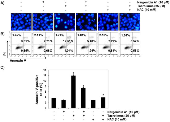
Figure 2.
Suppression of tacrolimus-induced apoptosis by nargenicin A1 in HINAE cells. HINAE cells were treated with 10 μM nargenicin A1 or 10 mM NAC for 1 h, and then stimulated with or without 25 μM tacrolimus for 24 h. (A) Cells were collected, fixed, stained with 4,6-diamidino-2-phenylindole (DAPI), and photographed under a fluorescence microscope (original magnification, ×400). Scale bar, 50 µm. (B,C) Cells cultured under the same conditions were collected and stained with fluorescein isothiocyanate (FITC)-conjugated annexin V and propidium iodide (PI) for flow cytometry. (B) Results showed necrosis, defined as annexin V-negative and PI-positive cells (lower upper quadrant); early apoptosis, defined as annexin V-positive and PI-negative cells (lower right quadrant); and late apoptosis, defined as annexin V-positive and PI-positive (upper right quadrant) cells. (C) Percentages of apoptotic cells were determined by expressing the number of annexin V-positive cells as a percentage of all cells present. Results are presented as the means ± SD of three independent experiments (* p < 0.05 versus non-treated controls, # p < 0.05 versus tacrolimus-treated cells).
Reference
- Park, C.; Kwon, D.H.; Hwang, S.J.; Han, M.H.; Jeong, J.-W.; Hong, S.H.; Cha, H.-J.; Hong, S.-H.; Kim, G.-Y.; Lee, H.-J.; et al. Protective Effects of Nargenicin A1 against Tacrolimus-Induced Oxidative Stress in Hirame Natural Embryo Cells. Int. J. Environ. Res. Public Health 2019, 16, 1044. [Google Scholar] [CrossRef] [PubMed]
Disclaimer/Publisher’s Note: The statements, opinions and data contained in all publications are solely those of the individual author(s) and contributor(s) and not of MDPI and/or the editor(s). MDPI and/or the editor(s) disclaim responsibility for any injury to people or property resulting from any ideas, methods, instructions or products referred to in the content. |
© 2025 by the authors. Licensee MDPI, Basel, Switzerland. This article is an open access article distributed under the terms and conditions of the Creative Commons Attribution (CC BY) license (https://creativecommons.org/licenses/by/4.0/).