Article

Environmental Monitoring of Legionella in Hospitals in the Campania Region: A 5-Year Study

1
Department of Public Health, University “Federico II”, Via Sergio Pansini N° 5, 80131 Naples, Italy
2
General Directorate of Health, Campania Region, Centro Direzionale C3, 80143 Naples, Italy
*
Author to whom correspondence should be addressed.
Int. J. Environ. Res. Public Health 2023, 20(8), 5526; https://doi.org/10.3390/ijerph20085526
Submission received: 6 March 2023 / Revised: 6 April 2023 / Accepted: 13 April 2023 / Published: 14 April 2023

Abstract

:
Legionella is a pathogen that colonizes soils, freshwater, and building water systems. People who are most affected are those with immunodeficiencies, so it is necessary to monitor its presence in hospitals. The purpose of this study was to evaluate the presence of Legionella in water samples collected from hospitals in the Campania region, Southern Italy. A total of 3365 water samples were collected from January 2018 to December 2022 twice a year in hospital wards from taps and showers, tank bottoms, and air-treatment units. Microbiological analysis was conducted in accordance with the UNI EN ISO 11731:2017, and the correlations between the presence of Legionella and water temperature and residual chlorine were investigated. In total, 708 samples (21.0%) tested positive. The most represented species was L. pneumophila 2–14 (70.9%). The serogroups isolated were 1 (27.7%), 6 (24.5%), 8 (23.3%), 3 (18.9%), 5 (3.1%), and 10 (1.1%). Non-pneumophila Legionella spp. represented 1.4% of the total. Regarding temperature, the majority of Legionella positive samples were found in the temperature range of 26.0–40.9 °C. An influence of residual chlorine on the presence of the bacterium was observed, confirming that chlorine disinfection is effective for controlling contamination. The positivity for serogroups other than serogroup 1 suggested the need to continue environmental monitoring of Legionella and to focus on the clinical diagnosis of other serogroups.

1. Introduction

Legionella is an aerobic, non-spore-forming, and Gram-negative pathogen [1], which was discovered in 1976 in Philadelphia following an outbreak of cases of pneumonia in a hotel [2]. Over 65 species belong to this genus [3], about 20 of which can cause disease in humans [4]. Moreover, Legionella pneumophila includes 15 serogroups, and serogroup 1 is the most dangerous to humans. Thus, the attention focused on this serogroup is high [4]. Furthermore, previous studies demonstrated human infections with serogroup 3 [5,6,7], serogroup 9 [7,8], and serogroup 6 [7,9].
Favourable factors to the growth of this bacterium are temperatures between 25 °C and 45 °C (but ranges between 5.7 °C and 63.0 °C can determine its survival); stagnant water [10], in which Legionella in amoebae copiously reproduces [11]; low flow [12]; pH values between 5.5 and 9.2; biofilm and protozoa [2]; inorganic elements (iron, zinc and potassium) and organic and inorganic compounds [13]; existence of water systems of old facilities [14]; dead branches in complex water structures [15]; low total chlorine levels [16]; and low free residual chlorine levels [17]. In addition, man-made disasters [18], natural disasters, flooding [19], water system interruptions, changes in disinfection methods of water systems, and water network breaks [11] are risk factors for Legionella growth.
Construction activities (such as demolition, repressurization, excavation, underground utility connections, commissioning at building opening, and water efficiency challenges) have been associated with healthcare-associated Legionella infections and deaths [20].
Hospitals need a water safety plan (WSP) to control Legionella proliferation, which includes the implementation of safety measures to ensure the water quality of facilities during demolition and construction activities and reduce the risk of exposure of patients [21]. Furthermore, hospitals need a WSP not only during construction or demolition activities. In fact, the World Health Organization (WHO) in 2004 recommended the creation of WSPs by all water suppliers: each of them should engage a group of water experts to assess the risks associated with water exposure, develop and implement strategies to prevent damage to public health, and evaluate the strategies’ effectiveness [22]. Such a plan aims to minimize colonization of Legionella from the source of water supply to the devices in contact with users [23].
Chlorine-based disinfection is the most common active measure used worldwide, and in Italy, against Legionella growth and spread in building water systems [24,25]. Levels of free residual oxidant (FRO), within 0.20 ppm and 4.0 ppm in potable water, have been correlated with reduced risks of growth and spread of disease outbreaks [26].
The presence of Legionella can be detected in soil and water sources, such as showers, hot tubs, air-conditioning systems [3], cooling towers, whirlpools, baths, fountains, ice machines, medical equipment such as aerosols from respiratory devices, eye-wash stations, dental units [27], hot-water recirculation systems [28], hot- and cold-water systems, faucets [29], heater–cooler units, and heater units for cardiac procedures [30].
Legionella infection occurs through inhalation or aspiration of droplets released from contaminated water [3]. In addition, potting soil can also transmit the bacterium, but the mechanism still remains unknown [27]. People who are most affected by Legionella infections are smokers [31], alcohol abusers [32], males, those of advanced age [33], and people with previous diseases, such as acquired immunodeficiency syndrome, hematologic malignancy [34], and diabetes mellitus [35]. Consequently, it is necessary to monitor the presence of Legionella in hospitals as these are places where immunocompromised patients reside [36]. In healthcare settings, the presence of Legionella has been linked to contaminated water reservoirs, cooling towers and air-conditioning systems [37,38,39]. The occurrence of Legionella spp. in the water distribution systems of hospitals and healthcare facilities is a possible concern for hospital populations, due to the vulnerable nature of patients admitted to specific wards, including intensive care, hematology, cardiology, hemodialysis, and pulmonology [40].
The objective to be achieved in healthcare facilities is to minimize colonization by Legionella or, in the case of facilities housing immunocompromised individuals, its total absence (not detectable with the analytical method used) [41].
Legionella infection may have several consequences: it may cause Legionnaires’ disease (LD) (a severe pneumonia), Pontiac fever (a flu-like condition), or it may remain asymptomatic. Because of the lack of specific symptoms and the absence of severity associated with this entity, Pontiac fever is often undiagnosed and under-notified [42].
There is a difference between patients who are hospitalized for LD [43] and others who acquire the infection in hospital due to susceptible medical conditions [44]. LD can be the cause of CAP (community-acquired pneumonia) and pneumonias that are acquired in situations other than travel, domestic environments, and hospitals, and it can be the cause of up to 30% of these types of pneumonias that require hospitalization [43]. Legionella infection can also be acquired in hospitals (hospital-associated infection): individuals most at risk from such an infection are those with prior situations that render them vulnerable, such as immunodeficiency, stem cell or organ transplantation, and obstructive pulmonary disease [44].
Legionella infections are becoming a public health problem due to their incidence and costs [45]. In order to prevent and control Legionella infections sourced from the colonization of water systems, many countries have developed guidelines or regulations. Several organizations, including the American Society of Heating, Refrigerating and Air-Conditioning Engineers (ASHRAE), the WHO, the Centers for Medicare and Medicaid Services (CMS), and the Centers for Disease Control and Prevention (CDC), recommend the creation of water management programs aimed at preventing the growth and spread of Legionella [46].
In the COVID-19 era, it was reported that 20% of patients had a Legionella co-infection during hospitalization [47]. COVID-19 patients have an increased risk for both hospitalization and residual lung impairment [48].
In 2021, the number of Italian individuals affected by Legionellosis was 2726, and its incidence was 46.0 cases per million population [49]. The incidence increased compared to 2020, when the value was 34.3 cases per million population. Of the 2726 notified cases, 83.6% had a community origin, 9.4% were associated with travel, 3.7% had a nosocomial origin, 3.1% were associated with closed communities (nursing homes for elderly people, and healthcare or rehabilitation facilities), and 0.2% had another exposure (prison or communities). Infections of nosocomial origin increased from 68 to 102 cases from 2020 to 2021 [49]. However, Legionella infections are underestimated, and it is reported that less than 5% are diagnosed [50].
Legionellosis is a condition that can be avoided if the bacterium is not present in the environment, so monitoring and preventing its presence is important [51]. Environmental monitoring of Legionella is an approach performed on several occasions in hospitals in Italian regions by Deiana et al. [40], Ditommaso et al. [52], Vincenti et al. [53], De Giglio et al. [54], Laganà et al. [14], Arrigo et al. [55], Pasquarella et al. [56], and Torre et al. [57]; in non-hospital facilities by Totaro et al. [58,59], Sabatini et al. [60], and De Filippis et al. [61]; and in both hospital and non-hospital facilities by Felice et al. [62], Leoni et al. [63], and Mazzotta et al. [64]. Moreover, several studies were also conducted using air samples by Montagna et al. [65,66,67].
To our knowledge, very few studies performed environmental monitoring of Legionella in the Campania region, Southern Italy. In particular, a study by Torre et al. monitored the bacterium in 50 hospitals in the Campania region during the period 2008–2014 [39]. As the new Italian National Guidelines [41] were published in 2015, it was decided to conduct this study to assess the effectiveness of the application of the new Guidelines in preventing Legionella colonization in water systems. Thus, the aim of this study was to analyze hospital environmental monitoring of Legionella in the Campania region over the 5-year study period. In detail, the purposes of the study were (i) to evaluate the presence of Legionella in tested water samples; (ii) to estimate the prevalence of species and of individual L. pneumophila serogroups; and (iii) to assess the influence of several parameters, such as temperature and free residual chlorine, on the presence of this bacterium.

2. Materials and Methods

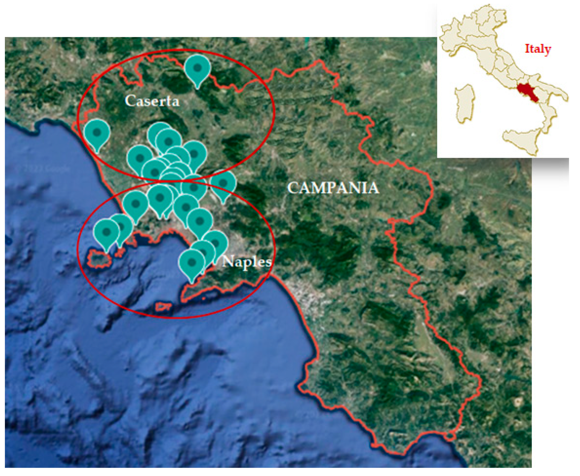
2.1. Study Area and Hospital Characteristics

The water sample collection was conducted from January 2018 to December 2022 in two provinces of the Campania region, Southern Italy (Naples and Caserta) (Figure 1). The samples were collected in hospitals which ask us to routinely perform Legionella testing every six months as part of WSPs as required by the Italian regulatory system for validation of a well-maintained building water system. The water samples were collected from 26 hospitals, all of which possessed the following characteristics: the presence of a single building, construction between 1970 and 1980, the presence of a maximum of five floors, and the presence of the number of beds served ranging between 20 and 200. These hospitals all have WSPs in place to respond to positive samples. We monitored 26 hospitals compared to the 50 hospitals in the study by Torre et al. [39], because some of the 50 hospitals were excluded since monitoring had not been carried out for all the years of the study, or these facilities were not comparable to the others (in terms of number of beds served, number of buildings, number of floors, construction period, or water treatment disinfection methods).
Water provided to the hospitals (which contains free chlorine as residual drinking water disinfectant) comes from the public supply system of the cities of Naples and Caserta, and it reaches the hospitals via a single pipeline. The hospital sites within the present study included buildings hosting patients considered at increased risk (Medicine, Pneumology, Geriatrics, Surgeries, etc.) [41] over the study period, inclusive of the time during the COVID-19 pandemic.
Samples were collected every six months, as required by the Italian National Guidelines for hospitals with this type of wards. If a sample tested positive, according to the Italian National Guidelines, the sampled point was decontaminated and re-sampled 1, 3, and 6 months later (these mandatory follow-up lab samples and corresponding data were excluded from the present study’s analysis, as the purpose of the work was to monitor Legionella during routine monitoring activities). Therefore, we included the initial positive samples and excluded the follow-up samples. The water treatment disinfection methods used in these hospitals are based on the application of hypochlorite.

2.2. Sample Collection

We chose the sampling locations described in the Italian National Guidelines [41]. In addition, this study used similar sampling locations (i.e., faucets, showers, and tank bottoms) within the building premise plumbing system of the study by Torre et al. [39]. Our study, in addition to the previous sampling locations, also analyzed samples collected from the cold-water circuit, as mentioned in the new Guidelines.
In detail, for each hot-water system, as described in the Italian National Guidelines [41], the following sampling locations were carried out: supply, recirculation, and tank bottoms, with at least 3 representative points (furthest in the water distribution and coldest). Tank bottom refers to a hot- or cold-water storage tank. For each cold-water system, the following sampling locations were carried out: tank bottoms, with at least 2 representative points (furthest in the water distribution and hottest). In addition, water was collected from air-treatment units (ATUs). Following the observation of the positivity rate for Legionella, it was decided to increase the sampled points. The COVID-19 pandemic prompted us to further increase the number of samplings, and with its end, we decided to decrease the number of sampled points. Of the total of 3365 water samples, 2065 originated from manual taps and showers (1485 from hot water and 580 from cold water), 780 from tank bottoms (in particular, 520 from hot water and 260 from cold water), and 520 from ATUs. The water samples were collected during the day (later in the morning, when the hospital was already in action). There were no point-of-use (POU) filters at any point of sampling.
According to the Italian National Guidelines, 1 L of water was collected by the laboratory staff in sterile polyethylene bottles enriched with 0.01% sodium thiosulfate to neutralize chlorine action [60]. All samples were collected without flushing and flaming or disinfecting at the point of discharge to simulate the common use of water, i.e., the exposure of a user. The water samples were univocally identified on a spreadsheet at the time of collection. At sampling, water temperature and residual chlorine were measured by our laboratory staff. Water temperature (expressed in °C) was obtained using a calibrated thermometer (TFA Digitales Einstichthermometer, TFA-Dostmann, GmbH & Co. KG, Wertheim-Reicholzheim, Germany), and free residual chlorine (expressed in mg/L) was monitored using a colorimetric diethyl-p-phenylenediamine (DPD) method (MQuant; Merck, Darmstadt, Germany). In detail, the test was based on a semi-quantitative measurement of free chlorine by visual comparison of the color of the measurement solution and a set of colors contained in a color card comparator.
The samples were transported, divided between hot- and cold-water samples at room temperature, and protected from light.
Calibrations (kits and equipment) and microbiological analysis were performed in our laboratory, which is accredited according to the ISO 17025 and periodically performs proficiency tests.
No samples were damaged during transport or showed a suspect appearance, such as different coloring or the presence of sediment or soil. Therefore, all samples collected were included and analyzed in the study.

2.3. Microbiological Analysis and Identification

The microbiological analysis was conducted within 2 hours after the collection of the samples, in accordance with the UNI EN ISO 11731:2017 [55,64]. Briefly, the samples were filtered on polycarbonate membrane filters with a pore size of 0.2 µm (Sartorius). The membrane was kept in 10 mL of the original water sample and vortexed. A total of 200 µL of water previously treated at (50 ± 1) °C for (30 ± 2) min. and the same volume of untreated water were inoculated on Petri plates containing Legionella Agar Base (Oxoid) medium and being supplemented with Legionella Growth Supplement (BCYE) (Oxoid) and Legionella Selective Supplement (GVPC) (Oxoid). The plates were incubated at (36 ± 2) °C with 2.5% CO2 and under a humid atmosphere. After 10 days, the presence or absence of colonies was evaluated. The presumed Legionella colonies were cultured both on BCYE agar and BCYE agar without L-cysteine (BCYE-cys agar). The growth of colonies on the BCYE agar and not on the BCYE-cys agar suggested Legionella positivity of the samples. Determination of species and serogroups was conducted by the latex agglutination test (Oxoid) and the anti-Legionella pneumophila monovalent serum (Biogenetics). The colony counts were reported in terms of CFU (colony-forming units)/L.
The test results were sent to the hospitals.

2.4. Statistical Analysis

A descriptive statistical analysis was performed as previously described [39]. It included, for bacterial load values of positive samples, detection of geometric mean (Log10 CFU/L), standard deviation, median, percentile range, and interquartile range [39]. This analysis was conducted using Microsoft Excel.
Normality tests were conducted using the Shapiro–Francia test to check for data distribution. The non-parametric Mann–Whitney U test was used to determine the connections between the presence of Legionella and residual chlorine (expressed in mg/L) and water temperature (expressed in °C) [68]. The statistical results were interpreted at the level of significance p < 0.05. The χ2 was calculated using Doornik–Hansen test. Multiple linear regression analysis (MLRA) was used to confirm the results of the Mann–Whitney U test. The statistical calculations were performed using the STATA MP v14.0 statistical software program (College Station, TX, USA).

3. Results

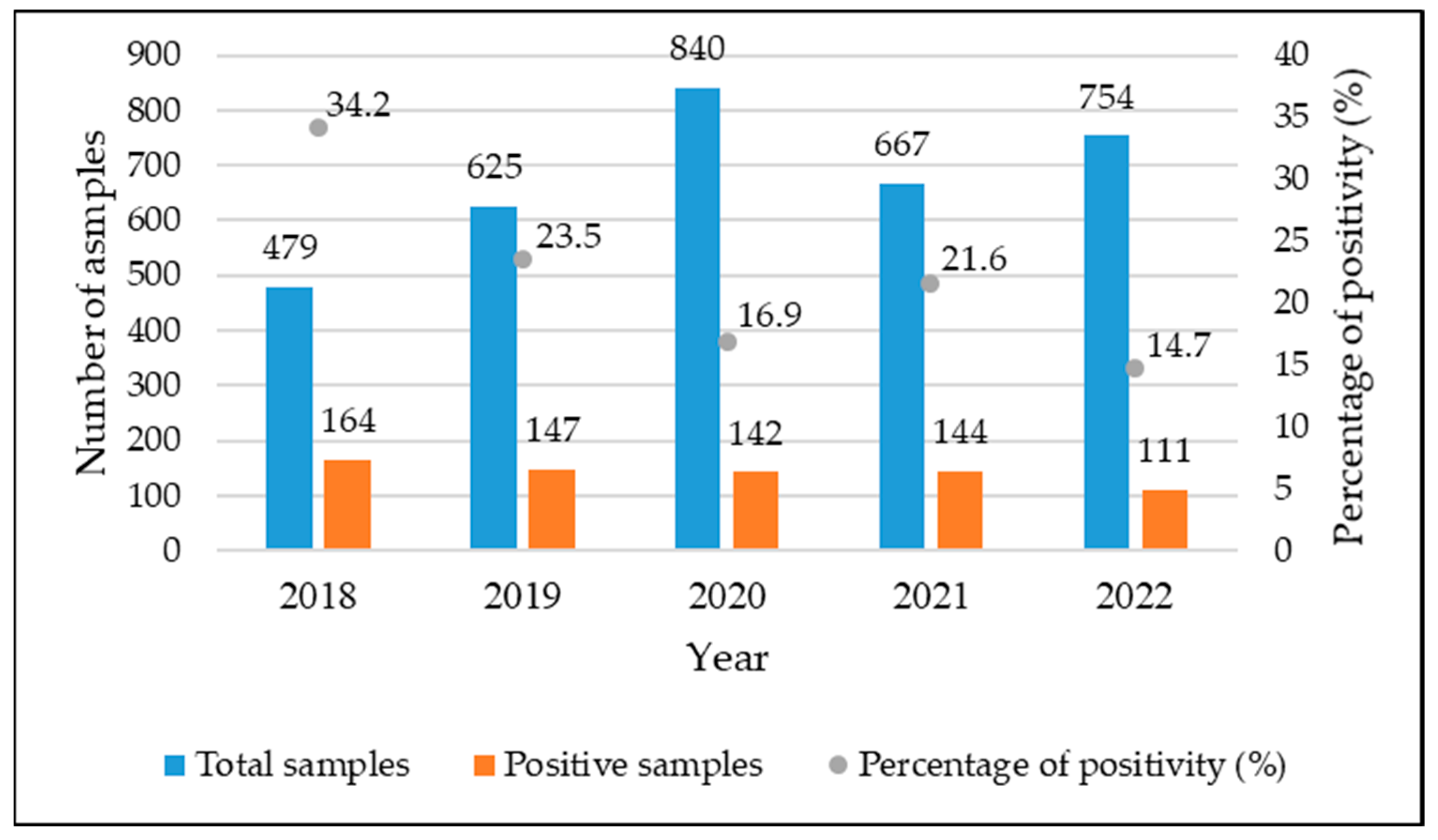
3.1. Positivity of Analyzed Samples

During the 5-year study period, the laboratory analyzed a total of 3365 water samples. Among these samples, positivity for Legionella was detected in 708 samples, representing 21.0% of the total. The number of samples per year, along with the number of positive samples per year, is shown in Figure 2 and Table 1. Specifically, the number of positive samples decreased from 164 (34.2%) to 111 (14.7%).
Table 2 shows the number of total samples, the number of positive samples, and the percentage for different sampling locations. Of the total number of cold-water samples (840), 37 (4.4%) tested positive. In particular, 11 originated from tank bottoms and 26 from taps and showers. In the hot-water samples, out of the 2005 total samples, 652 (32.5%) were positive. A total of 143 samples came from tank bottoms and 509 from taps and showers. In the ATU samples, a positivity rate of 3.7% was observed (19 positive samples out of a total of 520).

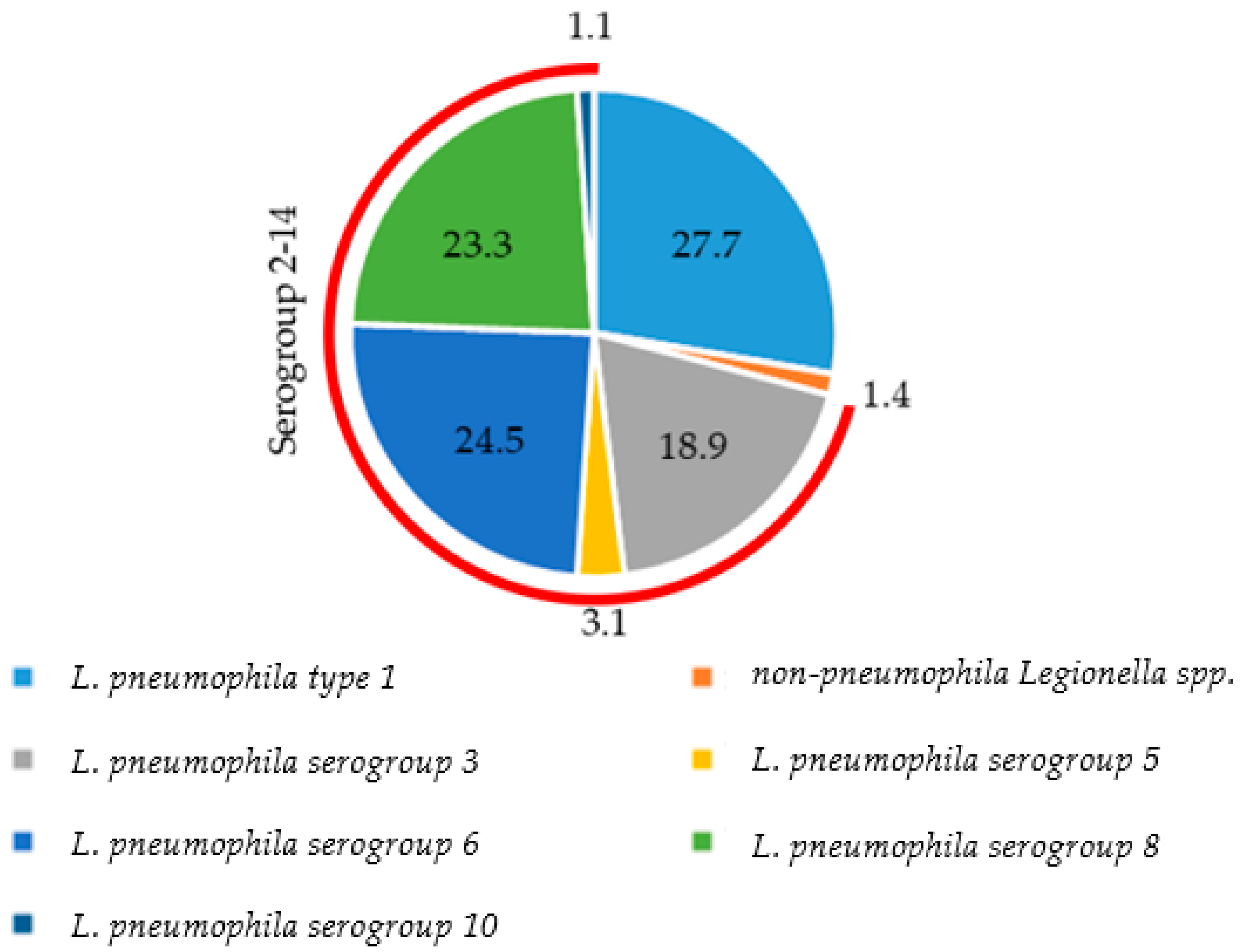
3.2. Distribution of Species and Serogroups among Positive Samples

Figure 3 exhibits the percentage of positivity of Legionella species and serogroups in the years 2018–2022. In detail, L. pneumophila was the most represented (98.6% versus 1.4% of non-pneumophila Legionella spp.). Serogroups 2–14 had a positivity percentage of 70.9%, and serogroup 1 has a positivity percentage of 27.7%. Among serogroups 2–14, out of the total number of isolated species and serogroups, the most represented were serogroups 6 (24.5%), 3 (18.9%), and 8 (23.3%). Serogroups 5 and 10, on the other hand, were detected at a rate of 3.1% and 1.1%, respectively (Figure 3).
In addition, in 27 samples of the 708 positive samples, the simultaneous presence of two species or serogroups was found. Particularly, out of these 27 samples, 22 samples tested positive for both serogroups 1 and 3 (3.1%), 1 sample tested positive for serogroups 1 and 6 (0.1%), 2 samples tested positive for serogroups 1 and 8 (0.3%), 1 sample tested positive for serogroups 1 and 10 (0.1%), and 1 sample tested positive for serogroup 5 and non-pneumophila Legionella spp. (0.1%).

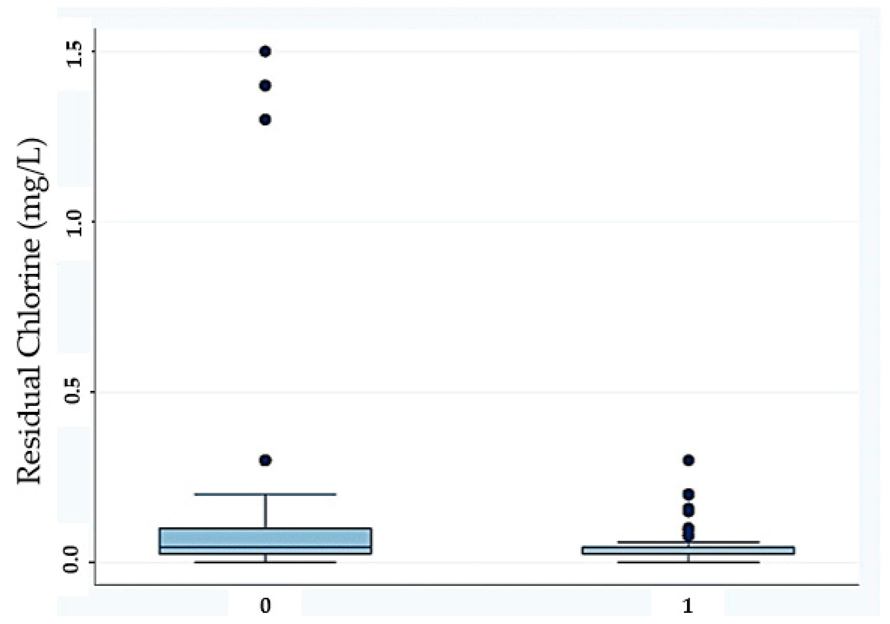
3.3. Water Temperature and Residual Chlorine

Of the 3365 analyzed samples, 520 were taken from the ATUs in which the temperature and residual chlorine parameters were not carried out, while the remaining 2845 samples were divided into hot-water samples (≥26.0 °C, n. 2,005) and cold-water samples (≤25.8 °C, n. 840), with a mean temperature of 37.5 °C (range of 11.0–91.0 °C) for all the samples collected.
Table 3 shows different ranges of temperature and, for each interval, the number of analyzed samples, the number of positive samples, the minimum Legionella concentration, the maximum Legionella concentration, and the geometric mean of the positive samples. The majority of Legionella positive samples were found in the temperature ranges of 26.0–30.9 °C, 31.0–35.9 °C, and 36.0–40.9 °C (57.4%, 46.9%, and 48.8%). The percentage of positive samples decreased with increasing temperatures (from 48.8% for the temperature range of 36.0–40.9 °C to 19.4% for temperatures ≥56.0 °C).
Regarding the bacterial concentration of the positive samples, a minimum value corresponding to 1.70 Log10 CFU/L was observed at all temperature ranges, while the highest maximum value was observed at the temperature range of 46.0–50.9 °C (4.36 Log10 CFU/L).
In addition, the minimum value of the geometric mean was 2.81 Log10 CFU/L in temperatures ≤20 °C, and it increased with increasing temperature, up to the range of 26.0–30.9 °C (3.10 Log10 CFU/L). For the temperature range of 41.0–45.9 °C and above, there was a decrease in the geometric mean value as temperature increased. At temperatures higher than 62 °C, no positivity for Legionella was observed.

3.4. Statistical Analysis

The results of the statistical analysis carried out to examine the bacterial load values of the positive samples are shown in Table 4. This table reports the number of total samples, the number of positive samples and percentage of positivity, the geometric mean (Log10 CFU/L), the standard deviation, the median, the percentile range, and the interquartile range. Moreover, the results show the normality of all data collected, and the Doornik–Hansen test for multivariate normality provides a χ2 = 1.09 × 10⁵.
The lowest Legionella concentration value recorded during the analysis was 1.70 Log10 CFU/L in all years, while the highest value was 4.36 Log10 CFU/L in 2019.
Table 4. Number of total samples, number of positive samples and percentage of positivity, geometric mean (Log10 CFU/L) of positive samples, and descriptive statistics. Descriptive statistical analysis was conducted for the positive samples (N = 708).
YearPos./Tot. (%)Mean (±SD 1)Min25th PercentileMedian75th PercentileMax
2018164/479 (34.2)3.02 ± 0.721.702.543.123.634.28
2019147/625 (23.5)2.71 ± 0.801.702.002.603.354.36
2020142/840 (16.9)2.82 ± 0.741.702.182.813.384.30
2021144/667 (21.6)2.96 ± 0.731.702.403.023.624.23
2022111/754 (14.7)3.14 ± 0.581.702.783.203.624.04
1 SD: standard deviation.
The results of the MRLA (Table 5) indicated that no statistically significances were recorded for the water samples’ temperature (p-value = 0.526), in contrast with residual chlorine (p-value < 0.05).
Furthermore, the MRLA results revealed the statistically significant correlation between residual chlorine and Legionella concentration, in which was negative, as suggested by the t-value (−6.19) and p-value (<0.05).
The boxplot regarding the influence of residual chlorine is shown in Figure 4.

4. Discussions

Legionella is a bacterium that colonizes soils, freshwater, and building water systems [51]. The aim of this paper was to analyze the prevalence of Legionella and its individual species and serogroups in hospitals of the Campania region over the period 2018–2022 and to assess the effectiveness of the Italian National Guidelines of 2015 in preventing Legionella colonization in water systems. In addition, the purpose was to test whether there was a relationship between the presence of Legionella with two variables, residual chlorine and water temperature.
In this study, we observed a 21.0% positivity rate. The positivity rate for Legionella decreased from 2018 (34.2%) to 2022 (14.7%). The number of positive samples gradually decreased, and this explains the decrease in the positivity percentage for this microorganism (Figure 2). Deiana et al. [40], on the other hand, monitored the presence of Legionella from 2010 to 2020 in a university hospital in the Sardinia region, Italy. Regarding the period 2018–2020, they also observed a decrease in the percentage of Legionella positivity in this region.
The authors posit that the decrease in Legionella positivity over the course of the study is likely attributed to the hospitals receiving the study’s results and performing environmental hazard controls. The COVID-19 pandemic in 2020 and 2021 decreased attention to environmental Legionella prevention, leading to an increase in Legionella positivity in 2021. This increase was not observed in 2020 because the increase in hospitalization led to the need to implement hygiene rules (e.g., hand washing) [69], which could have promoted water flow and prevented stagnation. Stagnation is known to be responsible for the disappearance and poor stability of residual disinfectant and microbial growth of Legionella [70]. Moreover, the monitored hospitals were all in constant activity during the pandemic. The pandemic also directed our attention to new locations to monitor for Legionella: monitoring more locations in hospitals meant preventing Legionella infections in patients.
The percentage of positivity observed at the different sampling locations was 19.7% for tank bottoms, 25.9% for taps and showers, and 3.7% for ATUs. This study, in general, used similar sampling locations (i.e., faucets, showers, and tank bottoms) within the building premise plumbing system of the study by Torre et al. [39], conducted in the years 2008–2014 in hospitals in the Campania region, which showed a percentage of positivity of 27.2% for tank bottoms, 31.9% for taps and showers, and 4.0% for ATUs. The authors supposed that this decrease could be explained by the publication of the Italian National Guidelines in 2015 and the implementation of a WSP by hospitals, which resulted in the collaboration of different personnel (laboratory staff, technical office, water disinfection staff, etc.) in order to ensure water safety. In addition, the highest percentage of Legionella detection was observed in taps and showers, which are a source of exposure for patients (taps and showers can generate aerosols containing the bacterium that can be inhaled) [71].
Out of a total of 840 cold-water samples (temperature range of 11.0–25.9 °C), 37 (4.4%) tested positive. The presence of Legionella in cold-water samples was also found in the study by Arvand et al., 2011 [72], who detected a percentage of positivity of 40% in cold-water samples (temperature range of 7–29 °C) [72]. Our results are not surprising, since out of the total number of cold-water samples taken (840), 458 (54.5%) showed a temperature between 21.0 and 25.9 °C, which are values near the range of temperatures that can create favorable conditions for Legionella multiplication.
The results indicated that L. pneumophila was the most isolated species. The serogroups 2–14 showed a positivity rate of 70.8% for the positive samples, which was similar to that found in Turkish hospitals (70.8%) by Yilmaz and Orhan [73], in Italian non-hospital facilities (71.7%) by Sabatini et al. [60], in hospital samples (68.7%) collected by Deiana et al. [40], and in Polish non-hospital samples (64.3%) collected by Stojek and Dutkiewicz [74].
Regarding the individual serogroups, the highest percentage of positivity was found for serogroup 1. This finding is congruent with what is found in clinical diagnosis, according to which the L. pneumophila type 1 is the serogroup that most commonly affects humans [7,75,76]. Amemura-Maekawa et al., in fact, isolated an 80.2% percentage of serogroup 1-positive samples in Japanese clinical specimens collected from 1980 to 2008 [77].
Regarding serogroups 2–14, the presence of serogroups different from serogroup 1 (6, 8, and 3) was the same as found previously [39]. Serogroups 3 and 6 were also among the serogroups isolated from humans in Italy during 1987 and 2009 [78], so these results had correspondence with what was previously reported in humans. These findings were also in agreement with the results determined by Perola et al., who, through a genotyping analysis, found an association between the isolates from two Legionella serogroup 5-positive patients residing for several days in a hospital and the environmental isolates from the hospital’s water supply [79].
Serogroups 3 and 6 were found in environmental water samples in several studies [54,80,81,82,83]. Particularly, serogroup 3 was isolated in hospitals in Iran [84], while Pignato et al. revealed the prevalence of this serogroup in Italian hospitals, which was sometimes associated with serogroup 6 [85]. Isolated serogroup 8 was reported by De Giglio et al. [54], Papadakis et al. [83], and Sakhaee et al. [84].
The presence of serogroups different from serogroup 1 suggests that attention should be focused on the clinical diagnosis of other serogroups, considering that the most widely used test for Legionella diagnosis in human specimens (the urinary antigen test) shows a low sensitivity for non-L. pneumophila type 1, resulting in underreporting of Legionella infections [76,86].
Regarding temperature, in our study, we observed that more hot-water samples were positive than cold-water samples (32.5% vs. 4.4%). This finding is consistent with that observed in the work by Stojek et al. conducted in Poland, who observed a higher frequency of Legionella isolation in hot-water samples (88.6%) [87]. In addition, the highest percentages of positive samples were found at temperatures ranging from 26.0 to 40.9 °C, which are temperatures corresponding to the optimal growth range of Legionella. For temperatures above this range, the percentage decreased. With regard to the bacterium’s concentrations, the highest values were found at a temperature range of 46.0–50.9 °C, and above this temperature range, these values decreased. In the study by Boppe et al. conducted in a hospital where the correlation between the concentration of L. pneumophila and the maximum water temperature at the point of use was investigated, a significant decrease in the bacterium’s concentrations was observed at temperatures ≥55 °C [88].
Our analysis revealed a Legionella concentration minimum value of 1.70 Log10 CFU/L in all years, and the highest value of 4.36 Log10 CFU/L was observed in 2019. In the study by Torre et al., the minimum and maximum values observed were 2.00 and 7.45 Log10 CFU/L [39]. On the other hand, in the study by Girolamini et al., the minimum and maximum concentration values observed in a hospital in the Emilia-Romagna region, Italy, which water was treated with a biocide, were between <1.70 and 5.80 Log10 CFU/L [89].
The Italian National Guidelines for the prevention and control of Legionellosis indicate the types of interventions required to be conducted in healthcare facilities, depending on the positivity rate, the Legionella concentration (expressed in CFU/L), and the presence or absence of clinical cases [41].
In addition, procedures must be performed to obtain non-detection of Legionella in air-treatment systems and water in wards housing profoundly immunocompromised and very high-risk patients (transplant centers, oncology, and hematology) and in the water for tank births.
If a sample tested positive, according to the Italian National Guidelines, the sampled point is decontaminated and re-sampled one, three, and six months later (these mandatory follow-up lab samples and corresponding data were excluded from the present study’s analysis).
Regarding the trends of Legionella concentration found, this study revealed a statistically significant negative correlation with residual chlorine. This finding was in agreement with D’Alessandro et al. [90], who observed a statistically significant reduction in positive samples following the increase in free chlorine in water. Furthermore, Totaro et al. found a moderate relationship between the presence of Legionella and a decrease in total chlorine concentration [58]. The same result was reported by Masaka et al., who found a weak statistically significant negative correlation with both L. pneumophila serogroups 1 and 2–14 [91]. A statistically significant negative correlation between Legionella concentration and total chlorine residual concentration was reported by Zhang et al. [92]. Moreover, in their study, Rafiee et al. argued that there was proportionality between residual chlorine content and the presence of Legionella [93]. The mechanism by which chlorine combats the presence of Legionella involves it interaction with the pathogen’s cell membrane [94]. This results in a dispersion of the cell’s macromolecules and subsequent modification of the cell’s chemical, physical, and biological processes [94].
Furthermore, no statistically significant correlation was found with water temperature, as reported by Masaka et al. [91] and Pierre et al. [46], and in contrast to the evidence found by Rakić et al. [68], who found a correlation with L. pneumophila. In addition, De Giglio et al. observed a weak correlation between Legionella concentrations and water temperature [95].
In summary, the results of the study confirm that chlorine disinfection is an effective method to control Legionella; this is in accordance with the results reported by Paranjape et al., who found that continuous application of chlorine inhibits the presence of Legionella in cooling towers, and by Orsi et al., who found that shock and continuous hyperchlorination significantly reduce the number of Legionella positive samples [16,96].

5. Limitations

The limitations of this study were that sampling covered only two provinces of the Campania region in the South of Italy. Thus, in the future, it would be interesting to expand the study to other provinces in order to have a complete overview of the situation in the study area. Moreover, the sampling covered only one type of facility. Consequently, a future goal could be to expand the analysis to other types of facilities, such as recreational centers, tourist sites, military bases, schools, and others, since it is possible to identify the bacterium in these structures [62]. In addition, only two variables were analyzed to evaluate the relationship with the presence of Legionella (water temperature and residual chlorine). Furthermore, we did not collect information about cases of Legionella infections in the monitored hospitals, and it would be interesting to investigate this aspect in future studies and assess whether there is a correlation between these two variables, as performed in the study by Perola et al. [79].
Finally, the circumstance that, in a high percentage of samples (79.0%), Legionella is not detected by the culture method does not mean that all samples are really negative for Legionella. There may be a percentage of samples that have viable but non-culturable (VBNC) Legionella, and the culture method cannot detect them. VBNC is a state of Legionella that can be established under unfavorable environmental conditions and retain virulence. Under favorable conditions and in free-living amoebae, the bacterium can resuscitate and become pathogenic again [97]. For example, a study conducted from 2010 to 2015 in an Italian university hospital subjected to continuous monochloramine treatment determined that 34.5% of the negative samples, as determined by the culture method, tested positive for the VBNC state. In addition, 18% of 22 tested samples were positive based on the resuscitation test [97]. This situation could occur in some of our negative samples. Future goals will include testing for the VBNC state.
This paper could be a starting point for future analyses, given the importance of environmental monitoring and the number of studies monitoring this bacterium in the Campania region in South of Italy.

6. Conclusions

Our environmental monitoring revealed bacterial contamination of water in this region, even though its presence decreased in percentage positivity from 2018 to 2022. Despite this decline, attention should not be reduced, especially in facilities housing immunocompromised individuals who are more susceptible to Legionella infection. The most represented species was confirmed to be L. pneumophila. Serogroups 2–14 were found at a high rate. Regarding total serogroups, the most represented was serogroup 1, followed by serogroups 6, 3, and 8. These results suggest the need for continuous environmental monitoring of Legionella and highlight the importance of focusing on the clinical diagnosis of serogroups different from serogroup 1 as well, since the most widely used test on human samples shows a high sensitivity only for this serogroup.
The highest concentration of positive samples was observed in the temperature range corresponding to the optimal growth temperature of the bacterium. In addition, the negative correlation between residual chlorine and the presence of Legionella confirmed that chlorine disinfection is an effective method for preventing Legionella contamination.
The implementation of a WSP, through the collaboration of different groups of professionals, is one of the main approaches to control Legionella contamination in hospitals and prevent infections.

Author Contributions

Conceptualization: A.L. and F.P.; methodology: T.B. and F.P.; software: A.L., E.D.R., F.D.D. and M.S; formal analysis: A.L., T.B. and F.P.; resources: I.T., P.M. and F.P.; data curation: A.L., T.B., E.D.R., F.D.D. and M.S.; writing—original draft preparation: A.L., T.B., U.T. and F.P.; writing—review and editing: A.L., E.D.R., F.D.D.; I.T., P.M. and F.P.; supervision: I.T., P.M. and F.P. All authors have read and agreed to the published version of the manuscript.

Funding

This research received no external funding.

Institutional Review Board Statement

Not applicable.

Informed Consent Statement

Not applicable.

Data Availability Statement

The datasets obtained and analyzed in the current study are available from the corresponding author on reasonable request.

Conflicts of Interest

The authors declare no conflict of interest.

References

  1. Younas, F.; Soltanmohammadi, N.; Knapp, O.; Benz, R. The major outer membrane protein of Legionella pneumophila Lpg1974 shows pore-forming characteristics similar to the human mitochondrial outer membrane pore, hVDAC1. Biochim. Biophys. Acta Biomembr. 2018, 1860, 1544–1553. [Google Scholar] [CrossRef] [PubMed]
  2. Gonçalves, I.G.; Fernandes, H.S.; Melo, A.; Sousa, S.F.; Simões, L.C.; Simões, M. LegionellaDB—A database on Legionella outbreaks. Trends Microbiol. 2021, 29, 863–866. [Google Scholar] [CrossRef] [PubMed]
  3. Mondino, S.; Schmidt, S.; Rolando, M.; Escoll, P.; Gomez-Valero, L.; Buchrieser, C. Legionnaires’ disease: State of the art knowledge of pathogenesis mechanisms of Legionella. Annu. Rev. Pathol. 2020, 15, 439–466. [Google Scholar] [CrossRef] [PubMed]
  4. Caicedo, C.; Rosenwinkel, K.H.; Exner, M.; Verstraete, W.; Suchenwirth, R.; Hartemann, P.; Nogueira, R. Legionella occurrence in municipal and industrial wastewater treatment plants and risks of reclaimed wastewater reuse. Water Res. 2019, 149, 21–34. [Google Scholar] [CrossRef] [PubMed]
  5. Herpers, B.L.; Bossink, A.W.; Cohen Stuart, J.W.; Hustinx, W.N.; Thijsen, S.F. A patient with Legionella pneumophila serogroup-3 pneumonia, detected by PCR. Ned. Tijdschr. Geneeskd. 2005, 149, 2009–2012. Available online: https://pubmed.ncbi.nlm.nih.gov/16171114 (accessed on 14 November 2022).
  6. Khanna, N.; Meikle, A.; Gillespie, L.; Edwards, G.; Lindsay, D. Legionella pneumophila serogroup 3 infection: Importance of serology. Scott. Med. J. 2012, 57, 182. [Google Scholar] [CrossRef]
  7. Ito, A.; Ishida, T.; Washio, Y.; Yamazaki, A.; Tachibana, H. Legionella pneumonia due to non-Legionella pneumophila serogroup 1: Usefulness of the six-point scoring system. BMC Pulm. Med. 2017, 17, 211. [Google Scholar] [CrossRef]
  8. Ito, A.; Ishida, T.; Tachibana, H.; Ito, Y.; Takaiwa, T.; Fujii, H.; Hashimoto, T.; Nakajima, H.; Amemura-Maekawa, J. A case of community-acquired pneumonia due to Legionella pneumophila serogroup 9 wherein initial treatment with single-dose oral azithromycin appeared useful. Jpn. J. Infect. Dis. 2017, 70, 660–662. [Google Scholar] [CrossRef]
  9. Chen, C.Y.; Chen, K.Y.; Hsueh, P.R.; Yang, P.C. Severe community-acquired pneumonia due to Legionella pneumophila serogroup 6. J. Formos. Med. Assoc. 2006, 105, 256–262. [Google Scholar] [CrossRef]
  10. De Giglio, O.; Fasano, F.; Diella, G.; Lopuzzo, M.; Napoli, C.; Apollonio, F.; Brigida, S.; Calia, C.; Campanale, C.; Marzella, A.; et al. Legionella and legionellosis in touristic-recreational facilities: Influence of climate factors and geostatistical analysis in Southern Italy (2001–2017). Environ. Res. 2019, 178, 108721. [Google Scholar] [CrossRef]
  11. Gleason, J.A.; Cohn, P.D. A review of legionnaires’ disease and public water systems–Scientific considerations, uncertainties and recommendations. Int. J. Hyg. Environ. Health 2022, 240, 113906. [Google Scholar] [CrossRef] [PubMed]
  12. Cunha, B.A.; Burillo, A.; Bouza, E. Legionnaires’ disease. Lancet 2016, 387, 376–385. [Google Scholar] [CrossRef] [PubMed]
  13. Sciuto, E.L.; Laganà, P.; Filice, S.; Scalese, S.; Libertino, S.; Corso, D.; Faro, G.; Coniglio, M.A. Environmental Management of Legionella in Domestic Water Systems: Consolidated and Innovative Approaches for Disinfection Methods and Risk Assessment. Microorganisms 2021, 9, 577. [Google Scholar] [CrossRef] [PubMed]
  14. Laganà, P.; Facciolà, A.; Palermo, R.; Delia, S. Environmental surveillance of legionellosis within an italian university hospital—Results of 15 years of analysis. Int. J. Environ. Res. Public Health 2019, 16, 1103. [Google Scholar] [CrossRef] [PubMed]
  15. Palazzolo, C.; Maffongelli, G.; D’Abramo, A.; Lepore, L.; Mariano, A.; Vulcano, A.; Ascoli Bartoli, T.; Bevilacqua, N.; Giancola, M.L.; Di Rosa, E.; et al. Legionella pneumonia: Increased risk after COVID-19 lockdown? Italy, May to June 2020. Eurosurveillance 2020, 25, 2001372. [Google Scholar] [CrossRef] [PubMed]
  16. Paranjape, K.; Bédard, É.; Whyte, L.G.; Ronholm, J.; Prévost, M.; Faucher, S.P. Presence of Legionella spp. in cooling towers: The role of microbial diversity, Pseudomonas, and continuous chlorine application. Water Res. 2020, 169, 115252. [Google Scholar] [CrossRef]
  17. Knežević, M.; Rončević, D.; Vukić Lušić, D.; Mihelčić, M.; Kogoj, R.; Keše, D.; Glad, M.; Cenov, A.; Ožanič, M.; Glažar Ivče, D.; et al. Decreasing Pasteurization Treatment Efficiency against Amoeba-Grown Legionella pneumophila—Recognized Public Health Risk Factor. Int. J. Environ. Res. Public Health 2022, 19, 1099. [Google Scholar] [CrossRef]
  18. Deshmukh Azhar, A.; Bhangale, P.P. Disaster Management for Cooling Tower-Case Study. Int. J. Eng. Res. Appl. 2016, 6, 09–14. [Google Scholar]
  19. Walker, J.T. The influence of climate change on waterborne disease and Legionella: A review. Perspect. Public Health 2018, 138, 282–286. [Google Scholar] [CrossRef]
  20. Scanlon, M.M.; Gordon, J.L.; McCoy, W.F.; Cain, M.F. Water management for construction: Evidence for risk characterization in community and healthcare settings: A systematic review. Int. J. Environ. Res. Public Health 2020, 17, 2168. [Google Scholar] [CrossRef]
  21. Scanlon, M.M.; Gordon, J.L.; Tonozzi, A.A.; Griffin, S.C. Reducing the Risk of Healthcare Associated Infections from Legionella and Other Waterborne Pathogens Using a Water Management for Construction (WMC) Infection Control Risk Assessment (ICRA) Tool. Infect. Dis. Rep. 2022, 14, 341–359. [Google Scholar] [CrossRef] [PubMed]
  22. Bartram, J.; Corrales, L.; Davison, A.; Deere, D.; Drury, D.; Gordon, B.; Howard, G.; Rinehold, A.; Stevens, M. Water Safety Plan Manual: Step-by-Step Risk Management for Drinking-Water Suppliers; World Heart Organization: Geneva, Switzerland, 2009; Available online: https://www.who.int/publications/i/item/9789241562638 (accessed on 12 December 2022).
  23. Coniglio, M.A.; Ferrante, M.; Yassin, M.H. Preventing healthcare-associated legionellosis: Results after 3 years of continuous disinfection of hot water with monochloramine and an effective water safety plan. Int. J. Environ. Res. Public Health 2018, 15, 1594. [Google Scholar] [CrossRef] [PubMed]
  24. Field, B.S. Minimizing the Risk of Legionellosis Associated with Building Water Systems. Ashrae Guidline. 2000. Available online: https://cir.nii.ac.jp/crid/1570291225346474368 (accessed on 14 November 2022).
  25. Muzzi, A.; Cutti, S.; Bonadeo, E.; Lodola, L.; Monzillo, V.; Corbella, M.; Scudeller, L.; Novelli, V.; Marena, C. Prevention of nosocomial legionellosis by best water management: Comparison of three decontamination methods. J. Hosp. Infect. 2020, 105, 766–772. [Google Scholar] [CrossRef] [PubMed]
  26. National Academies of Sciences, Engineering, and Medicine. Management of Legionella in Water Systems; National Academies Press: Washington, DC, USA, 2020. Available online: https://pubmed.ncbi.nlm.nih.gov/32200596/ (accessed on 12 December 2022).
  27. Van Heijnsbergen, E.; Schalk, J.A.; Euser, S.M.; Brandsema, P.S.; den Boer, J.W.; de Roda Husman, A.M. Confirmed and potential sources of Legionella reviewed. Environ. Sci. Technol. 2015, 49, 4797–4815. [Google Scholar] [CrossRef] [PubMed]
  28. Serrano-Suárez, A.; Dellundé, J.; Salvadó, H.; Cervero-Aragó, S.; Méndez, J.; Canals, O.; Blanco, S.; Arcas, A.; Araujo, R. Microbial and physicochemical parameters associated with Legionella contamination in hot water recirculation systems. Environ. Sci. Pollut. Res. 2013, 20, 5534–5544. [Google Scholar] [CrossRef]
  29. Chen, Y.S.; Liu, Y.C.; Lee, S.S.J.; Tsai, H.C.; Wann, S.R.; Kao, C.H.; Chang, C.L.; Huang, W.K.; Huang, T.S.; Chao, H.I.; et al. Abbreviated duration of superheat-and-flush and disinfection of taps for Legionella disinfection: Lessons learned from failure. Am. J. Infect. Control 2005, 33, 606–610. [Google Scholar] [CrossRef]
  30. Ditommaso, S.; Giacomuzzi, M.; Memoli, G.; Garlasco, J.; Zotti, C.M. Persistence of Legionella in routinely disinfected heater-cooler units and heater units assessed by propidium monoazide qPCR. Pathogens 2020, 9, 978. [Google Scholar] [CrossRef]
  31. Straus, W.L.; Plouffe, J.F.; File, T.M.; Lipman, H.B.; Hackman, B.H.; Salstrom, S.J.; Benson, R.F.; Breiman, R.F. Risk factors for domestic acquisition of Legionnaires disease. Arch. Intern. Med. 1996, 156, 1685–1692. [Google Scholar] [CrossRef]
  32. Viasus, D.; Di Yacovo, S.; Garcia-Vidal, C.; Verdaguer, R.; Manresa, F.; Dorca, J.; Gudiol, F.; Carratala, J. Community-acquired Legionella pneumophila pneumonia: A single-center experience with 214 hospitalized sporadic cases over 15 years. Medicine 2013, 92, 51–60. [Google Scholar] [CrossRef]
  33. Campese, C.; Bitar, D.; Jarraud, S.; Maine, C.; Forey, F.; Etienne, J.; Desenclos, J.C.; Saura, C.; Che, D. Progress in the surveillance and control of Legionella infection in France, 1998–2008. Int. J. Infect. Dis 2011, 15, e30–e37. [Google Scholar] [CrossRef]
  34. Marston, B.J.; Lipman, H.B.; Breiman, R.F. Surveillance for Legionnaires’ disease: Risk factors for morbidity and mortality. Arch. Intern. Med. 1994, 154, 2417–2422. [Google Scholar] [CrossRef] [PubMed]
  35. Den Boer, J.W.; Nijhof, J.; Friesema, I. Risk factors for sporadic community-acquired Legionnaires’ disease. A 3-year national case–control study. Public Health 2006, 120, 566–571. [Google Scholar] [CrossRef] [PubMed]
  36. Borella, P.; Bargellini, A.; Marchegiano, P.; Vecchi, E.; Marchesi, I. Hospital-acquired Legionella infections: An update on the procedures for controlling environmental contamination. Ann. Ig. 2016, 28, 98–108. [Google Scholar] [CrossRef]
  37. Nakamura, I.; Amemura-Maekawa, J.; Kura, F.; Kobayashi, T.; Sato, A.; Watanabe, H.; Matsumoto, T. Persistent Legionella contamination of water faucets in a tertiary hospital in Japan. Int. J. Infect. Dis. 2020, 93, 300–304. [Google Scholar] [CrossRef] [PubMed]
  38. Jalili, M.; Ehrampoush, M.H.; Zandi, H.; Ebrahimi, A.A.; Mokhtari, M.; Samaei, M.R.; Abbasi, F. Risk assessment and disease burden of Legionella presence in cooling towers of Iran’s central hospitals. Environ. Sci. Pollut. Res. 2021, 28, 65945–65951. [Google Scholar] [CrossRef]
  39. Torre, I.; Diana, M.V.; Iervolino, C.; Borriello, T.; Imperato, O.C.; Maccarino, S.; Pennino, F. Legionella contamination in hospitals of the Campania region: Five years of environmental surveillance results. Ann. Ig. 2014, 26, 89–96. [Google Scholar] [CrossRef]
  40. Deiana, G.; Arghittu, A.; Dettori, M.; Masia, M.D.; Deriu, M.G.; Piana, A.; Muroni, M.R.; Castiglia, P.; Azara, A. Environmental Surveillance of Legionella spp. In an Italian University Hospital Results of 10 Years of Analysis. Water 2021, 13, 2304. [Google Scholar] [CrossRef]
  41. Italian Health Ministry Guidelines for Prevention and Control of Legionellosis. 2015. Available online: https://www.salute.gov.it/imgs/C_17_pubblicazioni_2362_allegato.pdf (accessed on 14 November 2022).
  42. Diederen, B.M.W. Legionella spp. And Legionnaires’ disease. J. Infect. 2008, 56, 1–12. [Google Scholar] [CrossRef]
  43. Bartram, J.; Chartier, Y.; Lee, J.V.; Pond, K.; Surman-Lee, S. Legionella and the Prevention of Legionellosis; World Health Organization: Geneva, Switzerland, 2007; Available online: https://apps.who.int/iris/handle/10665/43233 (accessed on 27 October 2022).
  44. Decker, B.K.; Palmore, T.N. The role of water in healthcare-associated infections. Curr. Opin. Infect. Dis. 2013, 26, 345–351. [Google Scholar] [CrossRef]
  45. Viasus, D.; Gaia, V.; Manzur-Barbur, C.; Carratalà, J. Legionnaires’ disease: Update on diagnosis and treatment. Infect. Dis. Ther. 2022, 11, 973–986. [Google Scholar] [CrossRef]
  46. Pierre, D.; Baron, J.L.; Ma, X.; Sidari, F.P.; Wagener, M.M.; Stout, J.E. Water quality as a predictor of Legionella positivity of building water systems. Pathogens 2019, 8, 295. [Google Scholar] [CrossRef] [PubMed]
  47. Xing, Q.S.; Li, G.J.; Xing, Y.H.; Chen, T.; Li, W.J.; Ni, W.; Deng, K.; Gao, R.Q.; Chen, C.Z.; Gao, Y.; et al. Precautions are needed for COVID-19 patients with coinfection of common respiratory pathogens. medRxiv 2020. [Google Scholar] [CrossRef]
  48. Brunello, A.; Civilini, M.; De Martin, S.; Felice, A.; Franchi, M.; Iacumin, L.; Saccomanno, N.; Vitacolonna, N. Machine learning-assisted environmental surveillance of Legionella: A retrospective observational study in Friuli-Venezia Giulia region of Italy in the period 2002–2019. Inform. Med. Unlocked 2022, 28, 100803. [Google Scholar] [CrossRef]
  49. Rota, M.C.; Caporali, M.G.; Bella, A.; Scaturro, M.; Giannitelli, S.; Ricci, M.L. I risultati del sistema di sorveglianza della legionellosi nel 2021. Boll. Epidemiol. Naz. 2022, 3, 30–37. [Google Scholar] [CrossRef]
  50. Chauhan, D.; Shames, S.R. Pathogenicity and Virulence of Legionella: Intracellular replication and host response. Virulence 2021, 12, 1122–1144. [Google Scholar] [CrossRef] [PubMed]
  51. Fields, B.S.; Benson, R.F.; Besser, R.E. Legionella and Legionnaires’ disease: 25 years of investigation. Clin. Microbiol. Rev. 2002, 15, 506–526. [Google Scholar] [CrossRef] [PubMed]
  52. Ditommaso, S.; Giacomuzzi, M.; Rivera, S.R.A.; Raso, R.; Ferrero, P.; Zotti, C.M. Virulence of Legionella pneumophila strains isolated from hospital water system and healthcare-associated Legionnaires’ disease in Northern Italy between 2004 and 2009. BMC Infect. Dis. 2014, 14, 483. [Google Scholar] [CrossRef] [PubMed]
  53. Vincenti, S.; de Waure, C.; Raponi, M.; Teleman, A.A.; Boninti, F.; Bruno, S.; Boccia, S.; Damiani, G.; Laurenti, P. Environmental surveillance of Legionella spp. colonization in the water system of a large academic hospital: Analysis of the four–year results on the effectiveness of the chlorine dioxide disinfection method. Sci. Total Environ. 2019, 657, 248–253. [Google Scholar] [CrossRef]
  54. De Giglio, O.; Napoli, C.; Lovero, G.; Diella, G.; Rutigliano, S.; Caggiano, G.; Montagna, M.T. Antibiotic susceptibility of Legionella pneumophila strains isolated from hospital water systems in Southern Italy. Environ. Res. 2015, 142, 586–590. [Google Scholar] [CrossRef]
  55. Arrigo, I.; Galia, E.; Fasciana, T.; Diquattro, O.; Tricoli, M.R.; Serra, N.; Palermo, M.; Giammanco, A. Four-Year Environmental Surveillance Program of Legionella spp. in One of Palermo’s Largest Hospitals. Microorganisms 2022, 10, 764. [Google Scholar] [CrossRef]
  56. Pasquarella, C.; Veronesi, L.; Napoli, C.; Castiglia, P.; Liguori, G.; Rizzetto, R.; Torre, I.; Righi, E.; Farruggia, P.; Tesauro, M.; et al. Microbial environmental contamination in Italian dental clinics: A multicenter study yielding recommendations for standardized sampling methods and threshold values. Sci. Total Environ. 2012, 420, 289–299. [Google Scholar] [CrossRef] [PubMed]
  57. Torre, I.; Alfano, R.; Borriello, T.; De Giglio, O.; Iervolino, C.; Montagna, M.T.; Scamardo, M.S.; Pennino, F. Environmental surveillance and in vitro activity of antimicrobial agents against Legionella pneumophila isolated from hospital water systems in Campania, South Italy: A 5-year study. Environ. Res. 2018, 164, 574–579. [Google Scholar] [CrossRef] [PubMed]
  58. Totaro, M.; Valentini, P.; Costa, A.L.; Frendo, L.; Cappello, A.; Casini, B.; Miccoli, M.; Privitera, G.; Baggiani, A. Presence of Legionella spp. in hot water networks of different Italian residential buildings: A three-year survey. Int. J. Environ. Res. Public Health 2017, 14, 1296. [Google Scholar] [CrossRef] [PubMed]
  59. Totaro, M.; Costa, A.L.; Frendo, L.; Profeti, S.; Casini, B.; Gallo, A.; Privitera, G.; Baggiani, A. Evaluation of Legionella spp. colonization in residential buildings having solar thermal system for hot water production. Int. J. Environ. Res. Public Health 2020, 17, 7050. [Google Scholar] [CrossRef] [PubMed]
  60. Sabatini, L.; Sisti, M.; Campana, R. Isolation and identification of Legionella spp. from non-hospital facilities: A preliminary one-year surveillance study in the urban area of Pesaro-Urbino (Central Italy). Ann. Ig. 2022, 34, 177–183. [Google Scholar] [CrossRef] [PubMed]
  61. De Filippis, P.; Mozzetti, C.; Messina, A.; D’Alò, G.L. Prevalence of Legionella in retirement homes and group homes water distribution systems. Sci. Total Environ. 2018, 643, 715–724. [Google Scholar] [CrossRef]
  62. Felice, A.; Franchi, M.; De Martin, S.; Vitacolonna, N.; Iacumin, L.; Civilini, M. Environmental surveillance and spatio-temporal analysis of Legionella spp. in a region of northeastern Italy (2002–2017). PLoS ONE 2019, 14, e0218687. [Google Scholar] [CrossRef]
  63. Leoni, E.; De Luca, G.; Legnani, P.P.; Sacchetti, R.; Stampi, S.; Zanetti, F. Legionella waterline colonization: Detection of Legionella species in domestic, hotel and hospital hot water systems. J. Appl. Microbiol. 2005, 98, 373–379. [Google Scholar] [CrossRef]
  64. Mazzotta, M.; Salaris, S.; Pascale, M.R.; Girolamini, L.; Cristino, S. Occurrence of Legionella spp. in man-made water sources: Isolates distribution and phylogenetic characterization in the Emilia-Romagna region. Pathogens 2021, 10, 552. [Google Scholar] [CrossRef]
  65. Montagna, M.T.; De Giglio, O.; Napoli, C.; Cannova, L.; Cristina, M.L.; Deriu, M.G.; Delia, S.A.; Giuliano, A.; Guida, M.; Laganà, P.; et al. Legionella spp. contamination in indoor air: Preliminary results of an Italian multicenter study. Epidemiol. Prev. 2014, 38 (Suppl. 2), 62–65. Available online: https://pubmed.ncbi.nlm.nih.gov/25759346/ (accessed on 27 October 2022).
  66. Montagna, M.T.; Cristina, M.L.; De Giglio, O.; Spagnolo, A.M.; Napoli, C.; Cannova, L.; Deriu, M.G.; Delia, S.A.; Giuliano, A.; Guida, M.; et al. Serological and molecular identification of Legionella spp. isolated from water and surrounding air samples in Italian healthcare facilities. Environ. Res. 2016, 146, 47–50. [Google Scholar] [CrossRef]
  67. Montagna, M.T.; De Giglio, O.; Cristina, M.L.; Napoli, C.; Pacifico, C.; Agodi, A.; Baldovin, T.; Casini, B.; Coniglio, M.A.; D’Errico, M.M.; et al. Evaluation of Legionella air contamination in healthcare facilities by different sampling methods: An Italian multicenter study. Int. J. Environ. Res. Public Health 2017, 14, 670. [Google Scholar] [CrossRef] [PubMed]
  68. Rakić, A.; Perić, J.; Foglar, L. Influence of temperature, chlorine residual and heavy metals on the presence of Legionella pneumophila in hot water distribution systems. Ann. Agric. Environ. Med. 2012, 19, 431–436. Available online: https://pubmed.ncbi.nlm.nih.gov/23020035/ (accessed on 27 October 2022).
  69. WHO. Coronavirus Disease (COVID-19) Weekly Epidemiological Updates and Monthly Operational Updates. Situation Report-25. 14 February 2020. Available online: https://www.who.int/emergencies/diseases/novel-coronavirus-2019/situation-reports# (accessed on 27 February 2023).
  70. Proctor, C.R.; Rhoads, W.J.; Keane, T.; Salehi, M.; Hamilton, K.; Pieper, K.J.; Cwiertny, D.M.; Prévost, M.; Whelton, A.J. Considerations for large building water quality after extended stagnation. AWWA Water Sci. 2020, 2, e1186. [Google Scholar] [CrossRef] [PubMed]
  71. Federigi, I.; De Giglio, O.; Diella, G.; Triggiano, F.; Apollonio, F.; D’Ambrosio, M.; Cioni, L.; Verani, M.; Montagna, M.T.; Carducci, A. Quantitative Microbial Risk Assessment Applied to Legionella Contamination on Long-Distance Public Transport. Int. J. Environ. Res. Public Health 2022, 19, 1960. [Google Scholar] [CrossRef] [PubMed]
  72. Arvand, M.; Jungkind, K.; Hack, A. Contamination of the cold water distribution system of health care facilities by Legionella pneumophila: Do we know the true dimension? Eurosurveillance 2011, 16, 19844. Available online: http://www.eurosurveillance.org/ViewArticle.aspx?ArticleId=19844 (accessed on 24 November 2022). [CrossRef]
  73. Yilmaz, A.; Orhan, F. Investigation of the presence of Legionella pneumophila in water samples from Erzurum and surrounding provinces in Turkey. Ann. Agric. Environ. Med. 2021, 28, 255–259. [Google Scholar] [CrossRef]
  74. Stojek, N.M.; Dutkiewicz, J. Co-existence of Legionella and other Gram-negative bacteria in potable water from various 676 rural and urban sources. Ann. Agric. Environ. Med. 2011, 18, 330–334. Available online: https://pubmed.ncbi.nlm.nih.gov/22216808/ (accessed on 24 November 2022).
  75. Fontana, S.; Scaturro, M.; Rota, M.C.; Caporali, M.G.; Ricci, M.L. Molecular typing of Legionella pneumophila serogroup 1 clinical strains isolated in Italy. Int. J. Med. Microbiol. 2014, 304, 597–602. [Google Scholar] [CrossRef]
  76. Scaturro, M.; Rota, M.C.; Caporali, M.G.; Girolamo, A.; Magoni, M.; Barberis, D.; Romano, C.; Cereda, D.; Gramegna, M.; Piro, A.; et al. A community-acquired Legionnaires’ disease outbreak caused by Legionella pneumophila serogroup 2: An uncommon event, Italy, August to October 2018. Eurosurveillance 2021, 26, 2001961. [Google Scholar] [CrossRef]
  77. Amemura-Maekawa, J.; Kura, F.; Helbig, J.H.; Chang, B.; Kaneko, A.; Watanabe, Y.; Isobe, J.; Nukina, M.; Nakajima, H.; Kawano, K.; et al. Characterization of Legionella pneumophila isolates from patients in Japan according to serogroups, monoclonal antibody subgroups and sequence types. J. Med. Microbiol. 2010, 59, 653–659. [Google Scholar] [CrossRef]
  78. Istituto Superiore di Sanità. Legionellosi. Available online: https://www.epicentro.iss.it/legionellosi/documentazione-italia (accessed on 6 March 2023).
  79. Perola, O.; Kauppinen, J.; Kusnetsov, J.; Heikkinen, J.; Jokinen, C.; Katila, M.L. Nosocomial Legionella pneumophila serogroup 5 outbreak associated with persistent colonization of a hospital water system. APMIS 2002, 110, 863–868. [Google Scholar] [CrossRef]
  80. Borella, P.; Montagna, M.T.; Stampi, S.; Stancanelli, G.; Romano-Spica, V.; Triassi, M.; Marchesi, I.; Bargellini, A.; Tatò, D.; Napoli, C.; et al. Legionella contamination in hot water of Italian hotels. Appl. Environ. Microbiol. 2005, 71, 5805–5813. [Google Scholar] [CrossRef] [PubMed]
  81. Girolamini, L.; Mazzotta, M.; Lizzadro, J.; Pascale, M.R.; Dormi, A.; Salaris, S.; Cristino, S. Sit bath systems: A new source of Legionella infection. PLoS ONE 2020, 15, e0241756. [Google Scholar] [CrossRef] [PubMed]
  82. Katsiaflaka, A.; Pournaras, S.; Kristo, I.; Mouchtouri, V.A.; Kyritsi, M.; Velonakis, E.; Vatopoulos, A.C.; Hadjichristodoulou, C. Epidemiological investigation of Legionella pneumophila serogroup 2 to 14 isolates from water samples by amplified fragment length polymorphism and sequence-based typing and detection of virulence traits. Appl. Environ. Microbiol. 2016, 82, 6102–6108. [Google Scholar] [CrossRef] [PubMed]
  83. Papadakis, A.; Chochlakis, D.; Sandalakis, V.; Keramarou, M.; Tselentis, Y.; Psaroulaki, A. Legionella spp. risk assessment in recreational and garden areas of hotels. Int. J. Environ. Res. Public Health 2018, 15, 598. [Google Scholar] [CrossRef]
  84. Sakhaee, F.; Mafi, S.; Zargar, M.; Vaziri, F.; Hajiesmaeili, M.; Siadat, S.D.; Fateh, A. Correlation between Legionella pneumophila serogroups isolated from patients with ventilator-associated pneumonia and water resources: A study of four hospitals in Tehran, Iran. Environ. Sci. Pollut. Res. 2022, 29, 41368–41374. [Google Scholar] [CrossRef]
  85. Pignato, S.; Coniglio, M.A.; Faro, G.; Cantaro, P.; Carini, S.A.; Mangano, G.; Cunsolo, R.; Coco, G.; Giammanco, G. Legionella contamination in the hospital environment: Monitoring of the hot water distribution systems of three hospitals in Catania (Italy). Ig. Sanita Pubbl. 2006, 62, 635–652. Available online: https://pubmed.ncbi.nlm.nih.gov/17256019/ (accessed on 27 October 2022).
  86. Ito, A.; Yamamoto, Y.; Ishii, Y.; Okazaki, A.; Ishiura, Y.; Kawagishi, Y.; Takiguchi, Y.; Kishi, K.; Taguchi, Y.; Shinzato, T.; et al. Evaluation of a novel urinary antigen test kit for diagnosing Legionella pneumonia. Int. J. Infect. Dis. 2021, 103, 42–47. [Google Scholar] [CrossRef]
  87. Stojek, N.M.; Wójcik-Fatla, A.; Dutkiewicz, J. Efficacy of the detection of Legionella in hot and cold water samples by culture and PCR. II. Examination of native samples from various sources. Ann. Agric. Environ. Med. 2012, 19, 295–298. Available online: https://pubmed.ncbi.nlm.nih.gov/22742805/ (accessed on 16 January 2023).
  88. Boppe, I.; Bedard, E.; Taillandier, C.; Lecellier, D.; Nantel-Gauvin, M.A.; Villion, M.; Laferrière, C.; Prévost, M. Investigative approach to improve hot water system hydraulics through temperature monitoring to reduce building environmental quality hazard associated to Legionella. Build. Environ. 2016, 108, 230–239. [Google Scholar] [CrossRef]
  89. Girolamini, L.; Dormi, A.; Pellati, T.; Somaroli, P.; Montanari, D.; Costa, A.; Savelli, F.; Martelli, A.; Grottola, A.; Fregni Serpini, G.; et al. Advances in Legionella control by a new formulation of hydrogen peroxide and silver salts in a hospital hot water network. Pathogens 2019, 8, 209. [Google Scholar] [CrossRef]
  90. D’Alessandro, D.; Fabiani, M.; Cerquetani, F.; Orsi, G.B. Trend of Legionella colonization in hospital water supply. Ann. Ig. 2015, 27, 460–466. [Google Scholar] [CrossRef] [PubMed]
  91. Masaka, E.; Reed, S.; Davidson, M.; Oosthuizen, J. Opportunistic premise plumbing pathogens. A potential health risk in water mist systems used as a cooling intervention. Pathogens 2021, 10, 462. [Google Scholar] [CrossRef] [PubMed]
  92. Zhang, C.; Struewing, I.; Mistry, J.H.; Wahman, D.G.; Pressman, J.; Lu, J. Legionella and other opportunistic pathogens in full-scale chloraminated municipal drinking water distribution systems. Water Res. 2021, 205, 117571. [Google Scholar] [CrossRef] [PubMed]
  93. Rafiee, M.; Mesdaghinia, A.; Hajjaran, H.; Hajaghazadeh, M.; Miahipour, A.; Jahangiri-Rad, M. The efficacy of residual chlorine content on the control of Legionella spp. in hospital water systems. Iran. J. Public Health 2014, 43, 637–644. Available online: https://pubmed.ncbi.nlm.nih.gov/26060765 (accessed on 27 February 2023).
  94. Carlson, K.M.; Boczek, L.A.; Chae, S.; Ryu, H. Legionellosis and recent advances in technologies for Legionella control in premise plumbing systems: A review. Water 2020, 12, 676. [Google Scholar] [CrossRef]
  95. De Giglio, O.; Napoli, C.; Apollonio, F.; Brigida, S.; Marzella, A.; Diella, G.; Calia, C.; Scrascia, M.; Pacifico, C.; Pazzani, C.; et al. Occurrence of Legionella in groundwater used for sprinkler irrigation in Southern Italy. Environ. Res. 2019, 170, 215–221. [Google Scholar] [CrossRef]
  96. Orsi, G.B.; Vitali, M.; Marinelli, L.; Ciorba, V.; Tufi, D.; Del Cimmuto, A.; Ursillo, P.; Fabiani, M.; De Santis, S.; Protano, C.; et al. Legionella control in the water system of antiquated hospital buildings by shock and continuous hyperchlorination: 5 years experience. BMC Infect. Dis. 2014, 14, 394. [Google Scholar] [CrossRef]
  97. Casini, B.; Baggiani, A.; Totaro, M.; Mansi, A.; Costa, A.L.; Aquino, F.; Miccoli, M.; Valentini, P.; Bruschi, F.; Lopalco, P.L.; et al. Detection of viable but non-culturable Legionella in hospital water network following monochloramine disinfection. J. Hosp. Infect. 2018, 98, 46–52. [Google Scholar] [CrossRef]
Figure 1. Study area map.
Ijerph 20 05526 g001
Figure 2. Number of collected samples, number of positive samples, and positivity percentages by sampling year.
Ijerph 20 05526 g002
Figure 3. Percentage of positivity of each Legionella species and serogroup during the five-year study period (2018–2022).
Ijerph 20 05526 g003
Figure 4. Boxplot showing the influence of chlorine residue (expressed in mg/L) on the presence of Legionella. 0 = No Legionella detected (N = 2156), and 1 = Legionella detected (N = 689).
Ijerph 20 05526 g004
Table 1. Summary of the total number of samples collected during the 5-year study period (2018–2022) with the number of positive samples and related percentages.
Sampling YearTotal Number of SamplesNumber of Positive Samples (%)
2018479164 (34.2)
2019625147 (23.5)
2020840142 (16.9)
2021667144 (21.6)
2022754111 (14.7)
Table 2. Total samples, positive samples, and percentages for different sampling locations.
Tank BottomsTaps and ShowersATUs
ColdHotColdHot
N. (%)N. (%)N. (%)N. (%)N. (%)
11/260 (4.2%)143/520 (27.5%)26/580 (4.5%)509/1485 (34.3%)19/520 (3.7%)
Table 3. Number of analyzed samples, number of positive samples, percentage of positive samples, minimum and maximum values, and geometric mean of Legionella concentration of positive samples for different ranges of temperature.
Legionella Concentration
(Log10 CFU/L)
Temperature (°C)Analyzed
Samples
Positive
Samples (%)
MinMaxGeometric Mean
Cold-water
samples (n. 840)
≤20.938219 (5.0%)1.703.812.81
21.0–25.945818 (3.9%)1.704.002.83
Hot-water
samples
(n. 2005)
26.0–30.95431 (57.4%)1.704.183.10
31.0–35.917984 (46.9%)1.704.232.98
36.0–40.9240117 (48.8%)1.704.303.00
41.0–45.9558170 (30.5%)1.704.302.96
46.0–50.9549153 (27.9%)1.704.362.87
51.0–55.929672 (24.3%)1.703.962.67
≥56.012925 (19.4%)1.703.972.73
Table 5. Results of linear multiple regression.
Unstandardized Coefficients Standardized Coefficients
TStandard Errortp-Value
Dependent Variable: Bacterial Concentration (CFU/L)
Water Temperature (°C)0.00050.00080.630.526
Residual Chlorine (mg/L)−0.48440.0782−6.19<0.05
N = 2845.
Disclaimer/Publisher’s Note: The statements, opinions and data contained in all publications are solely those of the individual author(s) and contributor(s) and not of MDPI and/or the editor(s). MDPI and/or the editor(s) disclaim responsibility for any injury to people or property resulting from any ideas, methods, instructions or products referred to in the content.

