Article

Aspects of Dental Occlusion Assessed with the T-Scan System among a Group of Romanian Dental Students in a Cross-Sectional Study

by
Diana Elena Vlăduțu
1,†,
Mihaela Ionescu
2,*,
Lorenzo Noveri
3,
Horia Octavian Manolea
4,
Monica Scrieciu
1,†,
Sanda Mihaela Popescu
5,†,
Adina Andreea Turcu
6,†,
Alexandru Ştefârță
4,*,
Grigore Lăzărescu
1 and
Veronica Mercuț
1
1
Department of Prosthetic Dentistry, University of Medicine and Pharmacy of Craiova, 200349 Craiova, Romania
2
Department of Medical Informatics and Biostatistics, University of Medicine and Pharmacy of Craiova, 200349 Craiova, Romania
3
Studio Dentistico Noveri-Minelli, 18038 Sanremo, Italy
4
Department of Dental Materials, University of Medicine and Pharmacy of Craiova, 200349 Craiova, Romania
5
Department of Oral Rehabilitation, University of Medicine and Pharmacy of Craiova, 200349 Craiova, Romania
6
Department of Orodental Prevention, University of Medicine and Pharmacy of Craiova, 200349 Craiova, Romania
*
Authors to whom correspondence should be addressed.
These authors contributed equally to this work.
Int. J. Environ. Res. Public Health 2023, 20(6), 4877; https://doi.org/10.3390/ijerph20064877
Submission received: 7 February 2023 / Revised: 1 March 2023 / Accepted: 7 March 2023 / Published: 10 March 2023

Abstract

:
This study evaluated the occlusal relationships in students with bruxism, using the T-Scan III system, and their correlation with the activity of the masticatory muscles assessed through surface electromyography (sEMG). The study group was divided into two subgroups (based on self-reporting): 20 participants with possible bruxism and 20 participants without possible bruxism; all participants underwent the following evaluations: sEMG recordings using the dia-BRUXO device for masticatory muscles assessment, as well as static and dynamic occlusion using the T-SCAN III system. The analysis of the maximum intercuspidal (MI) position revealed a positive moderate association between the values of the occlusal forces in MI distributed along the two hemiarches, and the number of grinding events during daytime, which was statistically significant (p < 0.05). The analysis of protrusion movements reflected statistically significant differences between the non-working interferences and sEMG parameters specific to bruxism (p < 0.05). The analysis of laterotrusion movements indicated that participants with anterolateral guidance presented higher values of awake bruxism indexes and higher values of clenching events during nighttime. For all three mandibular movements, their duration was higher for the study group compared to the control group. Therefore, this study confirmed the utility of sEMG recordings in the bruxism diagnosis process, as well as the association between dental occlusion and bruxism.

1. Introduction

Bruxism is an intensely debated condition in the literature due to its clinical and research implications regarding etiology, assessment methods, and clinical manifestations.
Recently, specialists in the field have emphasized the importance of separating SB (sleep bruxism) from AB (awake bruxism), as they could have different etiologies, culminating with the first Consensus on bruxism [1]. In 2012, based on the definitions developed in the 8th edition of the Glossary of Prosthodontic Terms (GPT-8) [2], in the second edition of the International Classification of Sleep Disorders (ICSD-2) [3], and in the fourth edition of the Orofacial Pain Guidelines (OFPG-4) [4], published by the American Academy of Orofacial Pain, a new definition of bruxism was developed that could be used by all specialists. In 2018, the second Consensus on bruxism was developed in which separate definitions were formulated for the two forms of bruxism [5], and later, in 2020, the development of a standardized system for evaluating bruxism was proposed (STAB—Standardized Tool for the Assessment of Bruxism) [6,7,8,9,10,11,12]. According to this standardized system, future research in the field of bruxism will be directed along two axes: Axis A, which will include studies related to the evaluation of bruxism, and Axis B, which will include studies related to the etiology of bruxism, risk factors, and predisposing factors and the importance of the two forms of bruxism (SB and AB) due to different etiologies.
Over time, several etiological theories of bruxism have been formulated. Although these theories are difficult to confirm or disprove due to the controversial nature of the condition, most of them suggest a multifactorial etiology [13,14,15,16,17,18,19,20,21].
Initially, SB was thought to be caused by occlusal factors such as occlusal interferences and premature contacts, with tooth wear confirming the clinical diagnosis.
Currently, in addition to occlusal factors [22], sleep disorders [23,24,25], psycho-behavioral factors [24], and the involvement of central nervous mechanisms are considered [15,17].
The association of bruxism with temporomandibular disorder (TMD) is also studied without being able to specify whether bruxism is a manifestation of TMD or if bruxism causes TMD [26,27,28].
The role of dental occlusion in bruxism was invoked in 1961 by Ramfjord, and subsequent studies supported this concept, as occlusal balancing reduced SB [29,30]. Other studies have disputed the involvement of occlusal imbalances in SB and stated that occlusal therapy does not reduce bruxism [31,32]. Griffin, in 2003, referring to the role of occlusion in bruxism, stated that for the management of bruxism, it is essential to achieve harmony between the centric relation (CR) and maximum intercuspation (MI) [33].
Currently, there are no studies showing that occlusal adjustments (balancing) reduce muscle activity in bruxism, but several studies associate the use of occlusal mouth guard with reduced masticatory muscle activity in bruxism [22,34,35].
Moreover, Manfredini et al. [36] stated, based on a review of the specialized literature, that there is still a lack of methodological studies that definitively reject the importance of occlusal factors in the etiology of bruxism.
Starting from these data, the present study aims to evaluate static and dynamic dental occlusion, using the T-Scan III system to highlight aspects of dental occlusion that may be associated with the presence of bruxism. To establish an association between occlusal factors and bruxism, the results obtained by T-Scan analysis were correlated with those obtained by surface electromyographic (sEMG) recording of the masseter muscle [37]. The null hypothesis would be that there are no associations between occlusal factors and bruxism.
The present research is a cross-sectional study conducted on students from the Faculty of Dental Medicine Craiova, Romania.
This study falls under Axis B according to STAB, namely establishing the role of occlusal factors in the etiology of bruxism.

2. Materials and Methods

2.1. Participants’ Selection Process

The present research was carried out during the years 2021–2022 within the Faculty of Dental Medicine Craiova on a number of 328 students, aged between 21 and 41 years, of both genders [24,37]. Inclusion in the study was performed on a voluntary basis, and the diagnosis of “possible” and “certain” bruxism was made according to the recommendations of the Consensus on bruxism [5] and the recommendations of STAB [11]. The group formation of participants was limited by the conditions imposed by the COVID-19 pandemic, during which education took place in a hybrid manner.
Initially, based on a questionnaire that included data on the presence of bruxism episodes, the diagnosis of “possible” bruxism was established in 129 students; the difference of 199 students showed no signs of bruxism. From these, 20 students with “possible” bruxism were selected who constituted the study group, and 20 students without signs of bruxism constituted the control group [37].
In order to confirm the diagnosis of “certain” bruxism in the study group and to disprove muscle activity specific to bruxism in the control group, the 40 study participants were subjected to surface electromyographic (EMG) recordings of the masseter muscle using the electromyograph dia-BRUXO for 24 h. Subsequently, all 40 study participants had their occlusal relationships recorded using the T-Scan III system.
The inclusion criteria were based on informed consent for participation in the study, and the exclusion criteria were the presence of edentulous gaps or prosthetic restorations, periodontal diseases, active pathological processes in the dento-maxillary system, the presence of orthodontic appliances, the presence of general conditions that require the administration of anti-inflammatories, sedatives, or muscle relaxants. In addition, on the days when the recordings were made, the evaluated students did not consume alcohol, tobacco, or coffee.
The study was conducted after obtaining informed consent regarding the objectives and method of conducting the study, in compliance with the Declaration of Helsinki. The approval of the Ethics Commission of UMF Craiova (no. 84/03.06.2021) was obtained in advance.

2.2. sEMG Recordings

The dia-BRUXO electromyograph is a portable type; it has a single recording channel and is manufactured by Biotechnovations S.R.L., based in Via G. Matteotti, 189-18038 Sanremo IM [8,37,38].
The most important parameters exported by the dia-BRUXO device were: “BTI—Bruxism Time Index” which represents the time interval in which a person has bruxism episodes from the total recorded period, “BWI—Bruxism Effort Index” which represents a percentage between the effort exerted by the masseter muscle during bruxism episodes, compared to the maximum effort that the masseter muscle could exert in the same period of time, and the “BPI—Personal Bruxism Index”, which represents a parameter of the severity of bruxism for a given patient, so that different cases can be ordered according to severity. In addition to these parameters, it provided data describing the type of recorded events: tonic contractions, phasic contractions, mandibular bracing, mandibular thrusting, light grinding, and severe grinding [37].

2.3. Occlusal Recordings with the T-Scan System

T-Scan is a digital static and dynamic occlusion analysis system that records and measures the contacts between the teeth of the two arches in terms of occlusal force, dynamics of occlusal ratios, and even the duration of these occlusal contacts (expressed in seconds). The T-Scan III system from Tekscan Inc. (South Boston, MA, USA) was used in this study.
The T-Scan system consists of the Microsoft Windows T-Scan software (version 212243), associated hardware, the “Evolution Handle (EH-2)”, sensor holders, and special Tekscan sensors.
The “Evolution Handle (EH-2)” collects the data from the sensor and sends it to the computer for processing. The sensors are pressure sensitive and are embedded in a flexible, 0.1 mm thin device that is shaped like a dental arch [21].
In this research, the assessment of occlusion reports for each participant included in both the study group and the control group was carried out in the early hours of the morning.
For occlusal recordings, study participants were seated in the dental chair with the head in the spine extension. Participants were explained how the recordings would proceed, and then they were performed.
The occlusion ratios in the MI position (distribution of occlusal forces on the right or left hemiarch and occlusion and disocclusion time), protrusion movement (presence of incisal guidance, working side interferences and non-working side interferences, premature working side contacts and premature non-working (posterior) side contacts in edge-to-edge position) and right and left laterotrusion movement (type of lateral guidance, working-side (laterotrusive) interferences and non-working-side (mediotrusive) interferences, premature contacts of working and non-working side in edge-to-edge position, duration of movement) were evaluated. The data obtained by the occlusal recordings with the T-Scan device were entered into the same Excel table and later correlated with those obtained by sEMG.

2.4. Statistical Analysis

Statistical tests were applied using SPSS (Statistical Package for Social Sciences) software, version 20 (SPSS Inc., Chicago, IL, USA). Continuous variables were defined as mean ± standard deviation (SD), while frequency distributions and percentages were used to describe nominal and ordinal variables. Normality distribution was assessed using the Kolmogorov–Smirnov/Shapiro–Wilk test. Correlations and comparisons between the study group and the control group were performed with Kendall’s tau-b, Mann–Whitney U, and Kruskal–Wallis tests for continuous non-normally distributed data and the Chi-Square test for categorical data. All p-values less than 0.05 were considered statistically significant.

3. Results

3.1. Characteristics of the Study Groups

Following the application of the inclusion and exclusion criteria, the study group of 20 participants with “possible bruxism” and the control group of 20 participants without self-reported bruxism presented the following age and gender distributions (Table 1):
All 20 participants in the study group were diagnosed with “certain” bruxism following sEMG recordings.
The sEMG recordings provided the following values for the recorded parameters (Table 2):

3.2. Occlusal Recordings

Recorded data are presented in 2D, 3D, or as dynamic movies.

3.2.1. Evaluation of Recordings in the Position of MI

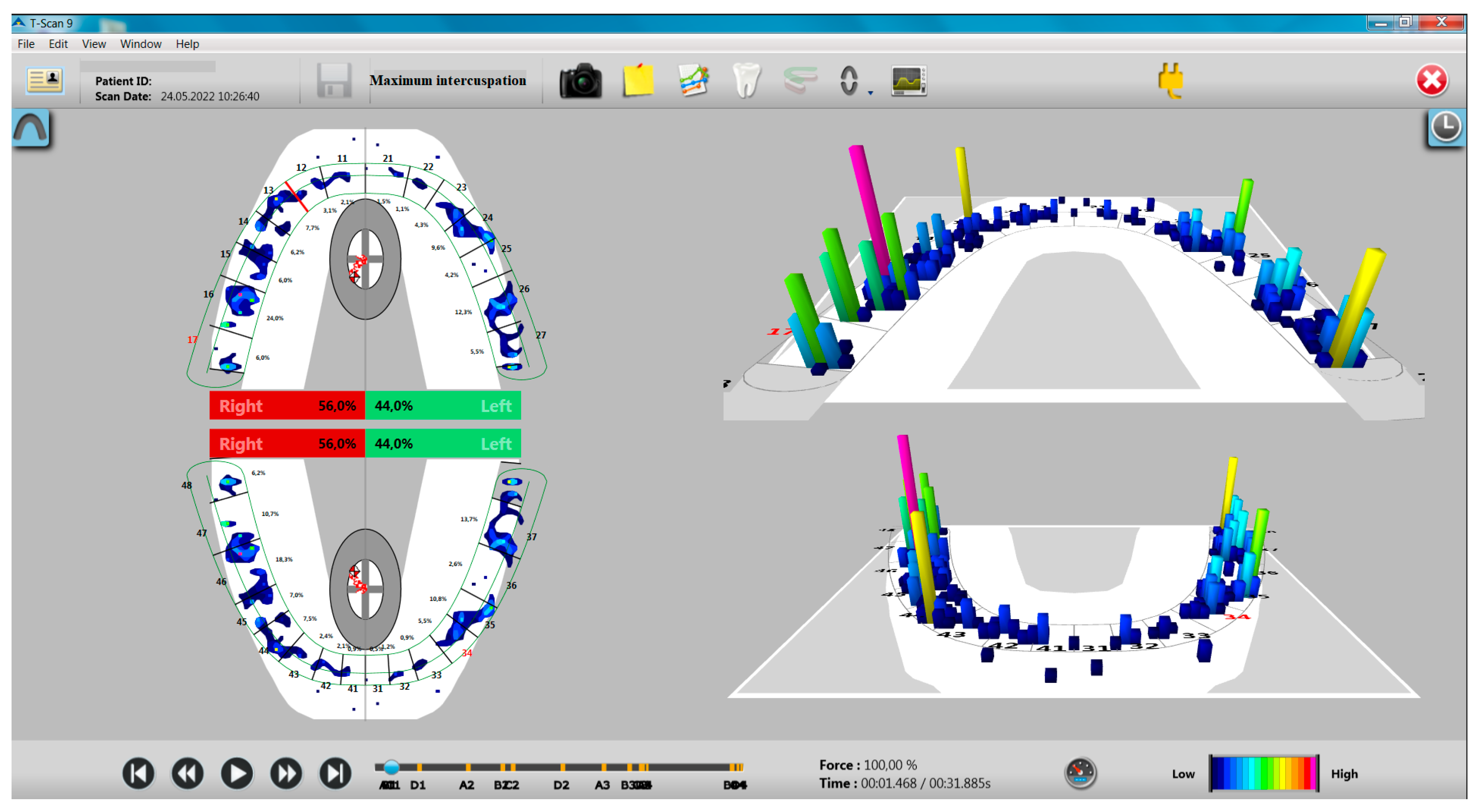
The distribution of occlusal forces at the level of the dental arches showed a higher share of occlusal forces at the level of the right hemiarch, especially in the study group with bruxism (Figure 1 and Figure 2).
Similarly, the gender distribution indicated a greater proportion of occlusal forces at the level of the right hemiarch in women, while men presented more balanced values (Table 3).
No statistically significant differences were identified between the study group and the control group or between women and men, regarding the distribution of occlusal forces at the level of the two hemiarches (p > 0.05).
For the statistical analysis of the distribution of occlusal forces in the MI between the two hemiarches and the data obtained by sEMG with the dia-BRUXO device, an index of the balance between the occlusal forces at the level of the two hemiarches was calculated, representing the difference of the two values (a higher value of the index represents a greater imbalance between the two occlusal forces). The correlation was analyzed using Kendall’s tau-b test.
A single moderate positive association was identified between the values of the occlusal forces in the MI distributed at the level of the two hemiarches and the number of “grinding” type events during the wakefulness period, which was statistically significant, τb = 0.226, p = 0.042.
Regarding the closing and opening time from the MI position, there are no statistically significant differences between the values recorded in the two groups (Table 4), although the closing/opening times were higher in the study group. There are no statistically significant differences by gender either (Table 4).
The correlation between the closing and opening time from the MI position and the parameters provided by the sEMG recordings with the dia-BRUXO device was analyzed using Kendall’s tau-b test.
A single moderate positive association was identified between the closing time and the number of “other” type events during the wakefulness period, which was statistically significant, τb = 0.276, p = 0.014. No association was identified relative to opening time (p > 0.05).

3.2.2. Evaluation of Recordings from Mandibular Movements

Evaluation of Protrusion Movement

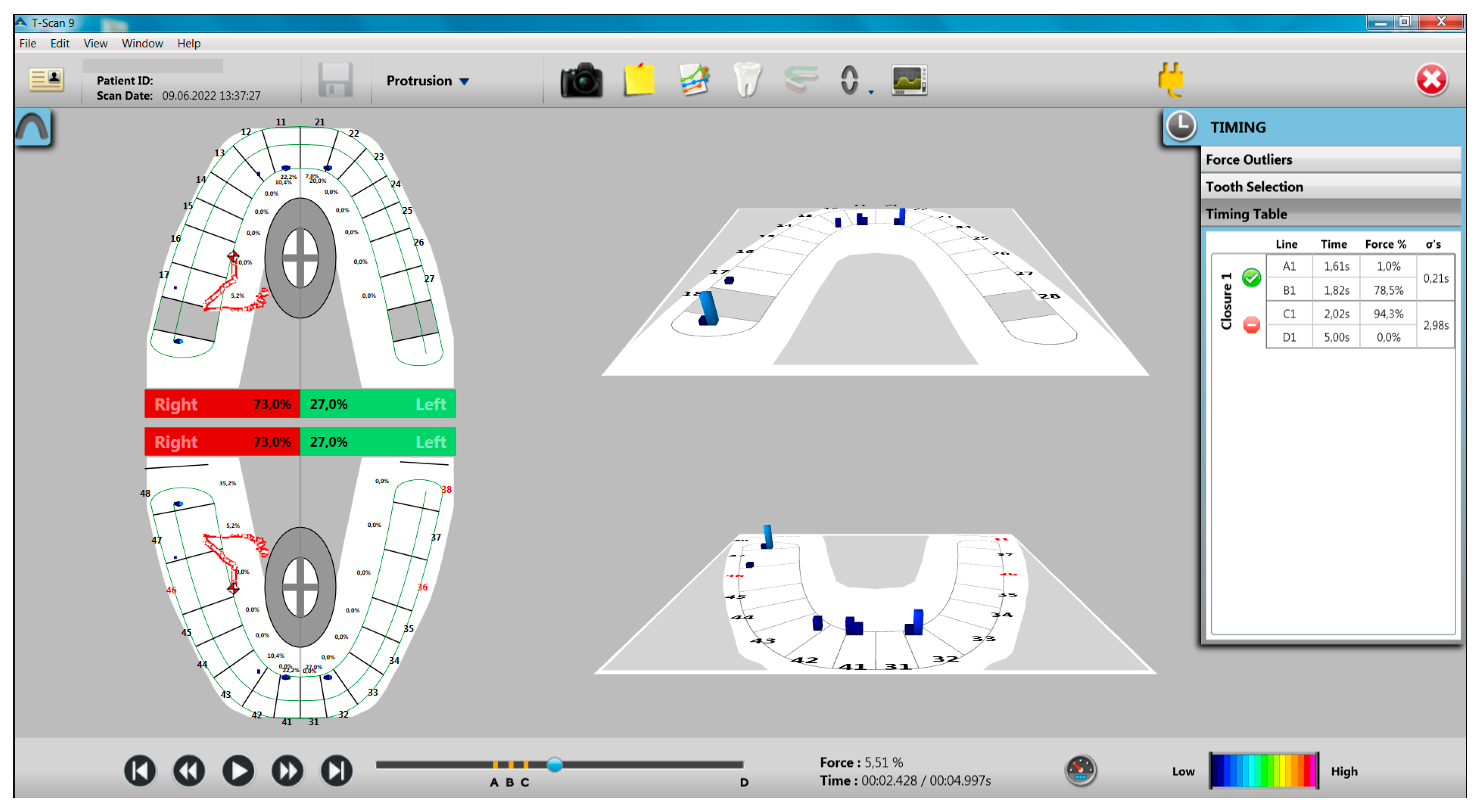
The evaluation of the protrusion movement showed that there are no differences between the two groups (study group and control group) in terms of the presence of incisal guidance, and in terms of working and non-working interferences, they were found to be present in higher numbers in the study group compared to the control group, but the differences are not statistically significant (Table 5). At the end of the protrusion movement, there was a reduced number of premature contacts on the working side and on the non-working side (Figure 3).
Associations between the presence of incisal guidance, working/non-working interferences, premature contacts, and parameters provided by sEMG recordings with the dia-BRUXO device were analyzed using the Mann–Whitney U test.
There are no statistically significant correlations between the presence of incisal guidance or the presence of working interferences and sEMG parameters, p > 0.05.
Instead, for the groups with and without non-working interferences in the protrusion movement, statistically significant differences were identified relative to several parameters acquired during the night: the SBTI index, the SBPI index, the number of “grinding” type events during the night (Table 6), so participants with non-working interferences had higher values of these parameters, compared to participants without non-working interferences.
The correlation between the duration of the protrusion movement and the parameters provided by the sEMG recordings with the dia-BRUXO device was analyzed using Kendall’s tau-b test.
The following statistically significant associations were identified: SBTI (moderate positive association, τb = 0.310, p = 0.005), SBPI (moderate positive association, τb = 0.307, p = 0.006), percentage of sleep hours (moderate positive association, τb = 0.288, p = 0.009), percentage of waking hours (moderate negative association, τb = −0.270, p = 0.015).
The analysis of the protrusion movement also highlighted the following aspects:
  • Participants with incisal guidance showed lower values of occlusal forces distributed on the right hemiarch in MI and higher values of occlusal forces distributed on the left hemiarch in MI, compared to participants without incisal guidance, differences between these groups being statistically significant (p < 0.0005);
  • Participants with work interferences presented similar values of the distribution of occlusal forces at the level of the right and left hemiarch compared to participants without work interferences;
  • Participants with non-working interferences presented higher values of forces distributed on the right hemiarch in MI and lower values of forces distributed on the left hemiarch in MI, compared to participants without non-working interferences, differences between these groups being statistically significant (p = 0.027 for forces from the level of the right hemiarch in MI and p = 0.025 for the forces from the level of the left hemiarch in MI) (Table 7).
The duration of the protrusion movement had similar values for participants without work interferences (2.39 ± 0.945), and with work interferences (2.36 ± 0.805), so the difference between the two groups was not statistically significant (Table 8). In addition, the duration of the protrusion movement had lower values for participants without non-working interferences (2.31 ± 0.845) compared to participants with non-working interferences (2.45 ± 0.929), and the difference between the two groups was not statistically significant (Table 8).
For a relevant evaluation of the duration of the protrusion movement according to the presence or absence of occlusal interferences, the study group was divided into four categories:
  • Group A—participants without working interferences and without non-working interferences;
  • Group B—participants without working interferences and with non-working interferences;
  • Group C—participants with working interferences and without non-working interferences;
  • Group D—participants with work interferences and with non-work interferences.
The distribution by groups relative to the duration of the protrusion movement and the values of the SBTI, ABTI, SBPI, and ABPI indices is highlighted in Table 9.
The longest mean duration of the protrusion movement was recorded for group B (participants without working interferences, but with non-working interferences) for participants with AB, followed by group D (participants with working interferences and with non-working interferences) for participants with SB (Table 9). It is noted that non-working interferences are involved in the increased duration of both movements.
It is worth noting the involvement of occlusal interferences, especially the non-working ones from the protrusion movement, during the mandibular movement, and during the contractions of the masticatory muscles.

Evaluation of Laterotrusion Movements

Analyzing the laterotrusion movements, the following aspects were found:
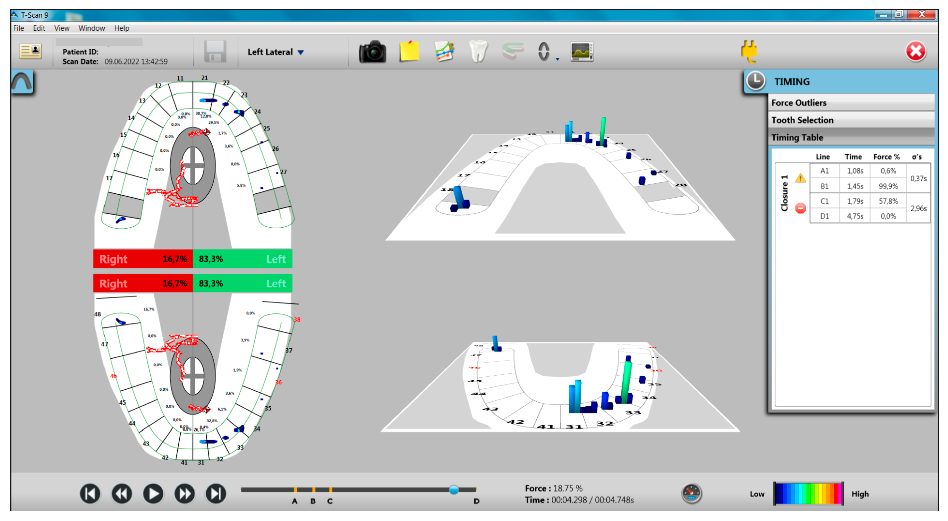
In the left laterotrusion movement from the study group, 50% of the participants presented canine guidance and 50% of the participants presented group guidance (40% antero-lateral, 10% lateral), compared to the control group that presented 90% canine guidance and only 10% showed group guidance (5% antero-lateral guidance and 5% group guidance) (Figure 4).
Clinically, this situation causes a longer disocclusion on the non-working hemiarch, respectively the right hemiarch, a situation also reflected by the recordings made and can generate a higher number of non-working interferences (75%) (Table 10).
Associations between the type of guidance in left laterotrusion movement, working/non-working interferences, premature contacts, and parameters provided by sEMG recordings with the dia-BRUXO device were analyzed using the Kruskal–Wallis H test.
There are no statistically significant correlations between the presence of working interferences or premature working/non-working contacts and the sEMG parameters recorded with the dia-BRUXO system, p > 0.05.
Instead, depending on the type of guidance: canine/antero-lateral group/lateral group, statistically significant differences were identified relative to some of the parameters acquired during the day: the ABPI index, the percentage duration of sleep, the percentage duration of the state of wakefulness, the number of “clenching” type events during night, and ABTI (Table 11).
Participants with antero-lateral guidance had higher values for ABTI index, ABPI, percent sleep duration, and number of “clenching” type events during the night.
An important variation between canine-guidance and side-group-guidance participants is the number of “clenching” type events during the night, where the side-group-guidance participants had twice as many events as the canine-guidance group (Table 11).
The correlation between the duration of the left laterotrusion movement and the parameters provided by the sEMG recordings with the dia-BRUXO device was analyzed using Kendall’s tau-b test. The following statistically significant associations were identified: SBTI (moderate positive association, τb = 0.305, p = 0.006), SBWI (moderate positive association, τb = 0.285, p = 0.010), SBPI (moderate positive association, τb = 0.294, p = 0.008), the number of “grinding” type events during the night (moderate positive association, τb = 0.259, p = 0.023), the number of “other” type events during the day (moderate negative association, τb = −0.297, p = 0.008).
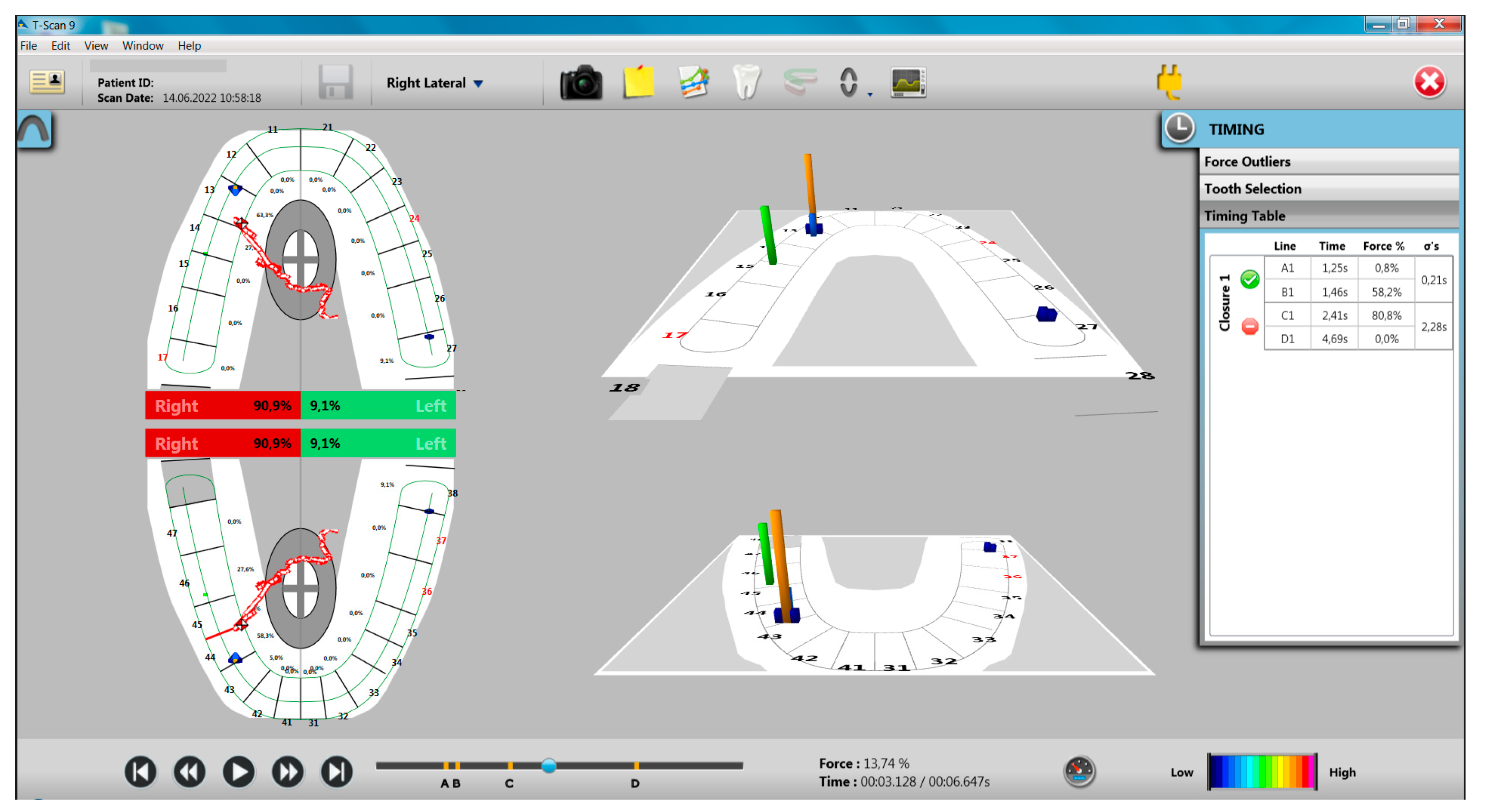
In the right laterotrusion movement, for the study group, the weight of the group guidance was lower compared to the left laterotrusion movement, respectively, 40% (30% antero-lateral guidance and 10% group guidance), but higher compared to the group control with 25% group guidance (20% antero-lateral guidance and 5% group guidance) compared to the left hemiarch with 10% (Figure 5, Table 12).
Associations between guidance type in right late laterotrusion movement, working/non-working interferences, premature contacts, and parameters provided by sEMG recordings with the dia-BRUXO device were analyzed using the Kruskal–Wallis H test.
No statistically significant correlations were revealed between the presence of working interferences or premature working/non-working contacts and the sEMG parameters recorded with the dia-BRUXO system, p > 0.05.
The correlation between the duration of the right laterotrusion movement and the parameters provided by the sEMG recordings with the dia-BRUXO device was analyzed using Kendall’s tau-b test.
The following statistically significant associations of the duration of the right laterotrusion movement were identified: SBTI (moderate positive association, τb = 0.325, p = 0.003), SBWI (moderate positive association, τb = 0.311, p = 0.005), SBPI (moderate positive association, τb = 0.325, p = 0.003), the number of “grinding” type events during the night (moderate positive association, τb = 0.243, p = 0.029), the number of “clenching” type events during the night (positive association moderate, τb = 0.250, p = 0.028), the number of “other” type events during the night (moderate positive association, τb = 0.354, p = 0.005).
Analyzing the duration of the three mandibular movements, it is found that, for all movements, the study group had higher values compared to the control group. The largest difference between the groups was for the protrusion movement (the study group had a mean of 2.60 ± 0.98 s, compared to the control group, with a mean of 2.16 ± 0.71 s).
In the case of laterotrusion movements, higher values were recorded for the study group, especially the laterotrusion movement had a longer duration for the study group with an average of 2.81 ± 0.80 s compared to the control group, with an average of 2.26 ± 0.78 s, the differences in the values recorded in both groups being statistically significant (Table 13).
Correlating the data obtained with the T-Scan device with those obtained by sEMG relative to time, the following aspects were found in the two groups of participants.
Analysis of BTI indices, both daytime and nighttime, revealed that there are moderate positive associations of BTI with the duration of protrusion and laterotrusion movements at night, but not during the day, or with other words, the duration of masticatory muscle contractions in participants with SB correlated with the duration of protrusion and laterotrusion movements (Table 14).
According to these results, there are greater changes in dental occlusion in the study participants who were diagnosed with bruxism. However, graphically representing BPI, the most representative index of bruxism, with the presence of occlusal non-working interferences in propulsion (Figure 6a) and BTI, the index that refers to the duration of bruxism episodes, with the duration of mandibular movements recorded with the T-Scan system (Figure 6b), it was found that there is no direct relationship (interdependence) between these parameters, which means that a number of other factors are also involved in bruxism.

4. Discussion

The masticatory system is a complex unit designed to perform the basic functions for life (mastication, swallowing, phonation). There is a close interdependence between all the components of the masticatory system so that a disorder in one of its components can induce changes in all components.
The role of occlusal imbalances in bruxism is a critical topic for dental specialists. If occlusal factors are indeed involved in the etiology of bruxism, then the dentist is responsible for providing appropriate therapy, as dentists are the only competent professionals who can improve dental occlusion [39].
The present cross-sectional study aimed to evaluate dental occlusion in a group of students diagnosed with bruxism compared to students without bruxism. The present research was focused, in particular, on highlighting premature contacts and occlusal interferences, which may be associated with bruxism, which is why closing and opening movements from the MI position, mandibular protrusion movements, and right and left laterotrusion movements were evaluated. The duration of closing–opening movements in the MI position as well as the duration of mandibular movements, were also evaluated.
Methods for evaluating occlusal contacts can be divided into qualitative analysis and quantitative analysis.
The qualitative analysis involves evaluating the contacts of the opposing teeth and their positions by means of an occlusal film, respectively, articulation paper, wax, and silicone [40]. The major disadvantage of these recordings is the lack of objectivity and reproducibility, as well as the difficulty in describing the state of occlusion.
The quantitative analysis involves the use of some indices to evaluate the occlusal state. The digital occlusal analysis is one of the best quantitative analysis methods because it measures occlusal forces with high precision and repeatability [41]. Most of the traditional methods analyze static occlusion, but occlusion is not static but rather a parameter of a dynamic structure.
The T-Scan III system developed by Tekscan (Boston, MA, USA) uses the computer to analyze the occlusion in dynamics, allowing both qualitative and quantitative occlusal analyses to be performed simultaneously and with high precision. In fact, during the past years, scientists have actively involved technology through software applications or medical devices to increase the accuracy of the diagnostic process in dentistry and in medicine in general [24,25,26]. The T-Scan system can measure the sequence of dental contacts and quantitative dynamics in relation to time, locate excessive occlusal forces, and record the time from the first contact to the end of the movement [42].
The evolution of T-Scan technology spans the past 40 years and started with T-Scan I, later with the development of Turbo recording in 2008, to the 2014 version known as T-Scan 8 (Tekscan Inc., South Boston, MA, USA) [43]. Sanjana Devi et al. carried out a review of occlusal analysis methods in which they described the evolution of the T-Scan system [44].
Other studies have also been conducted using this method and have highlighted its advantages. Popșor et al. in 2010 and in 2019 [45,46] and Komali G et al. in 2019 [47] showed that the T-Scan is a modern diagnostic tool that can measure relative occlusal forces in real time and is a significant step forward in the modern era of dentistry. It is a reliable indicator of occlusal interferences for their precise identification and occlusal balancing in TMD management, thus contributing to successful treatment. Kalachev et al. also showed that T-Scan is a valuable and reliable system for locating and distributing occlusal contacts in dynamics [48,49].
For bruxism, the T-Scan digital evaluation of occlusion relationships was described by Thanathornwong B and Suebnukarn S in 2017 [41] to show the effectiveness of the method
The results of the present study showed that the participants with bruxism presented greater imbalances at the level of dental occlusion, in terms of the distribution of occlusal forces at the level of the two hemiarches, the presence of non-working interferences in the propulsion movement and in terms of the duration of mandibular movements. Other studies have also addressed this aspect.
Thus, a recent study by Iacob et al. in 2022, by clinical assessment, showed that study participants with bruxism had greater occlusal imbalances [50].
Another study, conducted by Gümüş et al. in 2013, using the same method of occlusal analysis as in the present study, on the same number of participants, revealed that there are differences between occlusion relationships in participants with bruxism compared to those without bruxism [51].
The present study showed an asymmetric distribution of occlusal forces in the MI position, predominantly on the right hemiarch, especially in the participants with bruxism. The asymmetric distribution of occlusal forces has been associated with the presence of non-working interferences in protrusion movement, which has also been associated with bruxism.
However, the asymmetric distribution of the occlusal forces at the level of the two hemiarches is considered normal by some specialists, being determined by the masticatory type [52,53].
Regarding the evaluation of mandibular movements, the study revealed that in addition to non-working occlusal interferences from protrusion that were associated with bruxism, non-working interferences from laterotrusion movements were associated with increased bruxism indices, specifically for SB. Only the antero-lateral guidance in the left laterotrusion movement was associated with AB.
We have not found in the specialized literature T-Scan studies the association between non-working interferences and bruxism, but over time, this association has been made, in general. The role of non-working interferences from protrusion movement in bruxism was pointed out by Ramfjord in 1961 and Safari et al. in 2013 [21,54]. In the specialized literature, there are explanations of how occlusal interferences from mandibular movements influence the contraction of the masticatory muscles.
When the mandible moves in the protrusion motion, horizontal forces are generated that must be supported by the teeth. The front or anterior teeth can best absorb and dissipate these forces [39]. The anterior teeth must provide adequate guidance for the disocclusion of the posterior teeth. Interferences and non-working contacts from the protrusion movement generate unfavorable forces on the dento-maxillary system by their amplitude and direction [50,51]. Currently, anterior and posterior teeth are considered to function differently. The posterior teeth function effectively in accepting the forces applied during the mouth closing movement. They accept these forces well, primarily because their arched position ensures that the forces are directed into their long axes and are thus efficiently dissipated. The anterior teeth, however, are not favorably positioned on the arches to accept high forces. They are normally positioned with a vestibular incisal orientation to the closing direction so that axial loading is almost impossible. The consequence is that, in situations where the lateral teeth are lost, the front teeth move vestibularly.
The anterior teeth, unlike the posterior teeth, are in the proper position to accept the forces of the eccentric movements of the mandible. Therefore, in general, it can be stated that the posterior teeth function most effectively in fixing the mandible during closure, while the anterior teeth function most effectively in guiding the mandible during eccentric movements [39].
In the laterotrusion movement, regardless of the type of guidance, an immediate disocclusion of the teeth on the non-working hemiarch must occur. Occlusal interferences and premature non-working contacts can be destructive to the dento-maxillary system due to the magnitude and direction of forces that can be applied to the articular structures and teeth [39]. Some studies have shown that non-working interferences are perceived by the neuro-muscular system differently than other types of occlusal contacts. Electromyography studies have shown that all contacts with opposing teeth are inhibitory in nature [39]. In other words, the presence of interarch contacts tends to reduce muscle activity by inhibiting proprioceptors and nociceptors in the periodontal ligaments; however, other electromyographic studies have shown that the presence of non-working contacts on the posterior teeth increases muscle activity [55]. Although increased muscle activity can be demonstrated, the reason for its presence is currently not understood.
One of the most significant reasons why non-working interferences should be avoided relates to the effect they can have on the loading and stability of the temporo-mandibular joints (TMJ). When the mandible is displaced laterally, regardless of the type of guidance, the condyle on the balance side can be influenced by interferences or non-working contacts. When there is no interference or non-working contact, the condyle follows a fixed path, with the medial pole in contact with the medial wall of the glenoid fossa, moving downward and forward. If a non-working contact is present and this contact is significant enough to disable the lateral guidance from the working side, a very unstable mandibular relationship is created.
Remaining only the contact on the non-working side, the mandible can be in a situation where the masticatory muscles can be contracted bilaterally, and one of the condyles can be displaced, depending on the specific muscle activity. In this situation, the support point is established on the non-working contact that can cause the dislocation of one of the joints [39]. It should be noted that not all non-working contacts cause a dislocation, but in patients with bruxism, this situation can occur [39].
The study also highlighted an association between the duration of mandibular movements and bruxism indices, this association being logical because the muscle activity in bruxism also requires the mobilization of the mandible. We have not found any studies in the specialized literature that address this aspect.
In opposition to these studies, in 2003, Manfredini et al. [36] stated that there are not enough studies on the association between occlusal factors and bruxism; in 2004, they showed that there is no relationship between self-reported (clinically established) bruxism and occlusal factors in adults [56,57], a fact also supported by Demir A et al., 2004 [58], Gonçalves LPV et al., 2010 [59], and Cheng HJ et al., 2004 [60].
Later in 2012, Manfredini et al. [61] found a weak correlation between occlusal factors and bruxism, so it did not completely rule out this association.
Taking into account this situation and the lack of clear evidence regarding the involvement of occlusion in bruxism, the treatment of bruxism is indicated only when its complications occur [12,35].
Therapeutic guidelines do not recommend occlusal adjustment but recommend occlusal balancing and protection of tooth structures by means of an occlusal mouth guard.
Brocard et al., 2007 [62] recommended that the treatment of bruxism involves several stages: the behavioral stage with the therapy of psychosocial aspects, the pharmacological stage, the reversible and non-invasive dental stage (occlusal mouth guard), and the irreversible dental stage when oral rehabilitation is needed.
Matusz et al., 2022 [63] recommend educational therapy for a patient about harmful habits, biofeedback, muscle relaxation exercises, occlusal splint therapy (occlusal mouth guard), short-term drug therapy, botulinum toxin injections, psychotherapy (stress reduction, lifestyle change, hypnosis), electrical method, etc.
Paesani 2010 [64] showed that occlusal mouth guards reduce EMG activity but do not eliminate bruxism.
Other studies [65,66] have shown that occlusal mouthguards are effective in bruxism by reducing the activity of the masticatory muscles.
Summarizing the results of the study, we can state that dental occlusion is a component of the reflex mechanisms that control mandibular movements and implicit contractions of the masticatory muscles, but a number of other factors must be considered in the management of bruxism. The occlusal relationships and the forces developed by the oro-facial muscles are in a dynamic balance. Changes in occlusal relationships can influence muscle contraction and cause disturbances in the components of the dento-maxillary system.
Based on the results obtained in this study, we can state with certainty that there are occlusal imbalances in the participants with bruxism, but we cannot say whether these occlusal imbalances maintain the contraction disorders, the contraction disorders cause these occlusal imbalances, or simply the occlusal imbalances coexist with the disorders contraction of the masticatory muscles.
The limits of the study are represented by the number of participants in the study and the fact that occlusal interferences were not determined along the CR—MI pathway, which is also invoked in the etiology of bruxism.
A holistic approach should establish the role of the masticatory system in the etiology of bruxism. Following this, understanding that dysfunctions at the level of the dento-maxillary system (TMD) composed of the dental determinant (dental occlusion), the neuro-muscular determinant (masticatory muscles) and the articular determinant (TMJ) can be taken into account in the etiology of bruxism.

5. Conclusions

The study approached a very important and controversial aspect regarding the association between occlusion and bruxism.
The study demonstrated that study participants who had bruxism had a more “unbalanced” occlusion; regarding the distribution of occlusal forces on the two hemiarches, they had increased bruxism indices compared to those without bruxism. The presence of non-working interferences from protrusion movement and group guidance in lateral movements were also associated with increased bruxism indices.
In the current context in which the prevalence of bruxism is increasing, we consider that the approach to bruxism in this study is welcome and requires wider approaches, and the results to materialize in informational and preventive projects, in particular, for high-risk subjects, as well as for specialists involved in bruxism therapy.
Highlighting the risk factors involved in bruxism may be helpful in the management of bruxism.

Author Contributions

Conceptualization, D.E.V., A.A.T. and V.M.; methodology, D.E.V., M.S., S.M.P. and V.M.; software, M.I., L.N. and A.Ş.; validation, H.O.M. and G.L.; formal analysis, M.I., S.M.P. and A.A.T.; investigation, D.E.V., M.S., A.A.T. and A.Ş.; resources, L.N., H.O.M. and G.L.; data curation, L.N. and M.S.; writing—original draft preparation, D.E.V., M.S. and S.M.P.; writing—review and editing, H.O.M., A.A.T. and V.M.; visualization, M.I., A.Ş. and G.L.; supervision, V.M. and S.M.P.; project administration, V.M. All authors have read and agreed to the published version of the manuscript.

Funding

Article publication charges are supported by the University of Medicine and Pharmacy of Craiova.

Institutional Review Board Statement

The study was conducted in accordance with the Declaration of Helsinki and approved by the Ethics Commission of the University of Medicine and Pharmacy of Craiova, no 84/03.06.2021.

Informed Consent Statement

Informed consent was obtained from all subjects involved in the study.

Data Availability Statement

The authors declare that the data of this research are available from the corresponding authors upon reasonable request.

Acknowledgments

Special thanks to Lorenzo Noveri for providing the dia-BRUXO device.

Conflicts of Interest

The authors declare no conflict of interest.

References

  1. Lobbezoo, F.; Ahlberg, J.; Glaros, A.G.; Kato, T.; Koyano, K.; Lavigne, G.J.; Manfredini, D.; Svensson, P.; Winocur, E. Bruxism defined and graded: An international consensus. J. Oral Rehabil. 2013, 40, 2–4. [Google Scholar] [CrossRef] [PubMed]
  2. Ferro, K.J. The glossary of prosthodontic terms. J. Prosthet. Dent. 2005, 94, 10–92. [Google Scholar]
  3. International Classification of Sleep Disorders, 2nd ed.; American Academy of Sleep Medicine: Westchester, UK, 2005.
  4. De Leeuw, R. (Ed.) Orofacial Pain. Guidelines for Assessment, Diagnosis, and Management, 4th ed.; Quintessence Publishing Co., Inc.: Chicago, IL, USA, 2008; p. 316. [Google Scholar]
  5. Lobbezoo, F.; Ahlberg, J.; Raphael, K.G.; Wetselaar, P.; Glaros, A.G.; Kato, T.; Santiago, V.; Winocur, E.; De Laat, A.; De Leeuw, R.; et al. International consensus on the assessment of bruxism: Report of a work in progress. J. Oral Rehabil. 2018, 45, 837–844. [Google Scholar] [CrossRef] [PubMed]
  6. Manfredini, D.; Ahlberg, J.; Aarab, G.; Bender, S.; Bracci, A.; Cistulli, P.A.; Conti, P.C.; De Leeuw, R.; Durham, J.; Emodi-Perlman, A.; et al. Standardised Tool for the Assessment of Bruxism. J. Oral Rehabil. 2023, in press. [Google Scholar] [CrossRef]
  7. Manfredini, D.; Ahlberg, J.; Aarab, G.; Bracci, A.; Durham, J.; Emodi-Perlman, A.; Ettlin, D.; Gallo, L.M.; Häggman-Henrikson, B.; Koutris, M.; et al. The development of the Standardised Tool for the Assessment of Bruxism (STAB): An international road map. J. Oral Rehabil. 2022, 1–14. [Google Scholar] [CrossRef] [PubMed]
  8. Bracci, A.; Lobbezoo, F.; Häggman-Henrikson, B.; Colonna, A.; Nykänen, L.; Pollis, M.; Ahlberg, J.; Manfredini, D. International Network for Orofacial Pain and Related Disorders Methodology. Current Knowledge and Future Perspectives on Awake Bruxism Assessment: Expert Consensus Recommendations. J. Clin. Med. 2022, 11, 5083. [Google Scholar] [CrossRef]
  9. Manfredini, D.; Ahlberg, J.; Lobbezoo, F. Bruxism definition: Past, present, and future—What should a prosthodontist know? J. Prosthet. Dent. 2021, 128, 905–912. [Google Scholar] [CrossRef]
  10. Lavigne, G.; Kato, T.; Babiloni, A.H.; Huynh, N.; Fabbro, C.D.; Svensson, P.; Aarab, G.; Ahlberg, J.; Baba, K.; Carra, M.C.; et al. Research routes on improved sleep bruxism metrics: Toward a standardised approach. J. Sleep Res. 2021, 30, e13320. [Google Scholar] [CrossRef]
  11. Manfredini, D.; Ahlberg, J.; Aarab, G.; Bracci, A.; Durham, J.; Ettlin, D.; Gallo, L.M.; Koutris, M.; Wetselaar, P.; Svensson, P.; et al. Towards a Standardized Tool for the Assessment of Bruxism (STAB)-Overview and general remarks of a multidimensional bruxism evaluation system. J. Oral Rehabil. 2020, 47, 549–556. [Google Scholar] [CrossRef]
  12. Svensson, P.; Lavigne, G. Clinical bruxism semantics beyond academic debates: Normo- and patho-bruxism as a new proposal. J. Oral Rehabil. 2020, 47, 547–548. [Google Scholar] [CrossRef]
  13. Vlăduțu, D.E.; Ionescu, M.; Mercuț, R.; Stănuși, A.; Boțilă, M.R.; Nardin, M.; Popa, D.A.; Popescu, M.A.; Ionescu, A.G.; Mercuț, V. Bruxism as a contraction disorder of the masticatory muscles—Review. Rom. J. Oral Rehabil. 2022, 14, 26–34. [Google Scholar]
  14. Saczuk, K.; Lapinska, B.; Wawrzynkiewicz, A.; Witkowska, A.; Arbildo-Vega, H.I.; Domarecka, M.; Lukomska-Szymanska, M. Temporomandibular Disorders, Bruxism, Perceived Stress, and Coping Strategies among Medical University Students in Times of Social Isolation during Outbreak of COVID-19 Pandemic. Healthcare 2022, 10, 740. [Google Scholar] [CrossRef] [PubMed]
  15. Boscato, N.; Exposto, F.; Nascimento, G.G.; Svensson, P.; Costa, Y.M. Is bruxism associated with changes in neural pathways? A systematic review and meta-analysis of clinical studies using neurophysiological techniques. Brain Imaging Behav. 2022, 16, 2268–2280. [Google Scholar] [CrossRef] [PubMed]
  16. Boscato, N.; Nascimento, G.G.; Leite, F.R.M.; Horta, B.L.; Svensson, P.; Demarco, F.F. Role of occlusal factors on probable bruxism and orofacial pain: Data from the 1982 Pelotas birth cohort study. J. Dent. 2021, 113, 103788. [Google Scholar] [CrossRef] [PubMed]
  17. Bulanda, S.; Ilczuk-Rypuła, D.; Nitecka-Buchta, A.; Nowak, Z.; Baron, S.; Postek-Stefańska, L. Sleep Bruxism in Children: Etiology, Diagnosis, and Treatment-A Literature Review. Int. J. Environ. Res. Public Health 2021, 18, 9544. [Google Scholar] [CrossRef] [PubMed]
  18. Kuhn, M.; Türp, J.C. Risk factors for bruxism. Swiss. Dent. J. 2018, 128, 118–124. [Google Scholar] [PubMed]
  19. Klasser, G.D.; Rei, N.; Lavigne, G.J. Sleep bruxism etiology: The evolution of a changing paradigm. J. Can. Dent. Assoc. 2015, 81, f2. [Google Scholar] [PubMed]
  20. Lavigne, G.J.; Kato, T.; Kolta, A.; Sessle, B.J. Neurobiological mechanisms involved in sleep bruxism. Crit. Rev. Oral Biol. Med. 2003, 14, 30–46. [Google Scholar] [CrossRef] [Green Version]
  21. Ramfjord, S.P. Bruxism, a clinical and electromyographic study. J. Am. Dent. Assoc. 1961, 62, 21–44. [Google Scholar] [CrossRef]
  22. Lal, S.J.; Weber, D.D.S.K.K. Bruxism Management; StatPearls Publishing: Treasure Island, FL, USA, 2022. [Google Scholar]
  23. Huang, Z.; Zhou, N.; Lobbezoo, F.; Almeida, F.R.; Cistulli, P.A.; Dieltjens, M.; Huynh, N.T.; Kato, T.; Lavigne, G.J.; Masse, J.F.; et al. Dental sleep-related conditions and the role of oral healthcare providers: A scoping review. Sleep Med. Rev. 2023, 67, 101721. [Google Scholar] [CrossRef]
  24. Vlăduțu, D.; Popescu, S.M.; Mercuț, R.; Ionescu, M.; Scrieciu, M.; Glodeanu, A.D.; Stănuși, A.; Rîcă, A.M.; Mercuț, V. Associations between Bruxism, Stress, and Manifestations of Temporomandibular Disorder in Young Students. Int. J. Environ. Res. Public Health 2022, 19, 5415. [Google Scholar] [CrossRef]
  25. Ribeiro-Lages, M.B.; Jural, L.A.; Magno, M.B.; Vicente-Gomila, J.; Ferreira, D.M.; Fonseca-Gonçalves, A.; Maia, L.C. A world panorama of bruxism in children and adolescents with emphasis on associated sleep features: A bibliometric analysis. J. Oral Rehabil. 2021, 48, 1271–1282. [Google Scholar] [CrossRef] [PubMed]
  26. Manfredini, D.; Lobbezoo, F. Sleep bruxism and temporomandibular disorders: A scoping review of the literature. J. Dent. 2021, 111, 103711. [Google Scholar] [CrossRef] [PubMed]
  27. Ohlmann, B.; Waldecker, M.; Leckel, M.; Bömicke, W.; Behnisch, R.; Rammelsberg, P.; Schmitter, M. Correlations between Sleep Bruxism and Temporomandibular Disorders. J. Clin. Med. 2020, 9, 611. [Google Scholar] [CrossRef] [PubMed] [Green Version]
  28. Smardz, J.; Martynowicz, H.; Michalek-Zrabkowska, M.; Wojakowska, A.; Mazur, G.; Winocur, E.; Wieckiewicz, M. Sleep Bruxism and Occurrence of Temporomandibular Disorders-Related Pain: A Polysomnographic Study. Front. Neurol. 2019, 10, 168. [Google Scholar] [CrossRef] [PubMed]
  29. Lavigne, G.J.; Khoury, S.; Abe, S.; Yamaguchi, T.; Raphael, K. Bruxism physiology and pathology: An overview for clinicians. J. Oral Rehabil. 2008, 35, 476–494. [Google Scholar] [CrossRef]
  30. Lobbezoo, F.; Naeije, M. Bruxism is mainly regulated centrally, not peripherally. J. Oral Rehabil. 2001, 28, 1085–1091. [Google Scholar] [CrossRef]
  31. Tsukiyama, Y.; Baba, K.; Clark, G.T. An evidence-based assessment of occlusal adjustment as a treatment for temporomandibular disorders. J. Prosthet. Dent. 2001, 86, 57–66. [Google Scholar] [CrossRef]
  32. Clark, G.T.; Tsukiyama, Y.; Baba, K.; Watanabe, T. Sixty-eight years of experimental occlusal interference studies: What have we learned? J. Prosthet. Dent. 1999, 82, 704–713. [Google Scholar] [CrossRef]
  33. Griffin, K.M. Mandibular adaptive reposturing: The etiology of a common and multifaceted autodestructive syndrome. Gen. Dent. 2003, 51, 62–67. [Google Scholar]
  34. Goldstein, G.; DeSantis, L.; Goodacre, C. Bruxism: Best Evidence Consensus Statement. J. Prosthodont. 2021, 30, 91–101. [Google Scholar] [CrossRef] [PubMed]
  35. Beddis, H.; Pemberton, M.; Davies, S. Sleep bruxism: An overview for clinicians. Br. Dent. J. 2018, 225, 497–501. [Google Scholar] [CrossRef] [PubMed]
  36. Manfredini, D.; Cantini, E.; Romagnoli, M.; Bosco, M. Prevalence of bruxism in patients with different research diagnostic criteria for temporomandibular disorders (RDC/TMD) diagnoses. Cranio 2003, 21, 279–285. [Google Scholar] [CrossRef] [PubMed]
  37. Vlăduțu, D.E.; Ionescu, M.; Mercuț, R.; Noveri, L.; Lăzărescu, G.; Popescu, S.M.; Scrieciu, M.; Manolea, H.O.; Iacov Crăițoiu, M.M.; Ionescu, A.G.; et al. Ecological Momentary Assessment of Masseter Muscle Activity in Patients with Bruxism. Int. J. Environ. Res. Public Health 2023, 20, 581. [Google Scholar] [CrossRef] [PubMed]
  38. Colonna, A.; Noveri, L.; Ferrari, M.; Bracci, A.; Manfredini, D. Electromyographic Assessment of Masseter Muscle Activity: A Proposal for a 24 h Recording Device with Preliminary Data. J. Clin. Med. 2023, 12, 247. [Google Scholar] [CrossRef] [PubMed]
  39. Okeson, J.P. Management of Temporomandibular Disorders and Occlusion, 8th ed.; Elsevier, Inc.: Amsterdam, The Netherlands, 2020. [Google Scholar]
  40. Gu, D.A.; Miao, L.Y.; Liu, C. Application of Digital Occlusion Analysis System in Stomatological Clinical Medicine. Austin. J. Dent. 2022, 9, 1164. [Google Scholar]
  41. Thanathornwong, B.; Suebnukarn, S. Clinical Decision Support Model to Predict Occlusal Force in Bruxism Patients. Healthc. Inform. Res. 2017, 23, 255–261. [Google Scholar] [CrossRef] [Green Version]
  42. Kuć, J.; Szarejko, K.D.; Aleksandrowicz, K.; Gołębiewska, M. The role of soft tissue mobilization in reducing orofacial and general complaints in a patient with Kimmerle anomaly and temporomandibular joint disorder: A case report. Cranio 2021, 39, 74–87. [Google Scholar] [CrossRef]
  43. Kerstein, R.B. Handbook of Research on Computerized Occlusal Analysis Technology Applications in Dental Medicine; IGI Global Publishers: Hershey, PA, USA, 2015. [Google Scholar]
  44. Devi, S.; Nallaswamy, D.; Venugopalan, S. Prosthetic Occlusal Analyzers—A Comprehensive Review. Int. J. Dent. Oral Sci. 2021, 8, 3550–3554. [Google Scholar]
  45. Popsor, S.; Albert, L.E.; Sabau, D.C.; Mihai, A. Occlusal features in depressive patients with craniomandibular disfunction. Int. J. Med. Dent. 2019, 23, 106–111. [Google Scholar]
  46. Popsor, S.; Szasz, O.; Șoaita, L.C.; Liana Georgiana Hănțoiu, L.G. Adjusting of a Stabilization Splint by the Use of T-Scan III—BioEMG Integrated System. Rom. J. Oral Rehabil. 2010, 2, 14–20. [Google Scholar]
  47. Komali, G.; Ignatius, A.V.; Srivani, G.S.; Anuja, K. T-scan system in the management of temporomandibular joint disorders—A review. J. Indian Acad. Oral Med. Radiol. 2019, 31, 252–256. [Google Scholar]
  48. Agbaje, J.O.; Casteele, E.V.; Salem, A.S.; Anumendem, D.; Shaheen, E.; Sun, Y.; Politis, C. Assessment of occlusion with the T-Scan system in patients undergoing orthognathic surgery. Sci. Rep. 2017, 7, 5356. [Google Scholar] [CrossRef] [PubMed] [Green Version]
  49. Kalachev, Y.S.; Michailov, T.A.; Iordanov, P.I. Study of occlusal-articulation relationships with the help of T-SCAN apparatus. Folia Med. 2001, 43, 88–91. [Google Scholar]
  50. Iacob, S.M.; Chisnoiu, A.M.; Objelean, A.; Fluerașu, M.I.; Moga, R.R.; Buduru, S.D. Correlation between bruxism, occlusal dysfunction and musculo-articular status. Rom. J. Oral Rehabil. 2022, 14, 48–55. [Google Scholar]
  51. Gümüş, H.Ö.; Kılınç, H.İ.; Tuna, S.H.; Ozcan, N. Computerized analysis of occlusal contacts in bruxism patients treated with occlusal splint therapy. J. Adv. Prosthodont. 2013, 5, 256–261. [Google Scholar] [CrossRef] [Green Version]
  52. Wieczorek, A.; Loster, J.; Loster, B.W. Relationship between occlusal force distribution and the activity of masseter and anterior temporalis muscles in asymptomatic young adults. Biomed. Res. Int. 2013, 2013, 354017. [Google Scholar] [CrossRef] [Green Version]
  53. Suvinen, T.I.; Kemppainen, P. Review of clinical EMG studies related to muscle and occlusal factors in healthy and TMD subjects. J. Oral Rehabil. 2007, 34, 631–644. [Google Scholar] [CrossRef]
  54. Safari, A.; Jowkar, Z.; Farzin, M. Evaluation of the relationship between bruxism and premature occlusal contacts. J. Contemp. Dent. Pract. 2013, 14, 616–621. [Google Scholar] [CrossRef]
  55. Williamson, E.H.; Lundquist, D.O. Anterior guidance: Its effect on electromyographic activity of the temporal and masseter muscles. J. Prosthet. Dent. 1983, 49, 816–823. [Google Scholar] [CrossRef]
  56. Manfredini, D.; Landi, N.; Romagnoli, M.; Bosco, M. Psychic and occlusal factors in bruxers. Aust. Dent. J. 2004, 49, 84–89. [Google Scholar] [CrossRef]
  57. Manfredini, D.; Landi, N.; Tognini, F.; Montagnani, G.; Bosco, M. Occlusal features are not a reliable predictor of bruxism. Minerva Stomatol. 2004, 53, 231–239, (In English, Italian). [Google Scholar]
  58. Demir, A.; Uysal, T.; Guray, E.; Basciftci, F.A. The relationship between bruxism and occlusal factors among seven-to 19-year-old Turkish children. Angle Orthod. 2004, 74, 672–676. [Google Scholar] [PubMed]
  59. Goncalves, L.P.; Toledo, O.; Otero, S.M. The relationship between bruxism, occlusal factors and oral habits. Dent. Press J. Orthod. 2010, 15, 97–104. [Google Scholar]
  60. Cheng, H.J.; Chen, Y.Q.; Yu, C.H.; Shen, Y.Q. The influence of occlusion on the incidence of bruxism in 779 children in Shanghai. Shanghai Kou Qiang Yi Xue 2004, 13, 98–99. (In Chinese) [Google Scholar] [PubMed]
  61. Manfredini, D.; Winocur, E.; Guarda-Nardini, L.; Lobbezoo, F. Self-reported bruxism and temporomandibular disorders: Findings from two specialised centres. J. Oral Rehabil. 2012, 39, 319–325. [Google Scholar] [CrossRef] [PubMed]
  62. Brocard, D.; Laluque, J.F.; Knellesen, C. La Gestion du Bruxisme; Quintessence Internațional: Paris, France, 2007. [Google Scholar]
  63. Matusz, K.; Maciejewska-Szaniec, Z.; Gredes, T.; Pobudek-Radzikowska, M.; Glapiński, M.; Górna, N.; Przystańska, A. Common therapeutic approaches in sleep and awake bruxism—An overview. Neurol. Neurochir. Pol. 2022, 56, 455–463. [Google Scholar] [CrossRef] [PubMed]
  64. Paesani, D.A. Bruxism Theory and Practice, 1st ed.; Quintessence Publishing: Chicago, IL, USA, 2010. [Google Scholar]
  65. Shopova, D.; Bozhkova, T.; Yordanova, S.; Yordanova, M. Case Report: Digital analysis of occlusion with T-Scan Novus in occlusal splint treatment for a patient with bruxism. F1000Research 2021, 10, 915. [Google Scholar] [CrossRef] [PubMed]
  66. Hasegawa, K.; Okamoto, M.; Nishigawa, G.; Oki, K.; Minagi, S. The design of non-occlusal intraoral appliances on hard palate and their effect on masseter muscle activity during sleep. Cranio 2007, 25, 8–15. [Google Scholar] [CrossRef] [PubMed]
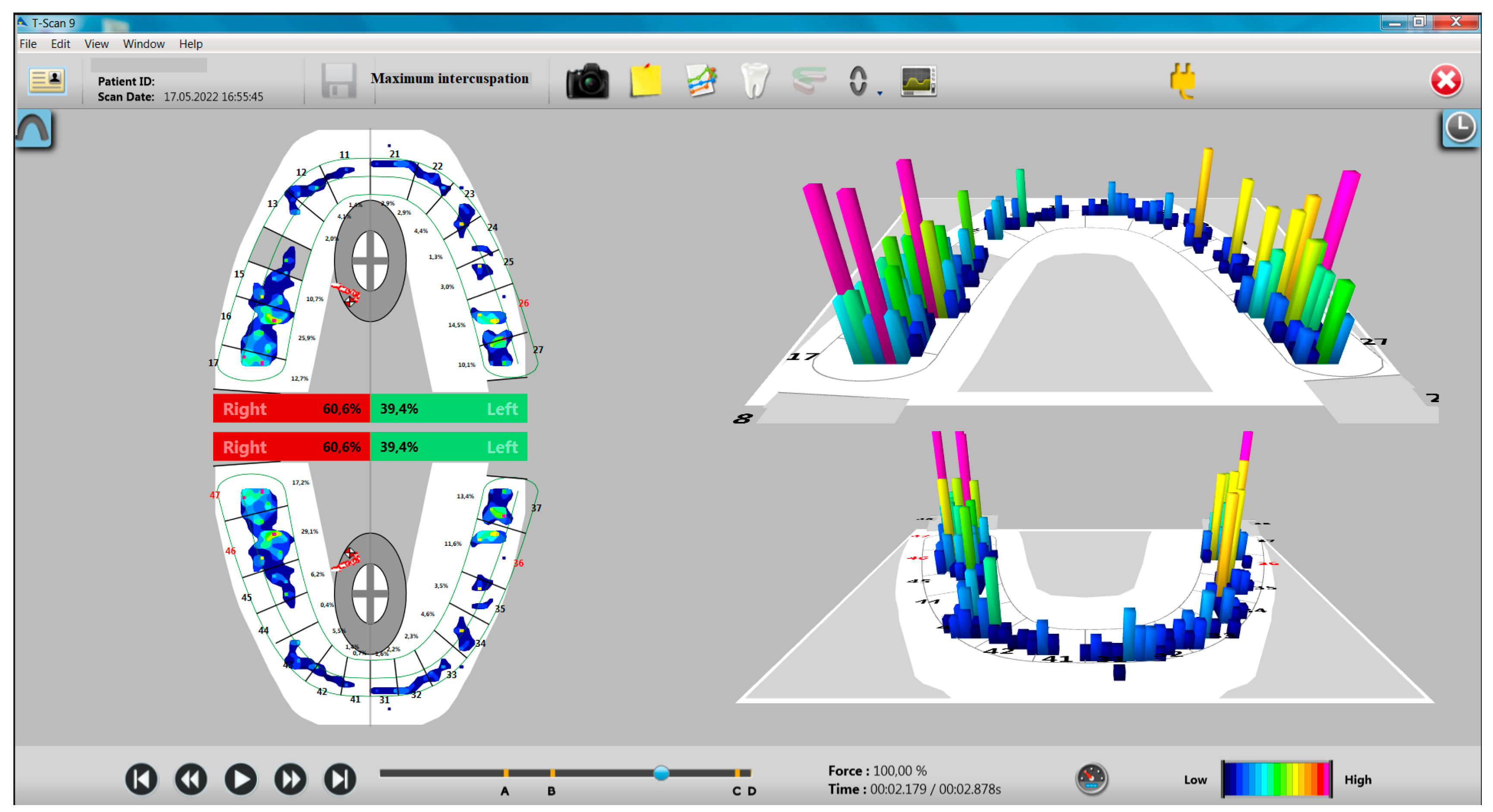
Figure 1. Aspects of T-Scan recording in MI for a participant with bruxism.
Ijerph 20 04877 g001
Figure 2. Aspects of T-Scan recording in MI for a participant without bruxism.
Ijerph 20 04877 g002
Figure 3. Aspects of the T-Scan recording of the protrusion movement for a participant with bruxism.
Ijerph 20 04877 g003
Figure 4. Aspects of T-Scan recording of left laterotrusion movement to a participant with bruxism. Note the antero-lateral group guidance and a non-working interference.
Ijerph 20 04877 g004
Figure 5. Aspects of T-Scan recording in right laterotrusion movement to a participant with bruxism. Canine guidance and the presence of working side interferences and non-working side interferences are noted.
Ijerph 20 04877 g005
Figure 6. Graphical representation of diaBRUXO and T-Scan indexes: (a) Sleep and awake BPI indexes and the presence of occlusal non-working interferences in propulsion; (b) sleep and awake BPI indexes and the duration of mandibular movements recorded with the T-Scan system.
Ijerph 20 04877 g006
Table 1. Demographic characteristics of the two groups.
Study Group Control Group
Participants
(N/%)
Age
Interval
Mean ± SDParticipants
(N/%)
Age
Interval
Mean ± SD
Females15 (75%)20–2924.2 ± 2.7815 (75%)21–5326.46 ± 7.82
Males5 (25%)25–3529 ± 4.245 (25%)26–4735 ± 9.11
Overall20 (100%)20–3525.4 ± 3.7420 (100%)21–5328.6 ± 8.76
Table 2. The main parameters provided by the dia-BRUXO device.
ParameterStudy Group (n = 20)
Mean ± SD
Control Group (n = 20)
Mean ± SD
BTI (%)Awake BTI (ABTI)2.788 ± 2.3810.722 ± 0.422
Sleep BTI (SBTI)1.055 ± 0.7900.233 ± 0.201
BWI (%)Awake BWI (ABWI)1.178 ± 1.0280.310 ± 0.214
Sleep BWI (SBWI)0.471 ± 0.3300.098 ± 0.085
BPI (%)Awake BPI (ABPI)2.251 ± 1.9260.585 ± 0.351
Sleep BPI (SBPI)0.860 ± 0.6330.188 ± 0.162
Table 3. Distribution of occlusal forces on the hemiarches in the MI position.
Subgroup (N)Right Hemiarcade (%)Left Hemiarcade (%)
Mean ± SDp *Mean ± SDp *
GroupStudy (20)57.080 ± 8.5660.11042.920 ± 8.5660.110
Control (20)52.505 ± 7.47647.425 ± 7.463
GenderFemales (30)55.856 ± 7.5510.187 144.110 ± 7.5330.198 1
Males (10)51.600 ± 9.86448.360 ± 9.854
* Mann–Whitney U Test. 1 Exact significance.
Table 4. Duration of occlusion and disocclusion from the MI position by group (seconds).
Subgroup (N)Occlusion Duration
in MI
Duration of Disocclusion
from MI
Mean ± SDp *Mean ± SDp *
GroupStudy (20)1.030 ± 0.6710.8390.785 ± 0.4440.925
Control (20)0.968 ± 0.3850.778 ± 0.418
GenderFemales (30)0.999 ± 0.5570.914 10.742 ± 0.4070.450 1
Males (10)1.000 ± 0.5190.896 ± 0.481
* Mann–Whitney U Test. 1 Exact significance.
Table 5. Evaluation of protrusion movement according to the presence of incisal guidance, working and non-working interferences, and premature contacts.
ParameterValueStudy Group (N = 20)Control Group (N = 20)p *
Protrusion—the presence of the
incisive guidance
Absent2 (10%)1 (5%)0.500
Present18 (90%)19 (95%)
Protrusion—working side
interference
No10 (50%)13 (65%)0.337
Yes10 (50%)7 (35%)
Protrusion—non-working side
interference
No8 (40%)13 (65%)0.113
Yes12 (60%)7 (35%)
Protrusion—edge-to-edge—premature contacts on the working sideNo14 (70%)14 (70%)1
Yes6 (30%)6 (30%)
Protrusion—edge-to-edge—
premature non-working side contacts
No17 (85%)14 (70%)0.225
Yes3 (15%)6 (30%)
* Chi-Square test.
Table 6. Association between bruxism indices and the presence of non-working interferences.
ParameterProtrusion without Non-Working Interference (N = 21)Protrusion with Non-Working Interference (N = 19)p *
SBPI (%)0.378 ± 0.4370.685 ± 0.6600.044 1
SBTI (%)0.461 ± 0.5360.845 ± 0.8210.044 1
Number of “grinding” events per night16.760 ± 43.38028.950 ± 35.4420.014 1
* Mann–Whitney U test. 1 Statistically significant results.
Table 7. Distribution of occlusal forces at the level of the two hemiarches in the MI position, according to the presence of the incisal guidance, the presence of working and non-working occlusal interferences in the protrusion movement.
Occlusal
Status
Distribution of Occlusal Forces on the Right
Hemiarch (%)
Distribution of Occlusal Forces on the Left
Hemiarch (%)
Mean ± SDp *Mean ± SDp *
Protrusion—incisive guidanceAbsent (N = 3)69.866 ± 0.814<0.0005 130.133 ± 0.814<0.0005 1,2
Present (N = 37)53.570 ± 7.31646.391 ± 7.304
Protrusion—working
interferences
No (N = 23)55.339 ± 6.6210.76344.617 ± 6.5970.774
Yes (N = 17)54.052 ± 10.26145.923 ± 10.250
Protrusion—non-working interferencesNo (N = 21)52.333 ± 6.5730.02747.619 ± 6.5690.025 2
Yes (N = 19)57.510 ± 9.23142.468 ± 9.212
* Mann–Whitney U Test. 1 Exact significance. 2 Statistically significant results.
Table 8. The duration of the protrusion movement according to the presence or absence of interferences (seconds).
ParameterGroup (N)Protrusion Duration
Mean ± SDp *
Protrusion—working
interferences
No (23)2.39 ± 0.9450.978 *
Yes (17)2.36 ± 0.805
Protrusion—non-working
interferences
No (21)2.31 ± 0.8450.357
Yes (19)2.45 ± 0.929
* Mann–Whitney U test.
Table 9. Duration of protrusion movement according to associations between working and non-working interferences.
ParameterGroup AGroup BGroup CGroup Dp *
The duration of the protrusion movement2.27 ± 0.9532.62 1 ± 0.9472.40 ± 0.5472.33 ± 0.9420.651
SBTI0.48 ± 0.5910.70 ± 0.4480.40 ± 0.4040.95 1 ± 1.0230.253
ABTI1.46 ± 1.4072.67 1 ± 3.5981.53 ± 1.2701.59 ± 1.3690.997
SBPI0.39 ± 0.4810.57 ± 0.3670.33 ± 0.3320.76 1 ± 0.8190.253
ABPI1.18 ± 1.1322.15 1 ± 2.9061.21 ± 0.9951.30 ± 1.1390.997
* Kruskal–Wallis test. 1 Highest duration
Table 10. Evaluation of the left laterotrusion movement according to the type of guidance, the presence of working and non-working interferences, and premature contacts.
ParameterGuidance Type, The Presence
of Interferences and
Premature Contacts
Study Group (N = 20)Control Group (N = 20)p *
Left laterotrusion movement, guidance typeCanine10 (50%)18 (90%)0.018 1
Antero-lateral group8 (40%)1 (5%)
Side group2 (10%)1 (5%)
Left laterotrusion movement, working side interferencesNo4 (20%)8 (40%)0.168
Yes16 (80%)12 (60%)
Left laterotrusion movement, non-working side interferencesNo5 (25%)10 (50%)0.102
Yes15 (75%)10 (50%)
Left laterotrusion movement, edge–edge position, premature working side contactsNo14 (70%)13 (65%)0.736
Yes6 (30%)7 (35%)
Left laterotrusion movement, edge–edge position, premature non-working side contactsNo13 (65%)18 (90%)0.064
Yes7 (35%)2 (10%)
* Chi-Square test. 1 Statistically significant results.
Table 11. The situation of the correlations between the type of guidance in the left laterotrusion movement and the indices of bruxism.
ParameterCanine Guidance (N = 28)Antero-Lateral Group Guidance (N = 9)Side Group Guidance (N = 3)p *
ABPI1.043 ± 1.0952.702 ± 2.4391.057 ± 0.8350.030 1
Sleep duration30.657 ± 8.19839.255 ± 6.17231.266 ± 6.2960.023 1
Duration of wakefulness68.917 ± 8.63160.744 ± 6.17268.7333 ± 6.2960.033 1
The number of nighttime clenching events16.610 ± 13.35645.440 ± 31.28534.670 ± 24.0070.015 1
ABTI1.286 ± 1.3483.360 ± 3.0141.310 ± 1.0130.027 1
* Kruskal–Wallis H test. 1 Statistically significant results.
Table 12. Evaluation of the right laterotrusion movement according to the type of guidance, the presence of working and non-working interferences, and premature contacts.
ParameterGuidance Type,
The Presence of Interferences and Premature Contacts
Study Group (N = 20)Control Group (N = 20)p *
Right laterotrusion movement, guidance type.Canine12 (60%)15 (75%)0.587
Antero-lateral group6 (30%)4 (20%)
Side group2 (10%)1 (5%)
Right side movement, working side
interferences.
No8 (40%)7 (35%)0.744
Yes12 (60%)13 (65%)
Right laterotrusion movement, non-working side interferences.No5 (25%)10 (50%)0.102
Yes15 (75%)10 (50%)
Right laterotrusion movement, edge–edge position—premature contacts on the working sideNo13 (65%)14 (70%)0.736
Yes7 (35%)6 (30%)
Right laterotrusion movement, edge–edge position—premature non-working side contactsNo12 (60%)14 (70%)0.507
Yes8 (40%)6 (30%)
* Chi-Square test.
Table 13. Duration of mandibular movements in the two groups of participants (seconds).
ParameterStudy Group
(N = 20)
Control Group
(N = 20)
p *
Duration of protrusion2.60 ± 0.982.16 ± 0.710.064
Duration of right laterotrusion2.81 ± 0.802.26 ± 0.780.036
Duration of left laterotrusion2.57 ± 0.872.21 ± 0.920.110
* Mann–Whitney U test.
Table 14. Correlations between duration of mandibular movements and bruxism time indices.
ParameterDuration (Value/p *)
Occlusion
MI
Disocclusion MIProtrusionLeft
Laterotrusion
Right
Laterotrusion
SBTI−0.046/0.675−0.054/0.6240.310/0.0050.305/0.006 10.325/0.003 1
ABTI0.018/0.870−0.023/0.8340.095/0.3880.113/0.3050.162/0.142
* Kendall’s tau-b test. 1 Statistically significant results
Disclaimer/Publisher’s Note: The statements, opinions and data contained in all publications are solely those of the individual author(s) and contributor(s) and not of MDPI and/or the editor(s). MDPI and/or the editor(s) disclaim responsibility for any injury to people or property resulting from any ideas, methods, instructions or products referred to in the content.