
Accidentally Swallowing a Toothbrush in a Patient during a Vomiting Attempt: Literature Review and Case Report


Abstract
1. Introduction
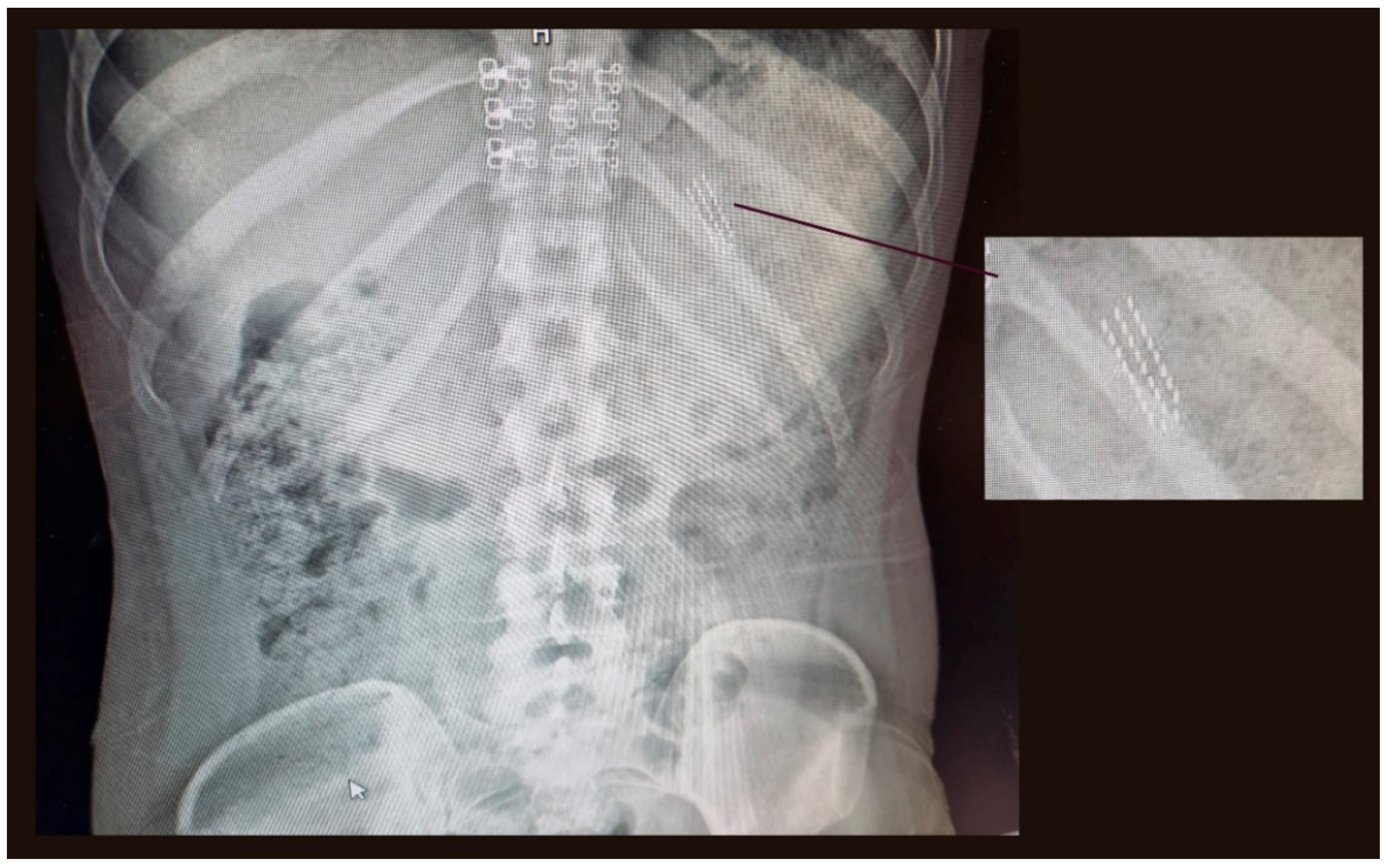
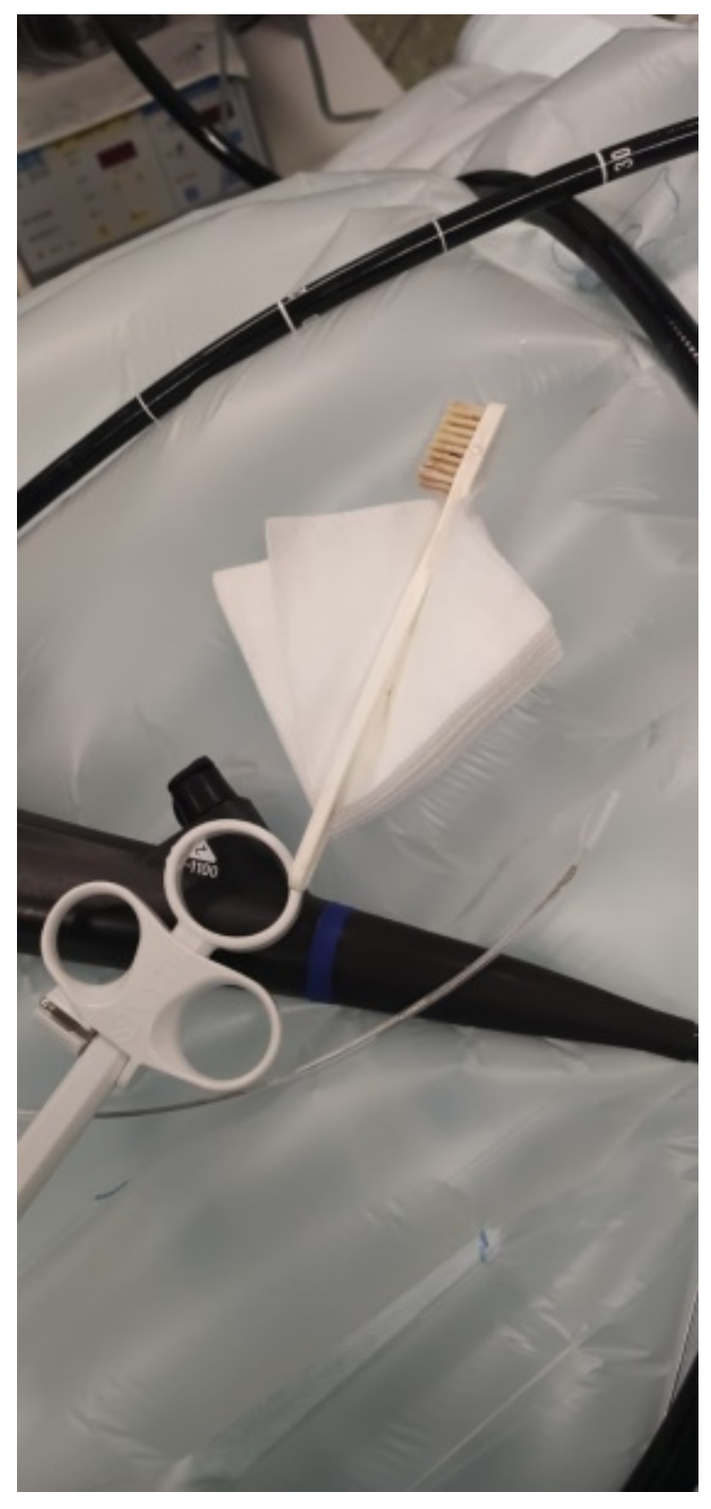
2. Case Report
3. Discussion
4. Conclusions
Author Contributions
Funding
Institutional Review Board Statement
Informed Consent Statement
Data Availability Statement
Conflicts of Interest
References
- Niknam, R.; Mahmoudi, L.; Nasseri-Moghaddam, S. An incidentally swallowed toothbrush. Arch. Iran. Med. 2012, 15, 177–178. [Google Scholar] [PubMed]
- Lee, M.R.; Hwang, Y.; Kim, J.H. A case of colohepatic penetration by a swallowed toothbrush. World J. Gastroenterol. 2006, 12, 2464–2465. [Google Scholar] [CrossRef] [PubMed]
- Yaosaka, T.; Chikama, T.; Ishi, S.; Miwa, K.; Murashima, Y. Endoscopic removal of a toothbrush from the second portion of the duodenum. Gastroenterol. Endosc. 1979, 21, 995. [Google Scholar]
- Mokánszki, I.; Adorján, T. Endoscopic removal of a toothbrush from a 13-year-old girl’s stomach. Orv. Hetil. 2002, 143, 1755–1757. [Google Scholar] [PubMed]
- Ertan, A.; Kedia, S.M.; Agrawal, N.M.; Akdamar, K. Endoscopic removal of a toothbrush. Gastrointest. Endosc. 1983, 29, 144–145. [Google Scholar] [CrossRef]
- Wishner, J.D.; Rogers, A.M. Laparoscopic removal of a swallowed toothbrush. Surg. Endosc. 1997, 11, 472–473. [Google Scholar] [CrossRef] [PubMed]
- Wilcox, D.T.; Karamanoukian, H.L.; Glick, P.L. Toothbrush ingestion by bulimics may require laparotomy. J. Pediatr. Surg. 1994, 29, 1596. [Google Scholar] [CrossRef]
- Kirk, A.D.; Bowers, B.A.; Moylan, J.A.; Meyers, W.C. Toothbrush swallowing. Arch. Surg. 1988, 123, 382–384. [Google Scholar] [CrossRef] [PubMed]
- Cox, D.; Donohue, P.; Costa, V. A swallowed toothbrush causing perforation 2 years after ingestion. Br. J. Hosp. Med. 2007, 68, 559. [Google Scholar] [CrossRef] [PubMed]
- American Psychiatric Association. Diagnostic and Statistical Manual of Mental Disorders: DSM-5, 5th ed.; American Psychiatric Association: Washington, DC, USA, 2013. [Google Scholar] [CrossRef]
- Ragnhildstveit, A.; Jackson, L.K.; Cunningham, S.; Good, L.; Tanner, Q.; Roughan, M.; Henrie-Barrus, P. Case Report: Unexpected Remission from Extreme and Enduring Bulimia Nervosa with Repeated Ketamine Assisted Psychotherapy. Front. Psychiatry 2021, 12, 764112. [Google Scholar] [CrossRef] [PubMed]
- Qian, J.; Wu, Y.; Liu, F.; Zhu, Y.; Jin, H.; Zhang, H.; Wan, Y.; Li, C.; Yu, D. An update on the prevalence of eating disorders in the general population: A systematic review and meta-analysis. Eat. Weight Disord. 2021, 1–14. [Google Scholar] [CrossRef] [PubMed]
- Riddlesberger, M.M.; Cohen, H.L.; Glick, P.L. The swallowed toothbrush: A radiographic clue of bulimia. Pediatr. Radiol. 1991, 21, 262–264. [Google Scholar] [CrossRef] [PubMed]
- Birk, M.; Bauerfeind, P.; Deprez, P.H.; Häfner, M.; Hartmann, D.; Hassan, C.; Hucl, T.; Lesur, G.; Aabakken, L.; Meining, A. Removal of foreign bodies in the upper gastrointestinal tract in adults: European Society of Gastrointestinal Endoscopy (ESGE) Clinical Guideline. Endoscopy 2016, 48, 489–496. [Google Scholar] [CrossRef] [PubMed]
- Benjamin, S.B.; Noguera, E.C. Foreign bodies. In The Esophagus, 4th ed.; Castell, D., Richter, J., Eds.; Lippincott Williams & Wilkins: Philadelphia, PA, USA, 2004; pp. 340–353. [Google Scholar]
- Ikenberry, S.O.; Jue, T.L.; Anderson, M.A.; Appalaneni, V.; Banerjee, S.; Ben-Menachem, T.; Decker, G.A.; Fanelli, R.D.; Fisher, L.R.; Fukami, N.; et al. Management of ingested foreing bodies and food impactations: Guideline ASGE. Gastrointest. Endosc. 2011, 73, 1085–1091. [Google Scholar] [CrossRef] [PubMed]
- Kim, Y.-H.; Cho, S.-I.; Do, N.-Y.; Park, J.-H. A case of pharyngeal injury in a patient with swallowed toothbrush: A case report. BMC Res. Notes 2014, 7, 788. [Google Scholar] [CrossRef] [PubMed]
- Magalhães-Costa, P.; Carvalho, L.; Rodrigues, J.P.; Túlio, M.A.; Marques, S.; Carmo, J.; Bispo, M.; Chagas, C. Endoscopic Management of Foreign Bodies in the Upper Gastrointestinal Tract: An Evidence-Based Review Article. GE Port. J. Gastroenterol. 2015, 23, 142–152. [Google Scholar] [CrossRef] [PubMed]
- Tonkic, A.; Bogdanovic, Z.; Grandić, L. Successful Endoscopic Removal of a Swallowed Toothbrush: Case Report. Case Rep. Gastroenterol. 2011, 5, 122–124. [Google Scholar] [CrossRef] [PubMed]


Publisher’s Note: MDPI stays neutral with regard to jurisdictional claims in published maps and institutional affiliations. |
© 2022 by the authors. Licensee MDPI, Basel, Switzerland. This article is an open access article distributed under the terms and conditions of the Creative Commons Attribution (CC BY) license (https://creativecommons.org/licenses/by/4.0/).
Share and Cite
Mesa-López, M.J.; Martínez-Camacho, C.; Mesa, F. Accidentally Swallowing a Toothbrush in a Patient during a Vomiting Attempt: Literature Review and Case Report. Int. J. Environ. Res. Public Health 2022, 19, 2682. https://doi.org/10.3390/ijerph19052682
Mesa-López MJ, Martínez-Camacho C, Mesa F. Accidentally Swallowing a Toothbrush in a Patient during a Vomiting Attempt: Literature Review and Case Report. International Journal of Environmental Research and Public Health. 2022; 19(5):2682. https://doi.org/10.3390/ijerph19052682
Chicago/Turabian StyleMesa-López, Maria José, Carina Martínez-Camacho, and Francisco Mesa. 2022. "Accidentally Swallowing a Toothbrush in a Patient during a Vomiting Attempt: Literature Review and Case Report" International Journal of Environmental Research and Public Health 19, no. 5: 2682. https://doi.org/10.3390/ijerph19052682
APA StyleMesa-López, M. J., Martínez-Camacho, C., & Mesa, F. (2022). Accidentally Swallowing a Toothbrush in a Patient during a Vomiting Attempt: Literature Review and Case Report. International Journal of Environmental Research and Public Health, 19(5), 2682. https://doi.org/10.3390/ijerph19052682