

Swallowing Apraxia Post Ischemic Stroke

 ,
, 
Abstract
:1. Introduction
2. Methods
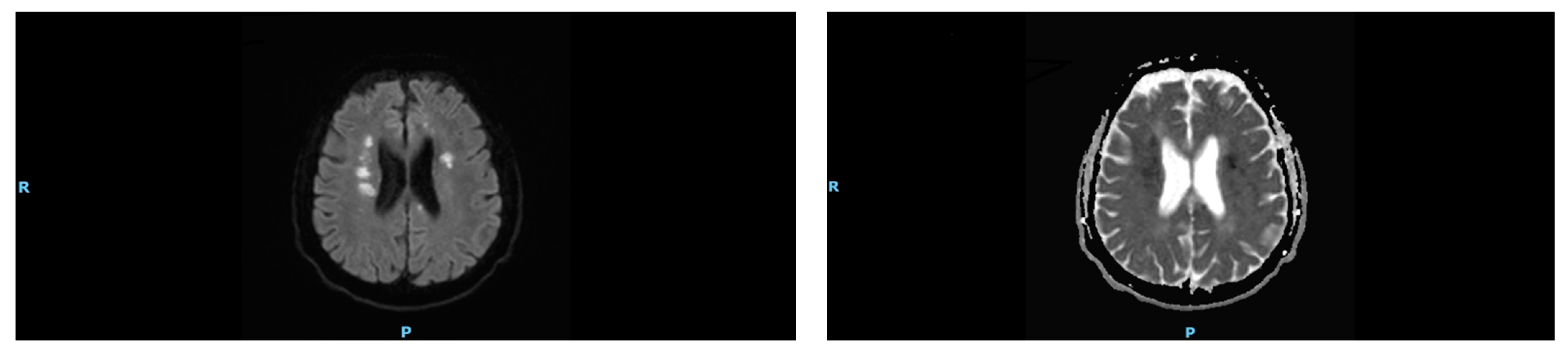
Case Description
3. Discussion
4. Conclusions
Author Contributions
Funding
Institutional Review Board Statement
Informed Consent Statement
Conflicts of Interest
References
- Mann, G.; Hankey, G.; Cameron, D. Swallowing Disorders following Acute Stroke: Prevalence and Diagnostic Accuracy. Cerebrovasc. Dis. 2000, 10, 380–386. [Google Scholar] [CrossRef] [PubMed]
- Trupe, L.A.; Mulheren, R.W.; Tippett, D.; Hillis, A.E.; González-Fernández, M. Neural mechanisms of swallowing dysfunction and apraxia of speech in acute stroke. Dysphagia 2018, 33, 610–615. [Google Scholar] [CrossRef] [PubMed]
- Meng, N.H.; Wang, T.G.; Lien, I.N. Dysphagia in patients with brainstem stroke: Incidence and outcome. Am. J. Phys. Med. Rehabil. 2000, 79, 170–175. [Google Scholar] [CrossRef] [PubMed]
- Rothi, L.J.G.; Heilman, K.M.A. The neuropsychology of action. In Apraxia: The Neuropsychology of Action; Psychology Press: London, UK, 2014. [Google Scholar]
- Daniels, S.K. Swallowing Apraxia: A Disorder of the Praxis System? Dysphagia 2000, 15, 159–166. [Google Scholar] [CrossRef] [PubMed]
- Robbins, J.; Levine, R.L. Swallowing after unilateral stroke of the cerebral cortex: Preliminary experience. Dysphagia 1988, 3, 11–17. [Google Scholar] [CrossRef] [PubMed]
- Yun, Y.J.; Na, Y.J.; Han, S.H. Swallowing apraxia in a patient with recurrent ischemic strokes: A case report. Medicine 2019, 98, e17056. [Google Scholar] [CrossRef] [PubMed]
- Sasegbon, A.; Hamdy, S. The anatomy and physiology of normal and abnormal swallowing in oropharyngeal dysphagia. Neurogastroenterol. Motil. 2017, 29, e13100. [Google Scholar] [CrossRef] [PubMed]
- Daniels, S.K.; Brailey, K.; Foundas, A.L. Lingual Discoordination and Dysphagia following Acute Stroke: Analyses of Lesion Localization. Dysphagia 1999, 14, 85–92. [Google Scholar] [CrossRef] [PubMed]
- Roy, E.A.; Square, P. Common considerations in the study of limb, oral, and verbal apraxia. In Neuropsychological Studies of Apraxia and Related Disorders, Roy, E.A., Ed.; North Holland: Amsterdam, The Netherland, 1985; pp. 111–162. [Google Scholar]
- Yuan, Y.; Wang, J.; Wu, D.; Huang, X.; Song, W. Effect of transcranial direct current stimulation on swallowing apraxia and cortical excitability in stroke patients. Top. Stroke Rehabil. 2017, 24, 503–509. [Google Scholar] [CrossRef] [PubMed]
- Yang, J.; Yuan, H. Application of transcranial direct current stimulation in cricopharyngeal dysfunction with swallowing apraxia caused by stroke: A case report. Medicine 2021, 100, 48. [Google Scholar] [CrossRef] [PubMed]
Publisher’s Note: MDPI stays neutral with regard to jurisdictional claims in published maps and institutional affiliations. |
© 2022 by the authors. Licensee MDPI, Basel, Switzerland. This article is an open access article distributed under the terms and conditions of the Creative Commons Attribution (CC BY) license (https://creativecommons.org/licenses/by/4.0/).
Share and Cite
Alfaris, A.M.; Alghamdi, A.S.; Almowalad, E.S.; Al Harbi, A.A.; Alghamdi, K.A.; Saeedi, J.; Al Awaji, N.N. Swallowing Apraxia Post Ischemic Stroke. Int. J. Environ. Res. Public Health 2022, 19, 16329. https://doi.org/10.3390/ijerph192316329
Alfaris AM, Alghamdi AS, Almowalad ES, Al Harbi AA, Alghamdi KA, Saeedi J, Al Awaji NN. Swallowing Apraxia Post Ischemic Stroke. International Journal of Environmental Research and Public Health. 2022; 19(23):16329. https://doi.org/10.3390/ijerph192316329
Chicago/Turabian StyleAlfaris, Abdullah Mohammed, Atheer Saeed Alghamdi, Enas Saad Almowalad, Awad Aweid Al Harbi, Khaled Abdulraheem Alghamdi, Jameelah Saeedi, and Nisreen Naser Al Awaji. 2022. "Swallowing Apraxia Post Ischemic Stroke" International Journal of Environmental Research and Public Health 19, no. 23: 16329. https://doi.org/10.3390/ijerph192316329
APA StyleAlfaris, A. M., Alghamdi, A. S., Almowalad, E. S., Al Harbi, A. A., Alghamdi, K. A., Saeedi, J., & Al Awaji, N. N. (2022). Swallowing Apraxia Post Ischemic Stroke. International Journal of Environmental Research and Public Health, 19(23), 16329. https://doi.org/10.3390/ijerph192316329