Article

The Effect of 13 Weeks Long-Distance Bicycle Riding on Inflammatory Response Indicators Related to Joint Cartilage and Muscle Damage

1
Center for Sport Science in Chungnam, Asan-si 31580, Republic of Korea
2
Department of Health and Sport Science, Korea National Sport University, Seoul 05541, Republic of Korea
*
Author to whom correspondence should be addressed.
Int. J. Environ. Res. Public Health 2022, 19(23), 16314; https://doi.org/10.3390/ijerph192316314
Submission received: 14 September 2022 / Revised: 30 November 2022 / Accepted: 2 December 2022 / Published: 6 December 2022
(This article belongs to the Special Issue 2nd Edition: Sports and Health)

Abstract

:
This study was to investigate the effects of 13 weeks of long-distance cycling on biomarkers of joint cartilage, muscle damage and inflammation. All subjects in this study were seven participants of the “One Korea New-Eurasia Peace Cycle Expedition”, in which they rode cycles from Berlin, Germany to Seoul, Korea for thirteen weeks. The total course of the expedition was divided into three sub-courses: course 1 (from Berlin to Moscow), course 2 (from Moscow to Ulaanbaatar) and course 3 (from Ulaanbaatar to Seoul). All the selected participants rode 87.4 km/day (course 1), 70.4 km/day (course 2) and 57.6 km/day (course 3) on average, respectively. We collected their blood samples before the expedition in Seoul (S1), after course 1 in Moscow (M), after course 2 in Ulaanbaatar (U) and after the expedition in Seoul (S2), to analyze biomarkers of joint cartilage damage (Cartilage Oligomeric Matrix Protein; COMP), muscle damage (Creatine Phosphokinase; CPK, Lactate Dehydrogenase; LDH, Myoglobin), inflammation (Interleukin-6; IL-6, Interleukin-1β; IL-1β, Tumor Necrosis Factor-α; TNF-α, C-Reactive Protein; CRP) and stress hormone (Cortisol). According to this result, COMP (S1; 188.37 ± 46.68 ng/mL) showed a significant increase after the expedition course 1 (M; 246.69 ± 51.69 ng/mL, p = 0.012) and course 2 (U; 237.09 ± 62.57 ng/mL, p = 0.047), and recovered to the stable state after expedition course 3 (S2; 218.46 ± 34.78. p = 0.047). Biomarkers of muscle damage (CPK, LDH and Myoglobin) and inflammation (IL-6, IL-1β, TNF-α and CRP) were not significantly changed in all courses, but CRP (S1; 1.07 ± 0.76 ng/mL) showed a tendency to decrease after the expedition course 1 (M; 0.3 ± 0.1 mg/mL, p = 0.044). Lastly, the Cortisol level significantly increased in all courses (per p < 0.05), but the Cortisol level after expedition course 3 (S2; 21.00 ± 3.65 mg/mL) was lower than that of after the expedition course 1 (M; 24.23 ± 2.47 mg/mL, p = 0.028). In summary, it seems that repetitive and continuous 50–90 km/day of cycling can increase joint cartilage damage risk and stress hormone temporarily. However, this result suggests that the appropriate intensity of cycling for thirteen weeks does not increase physical damage, and rather enhances the human body to adapt to exercise training.

1. Introduction

Endurance training improves cardiopulmonary endurance and immune function, and is also effective in preventing various metabolic diseases, such as obesity, diabetes and cardiovascular disease. Given these benefits, more people are participating in endurance exercise [1,2], but long-distance endurance training without proper preparation can increase the probability of sudden death, and can also cause acute liver damage, muscle damage, bone and joint damage, hematuria, dehydration and gastrointestinal disease [3,4]. Thus, the safety of participation in long-distance endurance competition and sports should be investigated.
Cartilage oligomeric matrix protein (COMP) is a non-collagenous extracellular matrix protein that maintains collagen structure in joint cartilage. It is secreted into the blood when joint cartilage is damaged, and is thus used as an index of joint cartilage damage [5,6]. In other words, the influence of long-distance endurance training on indicators of joint cartilage damage may differ depending on exercise method, intensity and duration. Moreover, research has been lacking on the influence of exercises in which the effects of bodyweight on joints are limited, such as cycling or swimming, on joint cartilage damage. Córdova et al. (2015) collected blood samples from athletes before and after a 3-day cycling competition (123 km for day 1, 128 km for day 2 and 100 km for day 3) to measure serum CPK, myoglobin and LDH concentrations; the authors found that CPK did not change significantly after the competition on day 1 and before the competition on day 2, but significantly increased after the competition on day 2 until recovery day 1 after the competition on day 3 [7]. Furthermore, Kim et al. (2009) showed that the concentration significantly increased up to 4 days after the completion of the 100–200 km ultramarathon [8]. In contrast, serum myoglobin concentration increased significantly after each competition, but returned to normal before the competition on the next day. Cytokines are regulatory proteins secreted by cells in response to various stimuli, which act as major regulatory factors in various aspects, such as immune response, muscle atrophy and development, insulin sensitivity and fat metabolism [9,10,11]. Interleukin 6 (IL-6) and 1 beta (IL-1β), which are pro-inflammatory cytokines, and tumor necrosis factor alpha (TNF-α), which is an inflammatory cytokine, are major indicators of muscle damage and inflammatory response [12,13]. IL-6 is known to be involved in various responses associated with inflammation and damage, including its role in increasing cortisol and C-reactive protein (CRP), another inflammatory marker [14]. Chiu et al. (2015) reported that in the concentration of IL-6 and TNF- α, CRP was significantly increased from immediately after exercise to 24 h after exercise in 24 men who completed a 100 km ultramarathon [15]. Although one-time long-distance endurance exercise has been reported to increase the serum concentrations of IL-6, TNF-α and CRP [12,16,17], changes in inflammatory markers with long-distance exercise still remain controversial.
In summary, with increases in the participation of non-professional athletes in endurance exercise, as well as increases in competitions and sports events, it is important to elucidate the influence of long-distance endurance exercise on markers of tissue damage in the body, in order to set safe exercise plans. Nevertheless, the influence of long-distance endurance exercise on joint cartilage damage, muscle damage and level of inflammation according to exercise type, distance and duration remains unclear, with a lack of relevant research. Therefore, this study aimed to investigate the influence of 13 weeks of long-distance cycling on inflammatory markers associated with muscle and joint cartilage damage.

2. Materials and Methods

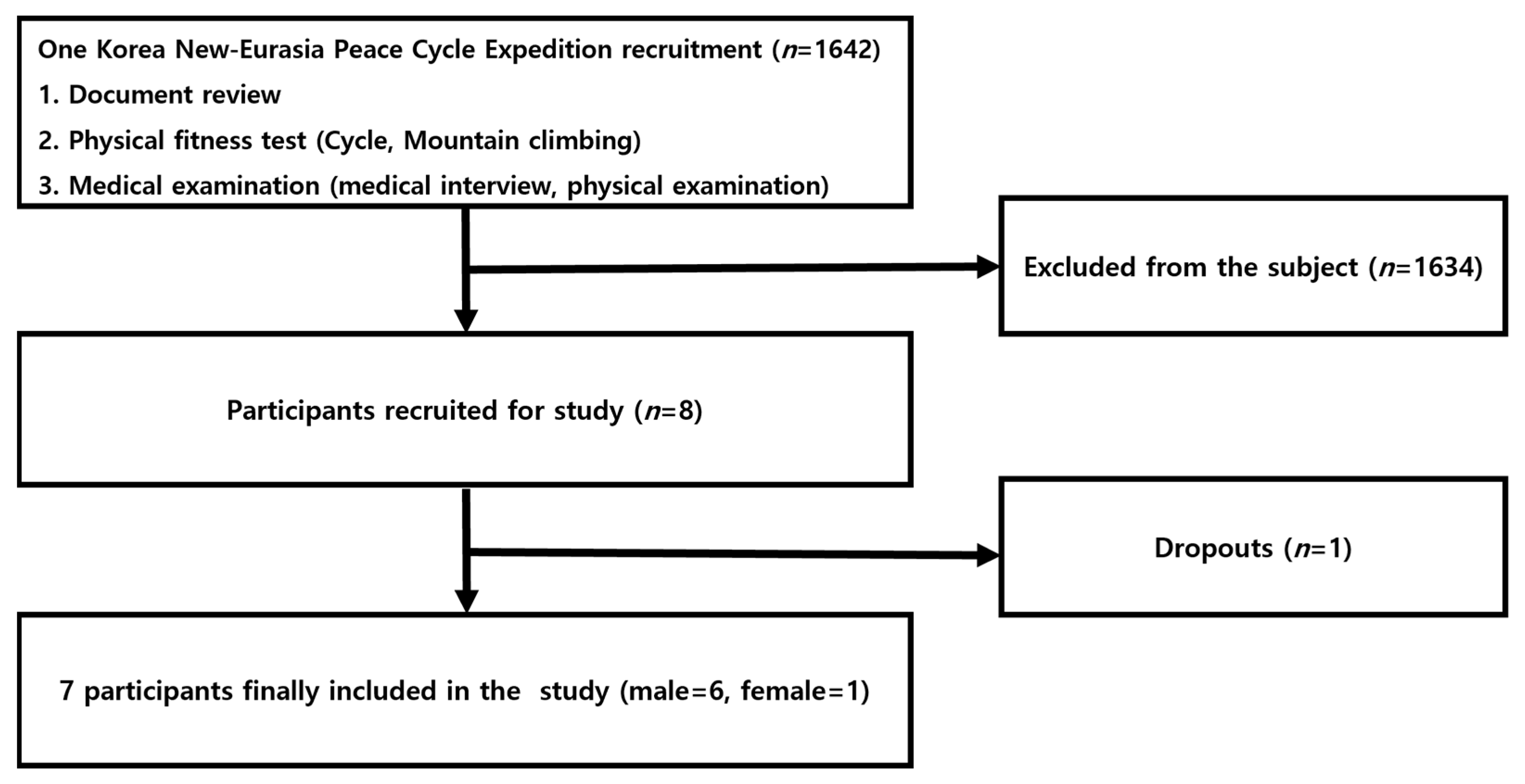
2.1. Study Participants

The participants of this study were adults aged from in their 20s to their 50s who did not have any musculoskeletal disorder, but had more than 7 years of experience with cycling. Furthermore, they were healthy individuals who had not participated in an exercise program or diet for the 3 months, and no specific disease was found in the medical examination. The participants were screened for their cycling abilities to complete the ultra-long-distance cycling event across Europe and Asia (One Korea New-Eurasia Peace Cycle Expedition, Figure 1). The participants consisted of 6 male and 1 female cyclist selected for the expedition (Table 1). The selected participants all received detailed explanation of the study purpose and procedures, and provided written consent. Ethical permission was granted from the Korea National Sport University’s Ethics Committee (Korea National Sport University Industry-Academic Cooperation Foundation-985), and all procedures were in accordance with the Declaration of Helsinki for research on human subjects.

2.2. Study Design and Participants
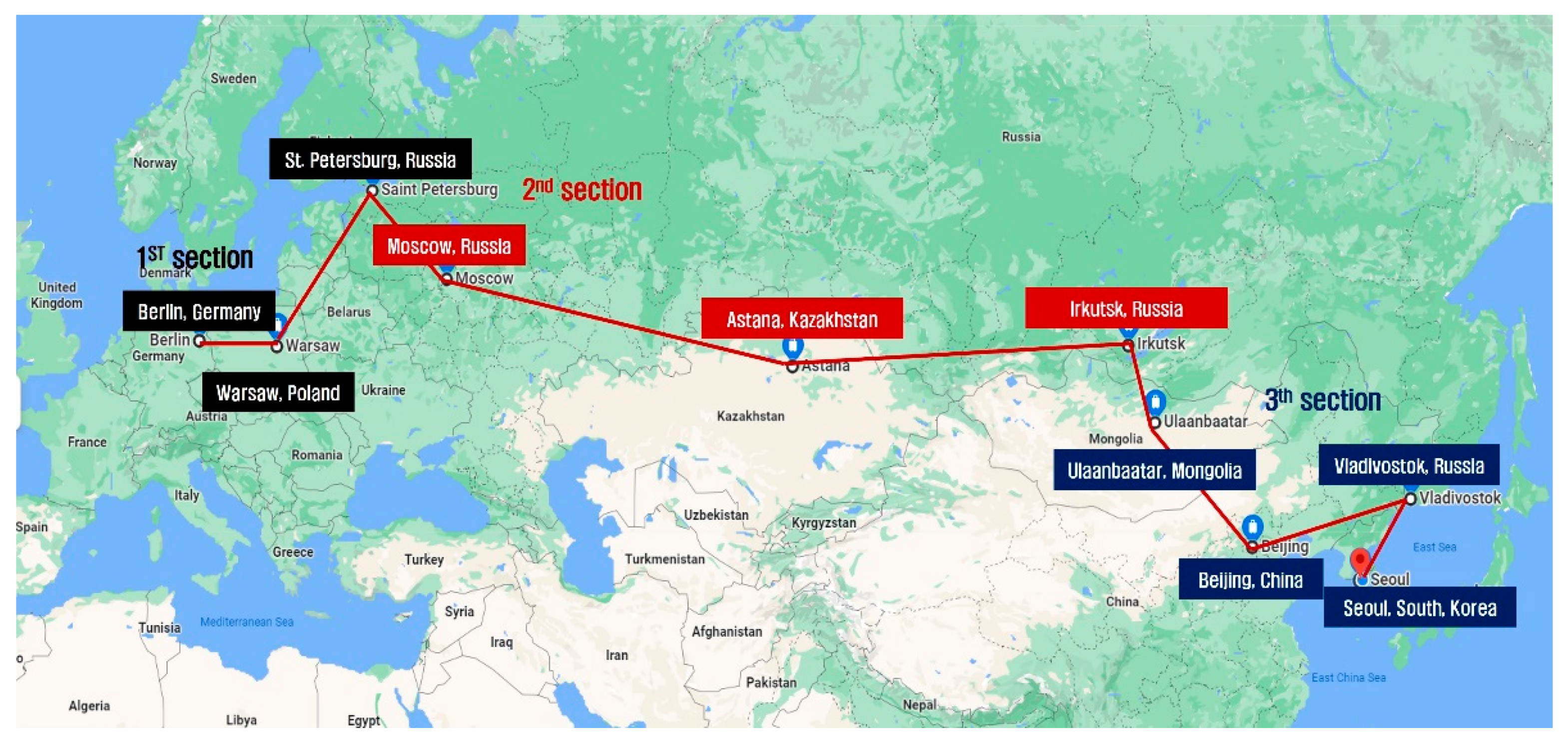
2.2.1. Cycling

According to the program of the One Korea New-Eurasia Peace Cycle Expedition, the participants cycled 4869 km from the 15,000 km-long expedition. The total course of the expedition was divided into three sub-courses: course 1 (from Berlin to Moscow); course 2 (from Moscow to Ulaanbaatar); and course 3 (from Ulaanbaatar to Seoul). Although the original plan was to cycle for 5 days and have 1 day of rest, some changes were made depending on environmental conditions (weather, route conditions and cycling permissions). Figure 2 (Google map) and Table 2 show the sub-courses and cycling content.

2.2.2. Time and Method for Sample Collection

Blood samples were collected in the morning of the day after cycling after fasting at the following time points: before the expedition (in Seoul); during the expedition in Moscow (day 29) and Ulaanbaatar (day 63); and on the last day of expedition in Seoul (day 93). The collected samples were immediately placed into serum tubes and EDTA tubes and centrifuged (Beckman, Brea, CA, USA, 15 min/3000 rpm). The supernatant serum and plasma were separated and carried on dry ice (−78.5 °C), and stored at −80 °C in a freezer until analysis.

2.2.3. Sample Analysis

In order to assess the level of joint cartilage and muscle damage from long-distance cycling, the concentration of COMP, an index of joint cartilage damage, was measured using Human COMP Quantikine ELISA kits (R&D systems Minneapolis, Minneapolis, MN, USA) through an enzyme-linked immunosorbent assay (ELISA). The levels of CPK, LDH and myoglobin, which are markers of muscle damage, were analyzed using Modular Analytics (Roche, Mannheim, Germany) through UV assay. Inflammatory markers (IL-6, IL-1β and TNF-α) were analyzed using Human High Sensitivity Cytokine kits (R&D systems Minneapolis, Minneapolis, MN, USA) through Luminex assay, and CRP was analyzed using Modular Analytics (Roche, Germany) through immunoturbidimetric assay. The concentration of cortisol, a stress hormone, was analyzed using a fully automated chemistry analyzer (Beckman, Brea, CA, USA). This study was conducted as a joint study. The results pertaining to serum cortisol have already been published through Gil’s study [18]. However, because the results were obtained from the same participants and because evidence for the influence of this study’s exercise type on tissue damage response was needed, the results have been cited in this study as well.

2.2.4. Data Analysis

Mean and standard deviation were calculated for the collected data using SPSS/PC+Version 20.0. One-way repeated measures ANOVA was used to assess the differences in serum markers according to cycling distance. When differences were present, they were analyzed through Duncan multiple test analysis, and the level of statistical significance was set as p < 0.05.

3. Results

3.1. Changes in Markers of Joint Cartilage Damage

When serum COMP concentration, which is an indicator of joint cartilage damage, was analyzed (Figure 3), the concentration was 188.37 ± 46.68 ng/mL before the expedition, 246.69 ± 51.69 ng/mL after course 1 (Berlin–Moscow), 237.09 ± 62.57 ng/mL after course 2 (Moscow–Ulaanbaatar) and 218.46 ± 34.78 ng/mL after course 3 (Ulaanbaatar–Seoul). Compared to pre-expedition value, the concentration increased after course 1 (p = 0.012) and course 2 (p = 0.047). Compared to course 1, the concentration decreased after course 3 (p = 0.036).

3.2. Changes in Markers of Muscle Damage

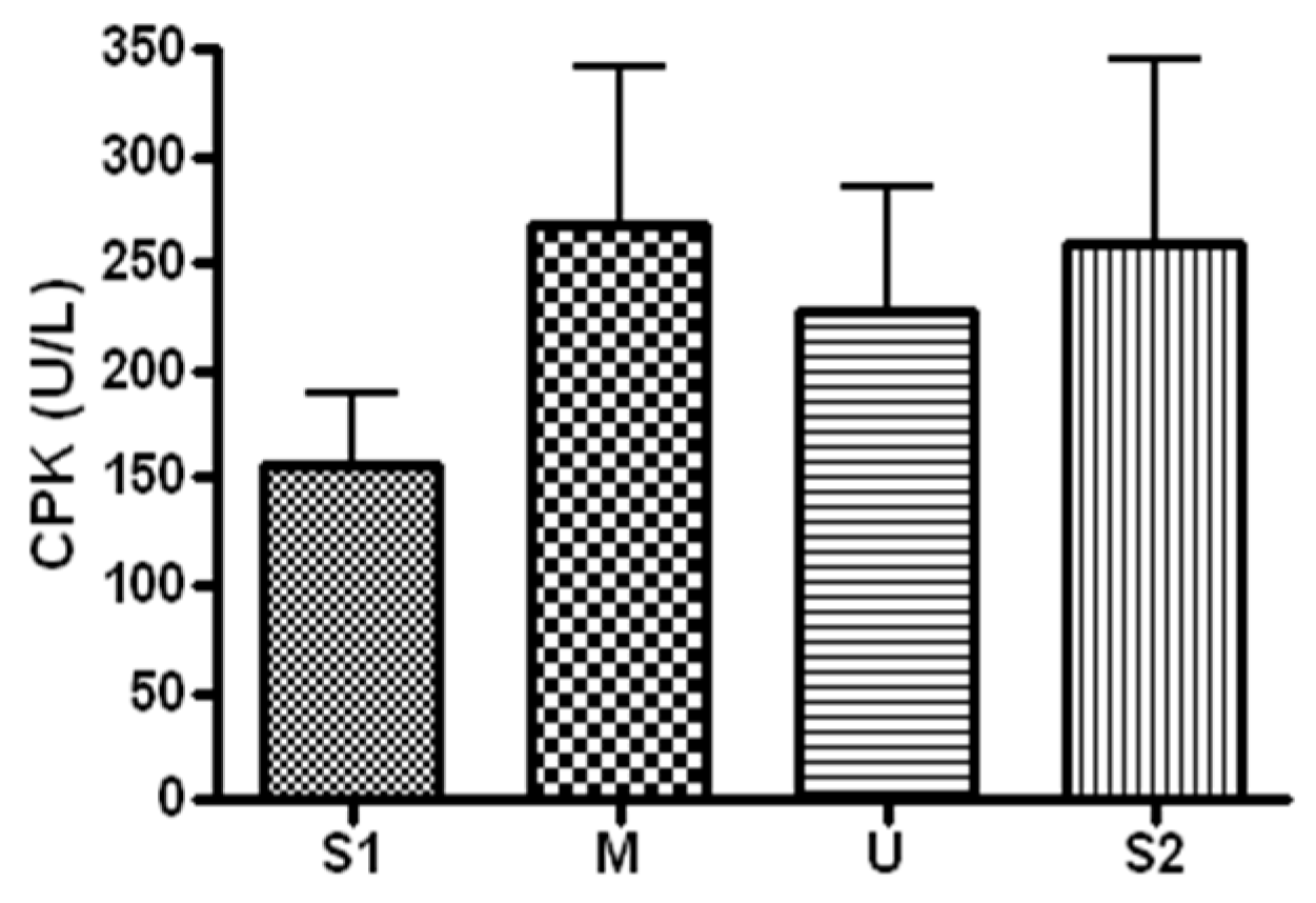
3.2.1. Changes in CPK

When serum CPK concentration, a marker of muscle damage, was analyzed (Figure 4), the concentration was 156 ± 85.84 U/L before the expedition, 267.28 ± 197.66 U/L after course 1 (Berlin–Moscow), 227.71 ± 155.22 U/L after course 2 (Moscow–Ulaanbaatar) and 259.57 ± 227.94 U/L after course 3 (Ulaanbaatar–Seoul). There was no significant difference noted at subsequent measurements compared to the baseline (p > 0.05).

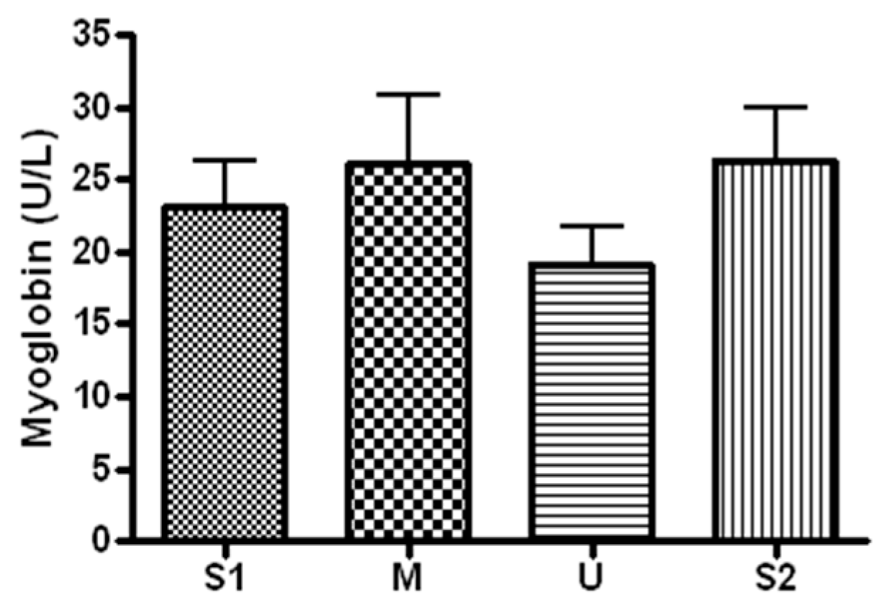
3.2.2. Changes in Myoglobin

When serum concentration of myoglobin, an indicator of muscle damage, was analyzed (Figure 5), the concentration was 23.14 ± 8.49 U/L before the expedition, 26.05 ± 12.83 U/L after course 1 (Berlin–Moscow), 19.17 ± 6.56 U/L after course 2 (Moscow–Ulaanbaatar) and 26.35 ± 9.71 U/L after course 3 (Ulaanbaatar–Seoul). The concentration did not differ significantly from the baseline at each of the subsequent measurements (p > 0.05).

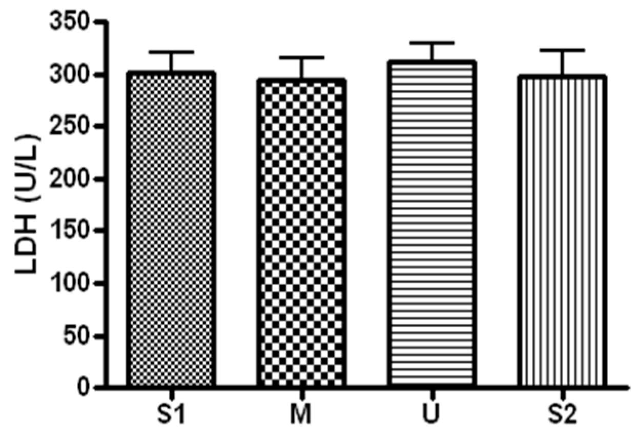
3.2.3. Changes in LDH

When serum concentration of LDH, an index of muscle damage, was analyzed (Figure 6), the concentration was 301.71 ± 49.21 before the expedition, 294.57 ± 54.93 U/L after course 1 (Berlin–Moscow), 311.28 ± 45.45 U/L after course 2 (Moscow–Ulaanbaatar) and 298.42 ± 62.83 U/L after course 3 (Ulaanbaatar–Seoul). There was no significant difference noted at subsequent measurements compared to the baseline (p > 0.05).

3.3. Changes in Inflammatory Markers

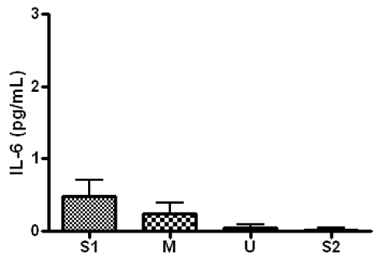
3.3.1. Changes in IL-6

When serum concentration of IL-6, an inflammatory marker, was analyzed (Figure 7), the concentration was 0.62 ± 0.44 pg/mL before the expedition, 0.37 ± 0.17 pg/mL after course 1 (Berlin–Moscow), 0.06 ± 0.09 pg/mL after course 2 (Moscow–Ulaanbaatar) and 0.04 ± 0.05 pg/mL after course 3 (Ulaanbaatar–Seoul). The concentration did not differ significantly from the baseline at each of the subsequent measurements (p > 0.05).

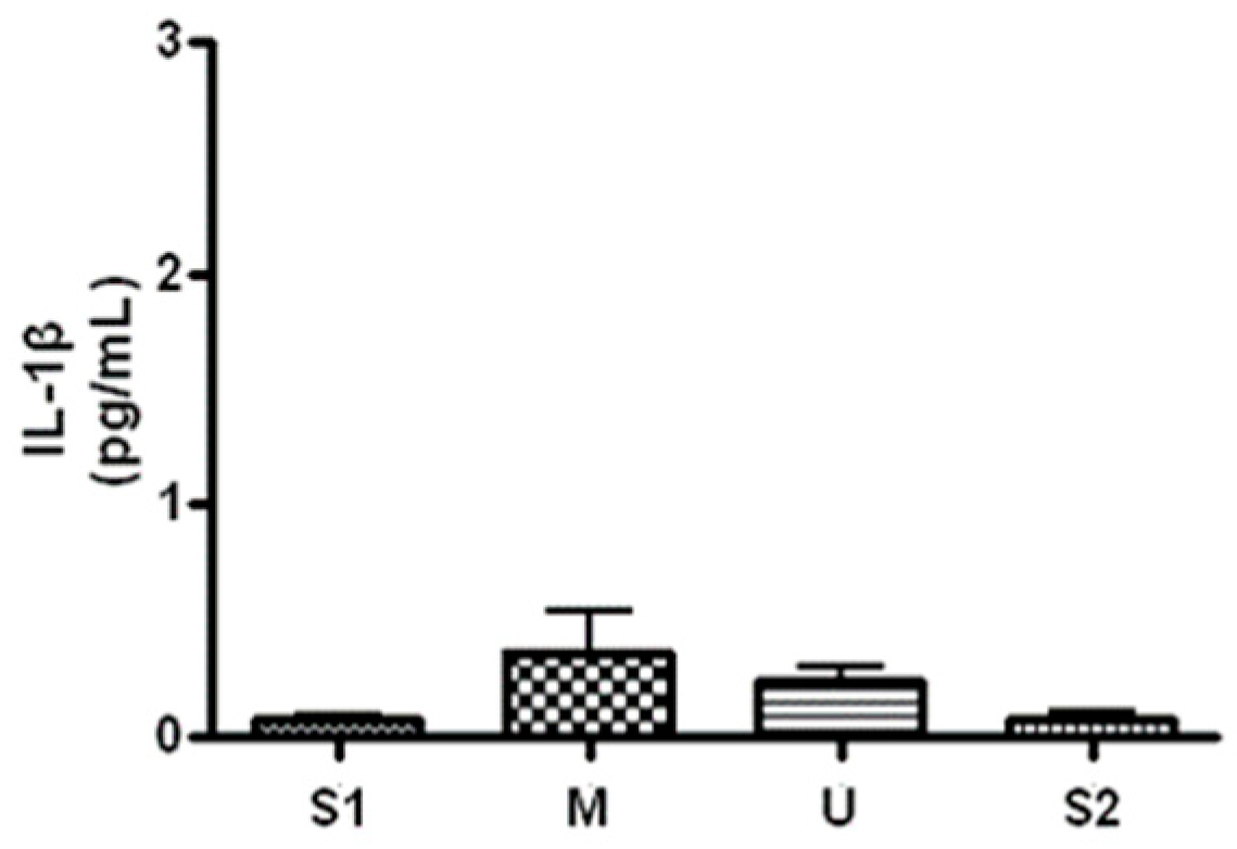
3.3.2. Changes in IL-1β

When serum concentration of IL-1β, an inflammatory marker, was analyzed (Figure 8), the concentration was 0.07 ± 0.02 pg/mL before the expedition, 0.36 ± 0.30 pg/mL after course 1 (Berlin–Moscow), 0.23 ± 0.11 pg/mL after course 2 (Moscow–Ulaanbaatar) and 0.07 ± 0.06 pg/mL after course 3 (Ulaanbaatar–Seoul). There was no significant difference noted at subsequent measurements compared to the baseline (p > 0.05).

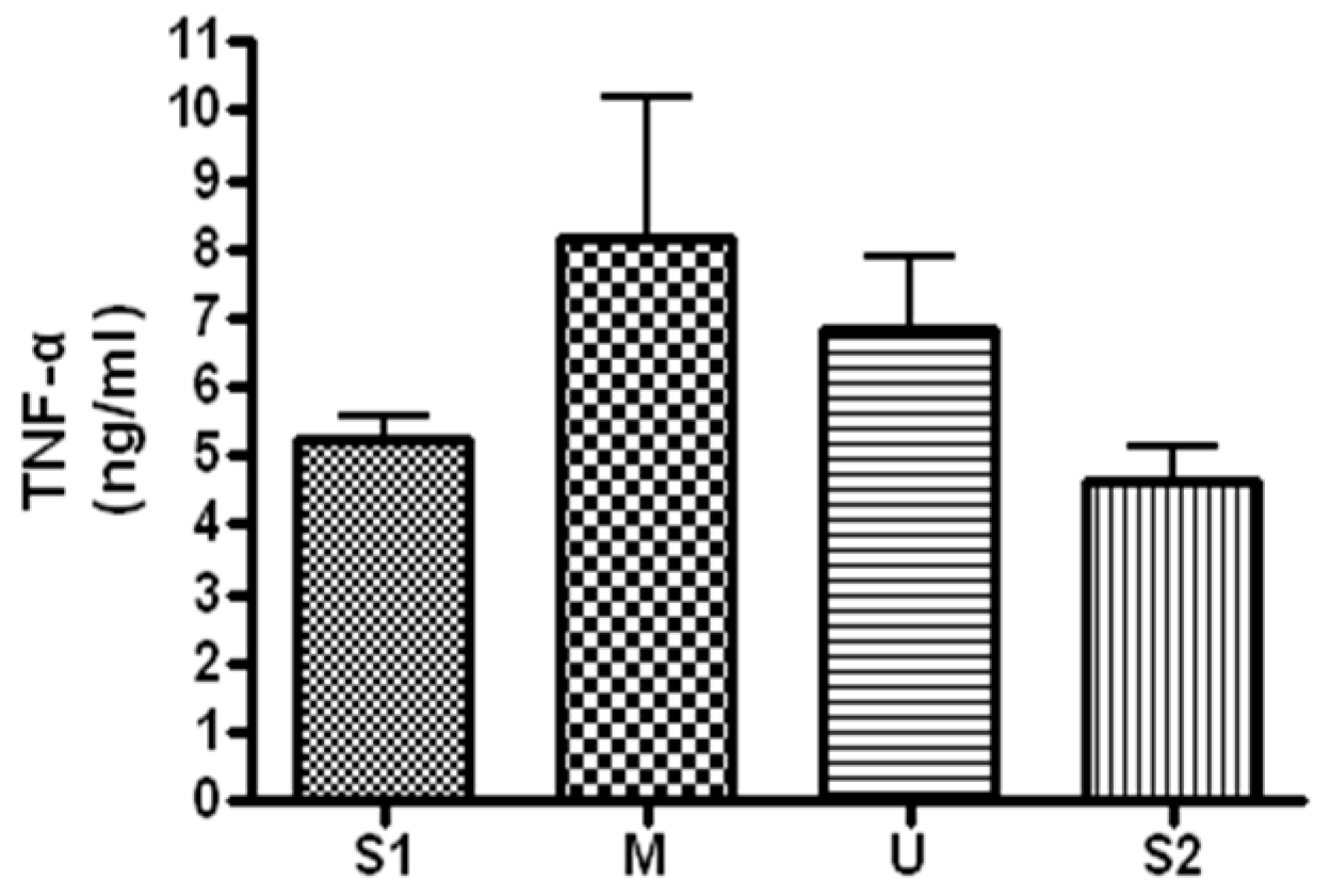
3.3.3. Changes in TNF-α

When serum concentration of TNF-α, an inflammatory marker, was analyzed (Figure 9), the concentration was 5.25 ± 0.78 ng/mL before the expedition, 8.16 ± 4.89 ng/mL after course 1 (Berlin–Moscow), 6.85 ± 2.56 ng/mL after course 2 (Moscow–Ulaanbaatar) and 4.61 ± 1.3 ng/mL after course 3 (Ulaanbaatar–Seoul). There was no significant difference noted at subsequent measurements compared to the baseline (p > 0.05).

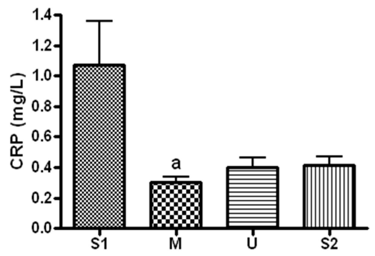
3.3.4. Changes in CRP

When serum concentration of CRP, an inflammatory marker, was analyzed (Figure 10), the concentration was 1.07 ± 0.76 mg/mL before the expedition, 0.3 ± 0.1 mg/mL after course 1 (Berlin–Moscow), 0.4 ± 0.16 mg/mL after course 2 (Moscow–Ulaanbaatar) and 0.41 ± 0.14 mg/mL after course 3 (Ulaanbaatar–Seoul). Compared to the pre-expedition measurement, the concentration decreased significantly after course 1 (p = 0.044).

3.4. Changes in Stress Hormone
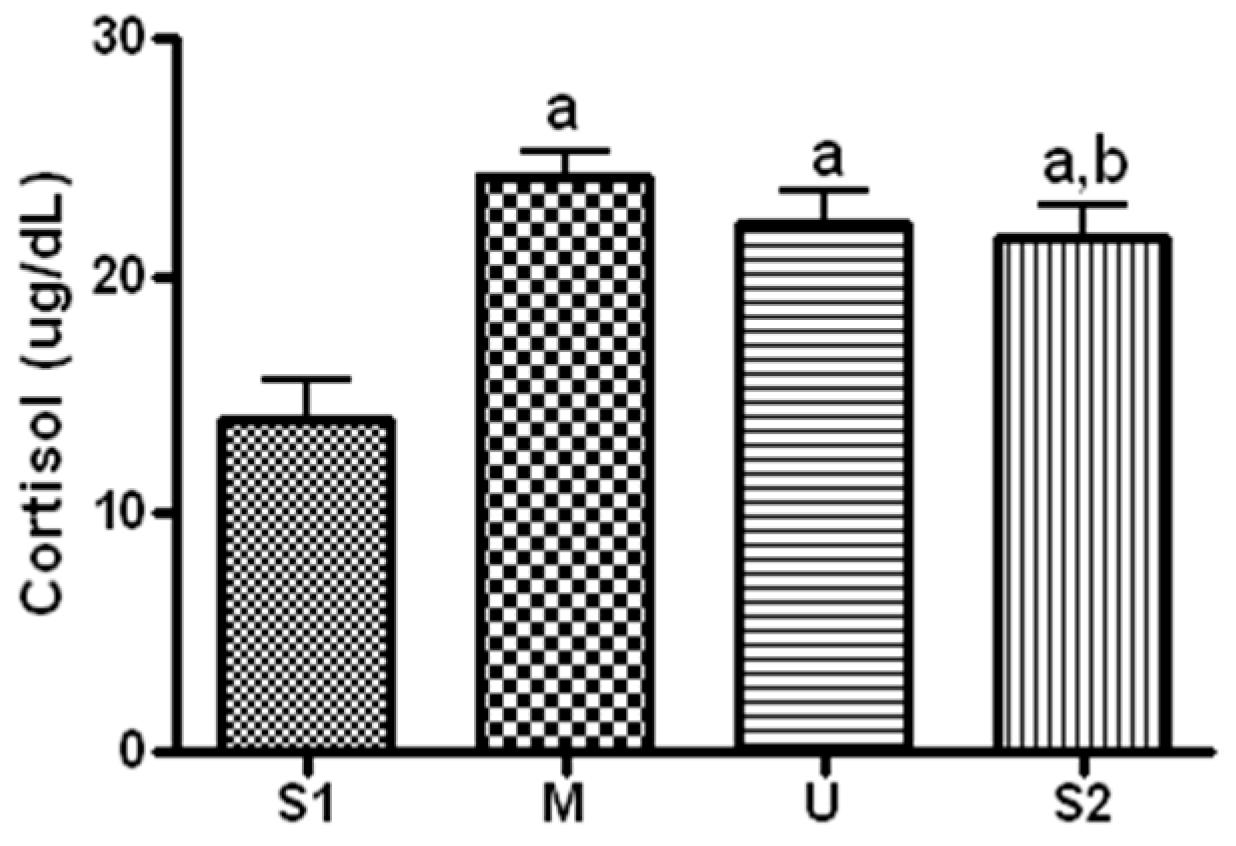
Changes in Cortisol

When serum concentration of cortisol, a stress hormone, was analyzed (Figure 11), the concentration was 13.92 ± 4.52 ug/dL before the expedition, 24.23 ± 2.47 ug/dL after course 1 (Berlin–Moscow), 22.18 ± 3.52 ug/dL after course 2 (Moscow–Ulaanbaatar) and 21 ± 3.65 mg/mL after course 3 (Ulaanbaatar–Seoul). Compared to the pre-expedition value, the values obtained from all of the subsequent measurements had significant increases (p < 0.05), and the concentration decreased significantly after course 3 compared to that after course 1 (p = 0.028).

4. Discussion

The present study was conducted to assess the influence of 13 weeks of long-distance cycling on serum markers of joint cartilage damage, muscle damage and inflammatory response after each of 3 sub-courses (course 1: Berlin–Moscow; course 2: Moscow–Ulaanbaatar; and course 3: Ulaanbaatar–Seoul). According to the results, joint cartilage damage and stress hormone increased in proportion to daily cycling distance in each course during the 13 weeks of long-distance cycling, and the values decreased with increases in cycling duration. Inflammatory response was found to decrease with long-distance cycling. In summary, although markers of damage had a positive correlation with daily cycling distance, with long-distance cycling, the participants adapted to the given exercise intensity, and had overall positive training effects in terms of muscle damage, joint cartilage damage and inflammatory response.
During the 13 weeks of long-distance cycling expedition, serum COMP concentration increased after course 1 and course 2. After course 3, the concentration returned to the normal range from the increased values measured after courses 1 and 2. The COMP concentration that increased after course 1 would have stayed elevated until after course 2, as the cycling distance was longer for courses 1 and 2 than course 3. Moreover, joint cartilage would have adapted to long-distance repeated exercise over the three courses. In this study, the participants cycled 87 km per day on average in course 1 for a total distance of 2097 km, 70 km per day on average in course 2 for a total of 1678 km and 57 km per day on average in course 3 for a total of 1094 km. Therefore, the daily cycling distance would have influenced the serum COMP concentration. Qi & Changlin (2006) measured COMP every 2 weeks after 10 weeks of exercise training in lab animals; the serum concentration was highest at weeks 4 and 6, and started to decrease from week 10 [19]. Corsetti et al. (2015) showed that the COMP of cyclists who rode a distance of 3500 km for 3 weeks decreased after 12 weeks [20]; this finding was explained by the fact that cartilage is capable of continuous regeneration. When cartilage damaged by an exercise stimulus regenerates, the regenerated cartilage is more resistant. Therefore, the improved resistance of cartilage at week 10 would have decreased joint damage from the exercise stimulus. Similarly, the decrease in serum COMP concentration after course 3 in the present study could be interpreted as adaptation of joint cartilage to long-distance cycling. However, since specific information on the influence of long-distance cycling on markers of joint cartilage damage has been lacking thus far, it may be difficult to generalize the present study’s findings.
High-intensity exercise is known to increase the serum concentration of muscle damage markers, and such changes are influenced by exercise intensity, duration, type and temperature during exercise [13,21,22,23,24]. In the present study, 13 weeks of long-distance cycling did not have any significant influence on the expression of serum CPK, LDH and myoglobin. Previous studies have reported that muscle damage markers are influenced by the participants’ exercise experience. In particular, trained athletes have smaller increases in muscle damage markers after exercise compared to untrained individuals, and trained individuals also recover faster [22,25,26,27]. The participants of this study are experienced cyclists capable of ultra-long-distance cycling who have been selected through tests of cycling ability. The daily cycling distance in this study may not have influenced changes in muscle damage markers, as the participants already had improved muscle resistance and resilience. Fallon et al. (1999) measured CPK concentration before 16 days of 1600 km ultramarathon, during the ultramarathon (days 4 and 11) and after the ultramarathon, and reported that the concentration increased on day 4 but decreased on day 11, and after the ultramarathon [28]. Ehlers et al. (2002) measured serum concentration of CPK before and during 10 days of training of identical intensity (on days 4, 7 and 10) in college soccer players, and reported that the concentration increased most on day 4 and returned to normal on day 10 [29]. In Chen et al.’s study (2001), 22 college students performed eccentric exercise every day for 7 days [30]. When serum CPK and LDH concentrations were measured, the concentration increased most on days 3 and 4, but returned to the baseline on day 7. Previous studies interpreted that long-distance exercise decreases serum markers of muscle damage, which increase after short-term exercise. The decrease was attributed to improved resistance of muscles as a result of exercise training. Therefore, muscle damage markers would not have changed in the present study after 13 weeks of long-distance cycling for the following reasons. First, the daily cycling distance may have been insufficient to influence muscle damage in our well-trained participants. Second, muscles may have metabolically adapted to long-distance cycling, and this may have increased their resistance to exercise stimulus or increased the half-life of damage markers.
IL-6, IL-1β, TNF-α and CRP are well-known markers of inflammation. They increase in specific conditions, and indicate the level of inflammation in the muscles and body [12,13,14]. Although these inflammatory markers are reported to increase with short-term intense exercise [31,32], their response to various types of external stimuli, including exercise type, target, intensity and duration, still remains controversial [33,34]. In the present study, the concentration of IL-6, IL-1β and TNF-α did not differ from baseline at every subsequent measurement according to cycling distance during the 13 weeks of long-distance cycling. In contrast, serum CRP concentration decreased after course 1. Although the changes were not significant, the concentration also tended to decrease after courses 2 and 3 [35,36,37]. In Meyer et al.’s study (2006), 33 obese adolescents performed aerobic exercise for 6 months [38]. When inflammatory markers were measured in these adolescents, serum CRP concentration was found to have decreased. Similarly, Mattusch et al. (2000) had 12 runners perform 10–20 km of running training every week for 9 months, and reported decreases in serum CRP concentration [39]. Moreover, various studies analyzing the relationship between physical activity and serum CRP concentration reported that CRP was lower in individuals who are physically active [40,41,42]. Previous studies attribute such a decrease in CRP with long-distance exercise directly to decreased production of cytokines, and indirectly to improved insulin sensitivity and antioxidative activity [42,43,44,45,46]. The present study’s findings agree with previous research reports. In other words, daily cycling distance during the 13 weeks of long-distance cycling seems to have improved inflammatory response by causing positive changes in the body, such as improved immune function. From a clinical point of view, daily cycling exercise for more than 12 weeks will reduce inflammation-related factors, and long-distance cycling will be useful for recovery and rehabilitation.
Cortisol is a stress hormone secreted by the adrenal cortex in response to various stimuli, which is known to suppress adrenocorticotropic hormone, promote gluconeogenesis and mediate inflammatory responses [47]. When serum cortisol concentration was analyzed during long-distance cycling, the concentration increased from the baseline at all subsequent measurements. The increase was greatest after course 1, compared to which concentration decreased significantly after course 3. Cortisol increases in proportion to exercise duration and intensity [48]. Therefore, 13 weeks of long-distance cycling could increase serum stress hormone, and daily cycling distance during each sub-course would have influenced changes in serum cortisol. However, cortisol is influenced by various factors in addition to exercise. Therefore, more comprehensive studies are required to attribute the changes in serum cortisol to exercise.
Regarding the limitations of the present study, for the results of this study to show a moderate difference, 80* power and 5% significance level, 24 subjects are needed [49]. However, the statistical significance is weak due to the small number of subjects, and the results of the study cannot be generalized. Therefore, a follow up study with an expanded number of subjects should be conducted. Additionally, several factors could not be controlled during the long-distance cycling expedition due to the inherent characteristics of field studies. The subjects’ diet, nutrition and psychological status were not controlled, which may have affected weight gain. Furthermore, only limited information could be collected from a small number of participants (n = 7), and the direct and indirect influence of changing geographic characteristics and climatic changes during the expedition on the participants could not be excluded or quantified objectively.

5. Conclusions

After 13 weeks of long-distance cycling, joint cartilage damage markers and stress hormone increased in proportion to daily cycling distance during each sub-course, and decreased with increases in exercise duration. Inflammatory response decreased with long-distance cycling. The results suggest that continued cycling for 50–90 km per day temporarily increases joint cartilage damage and stress hormone in the initial phases of adaptation. However, continued (13 weeks) long-distance cycling of appropriate intensity decreases the damage response through adaptation of tissues to exercise training, rather than increasing body damage.

Author Contributions

Conceptualization, H.-C.L. and H.-J.K.; methodology, H.-C.L. and H.-J.K.; software, H.-C.L.; formal analysis, H.-C.L. and H.-J.K.; investigation, H.-C.L. and H.-J.K.; data curation, H.-C.L.; writing—original draft preparation, H.-C.L. and H.-J.K.; writing—review and editing, H.-C.L. and H.-J.K.; visualization, H.-C.L.; supervision, H.-J.K.; and project administration, H.-J.K. All authors have read and agreed to the published version of the manuscript.

Funding

This research received no external funding.

Institutional Review Board Statement

This study was approved by the Korea National Sport University’s Ethics Committee, and all procedures involved were performed in accordance with the World Medical Association Declaration of Helsinki. In addition, the protocol for this study was approved by the IRB of Korea National Sport University (Korea National Sport University Industry-Academic Cooperation Foundation-985).

Informed Consent Statement

Written informed consent was obtained from all participants.

Data Availability Statement

Not applicable.

Acknowledgments

We thank all the participants and investigators who participated in the study.

Conflicts of Interest

The authors declare no conflict of interest.

References

  1. Shin, Y.A.; Lim, K.I.; Suk, M.H. The effects of a combined aerobic and resistance training program on leptin, adiponectin and metabolic syndrome factors in obese women. Exerc. Sci. 2005, 14, 569–582. [Google Scholar]
  2. Matsakas, A.; Narkar, V.A. Endurance exercise mimetics in skeletal muscle. Curr. Sports Med. Rep. 2010, 9, 227–232. [Google Scholar] [CrossRef]
  3. Kao, W.F.; Hou, S.K.; Chiu, Y.H.; Chou, S.L.; Kuo, F.C.; Wang, S.H.; Chen, J.J. Effects of 100-km ultramarathon on acute kidney injury. Clin. J. Sports Med. 2015, 25, 49–54. [Google Scholar] [CrossRef] [PubMed]
  4. Stuempfle, K.J.; Hoffman, M.D. Gastrointestinal distress is common during a 161-km ultramarathon. J. Sports Sci. 2015, 33, 1814–1821. [Google Scholar] [CrossRef]
  5. Saxne, T.; Heinegard, D. Cartilage oligomeric matrix protein: A novel marker of cartilage turover detectable in synovial fluid and blood. Br. J. Rheumatol. 1992, 31, 581–591. [Google Scholar] [CrossRef] [PubMed]
  6. Wislowska, M.; Jablonska, B. Serum cartilage oligomeric matrix protein (COMP) in rheumatoid arthritis and knee osteoarthritis. Clin. Rheumatol. 2005, 24, 278–284. [Google Scholar] [CrossRef] [PubMed]
  7. Córdova, M.A.; Martorell, P.M.; Sureda, G.A.; Tur Marí, J.A.; Pons, B.A. Changes in circulating cytokines and markers of muscle damage in elite cyclists during a multi-stage competition. Clin. Physiol. Funct. Imaging 2015, 35, 351–358. [Google Scholar] [CrossRef]
  8. Kim, H.J.; Lee, Y.H.; Kim, C.K. Changes in serum cartilage oligomeric matrix protein (COMP), plasma CPK and plasma hs-CRP in relation to running distance in a marathon (42.195 km) and an ultra-marathon (200 km) race. Eur. J. Appl. Physiol. 2009, 105, 765–770. [Google Scholar] [CrossRef]
  9. Zhang, J.M.; An, J. Cytokines, Inflammation and Pain. Int. Anesthesiol. Clin. 2007, 45, 27–37. [Google Scholar] [CrossRef] [PubMed] [Green Version]
  10. Festa, A.; Ralph, D.A.; Howard, G.; Mykkanen, L.; Tracy, R.P.; Haffner, S.M. Chronic subclinical inflammation as part of the insulin resistance syndrome-The insulin resistance atherosclerosis study (IRAS). Circulation 2000, 102, 42–47. [Google Scholar] [CrossRef] [Green Version]
  11. Peake, J.M.; Della Gatta, P.; Suzuki, K.; Nieman, D.C. Cytokine expression and secretion by skeletal muscle cells: Regulatory mechanisms and exercise effects. Exerc. Immunol. Rev. 2015, 21, 8–25. [Google Scholar] [PubMed]
  12. Bruunsgaard, H.; Galbo, H.; Halkjaer-Kristensen, J.; Johansen, T.L.; MacLean, D.A.; Pedersen, B.K. Exercise-induced increase in serum interleukin-6 in humans is related to muscle damage. J. Physiol. 1997, 499, 833–841. [Google Scholar] [CrossRef] [PubMed] [Green Version]
  13. Neubauer, O.; König, D.; Wagner, K.H. Recovery after an Ironman triathlon: Sustained inflammatory responses and muscular stress. Eur. J. Appl. Physiol. 2008, 104, 417–426. [Google Scholar] [CrossRef] [PubMed]
  14. Steensberg, A.; Fischer, C.P.; Keller, C.; Moller, K.; Pedersen, B.K. IL-6 enhances plasma IL-1ra, IL-10, and cortisol in humans. Am. J. Physiol. Endocrinol. Metab. 2003, 285, E433–E437. [Google Scholar] [CrossRef] [PubMed]
  15. Chiu, Y.H.; Lai, J.I.; Wang, S.H.; How, C.K.; Li, L.H.; Kao, W.F.; Yang, C.C.; Chen, R.J. Early changes of the anemia phenomenon in male 100 km ultramarathoners. J. Chin. Med. Assoc. 2015, 78, 108–113. [Google Scholar] [CrossRef] [Green Version]
  16. Schobersberger, W.; Hobisch-Hagen, P.; Fries, D.; Wiedermann, F.; Rieder-Scharinger, J.; Villiger, B.; Frey, W.; Herold, M.; Fuchs, D.; Jelkmann, W. Increase in immune activation, vascular endothelial growth factor and erythropoietin after an ultramarathon run at moderate altitude. Immunobiology 2000, 201, 611–620. [Google Scholar] [CrossRef]
  17. Nieman, D.C.; Dumke, C.I.; Henson, D.A.; McAnulty, S.R.; McAnulty, L.S.; Lind, R.H.; Morrow, J.D. Immune and oxidative changes during and following the Western States Endurance Run. Int. J. Sports Med. 2003, 24, 541–547. [Google Scholar]
  18. Gil, J.H. Analysis of Nutrient Intake and Blood Biomarkers during an Ultra-Distance Bicycling in Man. Ph.D. Thesis, Korea National Sport University, Seoul, Korea, 2016. [Google Scholar]
  19. Qi, C.; Changlin, H. Effects of moving training on histology and biomarkers levels of articular cartilage. J. Surg. Res. 2006, 135, 352–363. [Google Scholar] [CrossRef]
  20. Coresetti, R.; Perego, S.; Sansoni, V.; Xu, J.; Barassi, A.; Banfi, G.; Lombardi, G. Osteocartilaginous metabolic markers change over a 3-week stage race in pro-cyclists. Scand. J. Clin. Lab. Investig. 2015, 75, 523–530. [Google Scholar] [CrossRef]
  21. Janssen, G.M.; Kuipers, H.; Willems, G.M.; Does, R.J.; Janssen, M.P.; Geurten, P. Plasma activity of muscle enzymes: Quantification of skeletal muscle damage and relationship with metabolic variables. Int. J. Sports Med. 1989, 3, S160–S168. [Google Scholar] [CrossRef]
  22. Noakes, T.D. Heart disease in marathon runners. Med. Sci. Sports Exerc. 1987, 19, 187–194. [Google Scholar] [CrossRef]
  23. Brancaccio, P.; Maffulli, N.; Buonauro, R.; Limongelli, F.M. Serum enzyme monitoring in sports medicine. Clin. Sports Med. 2008, 27, 1–18. [Google Scholar] [CrossRef]
  24. Hassan, E.S. Muscle damage and immune responses to prolonged exercise in environmental estreme condition. J. Sports Med. Phys. Fit. 2016, 56, 1206–1213. [Google Scholar]
  25. Bass, A.; Vondra, K.; Rath, R.; Vítek, V.; Teisinger, J.; Macková, E.; Sprynarová, S.; Malkovská, M. Enzyme activity patterns of energy supplying metabolism in the quadriceps femoris muscle (vastus lateralis): Sedentary men and physically active men of different performance levels. Pflug. Arch. 1976, 361, 169–173. [Google Scholar] [CrossRef]
  26. Maxwell, J.H.; Bloor, C.M. Effects of conditioning on exertional rhabdomyolysis and serum creatine kinase after severe exercise. Enzyme 1981, 26, 177–181. [Google Scholar] [CrossRef]
  27. Vincent, H.K.; Vincent, K.R. The effect of training status on the serum creatine kinase response, soreness and muscle function following resistance exercise. Int. J. Sports Med. 1997, 18, 431–437. [Google Scholar] [CrossRef]
  28. Fallon, K.E.; Sivyer, G.; Sivyer, A.; Dare, A. The biochemistry of runners in a 1600 km ultramarathon. Br. J. Sports Med. 1999, 33, 264–269. [Google Scholar] [CrossRef] [Green Version]
  29. Ehlers, G.G.; Ball, T.E.; Liston, L. Creatine Kinase Levels are Elevated During 2-A-Day Practices in Collegiate Football Players. J. Athl. Train. 2002, 37, 151–156. [Google Scholar]
  30. Chen, T.C.; Hsieh, S.S. Effects of a 7-day eccentric training period on muscle damage and inflammation. Med. Sci. Sports Exerc. 2001, 33, 1732–1738. [Google Scholar] [CrossRef]
  31. Mooren, F.C.; Lechtermann, A.; Fobker, M.; Brandt, B.; Sorg, C.; Völker, K.; Nacken, W. The response of the novel pro-inflammatory molecules S100A8/A9 to exercise. Int. J. Sports Med. 2006, 27, 751–758. [Google Scholar] [CrossRef]
  32. Pedersen, B.K.; Steensberg, A.; Schjerling, P. Exercise and interleukin-6. Curr. Opin. Hematol. 2001, 8, 137–141. [Google Scholar] [CrossRef] [PubMed]
  33. Ploeger, H.E.; Takken, T.; de Greef, M.H.; Timmons, B.W. The effects of acute and chronic exercise on inflammatory markers in children and adults with a chronic inflammatory disease: A systematic review. Exerc. Immunol. Rev. 2009, 15, 6–41. [Google Scholar] [PubMed]
  34. Michigan, A.; Johnson, T.V.; Master, V.A. Review of the relationship between C-reactive protein and exercise. Mol. Diagn. Ther. 2011, 15, 265–275. [Google Scholar] [CrossRef]
  35. Adamopoulos, S.; Parissis, J.; Karatzas, D.; Kroupis, C.; Georgiadis, M.; Karavolias, G.; Paraskevaidis, J.; Koniavitou, K.; Coats, A.J.; Kremastinos, D.T. Physical training modulates proinflammatory cytokines and the soluble Fas/soluble Fas ligand systemin patients with chronic heart failure. J. Am. Coll. Cardiol. 2002, 39, 653–663. [Google Scholar] [CrossRef] [PubMed]
  36. Niebauer, J.; Clark, A.L.; Webb-Peploe, K.M.; Coats, A.J. Exercise training in chronic heart failure: Effects on pro-inflammatory markers. Eur. J. Heart Fail. 2005, 7, 189–193. [Google Scholar] [CrossRef]
  37. Belotto, M.F.; Magdalon, J.; Rodrigues, H.G.; Vinolo, M.A.; Curi, R.; Pithon-Curi, T.C.; Hatanaka, E. Moderate exercise improves leucocyte function and decreasesinflammation in diabetes. Clin. Exp. Immunol. 2010, 162, 237–243. [Google Scholar] [CrossRef] [Green Version]
  38. Meyer, A.A.; Kundt, G.; Lenschow, U.; Schuff-Werner, P.; Kienast, W. Improvement of early vascular changes and cardiovascular risk factors in obese children after a six-month exercise program. J. Am. Coll. Cardiol. 2006, 48, 1865–1870. [Google Scholar] [CrossRef] [Green Version]
  39. Mattusch, F.; Dufaux, B.; Heine, O.; Mertens, I.; Rost, R. Reduction of the plasma concentration of C-reactive protein following nine months of endurance training. Int. J. Sports Med. 2000, 21, 21–24. [Google Scholar] [CrossRef] [PubMed]
  40. Geffken, D.F.; Cushman, M.; Burke, G.L.; Polak, J.F.; Sakkinen, P.A.; Tracy, R.P. Association between physical activity and markers of inflammation in a healthy elderly population. Am. J. Epidemiol. 2001, 153, 242–250. [Google Scholar] [CrossRef] [PubMed]
  41. Wannamethee, S.G.; Lowe, G.D.; Whincup, P.H.; Rumley, A.; Walker, M.; Lennon, L. Physical activity and hemostatic and inflammatory variables in elderly men. Circulation 2002, 105, 1785–1790. [Google Scholar] [CrossRef] [Green Version]
  42. King, D.E.; Carek, P.; Mainous, A.G.; Pearson, W.S. Inflammatory markers and exercise: Differences related to exercise type. Med. Sci. Sports Exerc. 2003, 35, 575–581. [Google Scholar] [CrossRef] [PubMed]
  43. Mayer-Davis, E.J.; Karter, A.J. Intensity and amount of physical activity in relation to insulin sensitivity, the Insulin Resistance Atherosclerosis Study. JAMA 1998, 279, 669–674. [Google Scholar] [CrossRef]
  44. Shern-Brewer, R.; Santanam, N.; Wetzstein, C.; White-Welkley, J.; Parthasarathy, S. Exercise and cardiovascular disease: A new perspective. Arterioscler. Thromb. Vasc. Biol. 1998, 18, 1181–1187. [Google Scholar] [CrossRef] [Green Version]
  45. Gielen, S.; Adams, V.; Mobius-Winkler, S. Anti-inflammatory effects of exercise training in the skeletal muscle of patients with chronic heart failure. J. Am. Coll. Cardiol. 2003, 42, 861–868. [Google Scholar] [CrossRef] [PubMed]
  46. Kasapis, C.; Thompson, P.D. The effects of physical activity on serum C-reactive protein and inflammatory markers: A systematic review. J. Am. Coll. Cardiol. 2005, 45, 1563–1569. [Google Scholar] [CrossRef] [Green Version]
  47. Hoehn, K.; Marieb, E.N. Human Anatomy and Physiology, 8th ed.; Pearson: San Francisco, CA, USA, 2010; pp. 625–626. [Google Scholar]
  48. Galbo, H.; Christensen, N.J.; Mikines, K.J.; Sonne, B.; Hilsted, J.; Hagen, C.; Fahrenkrug, J. The effect of fasting on the hormonal response to graded exercise. J. Clin. Endocrinol. Metab. 1981, 52, 1106–1112. [Google Scholar] [CrossRef]
  49. Faul, F.; Erdfelder, E.; Lang, A.G.; Buchner, A. G*Power 3: A flexible statistical power analysis program for the social, behavior, and biomedical sciences. Behav. Res. Methods 2007, 39, 175–191. [Google Scholar] [CrossRef] [PubMed]
Figure 1. The participant recruitment and selection process.
Ijerph 19 16314 g001
Figure 2. The course of One Korea New-Eurasia Peace Cycle Expedition. 1st section, from Berlin to Moscow (black square), 2nd section, from Moscow to Ulaanbaatar (red square) and 3rd section, From Ulaanbaatar to Seoul (blue square).
Ijerph 19 16314 g002
Figure 3. Serum COMP concentration in different locations. Values are mean ± S.D. Significance (p < 0.05) is denoted by a: relative to S1 and b: relative to M. S1: Before start in Seoul, M: Moscow U: Ulaanbaatar S2: Return to Seoul.
Ijerph 19 16314 g003
Figure 4. Serum CPK concentration in different locations. Values are mean ± S.D. S1: Before start in Seoul, M: Moscow U: Ulaanbaatar S2: Return to Seoul.
Ijerph 19 16314 g004
Figure 5. Serum myoglobin concentration in different locations. Values are mean ± S.D. S1: Before start in Seoul, M: Moscow U: Ulaanbaatar S2: Return to Seoul.
Ijerph 19 16314 g005
Figure 6. Serum LDH concentration in different locations. Values are mean ± S.D. S1: Before start in Seoul, M: Moscow U: Ulaanbaatar S2: Return to Seoul.
Ijerph 19 16314 g006
Figure 7. Serum IL-6 concentration in different locations. Values are mean ± S.D. S1: Before start in Seoul, M: Moscow U: Ulaanbaatar S2: Return to Seoul.
Ijerph 19 16314 g007
Figure 8. Serum IL-1β concentration in different locations. Values are mean ± S.D. S1: Before start in Seoul, M: Moscow U: Ulaanbaatar S2: Return to Seoul.
Ijerph 19 16314 g008
Figure 9. Serum TNF-α concentration in different locations. Values are mean ± S.D. S1: Before start in Seoul, M: Moscow U: Ulaanbaatar S2: Return to Seoul.
Ijerph 19 16314 g009
Figure 10. Serum CRP concentration in different locations. Values are mean ± S.D. Significance (p < 0.05) is denoted by a: relative to S1. S1: Before start in Seoul, M: Moscow U: Ulaanbaatar S2: Return to Seoul.
Ijerph 19 16314 g010
Figure 11. Serum Cortisol concentration in different locations. Values are mean ± S.D. Significance (p < 0.05) is denoted by a: Relative to S1 and b: Relative to M. S1: Before start in Seoul, M: Moscow U: Ulaanbaatar S2: Return to Seoul.
Ijerph 19 16314 g011
Table 1. The characteristics of subjects.
No.SexAge (Yr.)Height (cm)Fat (%)S1 (kg)M (kg)U (kg)S2 (kg)
1Male23.0176.114.567.769.170.072.9
2Male51.6174.022.179.781.083.581.0
3Male22.8177.410.075.476.881.281.0
4Male45.1174.017.771.873.874.373.9
5Female33.9156.932.759.558.358.959.6
6Male29.7180.514.473.472.275.879.0
7Male25.5177.030.890.386.485.889.8
S1: Before start in Seoul, M: Moscow U: Ulaanbaatar S2: Return to Seoul.
Table 2. Cycling content.
1st Section (1)2nd Section (2)3rd Section (3)
Cycling distance (km)209716781094
Total days 293430
Cycling days242419
Mean cycling (km)87.47057.6
The longest cycling (km)132147117
The shortest cycling (km)201410
Mean temperature (°F)16.51.71.6
Maximum temperature (°F)221518
Minimum temperature (°F)−1−7−7
(1) From Berlin to Moscow, (2) From Moscow to Ulaanbaatar, (3) From Ulaanbaatar to Seoul.
Publisher’s Note: MDPI stays neutral with regard to jurisdictional claims in published maps and institutional affiliations.

