Article

Structural and Functional Neural Correlates in Individuals with Excessive Smartphone Use: A Systematic Review and Meta-Analysis

1
Division of Child and Adolescent Psychiatry & Division of Developmental and Behavioral Pediatrics, China Medical University Children’s Hospital, Taichung 404327, Taiwan
2
School of Post Baccalaureate Chinese Medicine, China Medical University, Taichung 406040, Taiwan
3
Division of Pediatric Neurology, China Medical University Children’s Hospital, Taichung 404327, Taiwan
4
Department of Diagnostic Radiology, Kaohsiung Chang Gung Memorial Hospital, Kaohsiung 83301, Taiwan
5
College of Medicine, Chang Gung University, Kaohsiung 83300, Taiwan
6
Department of Psychology, College of Medical and Health Science, Asia University, Taichung 41354, Taiwan
7
Department of Emergency Medicine, E-Da Hospital, I-Shou University, Kaohsiung 824005, Taiwan
8
School of Medicine for International Students, I-Shou University, Kaohsiung 82445, Taiwan
9
Clinical Psychology Center, Asia University Hospital, Taichung 41354, Taiwan
*
Authors to whom correspondence should be addressed.
These authors contributed equally to this work.
Int. J. Environ. Res. Public Health 2022, 19(23), 16277; https://doi.org/10.3390/ijerph192316277
Submission received: 6 November 2022 / Revised: 27 November 2022 / Accepted: 30 November 2022 / Published: 5 December 2022
(This article belongs to the Section Mental Health)

Abstract

:
Background: Despite known association of internet addiction with a reduced brain volume and abnormal connectivity, the impact of excessive smartphone use remains unclear. Methods: PubMed, Embase, ClinicalTrial.gov, and Web of Science databases were systematically searched from inception to July 2022 using appropriate keywords for observational studies comparing differences in brain volumes and activations between excessive smartphone users and individuals with regular use by magnetic resonance imaging. Results: Of the 11 eligible studies retrieved from 6993 articles initially screened, seven and six evaluated brain volumes and activations, respectively. The former enrolled 421 participants (165 excessive smartphone users vs. 256 controls), while the latter recruited 276 subjects with 139 excessive smartphone users. The results demonstrated a smaller brain volume in excessive smartphone users compared to the controls (g = −0.55, p < 0.001), especially in subcortical regions (p < 0.001). Besides, the impact was more pronounced in adolescents than in adults (p < 0.001). Regression analysis revealed a significant positive association between impulsivity and volume reduction. Regarding altered activations, the convergences of foci in the declive of the posterior lobe of cerebellum, the lingual gyrus, and the middle frontal gyrus were noted. Conclusions: Our findings demonstrated a potential association of excessive smartphone use with a reduced brain volume and altered activations.

1. Introduction

With the prevalence of smartphones worldwide, their excessive use has already become a social issue. In contrast to other forms of addiction such as gaming or gambling addiction that has been categorized as a distinct disease entity according to the International Classification of Disease (ICD) [1,2], excessive smartphone use is a more general behavioral addiction that has not been officially classified as a disorder [3]. Compared with drug dependence that affects structural and functional neural correlates through chemical pathways, changes associated with behavioral addiction are more likely through operant learning that involves rewards and punishments for behavioral impacts [4,5,6]. Excessive smartphone use has gradually replaced internet addiction as the most common behavioral addiction because of the need for communication and the convenience of its use [7]. Previous studies mainly addressed issues surrounding internet addiction [8] without focusing on the use of smartphones, which have become an indispensable communicating device in over 83% of the global population [8]. Although a previous meta-analysis has reported a reduction in gray matter volume (GMV) as well as significant activations in the medial/superior frontal gyri, the left anterior cingulate cortex/cingulate gyrus, and the left middle frontal/precentral gyri in individuals with internet addiction [9], the structural and functional impacts of excessive smartphone use on the central nervous system remains unclear. Therefore, the current meta-analysis aimed at investigating the difference in structural volume and functional connectivity between individuals with excessive smartphone use and their comparators with regular use.
Magnetic resonance imaging (MRI) is the most popular assessment tool for macroscopic neuroanatomical assessment of the brain (e.g., brain volume measurement) because of its excellent image resolution and quality of between-tissue contrast [10]. Consistently, MRI has been used to examine anatomic differences (e.g., gray matter volume) among individuals with behavioral addiction [9,11]. MRI systems of different magnetic strengths have their distinct merits in MRI acquisition. While 3-Tesla systems per se offer an improved resolution of between-tissue contrast, MRI scan using 1.5-Tesla systems has the advantage of providing sufficient data on quantifying relatively small brain structures [10]. Besides, several types of MR sequences are available for structural neuroimaging, such as T1-weighted and T2-weighted imaging. In contrast to T2-weighted imaging, the T1-weighted approach that provides the greatest clarity in distinguishing among gray matter, white matter, and cerebrospinal fluid (CSF) is frequently used for quantitative MRI studies of brain morphology (i.e., individual brain structures) [10]. Regarding the computation of structural volume, voxel-wise statistical analysis (e.g., voxel-based morphometry) [12] is commonly used. It is a fully automated MR image analysis technique that permits voxel-wise statistical comparison of the local concentration of GMV between two groups of subjects (e.g., patients vs. healthy controls). Hence, the current study focused on the use of voxel-based structural brain imaging for volumetric measurement (mm3) in assessing the difference in brain volume between individuals with excessive smartphone use and healthy controls. Furthermore, the associations of moderators (i.e., stages of life and brain areas) and mediators (i.e., demographic variables) with brain volume were examined and analyzed.
In addition to computing the structural neural correlate, we also combined coordinate-based meta-analysis to identify regions of consistent activation across functional MRI (fMRI) studies, followed by the determination of the convergence of foci reported from different studies (or experiments) using activation likelihood estimation (ALE) [13]. The current meta-analysis attempted to integrate brain structures with functional connectivity to provide neurobiological information about the differences between individuals with excessive smartphone use and healthy comparators.

2. Methods

2.1. Study Eligibility

The excellent resolution of MRI in differentiation among different structures makes it an ideal tool for discerning subtle anatomical differences in cortical and subcortical structures [10]. Regarding the evaluation of functional connectivity, fMRI is often used because of its ability to measure metabolic activity within anatomic structures. The eligibility criteria for study inclusion were: (i) Original, cross-sectional comparative studies that used MRI in individuals with excessive smartphone use versus their counterparts with regular use; (ii) Recruitment of individuals with excessive smartphone use confirmed through a psychiatric interview or a validated questionnaire; (iii) Studies that used a whole-brain or region of interest analysis; (v) those that reported the differences in structural volumes or peak coordinates between participants with excessive smartphone use and control groups. On the other hand, we excluded studies that focused on “non-smart” mobile phones as well as those involved participants with brain injury (e.g., traumatic or stroke), severe mental disorders, neurological or physical diseases, and those examining the treatment effects of drugs or non-drugs.

2.2. Electronic Searches

Following the PRISMA guidelines [14] (Table S1) and the MOOSE statement [15], we systematically searched for articles through the PubMed, Embase, ClinicalTrials.gov and Web of Science databases from inception to July 2022. For completeness of our literature search, we used different string terms, namely mobile phone addiction (or internet addiction or smartphone addiction or problematic phone use or overuse phone or nomophobia or excess phone use or phone addictive behaviors or pathological phone use or phone abuse) AND imaging (or magnetic resonance imaging or MRI or VBM or voxel-based morphometry) (Table S2). No restriction was placed on language, date of publication, and country of origin. The systematic review protocol was registered on the International Prospective Register of Systematic Reviews (PROSPERO) website (ID 359367).

2.3. Data Extraction

Five independent authors (Chen B.-S., Li L., Lee C.-Y., Sue Y.-R. and Sung T.-M.) completed title and abstract screening. Two other authors (Yeh P.-Y. and Liu S.-T.) independently screened the full text of the retrieved literature with disagreements resolved through discussion until consensus was reached. On encountering articles in which the necessary data were unavailable, we attempted to retrieve the information through electronically contacting the corresponding authors. For different studies using the same data, the article with more recent information or a larger sample size was selected for the current investigation.

2.4. Effect Size Analysis

For structural neuroimaging data synthesis, effect size (ES) was used as the measure for the primary outcome (i.e., brain volume) across the included studies. The current meta-analysis used Hedges’ g as the ES which was the difference in mean structural volumes between individuals with excessive smartphone use and their comparators with regular use divided by the pooled standard deviations of the two groups. To compute the ESs, we used the software “Comprehensive Meta-Analysis version for Windows (CMA, version 3.0)”. A negative ES represented a smaller brain volume in individuals with excessive smartphone use compared to their counterparts with regular use. If more than one dataset (e.g., different brain regions) were available in the eligible studies, a single ES denoting the mean volume was acquired through standardization and averaging of the results. In respect of the significance of findings, ESs of 0.8, 0.5, and 0.2 were deemed large, moderate, and small, respectively [16].
We used subgroup analyses to evaluate the effects of moderators on brain volume. Because the relatively small sample sizes commonly encountered in neuroimaging studies would decrease statistical power, subgroup analysis in the current meta-analysis was based on a random-effects model [17] on the assumption of an average distribution of ESs across the included studies [18] to minimize sample size bias. In contrast to the weights assessed with the fixed-effects model, those in the random-effects model are more similar to each other [19]. We also used Q statistics and the corresponding p values to detect the heterogeneity of ESs. As for the examination of the association between the mediators (e.g., age and prevalence of females) and brain volume, meta-regression with a mixed-effects model was performed.
We used different methods to evaluate publication bias according to the number of eligible studies. For outcomes reported in fewer than ten datasets, the corresponding funnel plots were inspected [20]. Egger’s regression test was used for outcomes described in ten or more datasets [21]. Using the Duval and Tweedie’s trim and fill method, potentially missing studies were imputed for evidence of funnel plot asymmetry [22]. The influence of individual studies on the overall outcome was assessed with leave-one-out sensitivity analysis [23].

2.5. ALE Analysis

ALE analysis was conducted with GingerALE v3.0.2 in the BrainMap environment [13,24,25]. There are two major statistical approaches to image analysis acquired with MRI, namely, cluster-based approach (CBA) and voxel-based approach (VBA). While CBA can detect a larger proportion of truly active brain areas (i.e., higher sensitivity to activation detection with large spatial extent) especially studies with moderate effect sizes and smaller sample sizes [26,27], VBA is suggested to avoid pitfalls [27] despite being too conservative to decrease power [28]. Therefore, CBA is recommended for ALE studies [28]. In addition, we used CBA to identify relative dominance because of the small number of eligible studies and their small sample sizes. Coordinate data were entered into Talairach space. Coordinates reported in MNI space were converted to Talairach space based on the icbm2tal transform [29], followed by GingerALE calculations. Modeled Activation (MA) maps were created for each foci group by modeling with Gaussians distribution. ALE map was constructed by combining a union of all of the MA maps with a table of p-value for ALE score. The ALE image and p-value table were used to create a 3D p-value image. Using the GingerALE software, the threshold of p-value image was then set utilizing the family-wise error (FWE) method with p < 0.05 being defined as statistically significant. We performed two separate ALE analyses in this study for two different conditions, namely, increased and decreased activations among individuals with excessive smartphone use compared to controls. Significant clusters were overlaid onto an anatomical Talairach template, colin_tlrc_2x2x2.nii (http://www.brainmap.org/ale, accessed on 16 May 2022), using the Mango software (version 4.1, Research Imaging Institute, University of Texas Health Science Center, Houston, TX, USA; http://www.ric.uthscsa.edu/mango, accessed on 16 May 2022).

3. Results

3.1. Study Characteristics and Participants

Figure 1 depicts the process of identification of eligible studies in the current meta-analysis. Of the 6993 full-text articles deemed eligible according to their abstracts and titles, 6982 were excluded because of failure to meet the inclusion criteria. Further, two studies were not qualified for this study [30,31] since they shared the same participants with the study by Horvath et al. [32]. Finally, 11 articles were included in this meta-analysis (Table 1). The definitions of excessive (problematic) smartphone users across the included studies are summarized in Table S3. Of the 11 articles, nine used questionnaires for excessive smartphone use including the smartphone addiction proneness scale (SAPS) (n = 5), smartphone addiction scale (SAS) (n = 3), and Mobile phone addiction index (MPAI) (n = 1) for evaluating the severity of excessive smartphone use as well as grouping participants into individuals with excessive smartphone use and comparators with regular use (Table 1). However, of the other two articles, one used the Facebook version of the compulsive internet use scale (CIUS) and the other adopted the Young’s internet addiction test (Table 1).
Of the 11 included articles, seven compared the brain volumes between individuals with excessive smartphone use (n = 165) and their comparators with regular use (n = 256) (Table 1). The mean age of the participants was 19.61 years (range, 15.26 to 29.80 years) with a female prevalence of 52.53% (range, 25.56.7 to 68.18%) (Table 1). With reference to brain activations, six studies investigated the functional connectivity of individuals with excessive smartphone use (n = 139, mean age = 24.18 years) and their counterparts with regular use (n = 137, mean age = 24.47 years) based on ALE analyses of their fMRI findings (Table 1). Because excessive smartphone use was found to be associated with activations of certain brain regions but decreased activities in others, data were collected from four studies that provided information about the regions with enhanced activities among excessive users compared to regular users (176 participants, 96 activation foci) [34,35,37,39] and four studies reporting the regions with suppressed activities (196 participants, 25 activation foci) [32,34,37,40].
Some studies used self-reported evaluation tools to investigate the severity of impulsivity (n = 8), depression (n = 6), and anxiety (n = 5). Focusing on the assessment of impulsivity, four studies [32,35,38,42] used the Barratt impulsiveness scale (BIS) [43] with 30 items, each of which was given a Likert score of 0 to 3 to indicate a progressive increase in severity. Regarding the evaluation of depression, three studies [32,33,38] adopted the Beck depression inventory (BDI) [44], which assesses the severity of depression based on the participant’s responses to 21 items, each of which is scored on a four-point Likert scale of 0, 1, 2, and 3 representing no, mild, moderate, and severe depression, respectively. In contrast, the five studies with self-reported information about anxiety used different scales for assessment that precluded the subsequent conduction of regression analysis.
The 11 eligible clinical trials were conducted mostly in Asian countries including Korea (n = 5), China (n = 3), and Malaysia (n = 2), while one study was performed in Germany. The sample size of the included studies ranged from 30 to 88.

3.2. Quantitative Brain Volume Data Synthesis

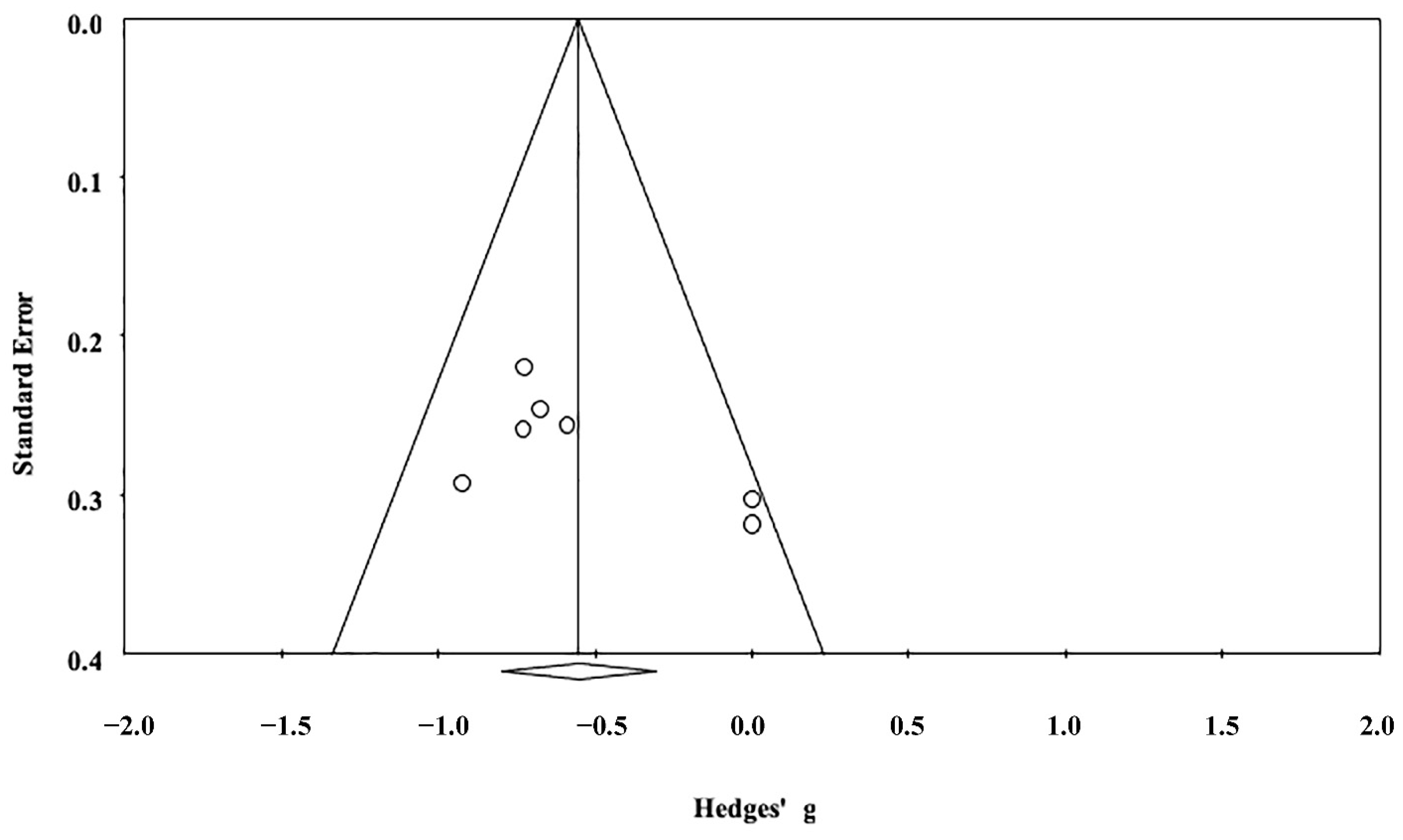
The results from seven studies demonstrated a significant anatomical difference between individuals with excessive smartphone use and their comparators with regular use (Hedges’ g = −0.55, 95% CI = −0.80 to −0.31, p < 0.001) (Figure 2), indicating a decreased brain volume in individuals with excessive smartphone use. The ES was unchanged on leave-one-out sensitivity analysis (p < 0.001), suggesting a non-significant impact of the results of any single study on the main outcome. The risks of publication bias reflected by funnel plot asymmetry for brain volume is shown in Figure 3. Because the ‘trim and fill’ method revealed zero potentially missing studies on the left side of the plot, the adjusted ES of −0.55 (−0.80 to −0.31) remained unchanged with the random-effects model.
Subgroup analysis focusing on age groups among individuals with excessive smartphone use (seven studies, 421 participants) revealed a significantly decreased brain volume in adolescents compared to the adults (p < 0.001), suggesting a more pronounced structural impact of excessive smartphone use among adolescents (Table 2). Subgroup analysis for cortical and subcortical regions in individuals with excessive smartphone use showed a significant reduction in size of the subcortical structures compared to that of the cerebral cortex (p < 0.001) (Table 2), implicating an association between excessive smartphone use and a reduced volume of subcortical structures. The results of meta-regression analysis demonstrated no correlation between most of the investigated mediators and brain volume. However, a significant correlation was noted between the BIS score and brain volume in individuals with excessive smartphone use (p = 0.03; Table 3), suggesting a reduced volume in those with a higher impulsivity.

3.3. The Difference in Brain Activations between Individuals with Excessive Smartphone Use and Their Counterparts with Regular Use

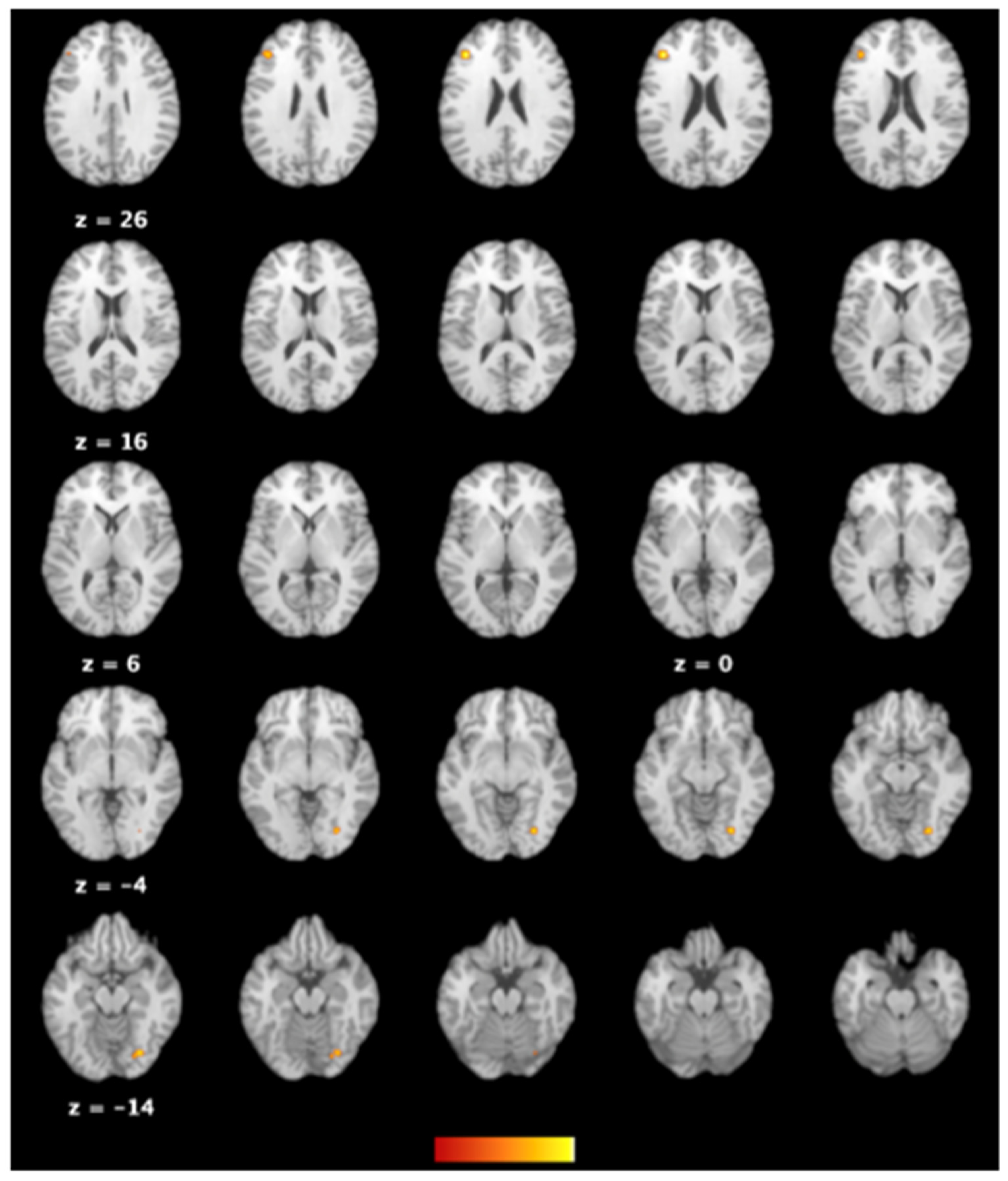
ALE analysis of the four studies [34,35,37,39] with available information about the anatomical locations with an enhancement of brain activities among individuals with excessive smartphone use showed consistent areas of activations, namely the declive of posterior lobe of the cerebellum, the lingual gyrus (BA 18), and the middle frontal gyrus (BA 46) (Table 4 and Figure 4). On the other hand, analysis of the four studies [32,34,37,40] providing data on the anatomical regions with suppressed activities in excessive cellphone users failed to pinpoint commonly affected areas.

4. Discussion

To our knowledge, the current study is the first meta-analysis to investigate the effect of excessive smartphone use on the structural and functional neural correlates. Our investigation revealed several findings that would be of clinical interest. First, individuals with excessive smartphone use showed a reduced brain volume, especially among the subcortical structures. Second, the impact on brain volume was more pronounced in adolescents than in adults. Third, our finding of a positive association between impulsivity and a reduced volume suggested a structural impact of impulsiveness on the brain. Fourth, our identification of significant spatial convergence in the declive of posterior lobe, the lingual gyrus (BA 18), and the middle frontal gyrus (BA 46) in individuals with excessive smartphone use indicated an anterior-posterior distribution of altered connectivity.
Focusing on the effects of excessive smartphone use on structural and functional neural correlates, the current study yielded several interesting results. Our finding of a relationship between excessive smartphone use and a decreased brain volume, especially subcortical structures, was consistent with that of a previous meta-analysis that demonstrated a correlation between internet addiction and a reduced GMV [9]. One of the possible mechanisms underlying the observation may be the emotional impacts associated with excessive smartphone use. Indeed, a body of evidence from systematic reviews has revealed a significant association between problematic smartphone use and mental health with anxiety and depression being the most common comorbid mental diseases [7,45]. In continuation with this finding, previous meta-analyses further showed a reduced GMV in the bilateral anterior cingulate cortex (ACC) among patients with major depressive disorder [46] as well as decreased volumes of the right ventral ACC and the left inferior frontal gyrus in individuals with anxiety disorders [47]. Besides anxiety and depression, other mental disorders have also been shown to be associated with a reduction in volume of the dorsal ACC and the bilateral insula [48].
Our ALE results revealed an association between excessive smartphone use and activation of several brain areas, namely the right posterior lobe of cerebellum, the right lingual gyrus (BA18), and the left middle frontal gyrus (BA46) (Figure 4 and Table 4). Regarding the relationship between the cerebellum and behavioral addiction, a previous observational study has reported a disruption of executive-cerebellar networks but increased occipital-putamen connectivity in internet addicts, probably resulting from addiction-sensitive cognitive control processes and bottom-up sensory plasticity [49]. With respect to the possible role of the lingual gyrus (BA18) in excessive smartphone use, a previous investigation has demonstrated that not only does the area receive information from the primary visual cortex but it also affects the cortical endophenotypes for anxiety and depression [50] which are common emotional comorbidities related to problematic smartphone use [7,45]. Consistent with the function of automatic emotion regulation of the cerebellum [49] as well as the role of the lingual gyrus (BA18) in modulating both voluntary and automatic emotions [50], a previous clinical trial has shown that the middle frontal gyrus (BA46) is also responsible for voluntary and automatic emotion control [51].
Another interesting finding of the present study was the variation in structural impacts of excessive smartphone use in different age groups on the brain. It has been reported that adolescents are more prone to excessive smartphone use compared to adults [52,53]. Previous studies on adolescents based on voxel-based morphometry demonstrated a lower GMV in multiple regions among those with internet addiction (especially male) [54,55], including in the bilateral dorsolateral prefrontal cortex, the supplementary motor area, the orbitofrontal cortex, the cerebellum, and the left rostral ACC. In addition to supporting the previous finding that addictive behavior is associated with a smaller GMV [54,55], our results further demonstrated a similar impact of excessive smartphone use as well as a more obvious effect in adolescents than in adults (Table 2). A change in GMV has been found to have a functional impact. In contrast to a reduction in GMV and cortical thickness from childhood to young adulthood, gray matter density (GMD) increases during this period [56] from GMV transformation to increase neuroreceptor and neurotransporter availability [57]. As there is a positive correlation between GMD and neurocognition [58], a smaller GMV in adolescents with excessive smartphone use may influence GMD and thereby weaken neurocognitive performance. Furthermore, our meta-regression analysis revealed a significant positive correlation between the BIS score (i.e., impulsivity levels) and a decreased brain volume (Table 3). Our finding was consistent was that of previous studies that reported a smaller brain volume in individuals diagnosed with depression, anxiety, or attention-deficit/hyperactivity disorder (ADHD) [7,45], taking into account the known positive associations of these disorders with impulsivity and excessive smartphone use [59]. Taken together, emotion dysregulation may play an important role in neurological development among adolescent problematic smartphone users.
One of the therapeutic implications of the present investigation is a restoration of brain volume. Given the low probability of a reduction in smartphone use, other measures may be taken. Neurofeedback (NF), which is a well-accepted technique for regulating brain activities, has been found to alter brain microstructures [60]. Nevertheless, a significant limitation of EEG-based NF is its confinement to the detection and modification of cortical instead of subcortical activities. In contrast, real-time fMRI-based NF, which can discern whole-brain activities, has been used to restore the size of hippocampus in patients with post-traumatic stress disorder through amplifying the NF signals from the left amygdala [61]. In addition, compared to fMRI-based NF, low-resolution electromagnetic tomography (LORETA) NF is another more economical approach based on the theory of brain volume conduction [62]. LORETA NF could be regarded as a form of neuropsychotherapy [63] to more accurately restore the reduced volume and downregulate the activity of neural mediators related to excessive smartphone use (i.e., BA 18 and BA46; Table 4).
The current meta-analysis had its limitations. First, because our findings of structural and functional neural correlates were based on data from a limited number of available studies (i.e., seven and six, respectively), further investigations are needed to support our findings. Second, heterogeneity in task-evoked stimuli (i.e., social media, entertainment, game playing) being used across the eligible fMRI studies may introduce bias to the localization of the brain areas being affected. Nevertheless, we identified consistent brain regions showing activations despite failure to specify the regions with decreased activities. Third, the current meta-analysis revealed a publication bias; while there were five studies on the left side of the funnel plot in support of a reduction in brain volume among individuals with excessive smartphone use (Figure 3), only two studies [32,35] were on the right side because of their concomitant findings of both increases and decreases in different brain regions resulting in a lack of overall change in brain volume in individuals with excessive smartphone use (Figure 2). One possible explanation may be a difference in gender distribution. While there was a male predominance in the five studies on the left side of the funnel plot, one study on the left side did not report its gender ratio [35] and the female prevalence in the other [32] was up to 69%. Fourth, variations in the criteria for defining excessive smartphone use across the eligible studies may predispose to discrepancies in severity-related structural and functional alterations in their findings which, in turn, may bias our study outcomes. Finally, a previous study has shown an association of depression, anxiety, attention deficit hyperactivity disorder (ADHD), and excessive internet use with brain volume [8]. For instance, a decreased volume has been reported to be related to excessive internet use, which could be associated with depression, anxiety, and ADHD. Taking into account that excessive smartphone use is a complex process that may involve interaction of different emotional components, merely attributing the observed structural alteration to smartphone use would not be inappropriate. Moreover, whether our finding suggests a decreased brain volume requires further well-designed longitudinal cohort studies for elucidation.

5. Conclusions

Through systematically reviewing the currently available clinical trials focusing on structural and functional neural correlates in individuals with excessive smartphone use, the current meta-analysis not only demonstrated a reduced brain volume (especially subcortical regions) but also identified the brain areas with enhanced activities, namely the right posterior lobe of cerebellum, the right lingual gyrus (BA18), and the left middle frontal gyrus (BA46) in individuals with excessive smartphone use. Moreover, the structural impact of excessive smartphone use on brain volume was more pronounced in adolescents than in adults. In addition, there was a positive correlation between size reduction and impulsivity among individuals with excessive smartphone use. Due to the limited number of available clinical trials in this study, further investigations are warranted to verify our findings.

Supplementary Materials

The following supporting information can be downloaded at: https://www.mdpi.com/article/10.3390/ijerph192316277/s1, Table S1: PRISMA 2020 Checklist; Table S2: Definitions of excessive (problematic) smartphone users across the included studies; Table S3: Search strategies.

Author Contributions

Conceptualization, H.-M.L., Y.-T.C. and P.-Y.Y.; methodology, P.-Y.Y. and C.-K.S.; software, P.-Y.Y.; validation, H.-M.L. and Y.-T.C.; formal analysis, H.-M.L., M.-H.C. and Y.-T.C.; investigation, S.-T.L., B.-S.C., C.-Y.L. and T.-M.S.; data curation, Y.-R.S., B.-S.C., L.L., C.-Y.L. and T.-M.S.; writing—original draft preparation, H.-M.L., Y.-T.C. and P.-Y.Y.; writing—review and editing, P.-Y.Y. and C.-K.S.; supervision, P.-Y.Y. and C.-K.S.; project administration, H.-M.L., Y.-T.C. and P.-Y.Y.; funding acquisition, P.-Y.Y. and Y.-T.C. All authors have read and agreed to the published version of the manuscript.

Funding

This work was supported by China Medical University Hospital Research Project Grants (Grant number: ASIA-110-CMUH-15).

Institutional Review Board Statement

Ethical review and approval were waived for this study due to meta-analysis.

Informed Consent Statement

Not applicable.

Data Availability Statement

Not applicable.

Conflicts of Interest

Pin-Yang Yeh has received a research grant from China Medical University Hospital (Grant number: ASIA-110-CMUH-15). Yu-Tzu Chang has gained a research grant from China Medical University Hospital (Grant number: DMR-105-034). The remaining authors declare no conflict of interest.

Abbreviations

The following abbreviations are used in this manuscript:
ACCAnterior cingulate cortex
ADHDAttention deficit hyperactivity disorder
ALEActivation likelihood estimation
BDIBeck depression inventory
BISBarratt impulsiveness scale
CBACluster-based approach
CIUSCompulsive internet use scale
CSFCerebrospinal fluid
ESEffect size
fMRIFunctional magnetic resonance imaging
FEWFamily-wise error
GMDGray matter density
GMVGray matter volume
ICDInternational Classification of Disease
LORETALow-resolution electromagnetic tomography
MAModeled Activation
MPAIMobile phone addiction index
MRIMagnetic resonance imaging
NFNeurofeedback
SAPSSmartphone addiction proneness scale
SASSmartphone addiction scale
VBAVoxel-based approach

References

  1. WHO Gaming Disorder. Available online: https://www.who.int/news/item/14-09-2018-inclusion-of-gaming-disorder-in-icd-11 (accessed on 5 November 2022).
  2. Alina, R.; Alexinschi, O. Gambling disorder in ICD-11—A review of the neurobiological mechanisms behind the inclusion in behavioral addictions. Psihiatru Ro 2019, 56, 22–23. [Google Scholar]
  3. Lin, Y.-H.; Lin, S.-H.; Yang, C.C.H.; Kuo, T.B.J. Psychopathology of Everyday Life in the 21st Century: Smartphone Addiction. In Internet Addiction: Neuroscientific Approaches and Therapeutical Implications Including Smartphone Addiction; Montag, C., Reuter, M., Eds.; Springer International Publishing: Berlin/Heidelberg, Germany, 2017; pp. 339–358. [Google Scholar]
  4. Cerniglia, L.; Zoratto, F.; Cimino, S.; Laviola, G.; Ammaniti, M.; Adriani, W. Internet Addiction in adolescence: Neurobiological, psychosocial and clinical issues. Neurosci. Biobehav. Rev. 2017, 76, 174–184. [Google Scholar] [CrossRef] [PubMed]
  5. Lewis, M. Brain change in addiction as learning, not disease. N. Engl. J. Med. 2018, 379, 1551–1560. [Google Scholar] [CrossRef] [PubMed] [Green Version]
  6. Olsen, C.M. Natural rewards, neuroplasticity, and non-drug addictions. Neuropharmacology 2011, 61, 1109–1122. [Google Scholar] [CrossRef] [PubMed] [Green Version]
  7. Ratan, Z.A.; Parrish, A.-M.; Zaman, S.B.; Alotaibi, M.S.; Hosseinzadeh, H. Smartphone addiction and associated health outcomes in adult populations: A systematic review. Int. J. Environ. Res. Public Health 2021, 18, 12257. [Google Scholar] [CrossRef] [PubMed]
  8. Bankmycell How Many Smartphones Are in the World? Available online: https://www.bankmycell.com/blog/how-many-phones-are-in-the-world (accessed on 26 November 2022).
  9. Solly, J.E.; Hook, R.W.; Grant, J.E.; Cortese, S.; Chamberlain, S.R. Structural gray matter differences in Problematic Usage of the Internet: A systematic review and meta-analysis. Mol. Psychiatry 2022, 27, 1000–1009. [Google Scholar] [CrossRef] [PubMed]
  10. Lerch, J.P.; Van Der Kouwe, A.J.; Raznahan, A.; Paus, T.; Johansen-Berg, H.; Miller, K.L.; Smith, S.M.; Fischl, B.; Sotiropoulos, S.N. Studying neuroanatomy using MRI. Nat. Neurosci. 2017, 20, 314–326. [Google Scholar] [CrossRef] [PubMed] [Green Version]
  11. Meng, Y.-J.; Deng, W.; Wang, H.-Y.; Guo, W.-J.; Li, T.; Lam, C.; Lin, X. Reward pathway dysfunction in gambling disorder: A meta-analysis of functional magnetic resonance imaging studies. Behav. Brain Res. 2014, 275, 243–251. [Google Scholar] [CrossRef]
  12. Ashburner, J.; Friston, K.J. Voxel-Based Morphometry—The Methods. NeuroImage 2000, 11, 805–821. [Google Scholar] [CrossRef] [PubMed] [Green Version]
  13. Eickhoff, S.B.; Bzdok, D.; Laird, A.R.; Kurth, F.; Fox, P.T. Activation likelihood estimation meta-analysis revisited. NeuroImage 2012, 59, 2349–2361. [Google Scholar] [CrossRef] [PubMed] [Green Version]
  14. Page, M.J.; McKenzie, J.E.; Bossuyt, P.M.; Boutron, I.; Hoffmann, T.C.; Mulrow, C.D.; Shamseer, L.; Tetzlaff, J.M.; Akl, E.A.; Brennan, S.E.; et al. The PRISMA 2020 statement: An updated guideline for reporting systematic reviews. BMJ 2021, 372, n71. [Google Scholar] [CrossRef] [PubMed]
  15. Stroup, D.F.; Berlin, J.A.; Morton, S.C.; Olkin, I.; Williamson, G.D.; Rennie, D.; Moher, D.; Becker, B.J.; Sipe, T.A.; Thacker, S.B. Meta-analysis of observational studies in epidemiology: A proposal for reporting. JAMA 2000, 283, 2008–2012. [Google Scholar] [CrossRef]
  16. Cohen, J. Statistical Power Analysis for the Behavioral Sciences; Routledge: Oxfordshire, UK, 2013. [Google Scholar]
  17. Borenstein, M.; Hedges, L.V.; Higgins, J.P.; Rothstein, H.R. Introduction to Meta-Analysis; John Wiley & Sons: Hoboken, NJ, USA, 2021. [Google Scholar]
  18. Riley, R.D.; Higgins, J.P.; Deeks, J.J. Interpretation of random effects meta-analyses. BMJ 2011, 342, d549. [Google Scholar] [CrossRef] [PubMed] [Green Version]
  19. Borenstein, M.; Hedges, L.V.; Higgins, J.P.T.; Rothstein, H.R. A basic introduction to fixed-effect and random-effects models for meta-analysis. Res. Synth. Methods 2010, 1, 97–111. [Google Scholar] [CrossRef]
  20. Higgins, J.P.; Thomas, J.; Chandler, J.; Cumpston, M.; Li, T.; Page, M.J.; Welch, V.A. Cochrane Handbook for Systematic Reviews of Interventions; John Wiley & Sons: Hoboken, NJ, USA, 2019. [Google Scholar]
  21. Egger, M.; Smith, G.D.; Schneider, M.; Minder, C. Bias in meta-analysis detected by a simple, graphical test. BMJ 1997, 315, 629–634. [Google Scholar] [CrossRef] [PubMed] [Green Version]
  22. Duval, S.; Tweedie, R. Trim and fill: A simple funnel-plot–based method of testing and adjusting for publication bias in meta-analysis. Biometrics 2000, 56, 455–463. [Google Scholar] [CrossRef]
  23. Higgins, J. Commentary: Heterogeneity in meta-analysis should be expected and appropriately quantified. Int. J. Epidemiol. 2008, 37, 1158–1160. [Google Scholar] [CrossRef] [Green Version]
  24. Eickhoff, S.B.; Laird, A.R.; Grefkes, C.; Wang, L.E.; Zilles, K.; Fox, P.T. Coordinate-based activation likelihood estimation meta-analysis of neuroimaging data: A random-effects approach based on empirical estimates of spatial uncertainty. Hum. Brain Mapp. 2009, 30, 2907–2926. [Google Scholar] [CrossRef] [Green Version]
  25. Turkeltaub, P.E.; Eickhoff, S.B.; Laird, A.R.; Fox, M.; Wiener, M.; Fox, P. Minimizing within-experiment and within-group effects in activation likelihood estimation meta-analyses. Hum. Brain Mapp. 2012, 33, 1–13. [Google Scholar] [CrossRef] [Green Version]
  26. Heller, R.; Stanley, D.; Yekutieli, D.; Rubin, N.; Benjamini, Y. Cluster-based analysis of FMRI data. NeuroImage 2006, 33, 599–608. [Google Scholar] [CrossRef] [Green Version]
  27. Woo, C.-W.; Krishnan, A.; Wager, T.D. Cluster-extent based thresholding in fMRI analyses: Pitfalls and recommendations. NeuroImage 2014, 91, 412–419. [Google Scholar] [CrossRef] [PubMed]
  28. Eickhoff, S.B.; Nichols, T.E.; Laird, A.R.; Hoffstaedter, F.; Amunts, K.; Fox, P.T.; Bzdok, D.; Eickhoff, C.R. Behavior, sensitivity, and power of activation likelihood estimation characterized by massive empirical simulation. NeuroImage 2016, 137, 70–85. [Google Scholar] [CrossRef] [PubMed] [Green Version]
  29. Lancaster, J.L.; Tordesillas-Gutiérrez, D.; Martinez, M.; Salinas, F.; Evans, A.; Zilles, K.; Mazziotta, J.C.; Fox, P.T. Bias between MNI and Talairach coordinates analyzed using the ICBM-152 brain template. Hum. Brain Mapp. 2007, 28, 1194–1205. [Google Scholar] [CrossRef] [Green Version]
  30. Hirjak, D.; Henemann, G.M.; Schmitgen, M.M.; Götz, L.; Wolf, N.D.; Kubera, K.M.; Sambataro, F.; Leménager, T.; Koenig, J.; Wolf, R.C. Cortical surface variation in individuals with excessive smartphone use. Dev. Neurobiol. 2022, 82, 277–287. [Google Scholar] [CrossRef] [PubMed]
  31. Schmitgen, M.M.; Horvath, J.; Mundinger, C.; Wolf, N.D.; Sambataro, F.; Hirjak, D.; Kubera, K.M.; Koenig, J.; Wolf, R.C. Neural correlates of cue reactivity in individuals with smartphone addiction. Addict. Behav. 2020, 108, 106422. [Google Scholar] [CrossRef] [PubMed]
  32. Horvath, J.; Mundinger, C.; Schmitgen, M.M.; Wolf, N.D.; Sambataro, F.; Hirjak, D.; Kubera, K.M.; Koenig, J.; Wolf, R.C. Structural and functional correlates of smartphone addiction. Addict. Behav. 2020, 105, 106334. [Google Scholar] [CrossRef]
  33. Cho, I.H.; Yoo, J.H.; Chun, J.-W.; Cho, H.; Kim, J.-Y.; Choi, J.; Kim, D.-J. Reduced volume of a brainstem substructure in adolescents with problematic smartphone use. J. Korean Acad. Child Adolesc. Psychiatry 2021, 32, 137. [Google Scholar] [CrossRef]
  34. Choi, J.; Cho, H.; Choi, J.-S.; Choi, I.Y.; Chun, J.-W.; Kim, D.-J. The neural basis underlying impaired attentional control in problematic smartphone users. Transl. Psychiatry 2021, 11, 1–10. [Google Scholar] [CrossRef]
  35. Rashid, A.A.; Suppiah, S.; Nasser, N.S.; Sharifat, H.; Mohamad, M.; Loh, J.L.; Ibrahim, B.; Ibrahim, N.S.N.; Azmi, N.H.M.; Rahim, E.A. The neurobiology of smartphone addiction in emerging adults evaluated using brain morphometry and resting-state functional MRI. Neurosci. Res. Notes 2021, 4, 19–28. [Google Scholar] [CrossRef]
  36. Yoo, J.H.; Chun, J.-W.; Choi, M.R.; Cho, H.; Kim, J.-Y.; Choi, J.; Kim, D.-J. Caudate nucleus volume mediates the link between glutamatergic neurotransmission and problematic smartphone use in youth. J. Behav. Addict. 2021, 10, 338–346. [Google Scholar] [CrossRef]
  37. Nasser, N.S.; Sharifat, H.; Rashid, A.A.; Hamid, S.A.; Rahim, E.A.; Loh, J.L.; Ching, S.M.; Hoo, F.K.; Ismail, S.I.F.; Tyagi, R.; et al. Cue-Reactivity among Young Adults With Problematic Instagram Use in Response to Instagram-Themed Risky Behavior Cues: A Pilot fMRI Study. Front. Psychol. 2020, 11, 556060. [Google Scholar] [CrossRef] [PubMed]
  38. Lee, D.; Namkoong, K.; Lee, J.; Lee, B.O.; Jung, Y.-C. Lateral orbitofrontal gray matter abnormalities in subjects with problematic smartphone use. J. Behav. Addict. 2019, 8, 404–411. [Google Scholar] [CrossRef] [PubMed]
  39. Lou, J.-H.; Zhang, Z.-Z.; Guan, M.; Gao, L.; Ma, L.; Li, Z.; Wang, E.; Guo, H.-H.; Yan, B.; Tong, L.; et al. Altered default-mode network functional connectivity in college students with mobile phone addiction. Int. J. Clin. Exp. Med. 2019, 12, 1877–1887. [Google Scholar]
  40. Chun, J.W.; Choi, J.; Kim, J.Y.; Cho, H.; Ahn, K.J.; Nam, J.H.; Choi, J.S.; Kim, D.J. Altered brain activity and the effect of personality traits in excessive smartphone use during facial emotion processing. Sci. Rep. 2017, 7, 12156. [Google Scholar] [CrossRef] [PubMed] [Green Version]
  41. He, Q.; Turel, O.; Brevers, D.; Bechara, A. Excess social media use in normal populations is associated with amygdala-striatal but not with prefrontal morphology. Psychiatry Res. Neuroimaging 2017, 269, 31–35. [Google Scholar] [CrossRef]
  42. Wang, Y.; Zou, Z.; Song, H.; Xu, X.; Wang, H.; d’Oleire Uquillas, F.; Huang, X. Altered gray matter volume and white matter integrity in college students with mobile phone dependence. Front. Psychology 2016, 7, 597. [Google Scholar] [CrossRef] [Green Version]
  43. Patton, J.H.; Stanford, M.S.; Barratt, E.S. Factor structure of the Barratt impulsiveness scale. J. Clin. Psychology 1995, 51, 768–774. [Google Scholar] [CrossRef]
  44. Richter, P.; Werner, J.; Heerlein, A.; Kraus, A.; Sauer, H. On the validity of the Beck Depression Inventory. Psychopathology 1998, 31, 160–168. [Google Scholar] [CrossRef]
  45. Wacks, Y.; Weinstein, A.M. Excessive Smartphone Use Is Associated With Health Problems in Adolescents and Young Adults. Front. Psychiatry 2021, 12, 669042. [Google Scholar] [CrossRef]
  46. Lai, C.-H. Gray matter volume in major depressive disorder: A meta-analysis of voxel-based morphometry studies. Psychiatry Res. NeuroImaging 2013, 211, 37–46. [Google Scholar] [CrossRef]
  47. Gold, A.L.; Steuber, E.R.; White, L.K.; Pacheco, J.; Sachs, J.F.; Pagliaccio, D.; Berman, E.; Leibenluft, E.; Pine, D.S. Cortical thickness and subcortical gray matter volume in pediatric anxiety disorders. Neuropsychopharmacology 2017, 42, 2423–2433. [Google Scholar] [CrossRef] [PubMed]
  48. McTeague, L.M.; Huemer, J.; Carreon, D.M.; Jiang, Y.; Eickhoff, S.B.; Etkin, A. Identification of common neural circuit disruptions in cognitive control across psychiatric disorders. Am. J. Psychiatry 2017, 174, 676–685. [Google Scholar] [CrossRef] [PubMed]
  49. Patil, A.U.; Madathil, D.; Huang, C.M. Age-related and individual variations in altered prefrontal and cerebellar connectivity associated with the tendency of developing internet addiction. Hum. Brain Mapp. 2021, 42, 4525–4537. [Google Scholar] [CrossRef] [PubMed]
  50. Couvy-Duchesne, B.; Strike, L.T.; de Zubicaray, G.I.; McMahon, K.L.; Thompson, P.M.; Hickie, I.B.; Martin, N.G.; Wright, M.J. Lingual Gyrus Surface Area Is Associated with Anxiety-Depression Severity in Young Adults: A Genetic Clustering Approach. eNeuro 2018, 5, ENEURO.0153-17.2017. [Google Scholar] [CrossRef] [PubMed] [Green Version]
  51. Phillips, M.L.; Ladouceur, C.D.; Drevets, W.C. A neural model of voluntary and automatic emotion regulation: Implications for understanding the pathophysiology and neurodevelopment of bipolar disorder. Mol. Psychiatry 2008, 13, 833–857. [Google Scholar] [CrossRef] [Green Version]
  52. Hwang, Y.; Park, N. Is smartphone addiction comparable between adolescents and adults? Examination of the degree of smartphone use, type of smartphone activities, and addiction levels among adolescents and adults. Int. Telecommun. Policy Rev. 2017, 24, 17. [Google Scholar]
  53. Madden, M.; Lenhart, A.; Duggan, M.; Cortesi, S.; Gasser, U. Teens and Technology; Pew Internet & American Life Project: Washington, DC, USA, 2005. [Google Scholar]
  54. Yuan, K.; Qin, W.; Wang, G.; Zeng, F.; Zhao, L.; Yang, X.; Liu, P.; Liu, J.; Sun, J.; von Deneen, K.M.; et al. Microstructure abnormalities in adolescents with internet addiction disorder. PLoS ONE 2011, 6, e20708. [Google Scholar] [CrossRef] [Green Version]
  55. Hong, S.-B.; Kim, J.-W.; Choi, E.-J.; Kim, H.-H.; Suh, J.-E.; Kim, C.-D.; Klauser, P.; Whittle, S.; Yűcel, M.; Pantelis, C.; et al. Reduced orbitofrontal cortical thickness in male adolescents with internet addiction. Behav. Brain Funct. 2013, 9, 11. [Google Scholar] [CrossRef] [Green Version]
  56. Gennatas, E.D.; Avants, B.B.; Wolf, D.H.; Satterthwaite, T.D.; Ruparel, K.; Ciric, R.; Hakonarson, H.; Gur, R.E.; Gur, R.C. Age-Related Effects and Sex Differences in Gray Matter Density, Volume, Mass, and Cortical Thickness from Childhood to Young Adulthood. J. Neurosci. 2017, 37, 5065–5073. [Google Scholar] [CrossRef] [Green Version]
  57. Manninen, S.; Karjalainen, T.; Tuominen, L.J.; Hietala, J.; Kaasinen, V.; Joutsa, J.; Rinne, J.; Nummenmaa, L. Cerebral grey matter density is associated with neuroreceptor and neurotransporter availability: A combined PET and MRI study. NeuroImage 2021, 235, 117968. [Google Scholar] [CrossRef]
  58. Breukelaar, I.A.; Antees, C.; Grieve, S.M.; Foster, S.L.; Gomes, L.; Williams, L.M.; Korgaonkar, M.S. Cognitive control network anatomy correlates with neurocognitive behavior: A longitudinal study. Hum. Brain Mapp. 2017, 38, 631–643. [Google Scholar] [CrossRef] [PubMed]
  59. Kim, S.-G.; Park, J.; Kim, H.-T.; Pan, Z.; Lee, Y.; McIntyre, R.S. The relationship between smartphone addiction and symptoms of depression, anxiety, and attention-deficit/hyperactivity in South Korean adolescents. Ann. Gen. Psychiatry 2019, 18, 1–8. [Google Scholar] [CrossRef] [PubMed] [Green Version]
  60. Ghaziri, J.; Tucholka, A.; Larue, V.; Blanchette-Sylvestre, M.; Reyburn, G.; Gilbert, G.; Lévesque, J.; Beauregard, M. Neurofeedback training induces changes in white and gray matter. Clin. EEG Neurosci. 2013, 44, 265–272. [Google Scholar] [CrossRef] [PubMed]
  61. Misaki, M.; Mulyana, B.; Zotev, V.; Wurfel, B.E.; Krueger, F.; Feldner, M.; Bodurka, J. Hippocampal volume recovery with real-time functional MRI amygdala neurofeedback emotional training for posttraumatic stress disorder. J. Affect Disord. 2021, 283, 229–235. [Google Scholar] [CrossRef] [PubMed]
  62. Kaur, C.; Singh, P.; Sahni, S. Towards efficacy of EEG neurofeedback from traditional to advanced approach: A review. Biomed. Pharmacol. J. 2019, 12, 619–627. [Google Scholar] [CrossRef]
  63. Walter, H.; Berger, M.; Schnell, K. Neuropsychotherapy: Conceptual, empirical and neuroethical issues. Eur. Arch. Psychiatry Clin. Neurosci. 2009, 259, 173–182. [Google Scholar] [CrossRef]
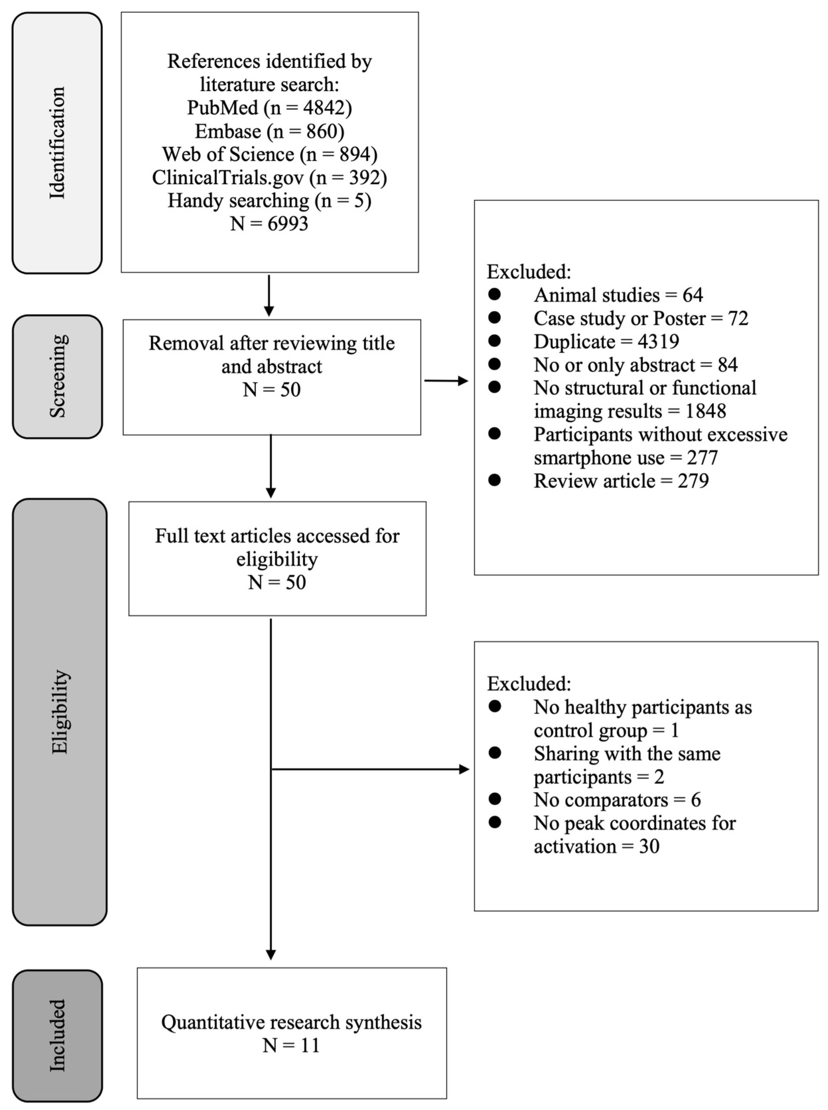
Figure 1. Flowchart summarizing the process of study selection.
Ijerph 19 16277 g001
Figure 2. Forest plot comparing the effect sizes for brain volumes between individuals with excessive smartphone use and their comparators with regular use [32,33,35,36,38,41,42].
Ijerph 19 16277 g002
Figure 3. Random-effects funnel plot for detecting publication bias in studies reporting the brain volumes between individuals with excessive smartphone use and their comparators with regular use.
Ijerph 19 16277 g003
Figure 4. Functional magnetic resonance imaging (fMRI) studies showing brain regions with enhanced activities in individuals with excessive smartphone use compared to their comparators with regular use.
Ijerph 19 16277 g004
Table 1. Comparison of structural and functional neural correlates between individuals with excessive smartphone use and their counterparts with regular use.
StudiesEXPCONEVAL of EXP MPA Definition Brain Volume Functional ConnectivityCountry
N% FAvg. Age (SD)N% FAvg. Age (SD)ANAT Meas.EXP < CONEXP > CONCMEXP < CONEXP > CON
Cho et al. [33]2060.0016.20 (1.11)6728.3615.27(1.69)SAPS≥42 on SAPS as EXPGMV and WMVSCPN/AN/AKorea
Choi et al. [34]3354.5525.21 (5.54)3354.5524.85(4.49)SAPSEXP score > CON scoreN/ABOLD signalLeft MOOC
  • Right: AG, FFG, SMG, MFG
  • Left: SMA, supPL, PreCG, IFG, MCG, Precuneus
  • Bilateral: ParaCG, IPL
Korea
Rashid et al. [35]20N/A18–2520N/A18–25SAS-MSubjects with scores of >98 were considered as EXPGMD and GMVLeft IPLRight insula and right PreCGBOLD signalN/A
  • Right: Calcarine gyrus, SFG, supMA, precuneus
  • Left: FFG, supPL, PCC
  • Bilateral: Cerebellum
Malaysia
Yoo et al. [36]2060.0016.20 (1.11)6827.9415.26 (1.68)SAPSA SAPS score of 42 and higher as EXPGMV and WMVBilateral CNN/AN/AKorea
Horvath et al. [32]2268.1822.5 (3.0)2669.2323.0 (3.2)SAS-SVscoring > 31 (M) and >33 (F) on SAS-SV GMVLeft anterior insula, IT, and PHCLeft SMGBOLD signalRight ACCN/AGermany
Nasser et al. [37]1533.0022.2
(0.86)
1553.0021.67 (1.18)SAS-M and IGATA cut-off score of ≥98 as EXPN/ABOLD signal
  • Right: MOOC, infOG
  • Left: Cuneus, supOG
  • Bilateral: Calcarine, FFG, LG
  • Right: LG, supPL, IFG, IT, MOOC, MFG, postCG, SMG, SFG, MCG
  • Left: FFG, MFG, MOOC, supPL, IPL, infOG, precuneus, insula, supOG, putamen, ACC, postCG
Malaysia
Lee et al. [38]3925.6422.9 (2.2)4934.6922.4
(2.7)
SAPSTotal SAPS score > 40, or subscale score > 14 for disturbance of adaptive function as EXPGMVRight OPFCN/AN/AKorea
Lou et al. [39]2454.1723.25 (1.33)1675.023.88 (0.86)YIATOver 5 “yes” to the 8 questions as EXPN/ABOLD signalN/A
  • Right: MTG, IT
  • Bilateral: ACC, MFG, IFG
China
Chun et al. [40]2548.0027.76 (5.97)2733.3028.93 (6.93)SAPSTotal > 44, or subscale > 15 (disturbance of adaptive function), >13(withdrawal), >13 (tolerance) N/ABOLD signal
  • Right: IT, supPL, dorsal ACC, SMA, cuneus, Thal
  • Left: MFG, LG, MOOC, DLPFC, cerebellum, supOG
  • Bilateral: preCG, MTG
N/AKorea
He et al. [41]2532.024.12 (6.15)2532.029.80 (10.9)FACIUIEXP > CONGMVRight VS,
bilateral amygdala
N/AN/AChina
Wang et al. [42]3461.7621.60 (2.10)3461.7621.73
(1.94)
MPAIOver 51 as EXPGMV&
DTI
  • Right: SFG, IFG, MOOC
  • Left: ACC
  • Bilateral: MFG, Thal, CgH(DTI)
N/AN/AChina
ACC = anterior cingulate cortex; AG = Angular gyrus; ANAT = Anatomy; BOLD = Blood-oxygen-level-dependent; CM = Connectivity measure; CN = Caudate nucleus; CON = Control group; DLPFC = Dorsolateral prefrontal cortex; EVAL = Evaluation; EXP = Experimental group; F = Female; FACIUI = Facebook-specific adaptation of the Compulsive Internet Use instrument; FFG = Fusiform gyrus; GMD = Gray matter density; GMV = Gray matter volume; IFG = Inferior frontal gyrus; IGAT = Instagram addiction test; IPL = inferior parietal lobule; IT = Inferior temporal cortex; LG = lingual gyrus; M = Male; MA = mobile phone addiction; MCG = Middle cingulate gyrus; MFG = Middle frontal gyrus; MOOC = Middle occipital cortex; MPAI = Mobile phone addiction index; MTG = Middle temporal gyrus; N/A = not available; InfOG = Inferior occipital gyrus; OPFC = Orbitofrontal cortex; PCC = Posterior cingulate cortex; PHC = Parahippocampal cortex; PreCG = Precentral gyrus; postCG = Postcentral gyrus; SAPS = Smartphone addiction proneness scale; SAS-M = Smartphone addiction scale-Malay version questionnaire; SAS-SV = Short version of smartphone addiction scale; SCP = Superior cerebellar peduncle; SFG = Superior frontal gyrus; SMA = Supplementary motor area; SMG = Supramarginal gyrus; SPAI = Smartphone addiction inventory; supMA = Superior motor area; supOG = superior occipital gyrus; supPL = Superior parietal lobule; Thal = Thalamus; VS = Ventral striatum; WMV = White matter volume; YIAT = Young’s internet addiction test.
Table 2. Comparison of brain volumes between individuals with excessive smartphone use and their counterparts with regular use: Hedges’ g.
Subgroup AnalysisNCompga95% CIZQbpc
Age group
Adolescents2−0.66−1.01 to −0.30−3.61 **0.410.00
Adults5−0.49−0.85 to −0.14−2.74 **
Location
Cerebral cortex d4−0.14−0.92 to 0.64−0.342.350.00
Subcortical structure e5−0.77−1.01 to −0.54−6.42 **
Comp Comparison; a According to the random effects model; b Cochran’s Q to measure the heterogeneity in accordance with random effects analysis; c The P levels in this column indicate whether the difference between the effect sizes in the subgroups is significant; d Encompassing Rashid et al. [35], Horvath et al. [32], Lee et al. [38], Wang et al. [42]; e Consisting of Cho et al. [33], Yoo et al. [36], Horvath et al. [32], He et al. [41], Wang et al. [42]. ** p < 0.01.
Table 3. Regression analysis of correlations between different mediators and changes in brain volume among individuals with excessive smartphone use.
Variable (Continuous)Coefficient (95% CI)p
Prevalence of female0.01 (−0.002 to 0.03) (n = 6)0.09
Age−0.006 (−0.07 to 0.06) (n = 6)0.86
Intelligence quotient−0.006 (−0.07 to 0.06) (n = 3)0.85
Mean Beck depression inventory score−0.08 (−0.36 to 0.19) (n = 3)0.55
Mean Barratt impulsiveness scale score0.03 (0.004 to 0.06) (n = 4)0.03
Table 4. Brain areas with increased activities in participants with excessive smartphone use compared with those with regular use.
ClusterSideBrain AreaBAVolume (mm3)ALExyz
1RDeclive of posterior lobe 5680.01435193428−76−14
1RLingual gyrus180.01406890228−76−8
2LMiddle frontal gyrus464720.016162576−383020
Note: All areas reported in Talairach space; Broadmann areas are defined by Brain Map Talairach atlas; ALE = activation likelihood estimation; BA = Broadmann area; L = Left; R = Right.
Publisher’s Note: MDPI stays neutral with regard to jurisdictional claims in published maps and institutional affiliations.