Article

Left Ventricular, Left Atrial and Right Ventricular Strain Modifications after Maximal Exercise in Elite Ski-Mountaineering Athletes: A Feasibility Speckle Tracking Study

by
Paul Zimmermann
1,2,3,†,
Max L. Eckstein
3,†,
Othmar Moser
3,*,
Isabelle Schöffl
2,4,5,
Lukas Zimmermann
2 and
Volker Schöffl
2,5,6,7,8
1
Department of Cardiology, Klinikum Bamberg, 96049 Bamberg, Germany
2
Interdisciplinary Center of Sportsmedicine Bamberg, Klinikum Bamberg, 96049 Bamberg, Germany
3
Division of Exercise Physiology and Metabolism, BaySpo-Bayreuth Center of Sport Science, University of Bayreuth, 95440 Bayreuth, Germany
4
Department of Pediatric Cardiology, Friedrich-Alexander-University Erlangen-Nurnberg, 91054 Erlangen, Germany
5
School of Clinical and Applied Sciences, Leeds Beckett University, Leeds LS1 3HE, UK
6
Department of Orthopedic and Trauma Surgery, Friedrich-Alexander-University Erlangen-Nurnberg, 91054 Erlangen, Germany
7
Department of Orthopedic and Trauma Surgery, Klinikum Bamberg, 96049 Bamberg, Germany
8
Section of Wilderness Medicine, Department of Emergency Medicine at the University of Colorado School of Medicine, Denver, CO 80045, USA
*
Author to whom correspondence should be addressed.
These authors contributed equally to this work.
Int. J. Environ. Res. Public Health 2022, 19(20), 13153; https://doi.org/10.3390/ijerph192013153
Submission received: 12 August 2022 / Revised: 3 October 2022 / Accepted: 10 October 2022 / Published: 13 October 2022
(This article belongs to the Special Issue Winter Sports Implications for Training, Environmental and Health)

Abstract

:
Eleven world elite ski-mountaineering (Ski-Mo) athletes were evaluated for pronounced echocardiographic physiological remodeling as the primary aim of our feasibility speckle tracking study. In this context, sports-related cardiac remodeling was analyzed by performing two-dimensional echocardiography, including speckle tracking analysis of the left atrium (LA), right ventricle (RV) and left ventricular (LV) global longitudinal strain (LV-GLS) at rest and post-peak performance. The feasibility echocardiographic speckle tracking analysis was performed on eleven elite Ski-Mo athletes, which were obtained in 2022 during the annual medical examination. The obtained data of the professional Ski-Mo athletes (11 athletes, age: 18–26 years) were compared for different echocardiographic parameters at rest and post-exercise. Significant differences were found for LV-GLS mean (p = 0.0036) and phasic LA conduit strain pattern at rest and post-exercise (p = 0.0033). Furthermore, negative correlation between LV mass and LV-GLS (p = 0.0195, r = −0.69) and LV mass Index and LV-GLS (p = 0.0253, r = −0.66) at rest were elucidated. This descriptive reporting provided, for the first time, a sport-specific dynamic remodeling of an entire elite national team of the Ski-Mo athlete’s left heart and elucidated differences in the dynamic deformation pattern of the left heart.

1. Introduction

Ski mountaineering (Ski-Mo) has been accepted as a new Olympic sport for the 2026 Milan-Cortina Olympics [1]. Ski-Mo has been rated among the most strenuous endurance sports with the highest “hypoxic dose” (i.e., time spent in a hypoxic environment) [2,3,4,5], as it demands both maximal endurance performance during the individual races as well as high-intensity bouts during the sprint and vertical races in elite winter sports athletes [2,3,4,6,7,8,9].
In comparison to other winter endurance sports, e.g., Nordic Cross Country skiing or biathlon, Ski-Mo involves long uphill and downhill passages that have highly varying muscular demands. This leads to significantly different athletic features in comparison to other professional winter sports athletes [8]. Ski-Mo athletes tend to be smaller and younger than other elite endurance winter sports athletes [6,7,8]. In this context, significant physiological differences in morphological and functional cardiac remodeling could be identified for Ski-Mo athletes, especially due to left atrial (LA) remodeling, speckle tracking analysis of the left ventricle (LV), LV ejection fraction (LV-EF) assessment and LV Mass index [6,7,8].
The term athlete’s heart describes adaptions, namely physiological, functional and electro-physiological, caused by variable physiological sport-specific demands [8]. A conventional morphological and functional echocardiographic assessment might fail to distinguish between an athlete’s heart and controls, whereby functional strain rate evaluation provides further information for subclinical abnormalities and risk stratification [8,10,11,12,13,14,15,16,17,18,19]. These relatively new, non-invasive imaging techniques enhance the understanding of an athlete’s heart through a comprehensive characterization of anatomical and functional adaption, providing novel insights into the assessment of cardio-physiological adaption [20].
Especially the analyzed innovative echocardiographic data on pre- and post-exercise conditions with particular attention to the application of speckle tracking as dynamic functional assessment of cardiac remodeling might provide further insights into the characterization of biventricular and LA function in athletes and the impact of sport-specific cardiocirculatory functional adaption in these athletes [20].
The scientific evidence on the physiological aspects of competitive Ski-Mo athletes is still sparse. However, so far sport specific cardiopulmonary remodeling due to structural and functional adaption of the athlete’s heart has already been proven [6,7,8,21,22,23]. Under resting conditions sport specific structural remodeling, such as left atrial volume index (LAVI) and LV global longitudinal strain (LV-GLS), could be detected [7,8]. The objective of the present scientific analysis is to investigate the feasibility of biventricular and LA phasic deformation patterns after maximal exercise, as earlier studies solely focused on strain analysis at rest. In this context, the aim of this feasibility study is to investigate the correlations between strain pattern at rest and their dynamic changes post-exercise to enhance our understanding of an athlete’s heart. This comprehensive characterization of biventricular and LA functions might provide novel insights into the physiological adaption of an athlete’s heart, especially under environmental conditions, such as specific altitude training conditions [20,24]. Previous research revealed a complex sport-specific interaction between physiological response and altitude training, whereby hypoxia and training stress are combined [25]. Alterations of the autonomous regulation of the nervous system and subsequent modifications of the heart rate variability (HRV) have been studied before, differently depending on the training intensity at altitude [25,26,27,28]. These peculiarities might be promising to provide further interesting data to distinguish sport-specific cardiac remodeling in these elite world winter sports professionals, who often perform their sports in altitude, from balanced cardiomyopathies in the future [7,8,20].

2. Materials and Methods

The local ethics committee of the University of Bayreuth approved the study protocol (O1305/1-GB). The study was conducted in conformity with the declaration of Helsinki and Good Clinical Practice [29]. Before any study-related activities, our participating athletes were informed about the study protocol, and athletes were asked to give their written informed consent.

2.1. Study Population

Eleven young elite Ski-Mo professionals, all active members of the German National Team, participating in World championships and the World Cup season, were examined and evaluated in the preseason preparation time in summer during the season 2022. No participating athlete had to be excluded from the study due to post-COVID-19 infection syndromes. All participants were evaluated for anthropometric data, 12 lead electrocardiogram (ECG) and two-dimensional transthoracic echocardiography, including strain analysis at rest and post-exercise. The obtained data of the athletes (n = 11; male n = 8, female n = 3) were compared for different echocardiographic parameters at rest and post-exercise. All participating athletes did not have any medical history, and their sports-related history revealed no potential risk factors for sudden cardiac death in all athletes and within their families.
All participating Ski-Mo athletes were winter sports professionals with a total amount of 20–30 training hours per week during high-volume training times and 5–10 training hours during low-volume training times. During low-volume training times, the athletes focus on continuous endurance training, such as running and cycling, as well as functional strength training and individual training to improve muscle disbalances [6]. An additional sample size estimation was not conducted since the included n = 11 represents the entire cohort of the German National Team competing at World Cup and World Championship levels.

2.2. Echocardiographic Examination

An echocardiographic functional and morphological assessment at rest and post-exercise using a commercially available echocardiographic system Phillips EPIQ 7 device with an X5-1 Matrix-array transducer (Phillips Healthcare, Eindhoven, The Netherlands), following a standard protocol, was performed [30]. In addition, heart rate and blood pressure were measured. Two-dimensional echocardiographic analyses were performed at rest following the general recommendations [8,30,31,32]. The systolic LV-EF was calculated using biplane Simpson rule, based on the apical two-chamber—as well as apical four-chamber view. Two-dimensional linear dimensions and LAVI were evaluated for both ventricles and both atria manually according to the recommendations [30,31,32,33]. An estimation of the RV systolic function at rest using the TAPSE (Tricuspide annular plane systolic excursion) was obtained in the apical four-chamber view. The LV Mass index and the relative wall thickness (RWT) of the LV were calculated using the formula recommended by the current guidelines [34]. We measured the pulse-wave Doppler in the apical four-chamber view referring to the peak early filling (E wave) and late diastolic filling (A wave) velocities to assess the LV diastolic function. Additionally, tissue Doppler imaging of the lateral mitral anulus in the apical four-chamber view was performed (peak early velocity E′) [30,31].
Furthermore, speckle tracking analysis of the athlete’s heart was recorded at rest as well as three minutes post maximal treadmill cardiopulmonary exercise testing (CPET) as post-exercise assessment, focusing on LV, RV and LA. In this context, we obtained the LV-GLS pattern, RV free wall longitudinal deformation (RV FW long.Def.), RV four chamber longitudinal deformation (RV 4C long.Def.) as well as phasic LA strain analysis by two-dimensional strain analysis in the apical views. The detailed phasic LA strain analysis was performed according to the recent European Association of Cardiovascular Imaging (EACVI) recommendations, including LA reservoir strain (LASr), LA conduit strain (LAScd) and LA contraction strain (LASct) assessment [35]. The LA strain assessment was not limited because no Ski-Mo athlete presented atrial fibrillation.
Each of the participating Ski-Mo athletes was evaluated for the prevalence of left and right heart valve regurgitation as part of the standard echocardiographic assessment [8,30,36].

2.3. Statistical Analyses

Data were analyzed with Graph Pad Prism 8.2.1(279) (Graph Pad Software; San Diego, CA, USA). All data were tested for normal distribution via Shapiro–Wilk test. Data were tested for differences with paired t-tests, with statistical significance being accepted at p ≤ 0.05. All data are presented as mean ± SD. Pearson correlations were conducted for anatomical echocardiographic parameters as independent parameter with GLS, RV deformation pattern and LA reservoir, LA conduit and LA contractile at resting conditions as well as post-exercise in our participating elite Ski-Mo athletes. Due to the relatively small number of participating elite winter sports athletes, who were enrolled as the uniqueness of this reporting, we are not able to draw reliable conclusions by uni- and multivariate regression analyses, but we might point out interesting trends in the cohort of enrolled world elite winter sports professionals.

3. Results

3.1. Baseline Athletes’ Characteristics and Echocardiographic Assessment at Rest

The baseline characteristics and anthropometric data of the participating male and female Ski-Mo athletes are presented in Table 1. The body surface areas were calculated with the Du Bois method.
In the two-dimensional echocardiographic assessment, all participating Ski-Mo athletes showed a low–normal to normal systolic LV-EF at rest estimated by the biplane Simpson method and did not show any relevant pathological regurgitation of the right and left heart valves. Only mild regurgitation at the tricuspid and mitral valves was revealed. There was no relevant systolic pulmonary artery pressure evaluated by tricuspid peak systolic velocity. The obtained LA and LV assessment did not differ in between the participating athletes. The baseline echocardiographic characteristics are presented in Table 2.
In comparison with data from sedentary control measurements presented by the German Society of Cardiology (DGK) [32] and with previous morphological and functional echocardiographic data from these athletes [7,8], the obtained sport specific morphological echocardiographic data [LVedd diameter, LV Mass index measurements, interventricular septal wall thickness at diastole (IVSd) and left ventricular posterior wall thickness at diastole (LVPWd), and systolic LV-EF] can be categorized to be in the normal range [7,8].

3.2. Speckle Tracking Analysis of the Right and Left Heart at Rest and Post-Exercise–Sport-Specific Functional Cardiac Remodeling

In the speckle tracking analysis, normal LV-GLS values at rest (−21.55 ± 3.44%) and slightly reduced values for LV-GLS post-exercise (−17.25 ± 3.39%) were elucidated. The difference between rest and post-exercise assessment was significant (p = 0.0036), as presented in Figure 1.
No significant differences were found for the RV free wall longitudinal deformation (RV FW long.Def. rest −28.17 ± 5.45% vs. RV FW long.Def. post-exercise −26.57 ± 6.24%, p = 0.48) nor for the RV apical four-chamber longitudinal deformation (RV 4C long.Def. rest −23.08 ± 2.09% vs. RV 4C long.Def. post-exercise −21.71 ± 4.89%, p = 0.40) at rest and post-exercise.
With respect to standard echocardiographic parameters, LA and LV geometric properties did not show significant interindividual differences. The evaluation of functional LA remodeling by the average phasic LA strain (LAS) during all three phases of the atrial cycle revealed significant differences in the comparison of resting and post-exercise conditions. In this context, across our participating athletes neither the LASr analysis at rest compared to post-exercise strain pattern (LASr rest 51.95 ± 11.55% vs. LASr post-exercise 43.92 ± 11.88%, p = 0.12) nor the LASct analysis at rest compared to post-exercise strain pattern (LASct rest −10.49 ± 7.45% vs. LASct post-exercise −15.46 ± 11.58%, p = 0.24) revealed significant differences.
Significant differences could be elucidated for the LAScd analysis at rest versus post-exercise parameters (LAScd rest −41.45 ± 5.46% vs. LAScd post-exercise −28.45 ± 10.33%, p = 0.0033, as presented in Figure 1).

3.3. Sport-Specific Functional Cardiac Remodeling–Univariante Relationships between Morphological Echocardiographic Characteristics and Functional Remodeling as Speckle Tracking Analyses

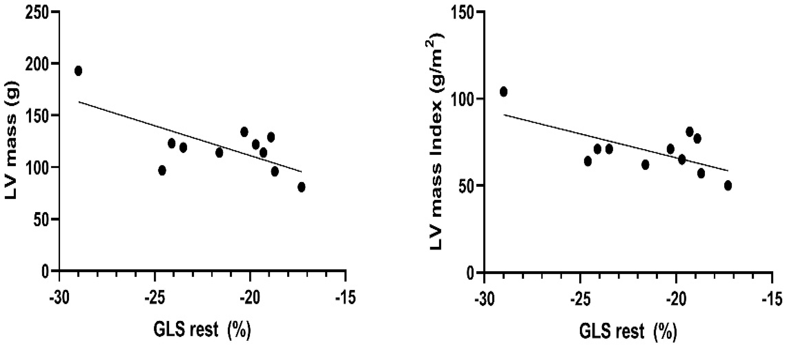
No significant correlations between LAVI and the athlete’s heart strain pattern could be revealed, whereby a negative correlation between LV mass and LV-GLS (p = 0.0195, r = −0.69) and LV-GLS and LV mass Index (p = 0.0253, r = −0.66) at rest could be proven, as presented in Figure 2.

4. Discussion

To the best of our knowledge, this is the first feasibility study to prove functional LA response due to exercise conditions in elite Ski-Mo athletes. These peculiarities provide novel insights into the assessment of Ski-mountaineers’ heart-to-exercise conditions at altitude and improve our understanding of physiological sport-specific demands in these winter sports professionals. Our previous research has shown that the heart of Ski-Mo athletes has adapted to physical exercise by individual sport-specific remodeling due to morphological and functional assessment [6,7,8]. Our additional novel presented results might provide further useful data to complete the understanding of the complex intraindividual Ski-Mo athlete’s cardio-physiological performance adaption and of their enhanced anaerobic capacity during race performance at altitude.
Cardiac and physiological adaptions due to exercise in elite athletes, mainly endurance athletes, represent a hot topic, and studies addressing this topic already exist [11,12,13,17,37]. The uniqueness of our study as a feasibility trial represents the performance evaluation of a small number of world elite winter sports athletes competing in one of the most strenuous endurance sports with the highest “hypoxic dose”, and their competitions are often performed in extreme altitudes, including vertical race components [3,4,6,7,8]. The detected strain rate measurements at rest for LV-GLS in our analyzed participating elite Ski-Mo athletes indicate normal values, despite mild differences compared to untrained controls and well-trained athletes of other sports [11,19,38,39]. However, up to now, LV strain assessment in sport professionals remain largely undefined and has to be interpreted in a multimodality approach assessing cardiac function following exercise [11,16,38,40,41]. Therefore, previous research revealed converse results with either progressive strain rate increase [15,42] or decreased LV-GLS and RV-GLS rates during exercise [42]. Our data elucidate normal LV-GLS strain rates at rest but a small decrease immediately post-exercise. Therefore, our study may serve as a pilot project to evaluate a Ski-Mo athlete’s heart due to altitude performance conditions. Previous research revealed the importance of energy storage during LV twisting to increase and to maintain stroke volume during exercise [24,43]. Prolonged strenuous variables, such as an athlete’s blood pressure response, variable preload conditions, heart rate response during exercise and exposure, especially to high-intensity interval training (HIIT) episodes at altitude, have to be considered in the interpretation and slight reduction of LV-GLS post-exercise in our Ski-Mo athletes’ data [44,45] and their potential negative impact on left and right ventricular function according to “exercise induced cardiac fatigue” [46,47,48].
RV longitudinal strain seems to provide an effective tool to assess sport-associated individual athletes’ adaption or alterations of the RV by exercise-related RV overload, even at an early subclinical stage [49,50,51,52,53,54,55]. Normal to slightly lower resting values of RV myocardial strain have to be shown to be a consequence of physiological cardiac remodeling rather than subclinical myocardial damage in elite endurance athletes [51,52,56,57,58]. No significant differences between pre- and post-exercise conditions in our small cohort could be proven. Although it might be difficult to derive reliable conclusions from our RV strain assessment, our descriptive reporting might contribute to a better understanding of sport-specific RV remodeling at altitude conditions. Several influencing environmental as well as physiological parameters, the impact of ethnicity, the beta-adrenergic desensitization, and the different levels of exposure to dynamic training on the RV, not all highlighted in our presented feasibility analyses, have to be taken into consideration for a focused and comprehensive assessment of right heart remodeling [48,59,60]. Next to the impact of the functional shift of RV as a novel marker of an athlete’s heart [48,49,61,62], we have to be aware that such RV remodeling might represent a proarrhythmogenic substrate in some highly trained athletes in the absence of known familiar predisposition-implicating the importance of long-term clinical assessment in the accompanying performance center [63,64,65].
Previous research demonstrated the importance of LA remodeling as a promising tool for the volumetric and functional characterization of the LA in athletes [66] as well as in patients with heart failure [67]. To the best of our knowledge, this is the first feasibility study to evaluate the acute LA response in Ski-Mo athletes. LA function, in general, has a great pathophysiological impact on the modulation of LV filling conditions as well as on LA response functions during exercise [67,68]. Our novel obtained data on exercise-induced LA adaption revealed significant differences for the phasic LAScd data at rest versus post-exercise values, whereas LASr and LASct strain patterns were not affected. The possibility to mobilize atrial functional “booster” capacity in response to exercise represents an important aspect of an athlete’s performance, whereby endurance training is known to be associated with an increase in venous return with a potential overstretching of the LA [67,69]. Although LA response according to a Frank–Starling mechanism up to a certain point has been described before [70], it has to be stated that LA response may vary to different exercise modalities, environmental conditions and loads [67]. Referring to this background, our observed LAScd reduction post-exercise might be interpreted due to high-intensity components mainly at altitude environmental conditions. Due to these extreme conditions causing hypoxia and modifications of the HRV [25,26,28], a certain impact on the specific Ski-Mo athlete’s LA response might be assumed. Functional LA properties have to be interpreted as a consequence of intense and chronic training in an athlete’s heart, as stated before [66]. Exercise-induced hypertension, variable preload conditions after endurance exercise and LVH are suggested to have the mean impact on alternated LAScd or the previously described LA intrinsic impairment [71]. Whether these LA remodeling persists during an athlete’s lifetime career or might be related to environmental conditions remains to be evaluated. Nevertheless, these alterations might be related to the higher risk of arrhythmias, especially atrial fibrillation (AF) known as paroxysmal AF, in young and middle-aged athletes (PAFIYAMA) [68,72]. Therefore, our feasibility study cannot appraise any chronological causalities on the LAS pattern and draw reliable conclusions, and further prospective research is necessary to strengthen the scientific evidence.
Sport-specific cardiac remodeling in extreme levels of endurance sports, such as Ski-Mo, is a previously known phenomenon displaying a continuous process with a broad spectrum of adaption during an athlete’s career [5,6,7,8,73,74,75,76]. Whether the LVH found in athletes is physiological adaption or a risk factor for the progression of an initial subclinical hypertrophy remains controversial in the literature [40,76]. Evaluating the impact of anatomical left heart remodeling on biventricular and LA deformation patterns at rest and post-exercise, we could reveal a negative correlation between higher LV Mass as well as LV Mass Index and lower LV-GLS strain values at rest in our elite Ski-Mo athletes. Due to the small sample size, data outliers might lead to a significant bias in the echocardiographic data assessment and consequently affect the correlation of LV mass index and LV-GLS. Although it is not possible to derive reliable conclusions from this small sample size of Ski-Mo professionals, our data assessment might be regarded as an additional descriptive characterization of an athlete’s altitude training-induced dynamic remodeling process, primarily appearing as a balanced process [76].
Our feasibility trial aims to elucidate sport-specific adaptions of elite Ski-Mo athletes, who compete as very endurant athletes with enhanced anaerobic capacity at altitude and under environmental influences. However, our study is not without limitations. Firstly, our measurements were acquired in the preseason preparation time in summer with respect to a certain deviation in individual training schedules. Secondly, the slightly anthropometric variability in the cohort of Ski-Mo athletes with a mixture of young and experienced athletes entails an intra-cohort variability and contributes to a certain standard deviation in our cardiac assessment, such as the LV Mass index. Furthermore, no comparable winter sports athletes with different ethnic backgrounds except Caucasian athletes were analyzed. Additionally, focusing on speckle tracking deformation pattern pre- and post-exercise, no complete echocardiographic examination and no calculation of average septal and lateral E’ data were obtained according to the current and latest recommendations [30,34]. During the treadmill CPET, no strain analyses were acquired because it was technically not feasible. Last, we were unable to draw reliable conclusions between echocardiographic assessment and parameters of CPET performance because the size of the entire elite German Ski-Mo team is small. This would not lead to statistically reliable conclusions. Future research might focus on a larger athlete’s sample size to verify and strengthen the scientific evidence base of the obtained findings of our feasibility reporting.

5. Conclusions

This report provides, for the first time, new evidence of sport-specific dynamic remodeling of the Ski-Mo athlete’s heart and elucidates differences at resting and post-exercise conditions in the strain pattern of the left heart against the sport-specific altitude training background of Ski-Mo athletes.
In conclusion, strain imaging is technically feasible in world elite Ski-Mo athletes and might contribute to a better understanding of sport-specific remodeling and probably enhanced physiological remodeling in these highly trained altitude athletes. Further data are warranted to characterize the sport-specific dynamic atrial and ventricular cardiac remodeling, and our obtained findings might pave the road to future studies with long-term follow-up and a greater number of athletes to verify and strengthen the scientific evidence base.

Author Contributions

Data curation, P.Z. and L.Z.; formal analysis, P.Z. and M.L.E.; investigation, P.Z., M.L.E. and V.S.; methodology, P.Z. and V.S.; project administration, P.Z. and V.S.; supervision, P.Z.; writing—original draft, P.Z. and M.L.E.; writing—review and editing, P.Z., M.L.E., V.S., L.Z., O.M. and I.S. All authors have read and agreed to the published version of the manuscript.

Funding

The article received no funding.

Institutional Review Board Statement

The study was conducted according to the guidelines of the Declaration of Helsinki and approved by the Ethics Committee of the University of Bayreuth (study protocol).

Informed Consent Statement

Informed consent was obtained from all subjects involved in the study.

Data Availability Statement

Individual anonymized data supporting the analyses of this study contained in this manuscript will be made available upon reasonable written request from researchers whose proposed use of data for a specific purpose has been approved.

Acknowledgments

We want to thank the participants, all of whom were examined in the Interdisciplinary Center of Sportsmedicine, Klinikum Bamberg.

Conflicts of Interest

The authors declare no conflict of interest.

References

  1. Ski Mountaineering Added to the Milano Cortina 2026 Sports Programme. Available online: https://olympics.com/Ioc/News/Ski-Mountaineering-Added-to-the-Milano-Cortina-2026-Sports-Programme (accessed on 20 July 2021).
  2. Faiss, R.; von Orelli, C.; Dériaz, O.; Millet, G.P. Responses to Exercise in Normobaric Hypoxia: Comparison of Elite and Recreational Ski Mountaineers. Int. J. Sports Physiol. Perform. 2014, 9, 978–984. [Google Scholar] [CrossRef]
  3. Praz, C.; Léger, B.; Kayser, B. Energy Expenditure of Extreme Competitive Mountaineering Skiing. Eur. J. Appl. Physiol. 2014, 114, 2201–2211. [Google Scholar] [CrossRef] [Green Version]
  4. Duc, S.; Cassirame, J.; Durand, F. Physiology of Ski Mountaineering Racing. Int. J. Sports Med. 2011, 32, 856–863. [Google Scholar] [CrossRef]
  5. Schöffl, V.R.; Bösl, T.; Lutter, C. Ski Mountaineering: Sports Medical Considerations for This New Olympic Sport. Br. J. Sports Med. 2022, 56, 2–3. [Google Scholar] [CrossRef] [PubMed]
  6. Zimmermann, P.; Wüstenfeld, J.; Zimmermann, L.; Schöffl, V.; Schöffl, I. Physiological Aspects of World Elite Competitive German Winter Sport Athletes. Int. J. Environ. Res. Public Health 2022, 19, 5620. [Google Scholar] [CrossRef] [PubMed]
  7. Zimmermann, P.; Schöffl, I.; Schöffl, V.; Zimmermann, L.; Eckstein, M.L.; Moser, O.; Wüstenfeld, J. Physiological Effects of Training in Elite German Winter Sport Athletes: Sport Specific Remodeling Determined Using Echocardiographic Data and CPET Performance Parameters. J. Cardiovasc. Dev. Dis. 2022, 9, 235. [Google Scholar] [CrossRef]
  8. Zimmermann, P.; Moser, O.; Eckstein, M.L.; Wüstenfeld, J.; Schöffl, V.; Zimmermann, L.; Braun, M.; Schöffl, I. Athlete’s Heart in Elite Biathlon, Nordic Cross—Country and Ski-Mountaineering Athletes: Cardiac Adaptions Determined Using Echocardiographic Data. J. Cardiovasc. Dev. Dis. 2021, 9, 8. [Google Scholar] [CrossRef]
  9. Schöffl, V.; Pöppelmeier, O.; Emmler, J.; Schöffl, I.; Küpper, T.; Lutter, C. Ski Mountaineering–Evaluation of a Sports Specific Performance Diagnosis. Sportverletz. Sportschaden 2018, 32, 233–242. [Google Scholar] [CrossRef]
  10. Unnithan, V.B.; Beaumont, A.; Rowland, T.W.; Sculthorpe, N.; George, K.; Lord, R.; Oxborough, D. The Influence of Training Status on Right Ventricular Morphology and Segmental Strain in Elite Pre-Adolescent Soccer Players. Eur. J. Appl. Physiol. 2021, 121, 1419–1429. [Google Scholar] [CrossRef] [PubMed]
  11. Caselli, S.; Montesanti, D.; Autore, C.; di Paolo, F.M.; Pisicchio, C.; Squeo, M.R.; Musumeci, B.; Spataro, A.; Pandian, N.G.; Pelliccia, A. Patterns of Left Ventricular Longitudinal Strain and Strain Rate in Olympic Athletes. J. Am. Soc. Echocardiogr. 2015, 28, 245–253. [Google Scholar] [CrossRef] [PubMed]
  12. Moneghetti, K.J.; Singh, T.; Hedman, K.; Christle, J.W.; Kooreman, Z.; Kobayashi, Y.; Bouajila, S.; Amsallem, M.; Wheeler, M.; Gerche, A.L.; et al. Echocardiographic Assessment of Left Ventricular Remodeling in American Style Footballers. Int. J. Sports Med. 2020, 41, 27–35. [Google Scholar] [CrossRef]
  13. D’Andrea, A.; Galderisi, M.; Sciomer, S.; Nistri, S.; Agricola, E.; Ballo, P.; Buralli, S.; D’Errico, A.; Losi, M.A.; Mele, D.; et al. Echocardiographic Evaluation of the Athlete’s Heart: From Morphological Adaptations to Myocardial Function. G Ital. Cardiol. 2009, 10, 533–544. [Google Scholar]
  14. Artis, N.J.; Oxborough, D.L.; Williams, G.; Pepper, C.B.; Tan, L.B. Two-Dimensional Strain Imaging: A New Echocardiographic Advance with Research and Clinical Applications. Int. J. Cardiol. 2008, 123, 240–248. [Google Scholar] [CrossRef]
  15. Utomi, V.; Oxborough, D.; Whyte, G.P.; Somauroo, J.; Sharma, S.; Shave, R.; Atkinson, G.; George, K. Systematic Review and Meta-Analysis of Training Mode, Imaging Modality and Body Size Influences on the Morphology and Function of the Male Athlete’s Heart. Heart 2013, 99, 1727–1733. [Google Scholar] [CrossRef]
  16. Gruca, M.M.; Cheema, B.; Garg, G.; Ryan, J.; Thomas, J.D.; Rigolin, V.H.; Zielinski, A.R.; Puthumana, J.J. Strain Echocardiography to Describe Left Ventricular Function Pre- and Postexercise in Elite Basketball Athletes: A Feasibility Study. Echocardiography 2021, 38, 1165–1172. [Google Scholar] [CrossRef]
  17. Forsythe, L.; George, K.; Oxborough, D. Speckle Tracking Echocardiography for the Assessment of the Athlete’s Heart: Is It Ready for Daily Practice? Curr. Treat. Options Cardiovasc. Med. 2018, 20, 83. [Google Scholar] [CrossRef] [Green Version]
  18. la Gerche, A.; Jurcut, R.; Voigt, J.-U. Right Ventricular Function by Strain Echocardiography. Curr. Opin. Cardiol. 2010, 25, 430–436. [Google Scholar] [CrossRef]
  19. Yingchoncharoen, T.; Agarwal, S.; Popović, Z.B.; Marwick, T.H. Normal Ranges of Left Ventricular Strain: A Meta-Analysis. J. Am. Soc. Echocardiogr. 2013, 26, 185–191. [Google Scholar] [CrossRef]
  20. D’Ascenzi, F.; Caselli, S.; Solari, M.; Pelliccia, A.; Cameli, M.; Focardi, M.; Padeletti, M.; Corrado, D.; Bonifazi, M.; Mondillo, S. Novel Echocardiographic Techniques for the Evaluation of Athletes’ Heart: A Focus on Speckle-Tracking Echocardiography. Eur. J. Prev. Cardiol. 2016, 23, 437–446. [Google Scholar] [CrossRef]
  21. Rupwate, R.U.; Chitaley, M.; Kamat, S.R. Cardiopulmonary Functional Changes in Acute Acclimatisation to High Altitude in Mountaineers. Eur. J. Epidemiol. 1990, 6, 266–272. [Google Scholar] [CrossRef]
  22. Gaston, A.-F.; Marti Peiro, A.; Hapkova, I.; Durand, F. Exploring Physiological Parameters in Ski Mountaineering during World Cup Races. Int. J. Perform. Anal. Sport 2019, 19, 275–288. [Google Scholar] [CrossRef]
  23. Lasshofer, M.; Seifert, J.; Wörndle, A.-M.; Stöggl, T. Physiological Responses and Predictors of Performance in a Simulated Competitive Ski Mountaineering Race. J. Sports Sci. Med. 2021, 20, 250–257. [Google Scholar] [CrossRef]
  24. Unnithan, V.B.; Rowland, T.; Lindley, M.R.; Roche, D.M.; Garrard, M.; Barker, P. Cardiac Strain during Upright Cycle Ergometry in Adolescent Males. Echocardiography 2015, 32, 638–643. [Google Scholar] [CrossRef] [PubMed] [Green Version]
  25. Schmitt, L.; Hellard, P.; Millet, G.P.; Roels, B.; Richalet, J.P.; Fouillot, J.P. Heart Rate Variability and Performance at Two Different Altitudes in Well-Trained Swimmers. Int. J. Sports Med. 2006, 27, 226–231. [Google Scholar] [CrossRef] [PubMed] [Green Version]
  26. Schmitt, L.; Fouillot, J.-P.; Millet, G.P.; Robach, P.; Nicolet, G.; Brugniaux, J.; Richalet, J.-P. Altitude, Heart Rate Variability and Aerobic Capacities. Int. J. Sports Med. 2008, 29, 300–306. [Google Scholar] [CrossRef] [PubMed]
  27. Sevre, K.; Bendz, B.; Hankø, E.; Nakstad, A.R.; Hauge, A.; Kåsin, J.I.; Lefrandt, J.D.; Smit, A.J.; Eide, I.; Rostrup, M. Reduced Autonomic Activity during Stepwise Exposure to High Altitude. Acta Physiol. Scand. 2001, 173, 409–417. [Google Scholar] [CrossRef]
  28. Schmitt, L.; Willis, S.J.; Coulmy, N.; Millet, G.P. Effects of Different Training Intensity Distributions Between Elite Cross-Country Skiers and Nordic-Combined Athletes During Live High-Train Low. Front. Physiol. 2018, 9, 932. [Google Scholar] [CrossRef] [PubMed]
  29. Harriss, D.J.; MacSween, A.; Atkinson, G. Ethical Standards in Sport and Exercise Science Research: 2020 Update. Int. J. Sports Med. 2019, 40, 813–817. [Google Scholar] [CrossRef] [Green Version]
  30. Evangelista, A.; Flachskampf, F.; Lancellotti, P.; Badano, L.; Aguilar, R.; Monaghan, M.; Zamorano, J.; Nihoyannopoulos, P. European Association of Echocardiography Recommendations for Standardization of Performance, Digital Storage and Reporting of Echocardiographic Studies. Eur. J. Echocardiogr. 2008, 9, 438–448. [Google Scholar] [CrossRef] [PubMed] [Green Version]
  31. Lang, R.; Bierig, M.; Devereux, R.; Flachskampf, F.; Foster, E.; Pellikka, P.; Picard, M.; Roman, M.; Seward, J.; Shanewise, J. Recommendations for Chamber Quantification. Eur. J. Echocardiogr. 2006, 7, 79–108. [Google Scholar] [CrossRef]
  32. Hagendorff, A.; Fehske, W.; Flachskampf, F.A.; Helfen, A.; Kreidel, F.; Kruck, S.; la Rosée, K.; Tiemann, K.; Voigt, J.-U.; von Bardeleben, R.S.; et al. Manual Zur Indikation U.Und Durchführung Der Echokardiographie–Update 2020 Der Deutschen Gesellschaft Für Kardiologie. Kardiologe 2020, 14, 396–431. [Google Scholar] [CrossRef]
  33. Galderisi, M.; Cosyns, B.; Edvardsen, T.; Cardim, N.; Delgado, V.; di Salvo, G.; Donal, E.; Sade, L.E.; Ernande, L.; Garbi, M.; et al. Standardization of Adult Transthoracic Echocardiography Reporting in Agreement with Recent Chamber Quantification, Diastolic Function, and Heart Valve Disease Recommendations: An Expert Consensus Document of the European Association of Cardiovascular Imaging. Eur. Heart J. Cardiovasc. Imaging 2017, 18, 1301–1310. [Google Scholar] [CrossRef] [PubMed] [Green Version]
  34. Lang, R.M.; Badano, L.P.; Mor-Avi, V.; Afilalo, J.; Armstrong, A.; Ernande, L.; Flachskampf, F.A.; Foster, E.; Goldstein, S.A.; Kuznetsova, T.; et al. Recommendations for Cardiac Chamber Quantification by Echocardiography in Adults: An Update from the American Society of Echocardiography and the European Association of Cardiovascular Imaging. J. Am. Soc. Echocardiogr. 2015, 28, 1–39.e14. [Google Scholar] [CrossRef] [Green Version]
  35. Badano, L.P.; Kolias, T.J.; Muraru, D.; Abraham, T.P.; Aurigemma, G.; Edvardsen, T.; D’Hooge, J.; Donal, E.; Fraser, A.G.; Marwick, T.; et al. Standardization of Left Atrial, Right Ventricular, and Right Atrial Deformation Imaging Using Two-Dimensional Speckle Tracking Echocardiography: A Consensus Document of the EACVI/ASE/Industry Task Force to Standardize Deformation Imaging. Eur. Heart J. Cardiovasc. Imaging 2018, 19, 591–600. [Google Scholar] [CrossRef]
  36. Zimmermann, P.; Moser, O.; Edelmann, F.; Schöffl, V.; Eckstein, M.L.; Braun, M. Electrical and Structural Adaption of Athlete’s Heart and the Impact on Training and Recovery Management in Professional Basketball Players: A Retrospective Observational Study. Front. Physiol. 2022, 13, 739753. [Google Scholar] [CrossRef]
  37. Klaeboe, L.G.; Edvardsen, T. Echocardiographic Assessment of Left Ventricular Systolic Function. J. Echocardiogr. 2019, 17, 10–16. [Google Scholar] [CrossRef] [Green Version]
  38. D’Andrea, A.; Radmilovic, J.; Carbone, A.; Mandoli, G.E.; Santoro, C.; Evola, V.; Bandera, F.; D’Ascenzi, F.; Bossone, E.; Galderisi, M.; et al. Speckle Tracking Evaluation in Endurance Athletes: The “Optimal” Myocardial Work. Int. J. Cardiovasc. Imaging 2020, 36, 1679–1688. [Google Scholar] [CrossRef]
  39. Schattke, S.; Xing, Y.; Lock, J.; Brechtel, L.; Schroeckh, S.; Spethmann, S.; Baumann, G.; Borges, A.C.; Knebel, F. Increased Longitudinal Contractility and Diastolic Function at Rest in Well-Trained Amateur Marathon Runners: A Speckle Tracking Echocardiography Study. Cardiovasc. Ultrasound 2014, 12, 11. [Google Scholar] [CrossRef] [PubMed] [Green Version]
  40. Binnetoğlu, F.K.; Babaoğlu, K.; Altun, G.; Kayabey, Ö. Effects That Different Types of Sports Have on the Hearts of Children and Adolescents and the Value of Two-Dimensional Strain-Strain-Rate Echocardiography. Pediatr. Cardiol. 2014, 35, 126–139. [Google Scholar] [CrossRef]
  41. Shave, R.; George, K.; Whyte, G.; Middleton, N.; Hart, E.; Artis, N.; Oxborough, D. A Comparison of Doppler, Tissue Doppler Imaging, and Strain Rate Imaging in the Assessment of Postexercise Left Ventricular Function. Appl. Physiol. Nutr. Metab. 2009, 34, 33–39. [Google Scholar] [CrossRef] [PubMed]
  42. Stewart, G.M.; Yamada, A.; Haseler, L.J.; Kavanagh, J.J.; Koerbin, G.; Chan, J.; Sabapathy, S. Altered Ventricular Mechanics after 60 Min of High-Intensity Endurance Exercise: Insights from Exercise Speckle-Tracking Echocardiography. Am. J. Physiol. Heart Circ. Physiol. 2015, 308, H875–H883. [Google Scholar] [CrossRef]
  43. Santoro, A.; Alvino, F.; Antonelli, G.; Cameli, M.; Bertini, M.; Molle, R.; Mondillo, S. Left Ventricular Strain Modifications after Maximal Exercise in Athletes: A Speckle Tracking Study. Echocardiography 2015, 32, 920–927. [Google Scholar] [CrossRef]
  44. Huang, Y.-C.; Tsai, H.-H.; Fu, T.-C.; Hsu, C.-C.; Wang, J.-S. High-Intensity Interval Training Improves Left Ventricular Contractile Function. Med. Sci. Sports Exerc. 2019, 51, 1420–1428. [Google Scholar] [CrossRef] [PubMed]
  45. Schneider, C.; Forsythe, L.; Somauroo, J.; George, K.; Oxborough, D. The Impact of Preload Reduction with Head-up Tilt Testing on Longitudinal and Transverse Left Ventricular Mechanics: A Study Utilizing Deformation Volume Analysis. Echo Res. Pract. 2018, 5, 11–18. [Google Scholar] [CrossRef] [PubMed] [Green Version]
  46. Żebrowska, A.; Mikołajczyk, R.; Waśkiewicz, Z.; Gąsior, Z.; Mizia-Stec, K.; Kawecki, D.; Rosemann, T.; Nikolaidis, P.T.; Knechtle, B. Left Ventricular Systolic Function Assessed by Speckle Tracking Echocardiography in Athletes with and without Left Ventricle Hypertrophy. J. Clin. Med. 2019, 8, 687. [Google Scholar] [CrossRef] [PubMed] [Green Version]
  47. Oxborough, D.; Birch, K.; Shave, R.; George, K. “Exercise-Induced Cardiac Fatigue”—A Review of the Echocardiographic Literature. Echocardiography 2010, 27, 1130–1140. [Google Scholar] [CrossRef]
  48. Banks, L.; Sasson, Z.; Busato, M.; Goodman, J.M. Impaired Left and Right Ventricular Function Following Prolonged Exercise in Young Athletes: Influence of Exercise Intensity and Responses to Dobutamine Stress. J. Appl. Physiol. 2010, 108, 112–119. [Google Scholar] [CrossRef] [PubMed] [Green Version]
  49. Knackstedt, C.; Hildebrandt, U.; Schmidt, K.; Syrocki, L.; Lang, A.; Bjarnason-Wehrens, B.; Schummers, G.; Stapf, D.; Becker, M.; Predel, H.G. Analysis of Right and Left Ventricular Deformation in Former World Class Swimmers: Evaluation Using Speckle Tracking. J. Sports Med. Phys. Fitness 2015, 55, 978–987. [Google Scholar]
  50. Smolarek, D.; Gruchała, M.; Sobiczewski, W. Echocardiographic Evaluation of Right Ventricular Systolic Function: The Traditional and Innovative Approach. Cardiol. J. 2017, 24, 563–572. [Google Scholar] [CrossRef] [PubMed] [Green Version]
  51. Bjerring, A.W.; Landgraff, H.E.; Leirstein, S.; Aaeng, A.; Ansari, H.Z.; Saberniak, J.; Murbræch, K.; Bruun, H.; Stokke, T.M.; Haugaa, K.H.; et al. Morphological Changes and Myocardial Function Assessed by Traditional and Novel Echocardiographic Methods in Preadolescent Athlete’s Heart. Eur. J. Prev. Cardiol. 2018, 25, 1000–1007. [Google Scholar] [CrossRef] [PubMed]
  52. Dawkins, T.G.; Curry, B.A.; Wright, S.P.; Meah, V.L.; Yousef, Z.; Eves, N.D.; Shave, R.E.; Stembridge, M. Right Ventricular Function and Region-Specific Adaptation in Athletes Engaged in High-Dynamic Sports: A Meta-Analysis. Circ. Cardiovasc. Imaging 2021, 14, e012315. [Google Scholar] [CrossRef]
  53. Christou, G.A.; Pagourelias, E.D.; Anifanti, M.A.; Sotiriou, P.G.; Koutlianos, N.A.; Tsironi, M.P.; Andriopoulos, P.I.; Christou, K.A.; Kouidi, E.J.; Deligiannis, A.P. Exploring the Determinants of the Cardiac Changes after Ultra-Long Duration Exercise: The Echocardiographic Spartathlon Study. Eur. J. Prev. Cardiol. 2020, 27, 1467–1477. [Google Scholar] [CrossRef]
  54. Oxborough, D.; Sharma, S.; Shave, R.; Whyte, G.; Birch, K.; Artis, N.; Batterham, A.M.; George, K. The Right Ventricle of the Endurance Athlete: The Relationship between Morphology and Deformation. J. Am. Soc. Echocardiogr. 2012, 25, 263–271. [Google Scholar] [CrossRef]
  55. Zaidi, A.; Knight, D.S.; Augustine, D.X.; Harkness, A.; Oxborough, D.; Pearce, K.; Ring, L.; Robinson, S.; Stout, M.; Willis, J.; et al. Echocardiographic Assessment of the Right Heart in Adults: A Practical Guideline from the British Society of Echocardiography. Echo Res. Pract. 2020, 7, G19–G41. [Google Scholar] [CrossRef] [Green Version]
  56. King, G.; Almuntaser, I.; Murphy, R.T.; la Gerche, A.; Mahoney, N.; Bennet, K.; Clarke, J.; Brown, A. Reduced Right Ventricular Myocardial Strain in the Elite Athlete May Not Be a Consequence of Myocardial Damage. “Cream Masquerades as Skimmed Milk”. Echocardiography 2013, 30, 929–935. [Google Scholar] [CrossRef]
  57. Rimensberger, C.; Carlen, F.; Brugger, N.; Seiler, C.; Wilhelm, M. Right Ventricular Adaptations and Arrhythmias in Amateur Ultra-Endurance Athletes. Br. J. Sports Med. 2014, 48, 1179–1184. [Google Scholar] [CrossRef]
  58. Ujka, K.; Bastiani, L.; D’Angelo, G.; Catuzzo, B.; Tonacci, A.; Mrakic-Sposta, S.; Vezzoli, A.; Giardini, G.; Pratali, L. Enhanced Right-Chamber Remodeling in Endurance Ultra-Trail Athletes Compared to Marathon Runners Detected by Standard and Speckle-Tracking Echocardiography. Front. Physiol. 2017, 8, 527. [Google Scholar] [CrossRef] [Green Version]
  59. Qasem, M.; George, K.; Somauroo, J.; Forsythe, L.; Brown, B.; Oxborough, D. Influence of Different Dynamic Sporting Disciplines on Right Ventricular Structure and Function in Elite Male Athletes. Int. J. Cardiovasc. Imaging 2018, 34, 1067–1074. [Google Scholar] [CrossRef]
  60. Zaidi, A.; Ghani, S.; Sharma, R.; Oxborough, D.; Panoulas, V.F.; Sheikh, N.; Gati, S.; Papadakis, M.; Sharma, S. Physiological Right Ventricular Adaptation in Elite Athletes of African and Afro-Caribbean Origin. Circulation 2013, 127, 1783–1792. [Google Scholar] [CrossRef] [Green Version]
  61. Lakatos, B.K.; Kiss, O.; Tokodi, M.; Tősér, Z.; Sydó, N.; Merkely, G.; Babity, M.; Szilágyi, M.; Komócsin, Z.; Bognár, C.; et al. Exercise-Induced Shift in Right Ventricular Contraction Pattern: Novel Marker of Athlete’s Heart? Am. J. Physiol. Heart Circ. Physiol. 2018, 315, H1640–H1648. [Google Scholar] [CrossRef] [Green Version]
  62. Sanz-de la Garza, M.; Rubies, C.; Batlle, M.; Bijnens, B.H.; Mont, L.; Sitges, M.; Guasch, E. Severity of Structural and Functional Right Ventricular Remodeling Depends on Training Load in an Experimental Model of Endurance Exercise. Am. J. Physiol. Heart Circ. Physiol. 2017, 313, H459–H468. [Google Scholar] [CrossRef] [PubMed] [Green Version]
  63. D’Andrea, A.; la Gerche, A.; Golia, E.; Teske, A.J.; Bossone, E.; Russo, M.G.; Calabrò, R.; Baggish, A.L. Right Heart Structural and Functional Remodeling in Athletes. Echocardiography 2015, 32 (Suppl. 1), S11–S22. [Google Scholar] [CrossRef] [PubMed]
  64. la Gerche, A.; Burns, A.T.; Mooney, D.J.; Inder, W.J.; Taylor, A.J.; Bogaert, J.; Macisaac, A.I.; Heidbüchel, H.; Prior, D.L. Exercise-Induced Right Ventricular Dysfunction and Structural Remodelling in Endurance Athletes. Eur. Heart J. 2012, 33, 998–1006. [Google Scholar] [CrossRef] [Green Version]
  65. Lord, R.; Somauroo, J.; Stembridge, M.; Jain, N.; Hoffman, M.D.; George, K.; Jones, H.; Shave, R.; Haddad, F.; Ashley, E.; et al. The Right Ventricle Following Ultra-Endurance Exercise: Insights from Novel Echocardiography and 12-Lead Electrocardiography. Eur. J. Appl. Physiol. 2015, 115, 71–80. [Google Scholar] [CrossRef]
  66. Nemes, A.; Domsik, P.; Kalapos, A.; Orosz, A.; Oszlánczi, M.; Török, L.; Balogh, L.; Márton, J.; Forster, T.; Lengyel, C. Volumetric and Functional Assessment of the Left Atrium in Young Competitive Athletes without Left Ventricular Hypertrophy: The MAGYAR-Sport Study. J. Sports Med. Phys. Fitness 2017, 57, 900–906. [Google Scholar] [CrossRef]
  67. Caminiti, G.; Perrone, M.A.; Iellamo, F.; D’Antoni, V.; Catena, M.; Franchini, A.; Volterrani, M. Acute Left Atrial Response to Different Eccentric Resistance Exercise Loads in Patients with Heart Failure with Middle Range Ejection Fraction: A Pilot Study. J. Pers. Med. 2022, 12, 689. [Google Scholar] [CrossRef]
  68. Cuspidi, C.; Tadic, M.; Sala, C.; Gherbesi, E.; Grassi, G.; Mancia, G. Left Atrial Function in Elite Athletes: A Meta-analysis of Two-dimensional Speckle Tracking Echocardiographic Studies. Clin. Cardiol. 2019, 42, 579–587. [Google Scholar] [CrossRef] [Green Version]
  69. Lentini, A.C.; McKelvie, R.S.; McCartney, N.; Tomlinson, C.W.; MacDougall, J.D. Left Ventricular Response in Healthy Young Men during Heavy-Intensity Weight-Lifting Exercise. J. Appl. Physiol. 1993, 75, 2703–2710. [Google Scholar] [CrossRef]
  70. Anwar, A.M.; Geleijnse, M.L.; Soliman, O.I.I.; Nemes, A.; Cate, F.J.t. Left Atrial Frank Starling Law Assessed by Real-Time, Three-Dimensional Echocardiographic Left Atrial Volume Changes. Heart 2007, 93, 1393–1397. [Google Scholar] [CrossRef] [Green Version]
  71. Hart, E.; Shave, R.; Middleton, N.; George, K.; Whyte, G.; Oxborough, D. Effect of Preload Augmentation on Pulsed Wave and Tissue Doppler Echocardiographic Indices of Diastolic Function after a Marathon. J. Am. Soc. Echocardiogr. 2007, 20, 1393–1399. [Google Scholar] [CrossRef]
  72. Sanchis-Gomar, F.; Perez-Quilis, C.; Lippi, G.; Cervellin, G.; Leischik, R.; Löllgen, H.; Serrano-Ostáriz, E.; Lucia, A. Atrial Fibrillation in Highly Trained Endurance Athletes-Description of a Syndrome. Int. J. Cardiol. 2017, 226, 11–20. [Google Scholar] [CrossRef]
  73. D’Andrea, A.; Bossone, E.; Radmilovic, J.; Caso, P.; Calabrò, R.; Russo, M.G.; Galderisi, M. The Role of New Echocardiographic Techniques in Athlete’s Heart. F1000Research 2015, 4, 289. [Google Scholar] [CrossRef]
  74. Perry, R.; Swan, A.L.; Hecker, T.; de Pasquale, C.G.; Selvanayagam, J.B.; Joseph, M.X. The Spectrum of Change in the Elite Athlete’s Heart. J. Am. Soc. Echocardiogr. 2019, 32, 978–986. [Google Scholar] [CrossRef] [PubMed]
  75. Gjerdalen, G.F.; Hisdal, J.; Solberg, E.E.; Andersen, T.E.; Radunovic, Z.; Steine, K. Atrial Size and Function in Athletes. Int. J. Sports Med. 2015, 36, 1170–1176. [Google Scholar] [CrossRef] [Green Version]
  76. Kooreman, Z.; Giraldeau, G.; Finocchiaro, G.; Kobayashi, Y.; Wheeler, M.; Perez, M.; Moneghetti, K.; Oxborough, D.; George, K.P.; Myers, J.; et al. Athletic Remodeling in Female College Athletes: The “Morganroth Hypothesis” Revisited. Clin. J. Sport Med. 2019, 29, 224–231. [Google Scholar] [CrossRef]
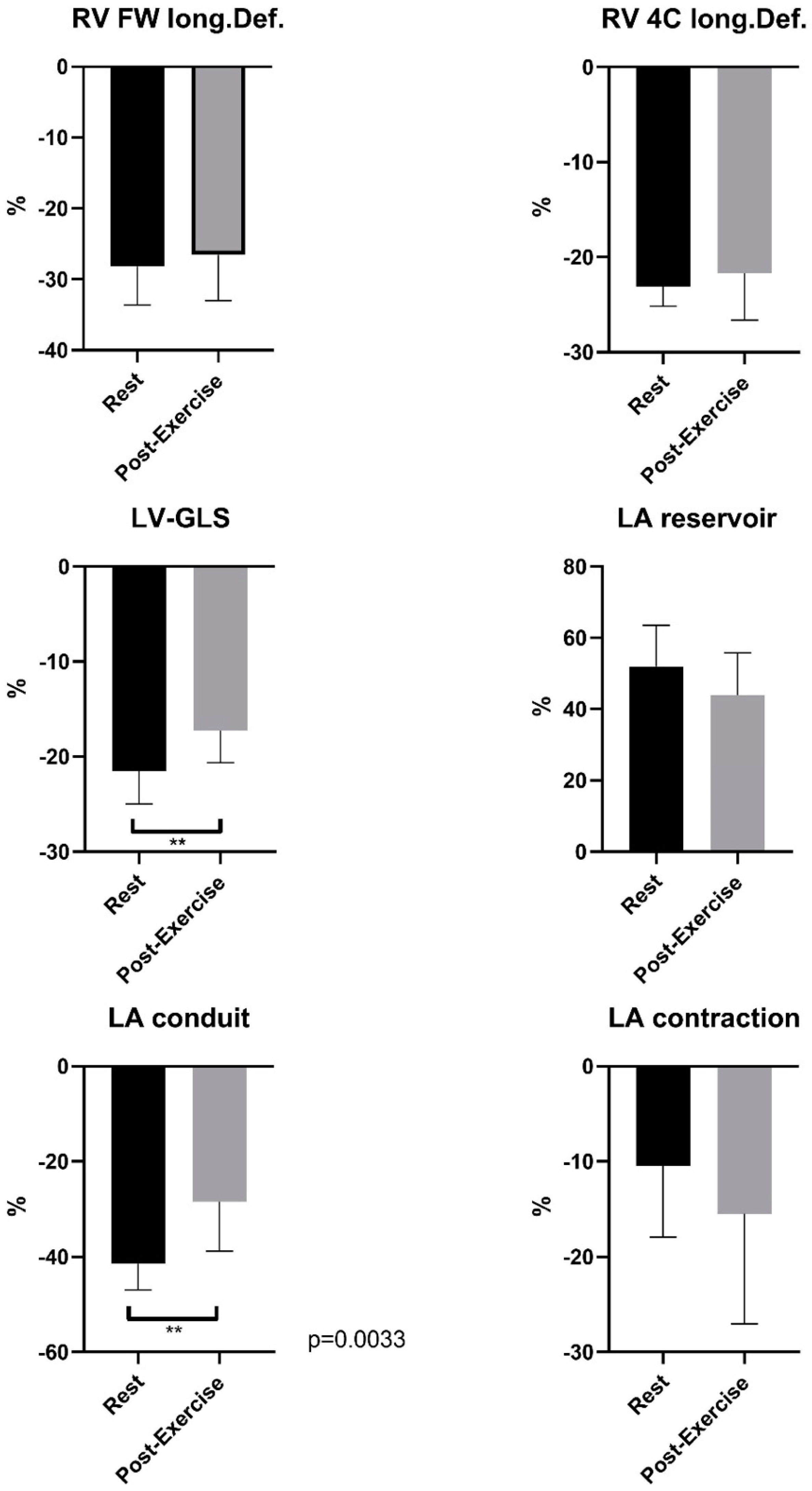
Figure 1. Speckle tracking analyses of world elite Ski-Mo professional–Comparison of mean LV-GLS, phasic LA strain and RV strain with significant differences between resting conditions and post-exercise. ** indicates p < 0.01.
Ijerph 19 13153 g001
Figure 2. Negative correlation between LV mass and LV mass Index and LV-GLS at rest in world elite Ski-Mo professionals.
Ijerph 19 13153 g002
Table 1. Baseline anthropometric Ski-Mo characteristics.
Ski-Mo
Male n = 8
Ski-Mo
Female n = 3
Age (years)20.5 ± 2.419 ± 0
Height (cm)177.8 ± 4.6164.0 ± 20.0
Weight (kg)63.6 ± 6.152.1 ± 5.9
BMI (kg/m2)20.1 ± 1.419.3 ± 0.4
resting blood pressure systolic/diastolic (mmHg)119 ± 6.1
85 ± 3.2
110 ± 5.2
71 ± 2.3
resting heart rate (bpm)41 ± 3.644 ± 2.5
BSA (body surface area m2)1.79 ± 0.11.79 ± 0.1
Data are presented as a median with standard deviation. Abbreviations: cm, centimeter; kg, kilogram; m2, square meter; bpm; beats per minute. Bold: anthropometric athlete’s characteristics.
Table 2. Baseline echocardiographic measurements (mean ± SD) of the Ski-mountaineering athletes and comparison to the stated control data of the position statement paper of the DGK (German Society of Cardiology, 2020).
Ski-Mo Male
n = 8
Ski-Mo Female
n = 3
Reference Value
Male
Reference Value
Female
LV edd (mm)47.13 ± 4.6443.67 ± 2.3142–5838–52
LV Mass Index (g/m2)71.38 ± 15.5067.33 ± 12.3549–11543–95
Relative wall Thickness RWT0.34 ± 0.050.37 ± 0.05
IVSd (mm)8.25 ± 1.287.33 ± 0.586–106–9
LVPWs (mm)8.00 ± 0.938.00 ± 1.006–106–9
E/A1.98 ± 0.321.90 ± 0.20
E/E′5.08 ± 2.185.53 ± 0.62
LAVI (mL/m2)28.13 ± 8.1736.00 ± 3.00
RA (cm2)15.00 ± 2.3914.00 ± 2.65
LV − EFrest (%)61.38 ± 4.1759.00 ± 1.0052–7254–72
LV − EFpost-stress (%)70.00 ± 2.8868.33 ± 0.58
Data are presented as a median with standard deviation. Abbreviations: LV edd, left ventricle enddiastolic size; LV, left ventricular; IVSd, interventricular septal wall thickness at diastole; LVPWd, left ventricular posterior wall thickness at diastole; E/A and E/E′, parameters for diastolic function of the left ventricle; LAVI, left atrial volume index; RA, right atrium; LV − EF, left ventricular systolic ejection fraction. Bold: echocardiographic parameters.
Publisher’s Note: MDPI stays neutral with regard to jurisdictional claims in published maps and institutional affiliations.