
Mucosal Melanoma of the Hard Palate: Surgical Treatment and Reconstruction

 ,
,  ,
,  , and
, and 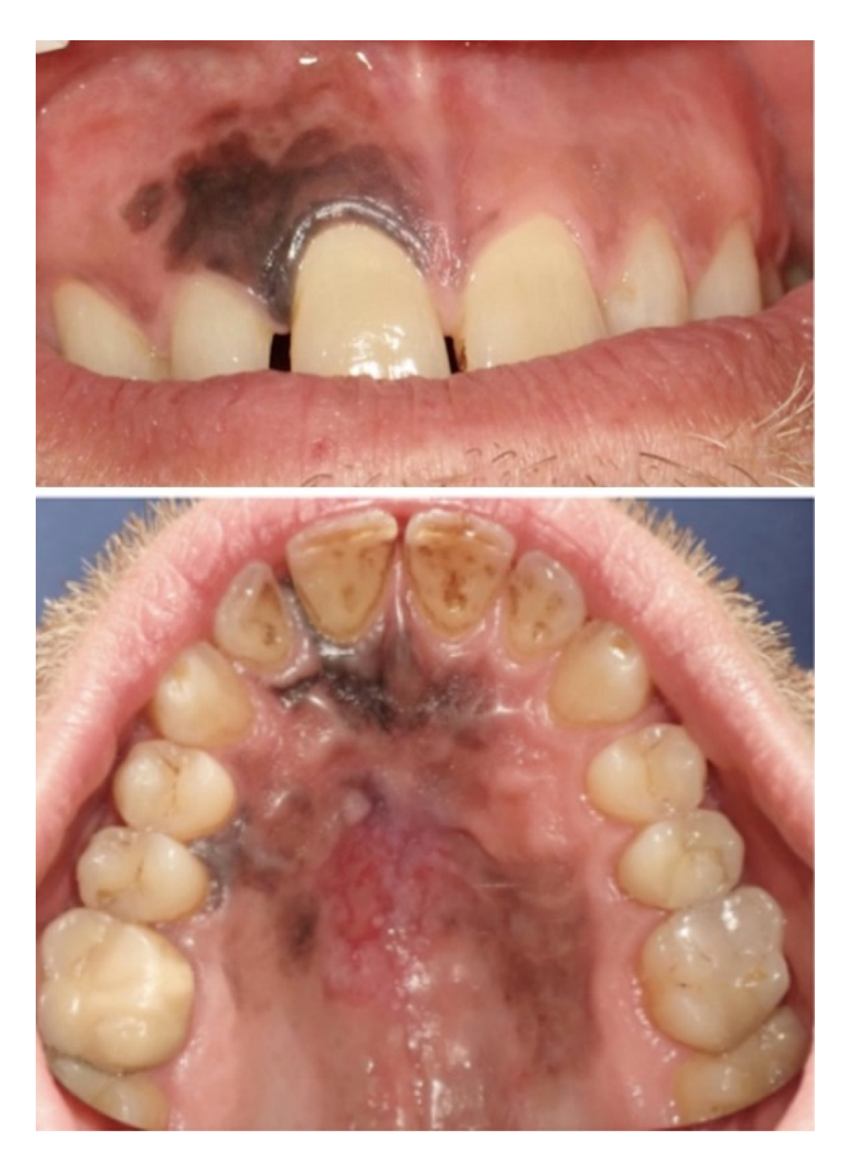
Abstract
1. Introduction
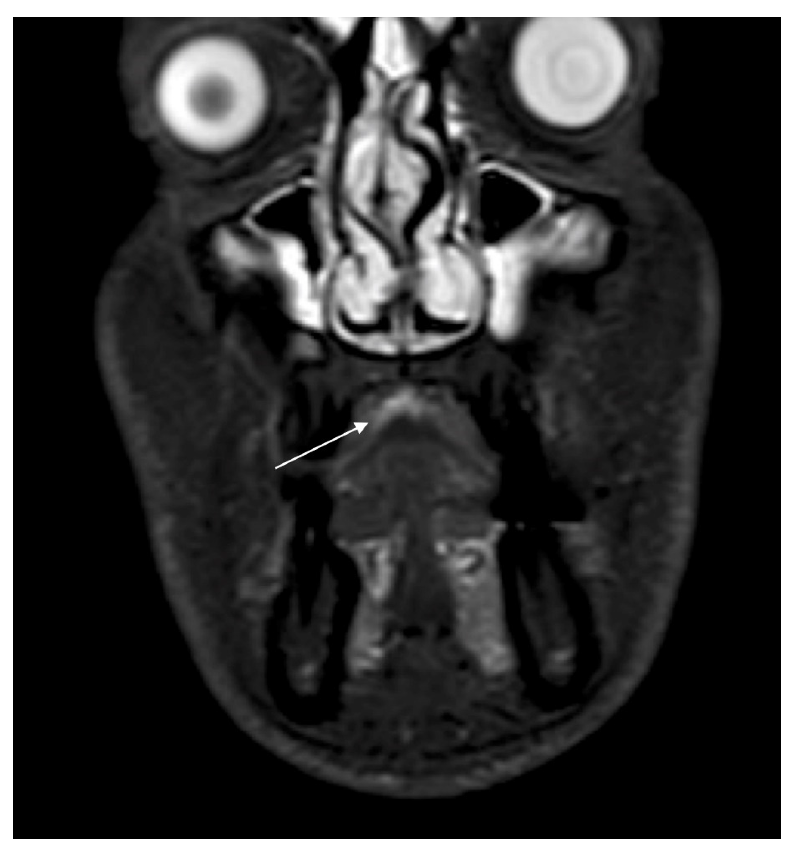
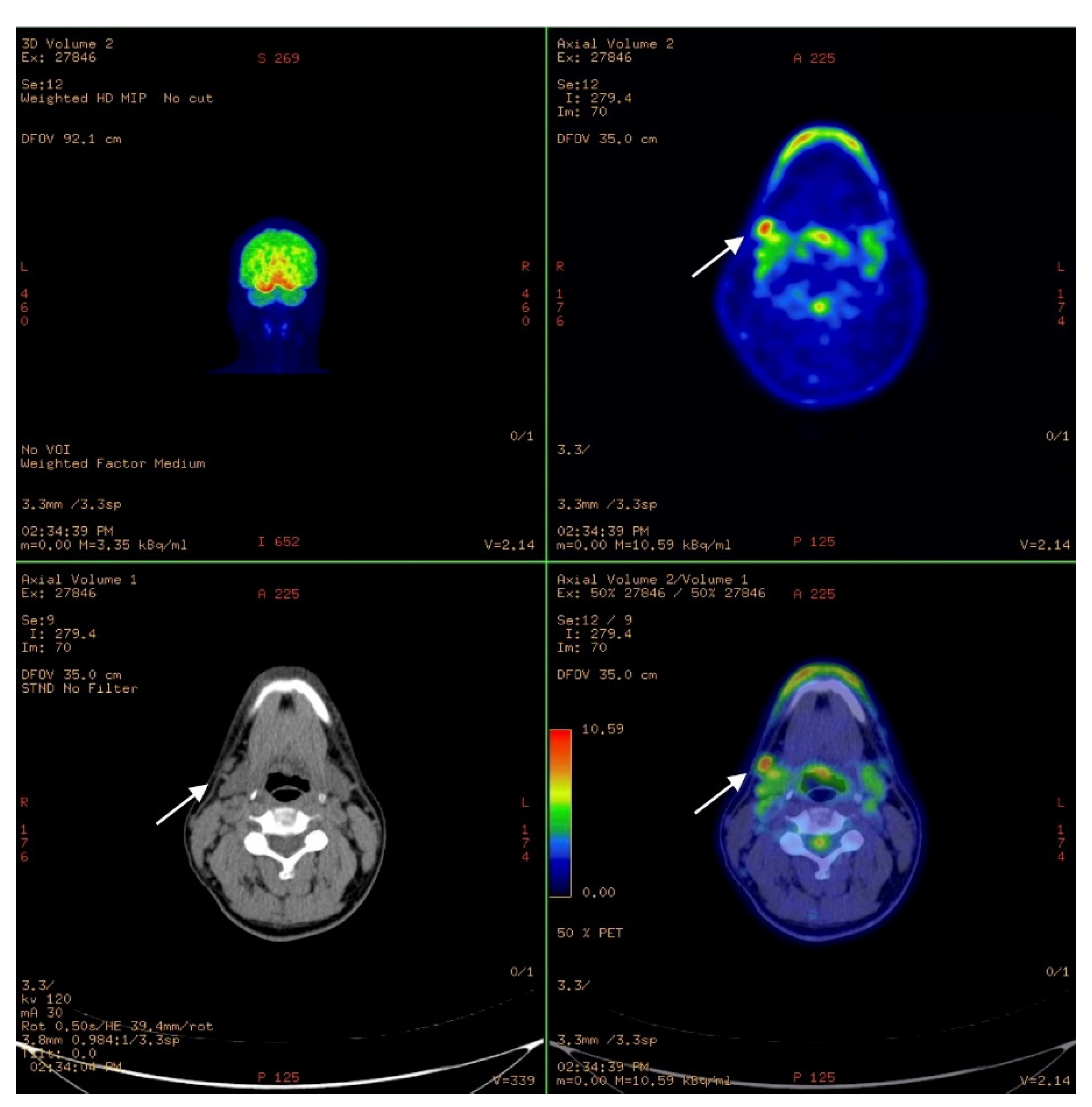
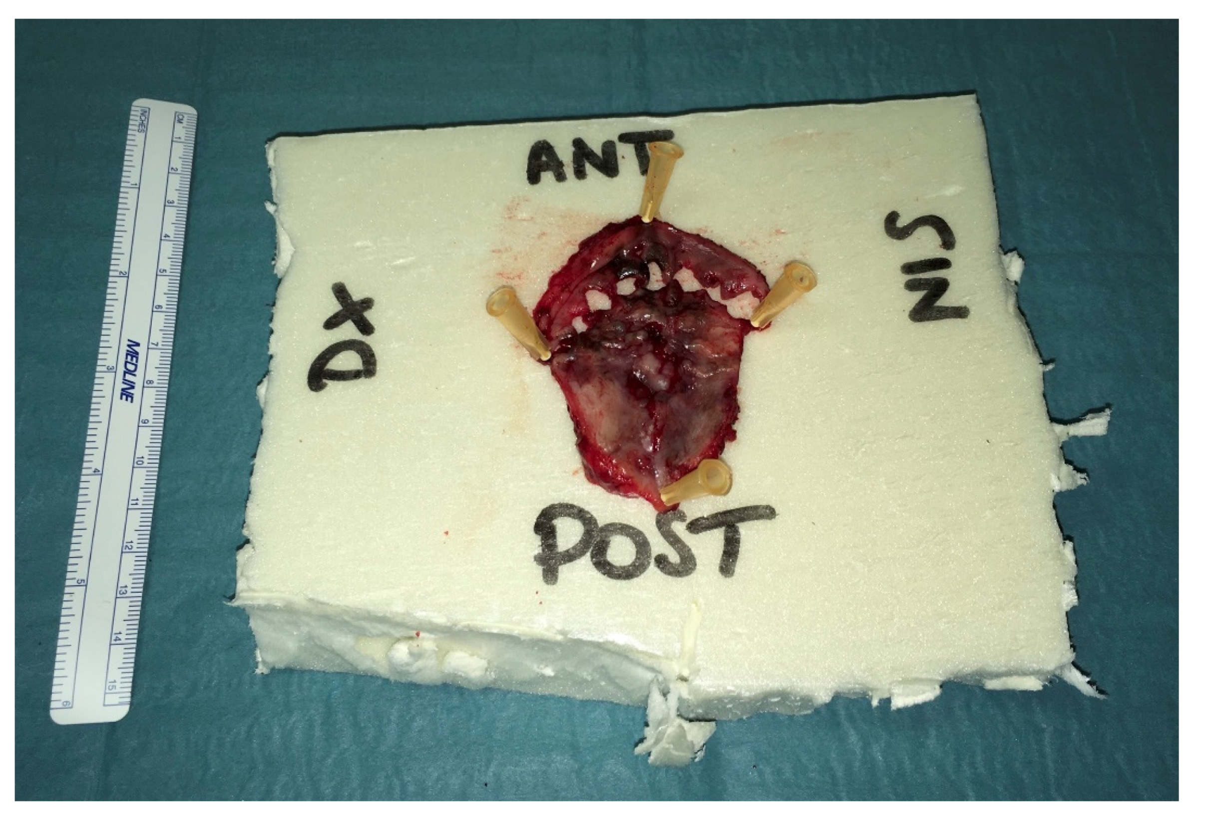
2. Case Report
3. Discussion
4. Conclusions
Author Contributions
Funding
Institutional Review Board Statement
Informed Consent Statement
Data Availability Statement
Conflicts of Interest
References
- Ascierto, P.A.; Accorona, R.; Botti, G.; Farina, D.; Fossati, P.; Gatta, G.; Gogas, H.; Lombardi, D.; Maroldi, R.; Nicolai, P.; et al. Mucosal melanoma of the head and neck. Crit. Rev. Oncol. 2017, 112, 136–152. [Google Scholar] [CrossRef] [PubMed]
- Chatzistefanou, I.; Kolokythas, A.; Vahtsevanos, K.; Antoniades, K. Primary mucosal melanoma of the oral cavity: Current therapy and future directions. Oral Surg. Oral Med. Oral Pathol. Oral Radiol. 2016, 122, 17–27. [Google Scholar] [CrossRef] [PubMed]
- Jarrom, D.; Paleri, V.; Kerawala, C.; Roques, T.; Bhide, S.; Newman, L.; Winter, S.C. Mucosal melanoma of the upper airways tract mucosal melanoma: A systematic review with meta-analyses of treatment. Head Neck 2017, 39, 819–825. [Google Scholar] [CrossRef]
- Gherlone, E.F.; Capparé, P.; Tecco, S.; Polizzi, E.; Pantaleo, G.; Gastaldi, G.; Grusovin, M.G. A Prospective Longitudinal Study on Implant Prosthetic Rehabilitation in Controlled HIV-Positive Patients with 1-Year Follow-Up: The Role of CD4+ Level, Smoking Habits, and Oral Hygiene. Clin. Implant. Dent. Relat. Res. 2015, 18, 955–964. [Google Scholar] [CrossRef] [PubMed]
- Della-Torre, E.; Campochiaro, C.; Cassione, E.B.; Albano, L.; Gerevini, S.; Bianchi-Marzoli, S.; Bozzolo, E.; Passerini, G.; Lanzillotta, M.; Terreni, M.; et al. Intrathecal rituximab for IgG4-related hypertrophic pachymeningitis. J. Neurol. Neurosurg. Psychiatry 2018, 89, 441–444. [Google Scholar] [CrossRef]
- Trimarchi, M.; Bellini, C.; Toma, S.; Bussi, M. Back-and-forth endoscopic septoplasty: Analysis of the technique and outcomes. Int. Forum Allergy Rhinol. 2011, 2, 40–44. [Google Scholar] [CrossRef] [PubMed]
- Biafora, M.; Bertazzoni, G.; Trimarchi, M. Maxillary Sinusitis Caused by Dental Implants Extending into the Maxillary Sinus and the Nasal Cavities. J. Prosthodont. 2014, 23, 227–231. [Google Scholar] [CrossRef]
- Mackintosh, A.J. The Antimicrobial Properties of Melanocytes, Melanosomes and Melanin and the Evolution of Black Skin. J. Theor. Biol. 2001, 211, 101–113. [Google Scholar] [CrossRef]
- Plonka, P.M.; Passeron, T.; Brenner, M.; Tobin, D.J.; Shibahara, S.; Thomas, A.; Slominski, A.; Kadekaro, A.L.; Hershkovitz, D.; Peters, E.M.J.; et al. What are melanocytesreallydoing all day long? Exp. Dermatol. 2009, 18, 799–819. [Google Scholar] [CrossRef]
- Juvekar, M.V.; Karle, R.R.; Wankhede, P.; Munde, A. Malignant melanoma of the oral cavity: Report of two cases. Contemp. Clin. Dent. 2014, 5, 227–230. [Google Scholar] [CrossRef]
- Axeix, T.; Hedin, C.A. Epidemiologic study of excessive oral melanin pigmentation with special reference to the influence of tobacco habits. Eur. J. Oral Sci. 1982, 90, 434–442. [Google Scholar] [CrossRef]
- Crespi, R.; Paolo, C.; Georgios, E.R.; Elisabetta, M.; Elisa, B.; Enrico, G. Corticocancellous porcine bone in the healing of human ex-traction sockets: Combining histomorphometry with osteoblast gene expression profiles in vivo. Int. J. Oral Maxillofac. Implants 2011, 26, 866–872. [Google Scholar] [PubMed]
- Lanzillotta, M.; Campochiaro, C.; Trimarchi, M.; Arrigoni, G.; Gerevini, S.; Milani, R.; Bozzolo, E.; Biafora, M.; Venturini, E.; Cicalese, M.P.; et al. Deconstructing IgG4-related disease involvement of midline structures: Comparison to common mimickers. Mod. Rheumatol. 2017, 27, 638–645. [Google Scholar] [CrossRef]
- Morassi, M.L.; Trimarchi, M.; Nicolai, P.; Gregorini, G.; Maroldi, R.; Specks, U.; Facchetti, F. Cocaine, ANCA, and Wegener’s granulomatosis. Pathologica 2001, 93, 581–583. [Google Scholar] [PubMed]
- Trimarchi, M.; Bondi, S.; Della Torre, E.; Terreni, M.; Bussi, M. Palate perforation differentiates cocaine-induced midline destructive lesions from granulomatosis with polyangiitis. Acta Otorhinolaryngol. Ital. 2017, 37, 281–285. [Google Scholar]
- Trimarchi, M.; Bellini, C.; Fabiano, B.; Gerevini, S.; Bussi, M. Multiple mucosal involvement in cicatricial pemphigoid. Acta Otorhinolaryngol. Ital. 2009, 29, 222–225. [Google Scholar] [PubMed]
- Sun, C.-Z.; Chen, Y.-F.; Jiang, Y.-E.; Hu, Z.-D.; Yang, A.-K.; Song, M. Treatment and prognosis of oral mucosal melanoma. Oral Oncol. 2012, 48, 647–652. [Google Scholar] [CrossRef]
- Penel, N.; Mallet, Y.; Mirabel, X.; Van, J.T.; Lefebvre, J.-L. Primary Mucosal Melanoma of Head and Neck: Prognostic Value of Clear Margins. Laryngoscope 2006, 116, 993–995. [Google Scholar] [CrossRef]
- Lyu, J.; Wu, Y.; Li, C.; Wang, R.; Song, H.; Ren, G.; Guo, W. Mutation scanning of BRAF, NRAS, KIT, and GNAQ/GNA11 in oral mucosal melanoma: A study of 57 cases. J. Oral Pathol. Med. 2016, 45, 295–301. [Google Scholar] [CrossRef]
- Tanaka, N.; Amagasa, T.; Iwaki, H.; Shioda, S.; Takeda, M.; Ohashi, K.; Reck, S.F. Oral malignant melanoma in Japan. Oral Surgery Oral Med. Oral Pathol. 1994, 78, 81–90. [Google Scholar] [CrossRef]
- Dupin, E.; Le Douarin, N.M. Development of melanocyte precursors from the vertebrate neural crest. Oncogene 2003, 22, 3016–3023. [Google Scholar] [CrossRef] [PubMed]
- Patel, S.G.; Prasad, M.L.; Escrig, M.; Singh, B.; Shaha, A.R.; Kraus, D.H.; Boyle, J.O.; Huvos, A.G.; Busam, K.; Shah, J.P. Primary mucosal malignant melanoma of the head and neck. Head Neck 2002, 24, 247–257. [Google Scholar] [CrossRef]
- Wu, Y.; Zhong, Y.; Li, C.; Song, H.; Guo, W.; Ren, G. Neck dissection for oral mucosal melanoma: Caution of nodular lesion. Oral Oncol. 2014, 50, 319–324. [Google Scholar] [CrossRef]
- Kim, S.S.; Han, M.H.; Kim, J.E.; Lee, C.H.; Chung, H.W.; Lee, J.S.; Chang, K.-H. Malignant melanoma of the sinonasal cavity: Explanation of magnetic resonance signal intensities with histopathologic characteristics. Am. J. Otolaryngol. 2000, 21, 366–378. [Google Scholar] [CrossRef] [PubMed]
- Bachar, G.; Loh, K.S.; O’Sullivan, B.; Goldstein, D.; Wood, S.; Brown, D.; Irish, J. Mucosal melanomas of the head and neck: The Princess Margaret Hospital experience. Head Neck 2008, 30, 1325–1331. [Google Scholar] [CrossRef] [PubMed]
- Michel, J.; Perret-Court, A.; Fakhry, N.; Braustein, D.; Monestier, S.; Richard, M.-A.; Grob, J.-J.; Giovanni, A.; Dessi, P. Sinonasal mucosal melanomas: The prognostic value of tumor classifications. Head Neck 2013, 36, 311–316. [Google Scholar] [CrossRef] [PubMed]
- Krengli, M.; Masini, L.; Kaanders, J.H.; Maingon, P.; Oei, S.B.; Zouhair, A.; Ozyar, E.; Roelandts, M.; Amichetti, M.; Bosset, M.; et al. Radiotherapy in the treatment of mucosal melanoma of the upper aerodigestive tract: Analysis of 74 cases. A Rare Cancer Network study. Int. J. Radiat. Oncol. 2006, 65, 751–759. [Google Scholar] [CrossRef] [PubMed]
- Medina, J.E.; Ferlito, A.; Pellitteri, P.K.; Shaha, A.R.; Khafif, A.; Devaney, K.O.; Fisher, S.R.; O’Brien, C.J.; Byers, R.M.; Robbins, K.T.; et al. Current management of mucosal melanoma of the head and neck. J. Surg. Oncol. 2003, 83, 116–122. [Google Scholar] [CrossRef]
- Musha, A.; Saitoh, J.-I.; Shirai, K.; Yokoo, S.; Ohno, T.; Nakano, T. Oral mucosal melanoma treated with carbon ion radiotherapy: A case report. J. Med Case Rep. 2016, 10, 284. [Google Scholar] [CrossRef]
- Grichnik, J.M. Melanoma, Nevogenesis, and Stem Cell Biology. J. Investig. Dermatol. 2008, 128, 2365–2380. [Google Scholar] [CrossRef]
- Keswell, D.; Davids, L.M.; Kidson, S.H. Migration of human melanocytes into keratinocyte monolayers in vitro. J. Dermatol. Sci. 2012, 66, 160–163. [Google Scholar] [CrossRef]
- Mihajlovic, M.; Vlajkovic, S.; Jovanovic, P.; Stefanovic, V. Primary mucosal melanomas: A comprehensive review. Int. J. Clin. Exp. Pathol. 2012, 5, 739–753. [Google Scholar]
- Crespi, R.; Capparè, P.; Gherlone, E. Sinus floor elevation by osteotome: Hand mallet versus electric mallet. A prospective clinical study. Int. J. Oral Maxillofac. Implant. 2012, 27, 1140–1150. [Google Scholar]
- Hu, S.; Fan, C.; Bs, B.P.; Rosenberg, J.D. Submental island flap vs free tissue transfer in oral cavity reconstruction: Systematic review and meta-analysis. Head Neck 2020, 42, 2155–2164. [Google Scholar] [CrossRef]
- Patel, U.A. The submental flap for head and neck reconstruction: Comparison of outcomes to the radial forearm free flap. Laryngoscope 2019, 130. [Google Scholar] [CrossRef]
- Jørgensen, M.G.; Tabatabaeifar, S.; Toyserkani, N.M.; Sørensen, J.A. Submental Island Flap versus Free Flap Reconstruction for Complex Head and Neck Defects. Otolaryngol. Neck Surg. 2019, 161, 946–953. [Google Scholar] [CrossRef] [PubMed]
- Martin, D.; Baudet, J.; Mondie, J.M.; Peri, G. The submental island skin flap. A surgical protocol. Prospects of use. Ann. Chir. Plast. Esthet. 1990, 35, 480–484. [Google Scholar] [PubMed]
- Sterne, G.; Januszkiewicz, J.; Hall, P.; Bardsley, A. The submental island flap. Br. J. Plast. Surg. 1996, 49, 85–89. [Google Scholar] [CrossRef]
- Martin, D.; Legaillard, P.; Bakhach, J.; Hu, W.; Baudet, J. Reverse flow YV pedicle extension: A method of doubling the arc of rotation of a flap under certain conditions. Ann. Chir. Plast. Esthétique 1994, 39, 403–414. [Google Scholar]
- Kudva, A.; Aramanadka, C.; D’Souza, C.; Lakshmi, R.; Karegowda, L.H. Anatomic Variation of Submental Artery: A Case of Submental Artery Coursing Through a Developmental Defect of Mylohyoid Muscle. J. Maxillofac. Oral Surg. 2020, 1–4. [Google Scholar] [CrossRef]
- Hayden, R.E.; Nagel, T.H.; Donald, C.B. Hybrid submental flaps for reconstruction in the head and neck: Part pedicled, part free. Laryngoscope 2013, 124, 637–641. [Google Scholar] [CrossRef]
- Gupta, V.; Cohan, D.M.; Arshad, H.; Kuriakose, M.A.; Hicks, W.L. Palatal reconstruction. Curr. Opin. Otolaryngol. Head Neck Surg. 2012, 20, 225–230. [Google Scholar] [CrossRef] [PubMed]
- Hammouda, Y.; Halily, S.; Oukessou, Y.; Rouadi, S.; Abada, R.; Roubal, M.; Mahtar, M. Malignant tumors of the hard palate: Report of 4 cases and review of the literature. Int. J. Surg. Case Rep. 2021, 78, 228–234. [Google Scholar] [CrossRef] [PubMed]
- Okay, D.J.; Genden, E.; Buchbinder, D.; Urken, M. Prosthodontic guidelines for surgical reconstruction of the maxilla: A classification system of defects. J. Prosthet. Dent. 2001, 86, 352–363. [Google Scholar] [CrossRef]
- Beier, U.S.; Salinas, T.; Puelacher, W. Resection of a primary oral malignant melanoma and rehabilitative management using nasolabial flap: A case report. Oral Maxillofac. Surg. 2011, 16, 141–145. [Google Scholar] [CrossRef] [PubMed]


Publisher’s Note: MDPI stays neutral with regard to jurisdictional claims in published maps and institutional affiliations. |
© 2021 by the authors. Licensee MDPI, Basel, Switzerland. This article is an open access article distributed under the terms and conditions of the Creative Commons Attribution (CC BY) license (http://creativecommons.org/licenses/by/4.0/).
Share and Cite
Bondi, S.; Vinciguerra, A.; Lissoni, A.; Rizzo, N.; Barbieri, D.; Indelicato, P.; Abati, S. Mucosal Melanoma of the Hard Palate: Surgical Treatment and Reconstruction. Int. J. Environ. Res. Public Health 2021, 18, 3341. https://doi.org/10.3390/ijerph18073341
Bondi S, Vinciguerra A, Lissoni A, Rizzo N, Barbieri D, Indelicato P, Abati S. Mucosal Melanoma of the Hard Palate: Surgical Treatment and Reconstruction. International Journal of Environmental Research and Public Health. 2021; 18(7):3341. https://doi.org/10.3390/ijerph18073341
Chicago/Turabian StyleBondi, Stefano, Alessandro Vinciguerra, Alessandra Lissoni, Nathalie Rizzo, Diego Barbieri, Pietro Indelicato, and Silvio Abati. 2021. "Mucosal Melanoma of the Hard Palate: Surgical Treatment and Reconstruction" International Journal of Environmental Research and Public Health 18, no. 7: 3341. https://doi.org/10.3390/ijerph18073341
APA StyleBondi, S., Vinciguerra, A., Lissoni, A., Rizzo, N., Barbieri, D., Indelicato, P., & Abati, S. (2021). Mucosal Melanoma of the Hard Palate: Surgical Treatment and Reconstruction. International Journal of Environmental Research and Public Health, 18(7), 3341. https://doi.org/10.3390/ijerph18073341