
Occurrence, Fate, Effects, and Risks of Dexamethasone: Ecological Implications Post-COVID-19

 , , , and
Abstract
:
1. Introduction
1.1. COVID-19 and Dexamethasone
1.2. Challenges of DEXA in Aquatic Systems
2. Occurrence of DEXA in the Environment
2.1. Sources of DEXA and Its Metabolites
2.2. Presence of DEXA in the Environment

| Country | Environmental Matrix | Analytical Methods | Concentrations (ng/L) | References |
|---|---|---|---|---|
| China | Surface water | SPE-LC-MS/MS; UPLC MS/MS | <0.02–22 | [71] |
| Malaysia | Surface water | ELISA kit | 0.02–8.74 | [65] |
| China | Urban river water | LC–MS/MS | 0–8.0 | [60] |
| China | Wastewater and river water | LC–ESI–MS/MS | 0.3–3.4 | [72] |
| Switzerland | Rivers and wastewaters | LC–MS/MS | 8–13; 15–1720 | [27] |
| The Netherlands | Various wastewater | LC–MS/MS | 0–90 | [67] |
| The Netherlands | Industry, hospital, STP, Surface water | CALUX reporter gene Bioassay | 243, 96, 11–38, 0.39–1.3 | [66] |
| China | River water | UHPLC–ESI–MS/MS | 0.3–3.5 | [61] |
| Germany | Treated wastewater, rivers, and streams. | LC–MS/MS | 0–0.4 | [73] |
| Singapore | Wastewater | UHPLC–(ESI)–MS/MS | 236 | [63] |
| Malaysia | Drinking water | ELISA kit | 0–0.37 | [65] |
| Malaysia | Drinking water | ELISA kit | 0.01–0.26 | [64] |
| Malaysia | Tap water | ELISA kit | 0.32 | [74] |
| South Africa | Effluent wastewater | LC-Orbitrap-HRMS | 0.92 | [75] |
| Malaysia | Drinking water | UHPLC–MS/MS | 0.36–2.11 | [76] |
| Hungary | River water | SPE–LC–MS/MS | 0.064–0.068 | [31] |
| France | Wastewater | SPE–LC–MS/MS | 7–15 | [77] |
| China | Sewage sludge | UHPLC–(ESI)–MS/MS | 0.02–0.81 | [78] |
| China | Surface water, Wastewater, and sludge | RRLC–MS/MS | 0–22 | [79] |
| China | Swine wastewater | RRLS–MS/MS | <1.27–260 | [80] |
| China | Surface water (downstream—1000 m) | RRLS–MS/MS | 37.8 | [80] |
| Italy | River and wastewater effluent | SPE–HPLC–ESI–MS/MS | 2–3 | [81] |
| Japan | Wastewater effluent | UHPLC–MS/MS | 0–1.3 | [82] |
| Malaysia | River water | LC–QTRAP MS/MS | 0–2.4 | [83] |
| China | Aquaculture water | UPLC–MS/MS | 0–41 | [84] |
| Portugal | Hospital effluents, WWTP influent and effluent | UPLC–QTrap–MS/MS | 0–352 | [70] |
| China | Surface watersheds | UPLC–ESI–TQD–MS/MS | 1.01–1.30 | [85] |
| USA | Surface water, groundwater and wastewater | (SPE) UHPLC–MS/MS | <1–94 | [76] |
| Spain | Hospital effluents | LC–(ESI)–MS/MS | 360 | [28] |
| Mexico | Hospital effluents | LC–MS/MS | 9.8 | [86] |
| Malaysia | Estuarine water | (SPE)-LC-MS/MS) | 1.0–1.51 | [29] |
| Spain | River waters, influent and effluent sewage | UHPLC–(ESI)–MS/MSa | <20 | [51] |
3. Ecotoxicological Effects of DEXA
3.1. Effects on Bacteria and Microbial Communities
3.2. Effects on Algae
3.3. Effects on Aquatic Invertebrates
3.4. Effects on Fish
4. Treatment Technologies of DEXA in Technical Systems
4.1. Conventional Removal of DEXA in Wastewater
4.2. Advanced Water Treatment Techniques
4.2.1. Physico-Chemical Treatments
4.2.2. Membrane Technology
4.2.3. Advanced Oxidation Processes
5. Environmental Risks of Detected DEXA
6. Concluding Remarks and Future Perspectives
Supplementary Materials
Author Contributions
Funding
Institutional Review Board Statement
Informed Consent Statement
Data Availability Statement
Conflicts of Interest
References
- Guan, W.; Ni, Z.; Hu, Y.; Liang, W.; Ou, C.; He, J.; Liu, L.; Shan, H.; Lei, C.; Hui, D.S.C.; et al. Clinical Characteristics of Coronavirus Disease 2019 in China. N. Engl. J. Med. 2020, 382, 1708–1720. [Google Scholar] [CrossRef] [PubMed]
- Zheng, Y.; Xiong, C.; Liu, Y.; Qian, X.; Tang, Y.; Liu, L.; Leung, E.L.-H.; Wang, M. Epidemiological and Clinical Characteristics Analysis of COVID-19 in the Surrounding Areas of Wuhan, Hubei Province in 2020. Pharmacol. Res. 2020, 157, 104821. [Google Scholar] [CrossRef] [PubMed]
- Zhu, N.; Zhang, D.; Wang, W.; Li, X.; Yang, B.; Song, J.; Zhao, X.; Huang, B.; Shi, W.; Lu, R.; et al. A Novel Coronavirus from Patients with Pneumonia in China, 2019. N. Engl. J. Med. 2020, 382, 727–733. [Google Scholar] [CrossRef]
- Rodriguez-Morales, A.J.; Bonilla-Aldana, D.K.; Balbin-Ramon, G.J.; Rabaan, A.A.; Sah, R.; Paniz-Mondolfi, A.; Pagliano, P.; Esposito, S. History Is Repeating Itself: Probable Zoonotic Spillover as the Cause of the 2019 Novel Coronavirus Epidemic. Infez Med. 2020, 28, 3–5. [Google Scholar] [PubMed]
- WHO. Director-General’s Opening Remarks at the Media Briefing on COVID-19—11 March 2020. Available online: https://www.who.int/director-general/speeches/detail/who-director-general-s-opening-remarks-at-the-media-briefing-on-covid-19---11-march-2020 (accessed on 14 August 2021).
- Dong, E.; Du, H.; Gardner, L. An Interactive Web-Based Dashboard to Track COVID-19 in Real Time. Lancet Infect. Dis. 2020, 20, 533–534. [Google Scholar] [CrossRef]
- Ksiazek, T.G.; Erdman, D.; Goldsmith, C.S.; Zaki, S.R.; Peret, T.; Emery, S.; Tong, S.; Urbani, C.; Comer, J.A.; Lim, W.; et al. A Novel Coronavirus Associated with Severe Acute Respiratory Syndrome. N. Engl. J. Med. 2003, 348, 1953–1966. [Google Scholar] [CrossRef] [PubMed]
- Zhang, W.; Zhao, Y.; Zhang, F.; Wang, Q.; Li, T.; Liu, Z.; Wang, J.; Qin, Y.; Zhang, X.; Yan, X.; et al. The Use of Anti-Inflammatory Drugs in the Treatment of People with Severe Coronavirus Disease 2019 (COVID-19): The Perspectives of Clinical Immunologists from China. Clin. Immunol. 2020, 214, 108393. [Google Scholar] [CrossRef] [PubMed]
- Wang, C.; Horby, P.W.; Hayden, F.G.; Gao, G.F. A Novel Coronavirus Outbreak of Global Health Concern. Lancet 2020, 395, 470–473. [Google Scholar] [CrossRef] [Green Version]
- Beigel, J.H.; Tomashek, K.M.; Dodd, L.E.; Mehta, A.K.; Zingman, B.S.; Kalil, A.C.; Hohmann, E.; Chu, H.Y.; Luetkemeyer, A.; Kline, S.; et al. Remdesivir for the Treatment of Covid-19—Final Report. N. Engl. J. Med. 2020, 383, 1813–1826. [Google Scholar] [CrossRef]
- Cao, B.; Wang, Y.; Wen, D.; Liu, W.; Wang, J.; Fan, G.; Ruan, L.; Song, B.; Cai, Y.; Wei, M.; et al. A Trial of Lopinavir-Ritonavir in Adults Hospitalized with Severe Covid-19. N. Engl. J. Med. 2020, 382, 1787–1799. [Google Scholar] [CrossRef]
- Costanzo, L.; Palumbo, F.P.; Ardita, G.; Antignani, P.L.; Arosio, E.; Failla, G. Coagulopathy, Thromboembolic Complications, and the Use of Heparin in COVID-19 Pneumonia. J. Vasc. Surg. Venous. Lymphat. Disord. 2020, 8, 711–716. [Google Scholar] [CrossRef]
- Choy, E.H.; de Benedetti, F.; Takeuchi, T.; Hashizume, M.; John, M.R.; Kishimoto, T. Translating IL-6 Biology into Effective Treatments. Nat. Rev. Rheumatol. 2020, 16, 335–345. [Google Scholar] [CrossRef] [PubMed] [Green Version]
- Hu, Z.; Lv, Y.; Xu, C.; Sun, W.; Chen, W.; Peng, Z.; Chen, C.; Cui, X.; Jiao, D.; Cheng, C.; et al. Clinical Use of Short-Course and Low-Dose Corticosteroids in Patients with Non-Severe COVID-19 During Pneumonia Progression. Front. Public Health 2020, 8, 355. [Google Scholar] [CrossRef] [PubMed]
- Wang, K.; Tan, F.; Zhou, R.; Liu, D.; Ni, Z.; Liu, J.; Luo, F. Therapeutic Response to Corticosteroids in a Critically Ill Patient with COVID-19. Medicine 2020, 99, e21597. [Google Scholar] [CrossRef]
- Shang, L.; Zhao, J.; Hu, Y.; Du, R.; Cao, B. On the Use of Corticosteroids for 2019-NCoV Pneumonia. Lancet 2020, 395, 683–684. [Google Scholar] [CrossRef] [Green Version]
- Group, T.R.C. Dexamethasone in Hospitalized Patients with COVID-19. N. Engl. J. Med. 2021, 384, 693–704. [Google Scholar] [CrossRef]
- Palumbo, A.; Chanan-Khan, A.; Weisel, K.; Nooka, A.K.; Masszi, T.; Beksac, M.; Spicka, I.; Hungria, V.; Munder, M.; Mateos, M.V.; et al. Daratumumab, Bortezomib, and Dexamethasone for Multiple Myeloma. N. Engl. J. Med. 2016, 375, 754–766. [Google Scholar] [CrossRef] [PubMed]
- Richardson, P.G.; Sonneveld, P.; Schuster, M.W.; Irwin, D.; Stadtmauer, E.A.; Facon, T.; Harousseau, J.-L.; Ben-Yehuda, D.; Lonial, S.; Goldschmidt, H.; et al. Bortezomib or High-Dose Dexamethasone for Relapsed Multiple Myeloma. N. Engl. J. Med. 2005, 352, 2487–2498. [Google Scholar] [CrossRef] [Green Version]
- Plint, A.C.; Johnson, D.W.; Patel, H.; Wiebe, N.; Correll, R.; Brant, R.; Mitton, C.; Gouin, S.; Bhatt, M.; Joubert, G.; et al. Epinephrine and Dexamethasone in Children with Bronchiolitis. N. Engl. J. Med. 2009, 360, 2079–2089. [Google Scholar] [CrossRef] [PubMed] [Green Version]
- Hughes, H.D.; Carroll, J.A.; Burdick Sanchez, N.C.; Roberts, S.L.; Broadway, P.R.; May, N.D.; Ballou, M.A.; Richeson, J.T. Effects of Dexamethasone Treatment and Respiratory Vaccination on Rectal Temperature, Complete Blood Count, and Functional Capacities of Neutrophils in Beef Steers1,2. J. Anim. Sci. 2017, 95, 1502–1511. [Google Scholar] [CrossRef]
- Divari, S.; de Lucia, F.; Berio, E.; Sereno, A.; Biolatti, B.; Cannizzo, F.T. Dexamethasone and Prednisolone Treatment in Beef Cattle: Influence on Glycogen Deposition and Gene Expression in the Liver. Domest. Anim. Endocrinol. 2020, 72, 106444. [Google Scholar] [CrossRef]
- Ward, S.A.; Kirkwood, R.N.; Plush, K.J. Administering Dexamethasone to Prepartum Sows: Effects on Sow and Piglet Performance. Livest. Sci. 2020, 239, 104171. [Google Scholar] [CrossRef]
- Oulton, R.L.; Kohn, T.; Cwiertny, D.M. Pharmaceuticals and Personal Care Products in Effluent Matrices: A Survey of Transformation and Removal during Wastewater Treatment and Implications for Wastewater Management. J. Environ. Monit. 2010, 12, 1956–1978. [Google Scholar] [CrossRef]
- Patel, M.; Kumar, R.; Kishor, K.; Mlsna, T.; Pittman, C.U.; Mohan, D. Pharmaceuticals of Emerging Concern in Aquatic Systems: Chemistry, Occurrence, Effects, and Removal Methods. Chem. Rev. 2019, 119, 3510–3673. [Google Scholar] [CrossRef] [Green Version]
- Bu, Q.; Cao, Y.; Yu, G.; He, X.; Zhang, H.; Sun, J.; Yun, M.; Cao, Z. Identifying Targets of Potential Concern by a Screening Level Ecological Risk Assessment of Human Use Pharmaceuticals in China. Chemosphere 2020, 246, 125818. [Google Scholar] [CrossRef]
- Ammann, A.A.; Macikova, P.; Groh, K.J.; Schirmer, K.; Suter, M.J.F. LC-MS/MS Determination of Potential Endocrine Disruptors of Cortico Signalling in Rivers and Wastewaters. Anal. Bioanal. Chem. 2014, 406, 7653–7665. [Google Scholar] [CrossRef]
- Cruz-Morató, C.; Lucas, D.; Llorca, M.; Rodriguez-Mozaz, S.; Gorga, M.; Petrovic, M.; Barceló, D.; Vicent, T.; Sarrà, M.; Marco-Urrea, E. Hospital Wastewater Treatment by Fungal Bioreactor: Removal Efficiency for Pharmaceuticals and Endocrine Disruptor Compounds. Sci. Total Environ. 2014, 493, 365–376. [Google Scholar] [CrossRef]
- Ismail, N.A.H.; Wee, S.Y.; Kamarulzaman, N.H.; Aris, A.Z. Quantification of Multi-Classes of Endocrine-Disrupting Compounds in Estuarine Water. Environ. Pollut. 2019, 249, 1019–1028. [Google Scholar] [CrossRef]
- Ismail, N.A.H.; Wee, S.Y.; Haron, D.E.M.; Kamarulzaman, N.H.; Aris, A.Z. Occurrence of Endocrine Disrupting Compounds in Mariculture Sediment of Pulau Kukup, Johor, Malaysia. Mar. Pollut. Bull. 2020, 150, 110735. [Google Scholar] [CrossRef] [PubMed]
- Tölgyesi, Á.; Verebey, Z.; Sharma, V.K.; Kovacsics, L.; Fekete, J. Simultaneous Determination of Corticosteroids, Androgens, and Progesterone in River Water by Liquid Chromatography-Tandem Mass Spectrometry. Chemosphere 2010, 78, 972–979. [Google Scholar] [CrossRef]
- Wee, S.Y.; Aris, A.Z.; Yusoff, F.M.; Praveena, S.M. Occurrence and Risk Assessment of Multiclass Endocrine Disrupting Compounds in an Urban Tropical River and a Proposed Risk Management and Monitoring Framework. Sci. Total Environ. 2019, 671, 431–442. [Google Scholar] [CrossRef] [PubMed]
- Zhang, H.-C.; Yu, X.; Yang, W.; Peng, J.; Xu, T.; Yin, D.-Q.; Hu, X. MCX Based Solid Phase Extraction Combined with Liquid Chromatography Tandem Mass Spectrometry for the Simultaneous Determination of 31 Endocrine-Disrupting Compounds in Surface Water of Shanghai. J. Chromatogr. B. 2011, 879, 2998–3004. [Google Scholar] [CrossRef]
- Johnson, T.L.; Tomanek, L.; Peterson, D.G. A Proteomic Analysis of the Effect of Growth Hormone on Mammary Alveolar Cell-T (MAC-T) Cells in the Presence of Lactogenic Hormones. Domest. Anim. Endocrinol. 2013, 44, 26–35. [Google Scholar] [CrossRef]
- Solinas, C.; Perra, L.; Aiello, M.; Migliori, E.; Petrosillo, N. A Critical Evaluation of Glucocorticoids in the Management of Severe COVID-19. Cytokine Growth Factor Rev. 2020, 54, 8–23. [Google Scholar] [CrossRef]
- Keller, M.J.; Kitsis, E.A.; Arora, S.; Chen, J.-T.; Agarwal, S.; Ross, M.J.; Tomer, Y.; Southern, W. Effect of Systemic Glucocorticoids on Mortality or Mechanical Ventilation in Patients With COVID-19. J. Hosp. Med. 2020, 15, 489–493. [Google Scholar] [CrossRef] [PubMed]
- Han, Y.; Jiang, M.; Xia, D.; He, L.; Lv, X.; Liao, X.; Meng, J. COVID-19 in a Patient with Long-Term Use of Glucocorticoids: A Study of a Familial Cluster. Clin. Immunol. 2020, 214, 108413. [Google Scholar] [CrossRef]
- Lu, S.; Zhou, Q.; Huang, L.; Shi, Q.; Zhao, S.; Wang, Z.; Li, W.; Tang, Y.; Ma, Y.; Luo, X.; et al. Effectiveness and Safety of Glucocorticoids to Treat COVID-19: A Rapid Review and Meta-Analysis. Ann. Transl. Med. 2020, 8, 10. [Google Scholar] [CrossRef]
- Stavreva, D.A.; George, A.A.; Klausmeyer, P.; Varticovski, L.; Sack, D.; Voss, T.C.; Schiltz, R.L.; Blazer, V.S.; Iwanowicz, L.R.; Hager, G.L. Prevalent Glucocorticoid and Androgen Activity in US Water Sources. Sci. Rep. 2012, 2, 937. [Google Scholar] [CrossRef] [Green Version]
- Deblonde, T.; Cossu-Leguille, C.; Hartemann, P. Emerging Pollutants in Wastewater: A Review of the Literature. Int. J. Hyg. Environ. Health 2011, 214, 442–448. [Google Scholar] [CrossRef]
- Runnalls, T.J.; Margiotta-Casaluci, L.; Kugathas, S.; Sumpter, J.P. Pharmaceuticals in the Aquatic Environment: Steroids and Anti-Steroids as High Priorities for Research. Hum. Ecol. Risk Assess. 2010, 16, 1318–1338. [Google Scholar] [CrossRef]
- Plotkin, B.J.; Roose, R.J.; Erikson, Q.; Viselli, S.M. Effect of Androgens and Glucocorticoids on Microbial Growth and Antimicrobial Susceptibility. Curr. Microbiol. 2003, 47, 514–520. [Google Scholar] [CrossRef]
- Kawabata, K.; Sugihara, K.; Sanoh, S.; Kitamura, S.; Ohta, S. Photodegradation of Pharmaceuticals in the Aquatic Environment by Sunlight and UV-A, -B and -C Irradiation. Toxicol. Sci. 2013, 38, 215–223. [Google Scholar] [CrossRef] [Green Version]
- DellaGreca, M.; Fiorentino, A.; Isidori, M.; Lavorgna, M.; Previtera, L.; Rubino, M.; Temussi, F. Toxicity of Prednisolone, Dexamethasone and Their Photochemical Derivatives on Aquatic Organisms. Chemosphere 2004, 54, 629–637. [Google Scholar] [CrossRef] [PubMed]
- LaLone, C.A.; Villeneuve, D.L.; Olmstead, A.W.; Medlock, E.K.; Kahl, M.D.; Jensen, K.M.; Durhan, E.J.; Makynen, E.A.; Blanksma, C.A.; Cavallin, J.E.; et al. Effects of a Glucocorticoid Receptor Agonist, Dexamethasone, on Fathead Minnow Reproduction, Growth, and Development. Environ. Toxicol. Chem. 2012, 31, 611–622. [Google Scholar] [CrossRef]
- Chen, Q.; Jia, A.; Snyder, S.A.; Gong, Z.; Lam, S.H. Glucocorticoid Activity Detected by In Vivo Zebrafish Assay and In Vitro Glucocorticoid Receptor Bioassay at Environmental Relevant Concentrations. Chemosphere 2016, 144, 1162–1169. [Google Scholar] [CrossRef]
- Jerez-Cepa, I.; Gorissen, M.; Mancera, J.M.; Ruiz-Jarabo, I. What Can We Learn from Glucocorticoid Administration in Fish? Effects of Cortisol and Dexamethasone on Intermediary Metabolism of Gilthead Seabream (Sparus aurata L.). Comp. Biochem. Physiol. Part A Mol. Integr. Physiol. 2019, 231, 1–10. [Google Scholar] [CrossRef]
- Guo, Z.; Guo, A.; Guo, Q.; Rui, M.; Zhao, Y.; Zhang, H.; Zhu, S. Decomposition of Dexamethasone by Gamma Irradiation: Kinetics, Degradation Mechanisms and Impact on Algae Growth. Chem. Eng. J. 2017, 307, 722–728. [Google Scholar] [CrossRef]
- Kugathas, S.; Sumpter, J.P. Synthetic Glucocorticoids in the Environment: First Results on Their Potential Impacts on Fish. Environ. Sci. Technol. 2011, 45, 2377–2383. [Google Scholar] [CrossRef]
- Sulaiman, S.; Khamis, M.; Nir, S.; Lelario, F.; Scrano, L.; Bufo, S.A.; Karaman, R. Stability and Removal of Dexamethasone Sodium Phosphate from Wastewater Using Modified Clays. Environ. Technol. 2014, 35, 1945–1955. [Google Scholar] [CrossRef] [PubMed]
- Herrero, P.; Borrull, F.; Pocurull, E.; Marcé, R.M. Determination of Glucocorticoids in Sewage and River Waters by Ultra-High Performance Liquid Chromatography–Tandem Mass Spectrometry. J. Chromatogr. A. 2012, 1224, 19–26. [Google Scholar] [CrossRef]
- Arsand, D.R.; Kümmerer, K.; Martins, A.F. Removal of Dexamethasone from Aqueous Solution and Hospital Wastewater by Electrocoagulation. Sci. Total Environ. 2013, 443, 351–357. [Google Scholar] [CrossRef]
- Tomlinson, E.S.; Maggs, J.L.; Park, B.K.; Back, D.J. Dexamethasone Metabolism in Vitro: Species Differences. J. Steroid Biochem. Mol. Biol. 1997, 62, 345–352. [Google Scholar] [CrossRef]
- Gentile, D.M.; Tomlinson, E.S.; Maggs, J.L.; Park, B.K.; Back, D.J. Dexamethasone Metabolism by Human Liver in Vitro. Metabolite Identification and Inhibition of 6-Hydroxylation. J. Pharmacol. Exp. Ther. 1996, 277, 105–112. [Google Scholar] [PubMed]
- Pervaiz, I.; Ahmad, S.; Mukhtar, M.F.; Arshad, A.; Imran, M.; Mahmood, W. Microbial Biotransformation of Dexamethasone by Bacillus Subtilis (ATCC 6051). Pharm. Chem. J. 2015, 49, 405–408. [Google Scholar] [CrossRef]
- Dumasia, M.C.; Houghton, E.; Moss, M.S.; Chakraborty, J.; Marks, V. The Biotransformation and Urinary Excretion of Dexamethasone in Equine Male Castrates. J. Steroid Biochem. Mol. Biol. 1986, 25, 547–553. [Google Scholar] [CrossRef]
- Sirés, I.; Brillas, E. Remediation of Water Pollution Caused by Pharmaceutical Residues Based on Electrochemical Separation and Degradation Technologies: A Review. Environ. Int. 2012, 40, 212–229. [Google Scholar] [CrossRef]
- Calza, P.; Pelizzetti, E.; Brussino, M.; Baiocchi, C. Ion Trap Tandem Mass Spectrometry Study of Dexamethasone Transformation Products on Light Activated TiO2 Surface. J. Am. Soc. Mass Spectrom. 2001, 12, 1286–1295. [Google Scholar] [CrossRef] [Green Version]
- Albini, A.; Fasani, E.; Albini, A.; Fasani, E. Photochemistry of drugs: An overview and practical problems. Drugs: Photochem. Photostability 1998, 225, 1–73. [Google Scholar]
- Chang, H.; Wan, Y.; Hu, J. Determination and Source Apportionment of Five Classes of Steroid Hormones in Urban Rivers. Environ. Sci. Technol. 2009, 43, 7691–7698. [Google Scholar] [CrossRef] [PubMed]
- Gong, J.; Lin, C.; Xiong, X.; Chen, D.; Chen, Y.; Zhou, Y.; Wu, C.; Du, Y. Occurrence, Distribution, and Potential Risks of Environmental Corticosteroids in Surface Waters from the Pearl River Delta, South China. Environ. Pollut. 2019, 251, 102–109. [Google Scholar] [CrossRef] [PubMed]
- Zhang, J.; Luo, J.; Chang, H. Aerobic Biodegradation of Four Groups of Steroid Hormones in Activated Sludge. J. Chem. 2020, 2020, e1309183. [Google Scholar] [CrossRef]
- Goh, S.X.L.; Goh, H.A.; Lee, H.K. Automation of Ionic Liquid Enhanced Membrane Bag-Assisted-Liquid-Phase Microextraction with Liquid Chromatography-Tandem Mass Spectrometry for Determination of Glucocorticoids in Water. Anal. Chim. Acta 2018, 1035, 77–86. [Google Scholar] [CrossRef] [PubMed]
- Mohd Nasir, F.A.; Praveena, S.M.; Aris, A.Z. Public Awareness Level and Occurrence of Pharmaceutical Residues in Drinking Water with Potential Health Risk: A Study from Kajang (Malaysia). Ecotoxicol. Environ. Saf. 2019, 185, 109681. [Google Scholar] [CrossRef] [PubMed]
- Praveena, S.M.; Shaifuddin, S.N.M.; Sukiman, S.; Nasir, F.A.M.; Hanafi, Z.; Kamarudin, N.; Ismail, T.H.T.; Aris, A.Z. Pharmaceuticals Residues in Selected Tropical Surface Water Bodies from Selangor (Malaysia): Occurrence and Potential Risk Assessments. Sci. Total Environ. 2018, 642, 230–240. [Google Scholar] [CrossRef]
- Van der Linden, S.C.; Heringa, M.B.; Man, H.-Y.; Sonneveld, E.; Puijker, L.M.; Brouwer, A.; van der Burg, B. Detection of Multiple Hormonal Activities in Wastewater Effluents and Surface Water, Using a Panel of Steroid Receptor CALUX Bioassays. Environ. Sci. Technol. 2008, 42, 5814–5820. [Google Scholar] [CrossRef] [PubMed]
- Schriks, M.; van Leerdam, J.A.; van der Linden, S.C.; van der Burg, B.; van Wezel, A.P.; de Voogt, P. High-Resolution Mass Spectrometric Identification and Quantification of Glucocorticoid Compounds in Various Wastewaters in The Netherlands. Environ. Sci. Technol. 2010, 44, 4766–4774. [Google Scholar] [CrossRef] [Green Version]
- Jha, R.R.; Singh, N.; Kumari, R.; Patel, D.K. Ultrasound-Assisted Emulsification Microextraction Based on a Solidified Floating Organic Droplet for the Rapid Determination of 19 Antibiotics as Environmental Pollutants in Hospital Drainage and Gomti River Water. J. Sep. Sci. 2017, 40, 2694–2702. [Google Scholar] [CrossRef]
- Kovalova, L.; Siegrist, H.; Singer, H.; Wittmer, A.; McArdell, C.S. Hospital Wastewater Treatment by Membrane Bioreactor: Performance and Efficiency for Organic Micropollutant Elimination. Environ. Sci. Technol. 2012, 46, 1536–1545. [Google Scholar] [CrossRef] [Green Version]
- Santos, L.H.M.L.M.; Gros, M.; Rodriguez-Mozaz, S.; Delerue-Matos, C.; Pena, A.; Barceló, D.; Montenegro, M.C.B.S.M. Contribution of Hospital Effluents to the Load of Pharmaceuticals in Urban Wastewaters: Identification of Ecologically Relevant Pharmaceuticals. Sci. Total Environ. 2013, 461–462, 302–316. [Google Scholar] [CrossRef] [PubMed] [Green Version]
- Shen, X.; Chang, H.; Sun, Y.; Wan, Y. Determination and Occurrence of Natural and Synthetic Glucocorticoids in Surface Waters. Environ. Int. 2020, 134, 105278. [Google Scholar] [CrossRef]
- Chang, H.; Hu, J.; Shao, B. Occurrence of Natural and Synthetic Glucocorticoids in Sewage Treatment Plants and Receiving River Waters. Environ. Sci. Technol. 2007, 41, 3462–3468. [Google Scholar] [CrossRef] [PubMed]
- Weizel, A.; Schlüsener, M.P.; Dierkes, G.; Ternes, T.A. Occurrence of Glucocorticoids, Mineralocorticoids, and Progestogens in Various Treated Wastewater, Rivers, and Streams. Environ. Sci. Technol. 2018, 52, 5296–5307. [Google Scholar] [CrossRef]
- Praveena, S.M.; Mohd Rashid, M.Z.; Mohd Nasir, F.A.; Wee, S.Y.; Aris, A.Z. Occurrence, Human Health Risks, and Public Awareness Level of Pharmaceuticals in Tap Water from Putrajaya (Malaysia). Expo. Health 2020, 13, 93–104. [Google Scholar] [CrossRef]
- Mhuka, V.; Dube, S.; Nindi, M.M. Occurrence of Pharmaceutical and Personal Care Products (PPCPs) in Wastewater and Receiving Waters in South Africa Using LC-OrbitrapTM MS. Emerg. Contam. 2020, 6, 250–258. [Google Scholar] [CrossRef]
- Anumol, T.; Merel, S.; Clarke, B.O.; Snyder, S.A. Ultra High Performance Liquid Chromatography Tandem Mass Spectrometry for Rapid Analysis of Trace Organic Contaminants in Water. Chem. Cent. J. 2013, 7, 104. [Google Scholar] [CrossRef] [Green Version]
- Piram, A.; Salvador, A.; Gauvrit, J.-Y.; Lanteri, P.; Faure, R. Development and Optimisation of a Single Extraction Procedure for the LC/MS/MS Analysis of Two Pharmaceutical Classes Residues in Sewage Treatment Plant. Talanta 2008, 74, 1463–1475. [Google Scholar] [CrossRef] [PubMed]
- Fan, Z.; Wu, S.; Chang, H.; Hu, J. Behaviors of Glucocorticoids, Androgens and Progestogens in a Municipal Sewage Treatment Plant: Comparison to Estrogens. Environ. Sci. Technol. 2011, 45, 2725–2733. [Google Scholar] [CrossRef] [PubMed]
- Liu, S.; Ying, G.-G.; Zhao, J.-L.; Chen, F.; Yang, B.; Zhou, L.-J.; Lai, H. Trace Analysis of 28 Steroids in Surface Water, Wastewater and Sludge Samples by Rapid Resolution Liquid Chromatography-Electrospray Ionization Tandem Mass Spectrometry. J. Chromatogr. A 2011, 1218, 1367–1378. [Google Scholar] [CrossRef]
- Liu, S.; Ying, G.-G.; Zhou, L.-J.; Zhang, R.-Q.; Chen, Z.-F.; Lai, H.-J. Steroids in a Typical Swine Farm and Their Release into the Environment. Water Res. 2012, 46, 3754–3768. [Google Scholar] [CrossRef] [PubMed]
- Merlo, F.; Speltini, A.; Maraschi, F.; Sturini, M.; Profumo, A. HPLC-MS/MS Multiclass Determination of Steroid Hormones in Environmental Waters after Preconcentration on the Carbonaceous Sorbent HA-C@silica. Arab. J. Chem. 2020, 13, 4673–4680. [Google Scholar] [CrossRef]
- Isobe, T.; Sato, K.; Joon-Woo, K.; Tanabe, S.; Suzuki, G.; Nakayama, K. Determination of Natural and Synthetic Glucocorticoids in Effluent of Sewage Treatment Plants Using Ultrahigh Performance Liquid Chromatography-Tandem Mass Spectrometry. Environ. Sci. Pollut. Res. 2015, 22, 14127–14135. [Google Scholar] [CrossRef]
- Omar, T.F.T.; Aris, A.Z.; Yusoff, F.M.; Mustafa, S. Risk Assessment of Pharmaceutically Active Compounds (PhACs) in the Klang River Estuary, Malaysia. Environ. Geochem. Health 2019, 41, 211–223. [Google Scholar] [CrossRef] [PubMed]
- Gao, G.; Li, S.; Li, S.; Wang, Y.; Zhao, P.; Zhang, X.; Hou, X. A Combination of Computational-Experimental Study on Metal-Organic Frameworks MIL-53(Al) as Sorbent for Simultaneous Determination of Estrogens and Glucocorticoids in Water and Urine Samples by Dispersive Micro-Solid-Phase Extraction Coupled to UPLC-MS/MS. Talanta 2018, 180, 358–367. [Google Scholar] [CrossRef]
- Xu, M.; Huang, H.; Li, N.; Li, F.; Wang, D.; Luo, Q. Occurrence and Ecological Risk of Pharmaceuticals and Personal Care Products (PPCPs) and Pesticides in Typical Surface Watersheds, China. Ecotoxicol. Environ. Saf. 2019, 175, 289–298. [Google Scholar] [CrossRef]
- Tenorio-Chávez, P.; Cerro-López, M.; Castro-Pastrana, L.I.; Ramírez-Rodrigues, M.M.; Orozco-Hernández, J.M.; Gómez-Oliván, L.M. Effects of Effluent from a Hospital in Mexico on the Embryonic Development of Zebrafish, Danio Rerio. Sci. Total Environ. 2020, 727, 138716. [Google Scholar] [CrossRef] [PubMed]
- Bal, N.; Kumar, A.; Du, J.; Nugegoda, D. Multigenerational Effects of Two Glucocorticoids (Prednisolone and Dexamethasone) on Life-History Parameters of Crustacean Ceriodaphnia Dubia (Cladocera). Environ. Pollut. 2017, 225, 569–578. [Google Scholar] [CrossRef] [PubMed]
- Li, X.; Ma, M.; Rene, E.R.; Ma, W.; Zhang, P. Changes in Microbial Communities during the Removal of Natural and Synthetic Glucocorticoids in Three Types of River-Based Aquifer Media. Environ. Sci. Pollut. Res. 2019, 26, 33953–33962. [Google Scholar] [CrossRef] [PubMed]
- Dubey, D.; Dutta, V. Nutrient enrichment in lake ecosystem and its effects on algae and macrophytes. In Environmental Concerns and Sustainable Development: Biodiversity, Soil and Waste Management; Shukla, V., Kumar, N., Eds.; Springer: Berlin/Heidelberg, Germany, 2020; Volume 2, pp. 81–126. ISBN 9789811363580. [Google Scholar]
- Silva, S.R.; Barbosa, F.A.R.; Mol, M.P.G.; Magalhães, S.M.S. Toxicity for Aquatic Organisms of Antiretroviral Tenofovir Disoproxil. J. Environ. Prot. Sci. 2019, 10, 1565. [Google Scholar] [CrossRef] [Green Version]
- Inaba, H.; Pui, C.-H. Glucocorticoid Use in Acute Lymphoblastic Leukaemia. Lancet Oncol. 2010, 11, 1096–1106. [Google Scholar] [CrossRef] [Green Version]
- Minguez, L.; Ballandonne, C.; Rakotomalala, C.; Dubreule, C.; Kientz-Bouchart, V.; Halm-Lemeille, M.-P. Transgenerational Effects of Two Antidepressants (Sertraline and Venlafaxine) on Daphnia Magna Life History Traits. Environ. Sci. Technol. 2015, 49, 1148–1155. [Google Scholar] [CrossRef]
- Pierce, A.L.; Dickey, J.T.; Felli, L.; Swanson, P.; Dickhoff, W.W. Metabolic Hormones Regulate Basal and Growth Hormone-Dependent Igf2 MRNA Level in Primary Cultured Coho Salmon Hepatocytes: Effects of Insulin, Glucagon, Dexamethasone, and Triiodothyronine. J. Endocrinol. 2010, 204, 331–339. [Google Scholar] [CrossRef] [Green Version]
- Lutton, B.V.; Callard, I.P. Influence of Reproductive Activity, Sex Steroids, and Seasonality on Epigonal Organ Cellular Proliferation in the Skate (Leucoraja erinacea). Gen. Comp. Endocrinol. 2008, 155, 116–125. [Google Scholar] [CrossRef]
- Li, D.; Yang, X.-L.; Zhang, S.-J.; Lin, M.; Yu, W.-J.; Hu, K. Effects of Mammalian CYP3A Inducers on CYP3A-Related Enzyme Activities in Grass Carp (Ctenopharyngodon idellus): Possible Implications for the Establishment of a Fish CYP3A Induction Model. Comp. Biochem. Physiol. Part C 2008, 147, 17–29. [Google Scholar] [CrossRef]
- Guiloski, I.C.; Ribas, J.L.C.; da Pereira, L.S.; Neves, A.P.P.; Silva de Assis, H.C. Effects of Trophic Exposure to Dexamethasone and Diclofenac in Freshwater Fish. Ecotoxicol. Environ. Saf. 2015, 114, 204–211. [Google Scholar] [CrossRef]
- Milla, S.; Wang, N.; Mandiki, S.N.M.; Kestemont, P. Corticosteroids: Friends or Foes of Teleost Fish Reproduction? Comp. Biochem. Physiol. Part A Mol. Integ. Physiol. 2009, 153, 242–251. [Google Scholar] [CrossRef]
- Sakalli, S.; Burkina, V.; Pilipenko, N.; Zlabek, V.; Zamaratskaia, G. In Vitro Effects of Diosmin, Naringenin, Quercetin and Indole-3-Carbinol on Fish Hepatic CYP1A1 in the Presence of Clotrimazole and Dexamethasone. Chemosphere 2018, 192, 105–112. [Google Scholar] [CrossRef]
- Rajashree, S.; Puvanakrishnan, R. Dexamethasone Induced Alterations in the Levels of Proteases Involved in Blood Pressure Homeostasis and Blood Coagulation in Rats. Mol. Cell. Biochem. 1999, 197, 203–208. [Google Scholar] [CrossRef] [PubMed]
- Das, C.; Thraya, M.; Vijayan, M.M. Nongenomic Cortisol Signaling in Fish. Gen. Comp. Endocrinol. 2018, 265, 121–127. [Google Scholar] [CrossRef]
- Qi, X.-Z.; Li, D.-L.; Tu, X.; Song, C.-G.; Ling, F.; Wang, G.-X. Preliminary Study on the Relationship between Dexamethasone and Pathogen Susceptibility on Crucian Carp (Carassius auratus). Fish Shellfish Immunol. 2016, 59, 18–24. [Google Scholar] [CrossRef]
- Kumari, Y.; Choo, B.K.M.; Shaikh, M.F.; Othman, I. Melatonin Receptor Agonist Piper Betle L. Ameliorates Dexamethasone-induced Early Life Stress in Adult Zebrafish. Exp. Ther. Med. 2019, 18, 1407–1416. [Google Scholar] [CrossRef]
- Jia, A.; Wu, S.; Daniels, K.D.; Snyder, S.A. Balancing the Budget: Accounting for Glucocorticoid Bioactivity and Fate during Water Treatment. Environ. Sci. Technol. 2016, 50, 2870–2880. [Google Scholar] [CrossRef] [PubMed]
- Bain, P.A.; Williams, M.; Kumar, A. Assessment of Multiple Hormonal Activities in Wastewater at Different Stages of Treatment. Environ. Toxicol. Chem. 2014, 33, 2297–2307. [Google Scholar] [CrossRef] [PubMed]
- Brnardić, I.; Ćurković, L.; Sofilić, T.; Pavlović, D.M.; Matijašić, G.; Grčić, I.; Rađenović, A. Removal of Heavy Metals and Pharmaceuticals from Contaminated Water Using Waste Sludge-Kinetics and Mechanisms. Clean Soil Air Water 2017, 45, 1600509. [Google Scholar] [CrossRef]
- Pflug, N.C.; Kupsco, A.; Kolodziej, E.P.; Schlenk, D.; Teesch, L.M.; Gloer, J.B.; Cwiertny, D.M. Formation of Bioactive Transformation Products during Glucocorticoid Chlorination. Environ. Sci. Water Res. Technol. 2017, 3, 450–461. [Google Scholar] [CrossRef]
- Khetan, S.K.; Collins, T.J. Human Pharmaceuticals in the Aquatic Environment: A Challenge to Green Chemistry. Chem. Rev. 2007, 107, 2319–2364. [Google Scholar] [CrossRef]
- Metcalfe, C.; Miao, X.-S.; Hua, W.; Letcher, R.; Servos, M. Pharmaceuticals in the Canadian environment. In Pharmaceuticals in the Environment; Kümmerer, K., Ed.; Springer: Berlin/Heidelberg, Germany, 2004; pp. 67–90. ISBN 978-3-662-09261-3. [Google Scholar]
- Verlicchi, P.; Galletti, A.; Petrovic, M.; Barceló, D. Hospital Effluents as a Source of Emerging Pollutants: An Overview of Micropollutants and Sustainable Treatment Options. J. Hydrol. 2010, 389, 416–428. [Google Scholar] [CrossRef]
- Luo, Y.; Guo, W.; Ngo, H.H.; Nghiem, L.D.; Hai, F.I.; Zhang, J.; Liang, S.; Wang, X.C. A Review on the Occurrence of Micropollutants in the Aquatic Environment and Their Fate and Removal during Wastewater Treatment. Sci. Total Environ. 2014, 473–474, 619–641. [Google Scholar] [CrossRef] [PubMed]
- Kenaga, E.E.; Goring, C.A. Relationship between water solubility, soil sorption, octanol-water partitioning, and concentration of chemicals in biota. In Aquatic Toxicology; ASTM International: West Conshohocken, PA, USA, 1980. [Google Scholar]
- Halfon, E.; Galassi, S.; Brüggemann, R.; Provini, A. Selection of Priority Properties to Assess Environmental Hazard of Pesticides. Chemosphere 1996, 33, 1543–1562. [Google Scholar] [CrossRef]
- Tahar, A.; Choubert, J.-M.; Coquery, M. Xenobiotics Removal by Adsorption in the Context of Tertiary Treatment: A Mini Review. Environ. Sci. Pollut. Res. 2013, 20, 5085–5095. [Google Scholar] [CrossRef] [PubMed]
- Pal, A.; Gin, K.Y.-H.; Lin, A.Y.-C.; Reinhard, M. Impacts of Emerging Organic Contaminants on Freshwater Resources: Review of Recent Occurrences, Sources, Fate and Effects. Sci. Total Environ. 2010, 408, 6062–6069. [Google Scholar] [CrossRef]
- Gomes, R.L.; Scrimshaw, M.D.; Lester, J.N. Fate of Conjugated Natural and Synthetic Steroid Estrogens in Crude Sewage and Activated Sludge Batch Studies. Environ. Sci. Technol. 2009, 43, 3612–3618. [Google Scholar] [CrossRef] [Green Version]
- Sui, Q.; Huang, J.; Deng, S.; Yu, G.; Fan, Q. Occurrence and Removal of Pharmaceuticals, Caffeine and DEET in Wastewater Treatment Plants of Beijing, China. Water Res. 2010, 44, 417–426. [Google Scholar] [CrossRef] [PubMed]
- Dolar, D.; Vuković, A.; Ašperger, D.; Košutić, K. Effect of Water Matrices on Removal of Veterinary Pharmaceuticals by Nanofiltration and Reverse Osmosis Membranes. J. Environ. Sci. 2011, 23, 1299–1307. [Google Scholar] [CrossRef]
- Forteza, M.; Galán, E.; Cornejo, J. Interaction of Dexamethasone and Montmorillonite—Adsorption-Degradation Process. Appl. Clay Sci. 1989, 4, 437–448. [Google Scholar] [CrossRef] [Green Version]
- Ghenaatgar, A.; Tehrani, R.M.A.; Khadir, A. Photocatalytic Degradation and Mineralization of Dexamethasone Using WO3 and ZrO2 Nanoparticles: Optimization of Operational Parameters and Kinetic Studies. J. Water Process Eng. 2019, 32, 100969. [Google Scholar] [CrossRef]
- Vadi, M.; Hossinie, N.; Shekari, Z. Comparative Study of Adsorption Isotherms Steroidal Anti-Inflammatory Drug Dexamethasone on Carbon Nanotube and Activated Carbon. Orient. J. Chem. 2013, 29, 491–496. [Google Scholar] [CrossRef]
- Mohseni, S.N.; Amooey, A.A.; Tashakkorian, H.; Amouei, A.I. Removal of Dexamethasone from Aqueous Solutions Using Modified Clinoptilolite Zeolite (Equilibrium and Kinetic). Int. J. Environ. Sci. Technol. 2016, 13, 2261–2268. [Google Scholar] [CrossRef] [Green Version]
- Vieno, N.; Tuhkanen, T.; Kronberg, L. Removal of Pharmaceuticals in Drinking Water Treatment: Effect of Chemical Coagulation. Environ. Technol. 2006, 27, 183–192. [Google Scholar] [CrossRef]
- Babu, B.R.; Venkatesan, P.; Kanimozhi, R.; Basha, C.A. Removal of Pharmaceuticals from Wastewater by Electrochemical Oxidation Using Cylindrical Flow Reactor and Optimization of Treatment Conditions. J. Environ. Sci. Health A 2009, 44, 985–994. [Google Scholar] [CrossRef]
- Schäfer, A.I.; Akanyeti, I.; Semião, A.J.C. Micropollutant Sorption to Membrane Polymers: A Review of Mechanisms for Estrogens. Adv. Colloid Interface Sci. 2011, 164, 100–117. [Google Scholar] [CrossRef]
- Sorell, T.L. Approaches to the Development of Human Health Toxicity Values for Active Pharmaceutical Ingredients in the Environment. AAPS J. 2016, 18, 92–101. [Google Scholar] [CrossRef] [PubMed] [Green Version]
- Besha, A.T.; Gebreyohannes, A.Y.; Tufa, R.A.; Bekele, D.N.; Curcio, E.; Giorno, L. Removal of Emerging Micropollutants by Activated Sludge Process and Membrane Bioreactors and the Effects of Micropollutants on Membrane Fouling: A Review. J. Environ. Chem. Eng. 2017, 5, 2395–2414. [Google Scholar] [CrossRef]
- Ioannou-Ttofa, L.; Michael-Kordatou, I.; Fattas, S.C.; Eusebio, A.; Ribeiro, B.; Rusan, M.; Amer, A.R.B.; Zuraiqi, S.; Waismand, M.; Linder, C.; et al. Treatment Efficiency and Economic Feasibility of Biological Oxidation, Membrane Filtration and Separation Processes, and Advanced Oxidation for the Purification and Valorization of Olive Mill Wastewater. Water Res. 2017, 114, 1–13. [Google Scholar] [CrossRef] [PubMed]
- Goswami, L.; Vinoth Kumar, R.; Borah, S.N.; Arul Manikandan, N.; Pakshirajan, K.; Pugazhenthi, G. Membrane Bioreactor and Integrated Membrane Bioreactor Systems for Micropollutant Removal from Wastewater: A Review. J. Water Process Eng. 2018, 26, 314–328. [Google Scholar] [CrossRef]
- Grandclément, C.; Seyssiecq, I.; Piram, A.; Wong-Wah-Chung, P.; Vanot, G.; Tiliacos, N.; Roche, N.; Doumenq, P. From the Conventional Biological Wastewater Treatment to Hybrid Processes, the Evaluation of Organic Micropollutant Removal: A Review. Water Res. 2017, 111, 297–317. [Google Scholar] [CrossRef] [Green Version]
- Wardenier, N.; Liu, Z.; Nikiforov, A.; van Hulle, S.W.H.; Leys, C. Micropollutant Elimination by O3, UV and Plasma-Based AOPs: An Evaluation of Treatment and Energy Costs. Chemosphere 2019, 234, 715–724. [Google Scholar] [CrossRef]
- Prieto-Rodríguez, L.; Oller, I.; Klamerth, N.; Agüera, A.; Rodríguez, E.M.; Malato, S. Application of Solar AOPs and Ozonation for Elimination of Micropollutants in Municipal Wastewater Treatment Plant Effluents. Water Res. 2013, 47, 1521–1528. [Google Scholar] [CrossRef]
- Pazoki, M.; Parsa, M.; Farhadpour, R. Removal of the Hormones Dexamethasone (DXM) by Ag Doped on TiO2 Photocatalysis. J. Environ. Chem. Eng. 2016, 4, 4426–4434. [Google Scholar] [CrossRef]
- Vestel, J.S.; Hong, J.-Y.; Meng, Q.; Naumann, B.D.; Robson, M.G.; Sargent, E.V. The Endocrine Disruption Potential of Betamethasone Using Japanese Medaka as a Fish Model. Hum. Ecol. Risk Assess. 2017, 23, 879–894. [Google Scholar] [CrossRef]
- Hernando, M.D.; Mezcua, M.; Fernández-Alba, A.R.; Barceló, D. Environmental Risk Assessment of Pharmaceutical Residues in Wastewater Effluents, Surface Waters and Sediments. Talanta 2006, 69, 334–342. [Google Scholar] [CrossRef] [PubMed]
- Al Aukidy, M.; Verlicchi, P.; Voulvoulis, N. A Framework for the Assessment of the Environmental Risk Posed by Pharmaceuticals Originating from Hospital Effluents. Sci. Total Environ. 2014, 493, 54–64. [Google Scholar] [CrossRef] [PubMed]
- Ankley, G.T.; Bennett, R.S.; Erickson, R.J.; Hoff, D.J.; Hornung, M.W.; Johnson, R.D.; Mount, D.R.; Nichols, J.W.; Russom, C.L.; Schmieder, P.K.; et al. Adverse Outcome Pathways: A Conceptual Framework to Support Ecotoxicology Research and Risk Assessment. Environ. Toxicol. Chem. 2010, 29, 730–741. [Google Scholar] [CrossRef] [PubMed]
- Musee, N. Environmental Risk Assessment of Triclosan and Triclocarban from Personal Care Products in South Africa. Environ. Pollut. 2018, 242, 827–838. [Google Scholar] [CrossRef] [PubMed]
| Drug Category | CAS No | Drug Name | Formula | Molecular Weight (g/Mol) | Molecular Structure |
|---|---|---|---|---|---|
| Antivirals | 1809249-37-3 | Remdesivir | C27H35N6O8P | 602.6 | 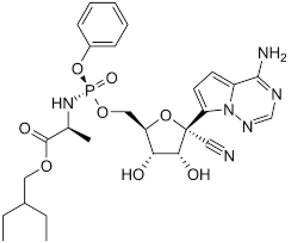 |
| 159989-64-7 | Nelfinavir | C32H45N3O4S | 663.9 | 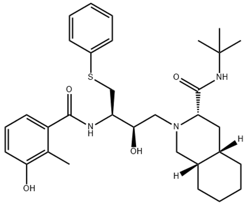 | |
| 259793-96-9 | Favipiravir | C5H4FN3O2 | 157.104 | 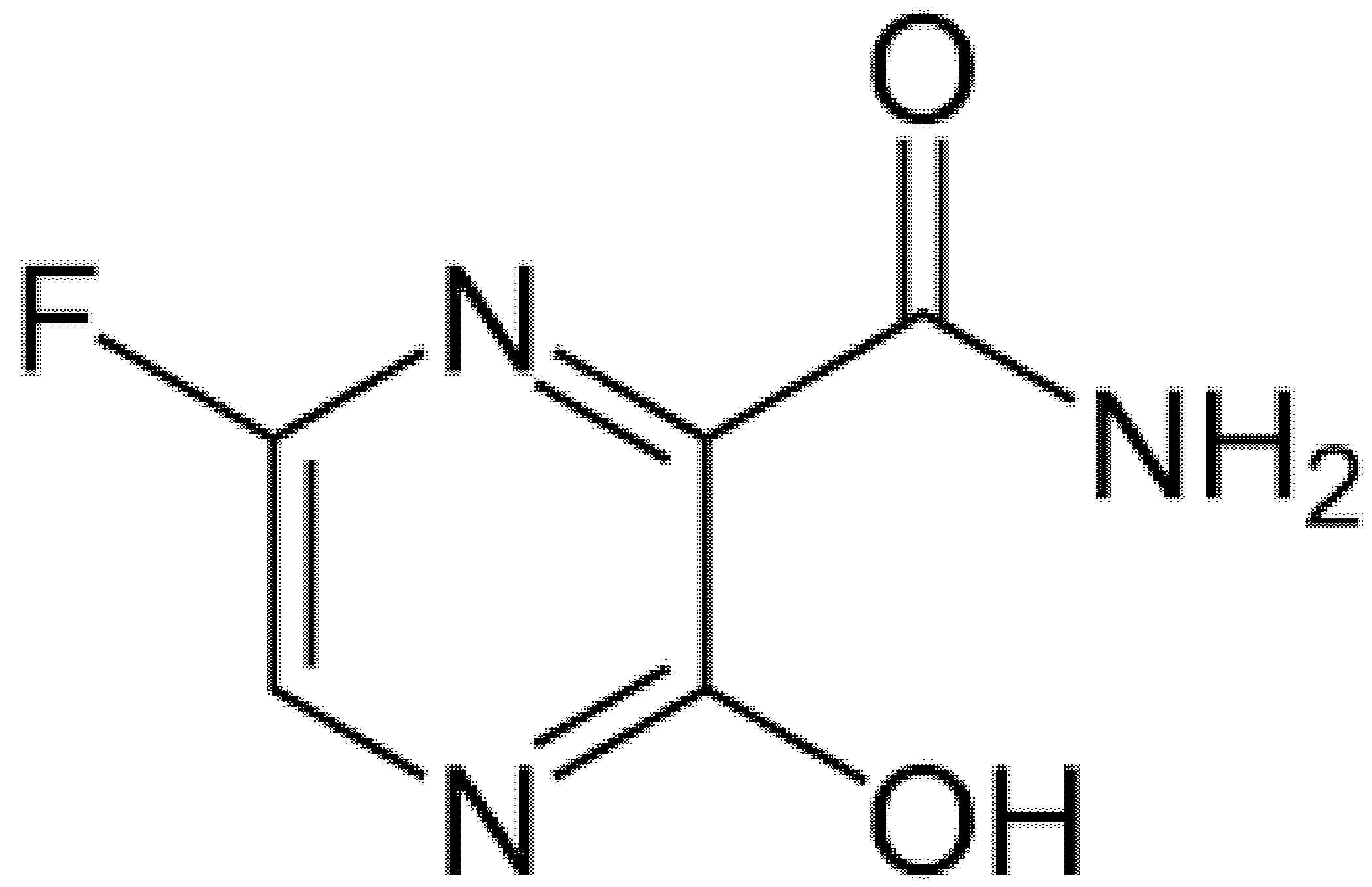 | |
| 192725-17-0 | Lopinavir- | C37H48N4O5 | 628.814 | 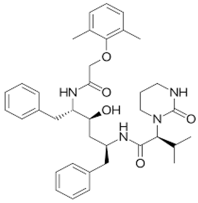 | |
| 155213-67-5 | Ritonavir | C37H48N6O5S2 | 720.948 | 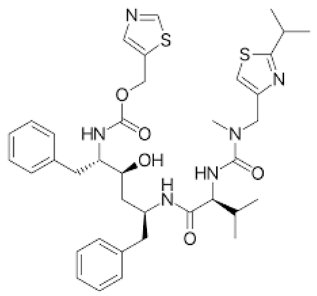 | |
| 196618-13-0 | Oseltamivir | C16H28N2O4 | 312.4 | 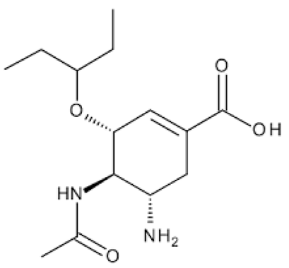 | |
| Antimalarial | 54-05-7 | Chloroquine | C18H26ClN3 | 319.872 |  |
| 118-42-3 | Hydroxycloroquine | C18H26ClN3O | 335.872 |  | |
| Anti-Inflammatory | 50-23-7 | Hydrocortisone | C21H30O5 | 362.46 | 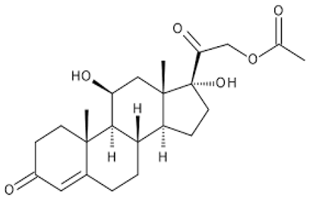 |
| 50-02-2 | Dexamethasone | C22H29FO5 | 392.46 | 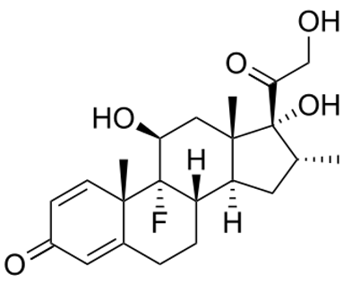 | |
| Antiparasitic | 70288-86-7 | Ivermectin | C48H74O14 | 875.1 |  |
| Antibacterial | Azithromycin | C38H72N2O12 | 749.0 |  |
Publisher’s Note: MDPI stays neutral with regard to jurisdictional claims in published maps and institutional affiliations. |
© 2021 by the authors. Licensee MDPI, Basel, Switzerland. This article is an open access article distributed under the terms and conditions of the Creative Commons Attribution (CC BY) license (https://creativecommons.org/licenses/by/4.0/).
Share and Cite
Musee, N.; Kebaabetswe, L.P.; Tichapondwa, S.; Tubatsi, G.; Mahaye, N.; Leareng, S.K.; Nomngongo, P.N. Occurrence, Fate, Effects, and Risks of Dexamethasone: Ecological Implications Post-COVID-19. Int. J. Environ. Res. Public Health 2021, 18, 11291. https://doi.org/10.3390/ijerph182111291
Musee N, Kebaabetswe LP, Tichapondwa S, Tubatsi G, Mahaye N, Leareng SK, Nomngongo PN. Occurrence, Fate, Effects, and Risks of Dexamethasone: Ecological Implications Post-COVID-19. International Journal of Environmental Research and Public Health. 2021; 18(21):11291. https://doi.org/10.3390/ijerph182111291
Chicago/Turabian StyleMusee, Ndeke, Lemme Prica Kebaabetswe, Shepherd Tichapondwa, Gosaitse Tubatsi, Ntombikayise Mahaye, Samuel Keeng Leareng, and Philiswa Nosizo Nomngongo. 2021. "Occurrence, Fate, Effects, and Risks of Dexamethasone: Ecological Implications Post-COVID-19" International Journal of Environmental Research and Public Health 18, no. 21: 11291. https://doi.org/10.3390/ijerph182111291
APA StyleMusee, N., Kebaabetswe, L. P., Tichapondwa, S., Tubatsi, G., Mahaye, N., Leareng, S. K., & Nomngongo, P. N. (2021). Occurrence, Fate, Effects, and Risks of Dexamethasone: Ecological Implications Post-COVID-19. International Journal of Environmental Research and Public Health, 18(21), 11291. https://doi.org/10.3390/ijerph182111291