Article

Macrocarpane-Type Sesquiterpenes from Laurencia microcladia

by
Adrián Gutiérrez-Cepeda
1,2,
Jorge J. Cabrera-Trujillo
1,3,
José J. Fernández
1,3,4,*,
Antonio Hernández-Daranas
5,
Manuel Norte
1,3 and
María L. Souto
1,3,4,*
1
Instituto Universitario de Bio-Orgánica Antonio González (IUBO AG), Universidad de La Laguna (ULL), Avenida Astrofísico Francisco Sánchez 2, 38206 La Laguna, Spain
2
Department of Chemistry, Chemistry Institute, Sciences Faculty, Autonomous University of Santo Domingo, University City, Santo Domingo 1355, Dominican Republic
3
Departamento de Química Orgánica, Universidad de La Laguna (ULL), Avenida Astrofísico Francisco Sánchez 3, 38206 La Laguna, Spain
4
Biotecnología Marina, IUBO AG-ULL, Unidad Asociada al IPNA-CSIC, 38206 La Laguna, Spain
5
Instituto de Productos Naturales y Agrobiología (IPNA), Consejo Superior de Investigaciones Científicas (CSIC), Avenida Astrofísico Francisco Sánchez 3, 38206 La Laguna, Spain
*
Authors to whom correspondence should be addressed.
Mar. Drugs 2026, 24(9), 316; https://doi.org/10.3390/md24090316
Submission received: 30 July 2026 / Revised: 28 August 2026 / Accepted: 4 September 2026 / Published: 9 September 2026
(This article belongs to the Section Structural Studies on Marine Natural Products)

Abstract

Seven undescribed sesquiterpenes featuring an uncommon macrocarpane skeleton, laurocarpanes A–G (17), were isolated from specimens of Laurencia microcladia collected in Fuerteventura, the Canary Islands. The identification of these macrocarpane-type metabolites is of particular interest from both chemical and chemotaxonomic perspectives, as it highlights distinctive biosynthetic capabilities within the studied species and further expands current knowledge regarding the distribution of this uncommon structural class in marine organisms. Their structures and configurational assignments were determined by a comprehensive NMR study, high-resolution mass spectrometry, and coupling constant evaluations. A biosynthetic model was proposed linking the assembly of the macrocarpane skeleton directly to an (S)-β-bisabolene monocyclic precursor, with the process mediated by vanadium-dependent bromoperoxidase (V-BPO). The plausibility of the proposed biogenetic formation of laurocarpanes A–G (17) was assessed through Density Functional Theory (DFT) calculations.

1. Introduction

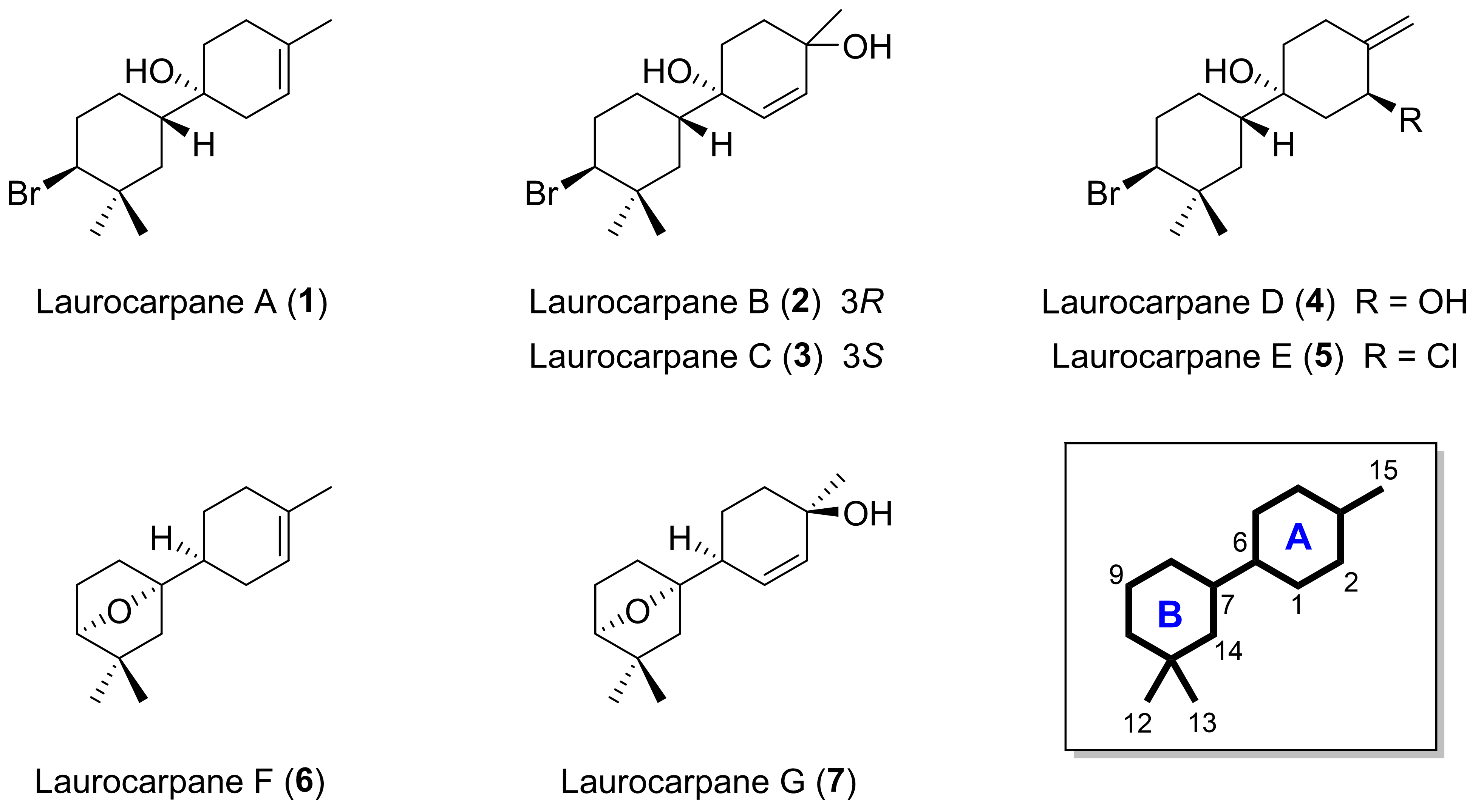
The bisabolyl-type cation plays a central role in the biosynthesis of various sesquiterpenes, acting as the precursor to several major skeletal types, such as cuparanes, lauranes, chamigranes, etc. [1]. Furthermore, cyclization between the C-14 and C-11 positions of the deprotonated bisabolyl cation intermediate is responsible for the formation of a rare class of bicyclic sesquiterpenes, the macrocarpanes (Figure 1). This skeleton was first proposed by Cool in 2005 [2] for a series of five new hydrocarbon sesquiterpenes isolated from the leaves of Cupressus macrocarpa, hence the common name for this class of compounds. The number of macrocarpane skeleton metabolites that have been isolated from natural sources is limited. In addition to eight examples obtained from terrestrial sources [2,3], only nine sesquiterpenes and one norderivative have been isolated, and all from Laurencia species [4,5,6].
As part of our continuing interest in the chemistry of the genus Laurencia [7,8,9,10,11,12,13] and during the course of a marine bioactive compound discovery program, seven new sesquiterpenes, laurocarpanes A–G (17), presenting this uncommon framework, were obtained from Laurencia microcladia (Figure 1).

2. Results and Discussion

2.1. Structural Elucidation of Macrocarpane-Type Sesquiterpenes

Fresh specimens of the alga Laurencia microcladia (2.0 kg) collected in El Cotillo, Fuerteventura (the Canary Islands, Spain) were extracted at room temperature using CH2Cl2/MeOH (1:1, v/v). The resultant extract was subjected to a multi-step chromatographic fractionation sequence to yield compounds 17 (Figure 1).
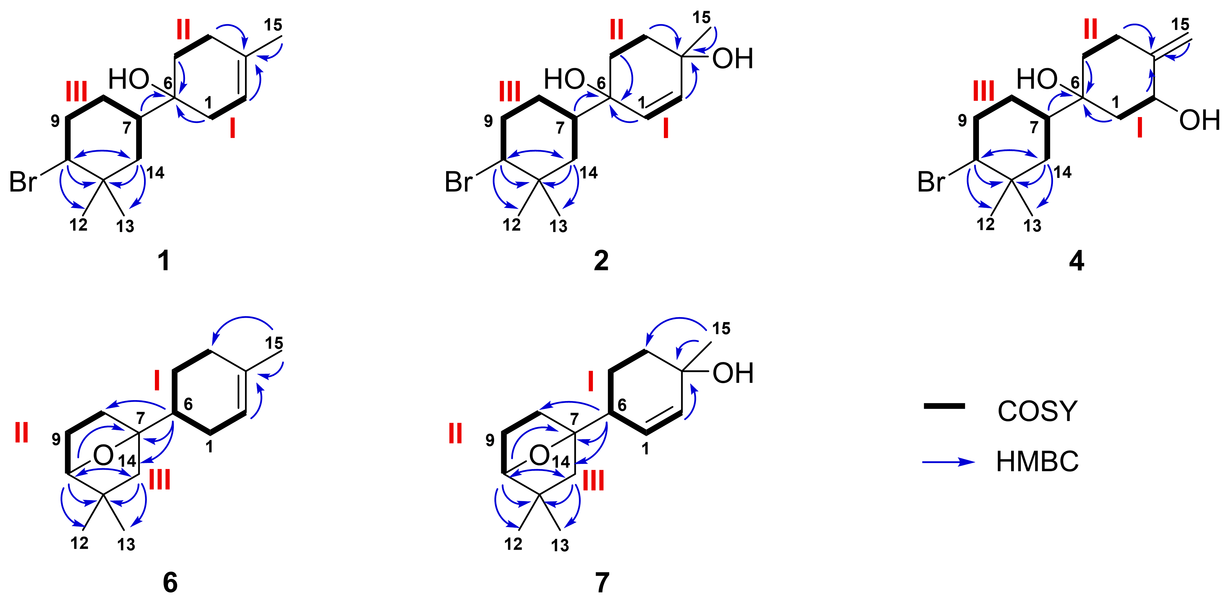
Laurocarpane A (1) was isolated as a colorless oil. ESI-FTICR (ion peaks at m/z 301.1410 and 303.1390 ) revealed a molecular formula of C15H25BrO, indicating the presence of three degrees of unsaturation. The 13C NMR and HSQC-edited spectra established the presence of three methines (δC 118.1, 66.5, 41.3), six methylenes (δC 40.3, 35.2, 34.2, 31.0, 28.1, 26.9), three methyls (δC 31.9, 23.3, 20.4), and three non-protonated carbons (δC 134.0, 71.0, 36.6) (Table 1). The 1H NMR spectrum showed a broad vinylic signal at δH 5.27, a brominated methine at δH 3.94 (dd, J = 12.8, 4.4 Hz), a vinylic methyl at δH 1.68, and two singlet methyls at δH 1.07 and 1.04. These data, combined with COSY correlations, enabled the assignment of three independent spin systems: I [H2-1 (δH 2.14 and 1.89) ⟶ H-2 (δH 5.27)], II [H2-4 (δH 2.13 and 1.94) ⟶ H2-5 (δH 1.66 and 1.51)], and III [H-10 (δH 3.94)/H2-9 (δH 2.20 and 2.00)/H2-8 (δH 1.78 and 1.19)/H-7 (δH 1.61)/H2-14 (δH 1.82 and 1.21)] (Figure S1). Additionally, the HMBC correlations of H-2, H2-4, and H3-15 (δH 1.68) with the quaternary carbon C-3 (δC 134.0), as well as those of the methylenes H2-1 and H2-5 with the quaternary carbon C-6 (δC 71.0), connected the spin systems I and II in one of the molecule’s rings. Likewise, C-6 showed an HMBC correlation with H-7 (δH 1.61) that belonged to system III. Finally, the HMBC correlations from H-10, H2-14, and the gem-dimethyl groups H3-12 (δH 1.07) and H3-13 (δH 1.04) to C-11 (δC 36.6) completed the planar structure of 1, establishing the existence of another cyclohexane ring (Figure 2).
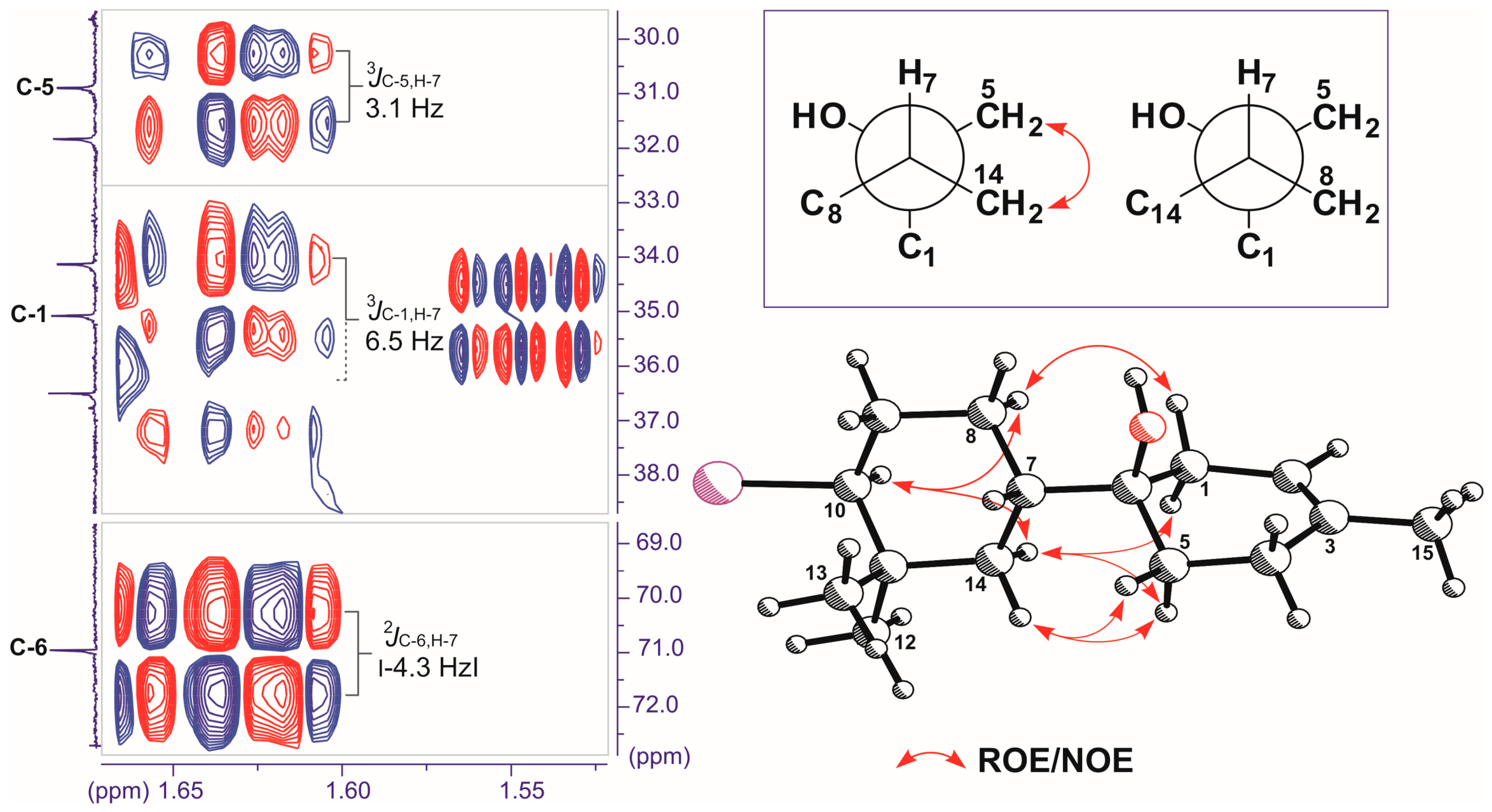
The relative configuration of metabolite 1 was determined by comparison of TROESY data and the analysis of homo- and heteronuclear J couplings [14]. According to the values of the H-10 homonuclear coupling constants (JH-H 12.8 and 4.4 Hz), its orientation must be axial, leaving the bromine atom in the equatorial position. In addition, a strong ROE enhancement observed between H-10 and one of the diastereotopic H-14 protons (δH 1.21), as well as its subsequent large coupling constant with H-7 (3JH-14α,H-7 = 12.6 Hz), was consistent with a trans-diaxial arrangement of H-14α/H-7 and an equatorial configuration of the bond that connects the two cyclohexane rings. Through a J-HMBC experiment, two rotamers around the C-6/C-7 bond were deduced (Figure 3). The large absolute value of 4.3 Hz obtained for the constant 2JC-6,H-7 indicates that the OH group on C-6 and H-7 are in a gauche relative position. Likewise, the values 3JC-5,H-7 = 3.1 Hz and 3JC-1,H-7 = 6.5 Hz agree with gauche (C-5/H-7) and anti (C-1/H-7) orientations. Finally, from the TROESY data and the 1D-NOE experiments for the H2-5 protons, it was possible to establish that they correlate exclusively with H2-14, which implies a 6R*, 7S* relative configuration.
Laurocarpane B (2) displayed a close structural relationship with 1. The molecular formula was determined to be C15H25BrO2 on the basis of ESI-HRMS data. The IR spectrum showed bands attributable to hydroxyl (3402 cm−1) and alkene (1666 cm−1) type functional groups.
In fact, the comparison of the 1H and 13C NMR spectroscopic data (Table 1) shows a correspondence in the assignments between the positions C-7/C-11,C-14 (ring B), locating the difference between both compounds in ring A, more specifically in the presence of a disubstituted double bond (δH/C 5.77/136.8 and 5.67/131.3 in 2) instead of a trisubstituted (δH/C 5.27/118.1 and 134.0 in 1), and in the upfield shift of the H3-15 methyl protons from a vinylic methyl (δH 1.68 in 1) to a methylcarbinol (δH 1.37 in 2). The key differences between them were confirmed by the 1H-1H COSY correlations of I: H-1 (δH 5.77) ⟶ H-2 (δH 5.67) and II: H2-4 (δH 2.08/1.69) ⟶ H2-5 (δH 1.93/1.51); and the HMBC correlations of H3-15, H-2, H2-4/C-3 (δC 78.8) and H-1, H2-5, H-7 (δH 1.71)/C-6 (δC 70.9) (Figure 2). The double bond geometry was established as Z based on the value of the coupling constant 3JH-1,H-2 = 10.1 Hz. The relative configurations of the C-10, C-7 and C-6 centers matched that proposed for laurocarpane A (1), while that of C-3 was established as 3R* based on the observed ROE correlations of one of the methylene protons H2-4 at δH 1.69 with the more deshielded diastereotopic proton H-5 (δH 1.93) and methyl H3-15 (Table 1).
The (+)-HRESIMS spectrum of laurocarpane C (3) exhibited a [M + Na]+ ion at m/z 339.0940; 341.0922 (100:76), indicating that it is an isomer of 2. Moreover, the NMR data of 3 were very similar to those of 2 (Table 1 and Table S1), with the exception of the chemical shifts at positions C-3 and C-15 (ΔδC > 0.5 ppm). The analysis of the 2D NMR spectra (HSQC, HMBC, and COSY) for 3 established the same planar structure as that of 2. Consequently, a different relative configuration at C-3 between 2 and 3 was proposed.
Laurocarpane D (4) shared the same molecular formula with compounds 2 and 3 according to analysis of ESI-FTICR (m/z 315.0954 and 317.0934 [M]+). A comparison of the 13C and 1H NMR spectra (Table 1 and Table S1) suggested the main differences among these isomers are found in ring A being the methylcarbinol in 2 and 3 replaced by an exocyclic double bond [δH 4.93 (s)/4.85 (s)], and an allylic oxymethine (δH 4.73, dd, J = 11.3, 4.6 Hz) in 4. Analysis of the C-1/C-6 fragment through HSQC and COSY experiments revealed two 1H-1H spin systems [I: H2-1 (δH 2.10 and 1.43) → H-2 (δH 4.73); II: H2-4 (δH 2.40 and 2.26) → H2-5 (δH 1.64 and 1.47)]. Conversely, the HMBC study connected the H2-4 methylene and H-2 methine protons with the C-3 quaternary carbon signal (δC 146.4), which was also correlated with the exo olefinic protons (H2-15). Additionally, the connectivity of the H2-1, H2-5, and H-7 protons (δH 1.58) with the C-6 carbon (δC 74.9), which is adjacent to the hydroxyl group, was observed. This completed the structure of laurocarpane D (4) (Figure 2). As in the previous metabolites, the relative arrangement of the substituents at C-7 and C-10 on the dimethylcyclohexyl ring and the hydroxyl group at C-6 on ring A was determined to be trans-diequatorial and axial, respectively. Based on the coupling constants exhibited by the axial methine H-2 (J = 11.3, 4.6 Hz) with the two methylene protons H2-1, the spatial arrangement of the hydroxyl group on C-2 was assigned as equatorial. Therefore, the relative configuration at this center was proposed as 2S*.
Laurocarpane E (5) was isolated as a colorless, amorphous solid. Based on the pseudomolecular isotopic ions [M + Na]+ observed in the high-resolution mass spectrum (m/z 357.2040, 359.2204), its molecular formula was established as C15H24BrClO. The NMR spectra of laurocarpane E (5) were very similar to those of 4 (Table 1). The most significant differences were observed at the 13C NMR chemical shift of C-2 (δC 69.2 in 5 vs. δC 82.2 in 4), which revealed the replacement of the hydroxy group of 4 by a chlorine atom in 5. This finding is in agreement with the molecular formula and the relative intensities of isotope pseudomolecular peaks observed. The coupling constants observed for the axial proton H-2 (J = 10.7, 4.2 Hz) confirmed that the configuration at this stereocenter in 5 was identical to that of 4.
The molecular formula of laurocarpane F (6) was assigned by ESI-FTICR (m/z 259.1743 [M + K]+) as C15H24O, implying four degrees of unsaturation. The absence of characteristic absorptions of hydroxyl and/or carbonyl groups in the infrared spectrum indicated that the oxygen atom must correspond to an ether bridge. Through COSY, HSQC, and HMBC experiments, three spin systems could be established. The first system (I) begins with the olefinic methine H-2 (δH 5.39), which couples with the protons of the methylene H2-1 (δH 2.03 and 1.81). This sequence continues with the methine H-6 (δH 1.90) and the methylenes H2-5 (δH 1.82 and 1.29) and H2-4 (δH 2.04 and 1.92). The second system (II) comprises the methylenes H2-8 (δH 1.50 and 1.44) and H2-9 (δH 1.92 and 1.62), as well as the oxygenated methine H-10 (δH 3.84). The third spin system (III) consists solely of the isolated methylene H2-14 (δH 1.34 and 1.20) (Table 2; Figure 2). An analysis of the HMBC experiment revealed the ring A of the molecule through correlations observed between the methyl group at δH 1.65 (H3-15) and the olefinic carbons at δC 134.0 (C-3)/120.3 (C-2), as well as the terminal carbon of fragment I at δC 30.3 (C-4). Furthermore, methine H-10 connects to carbon at δC 90.5 (C-7) via an ether bridge. Additionally, correlations of this proton with carbons at δC 40.9 (C-11), 30.1 (C-12), 25.0 (C-13) and 48.6 (C-14) connect the gem-dimethyl system at C-11 to fragments II and III. HMBC correlations of H-6 with carbons at C-7, C-14, and C-8 (δC 31.2) confirm the final structure of this metabolite (Figure 2). The relative stereochemistry of this compound is proposed on the basis of biogenetic considerations. As mentioned in the background, and will be discussed in more detail subsequently, it is logical to hypothesize that the stereochemistry of laurocarpane F in ring A is derived from the chiral precursor (S)-β-bisabolene, while the carbocation on C-7 evolves by the addition of water and subsequent cyclization on C-10 on the opposite side to the bromine loss (Figure 4). Consequently, a relative configuration of 6S*, 7S*, 10S* is proposed.
Laurocarpane G (7) showed an ion [M]+ at m/z 236.1766 in its ESI-HRMS mass spectrum, indicating a molecular formula of C15H24O2. Given the similarity of the 1H and 13C NMR data for 7 to those of 6, an analogous assignment strategy was used to elucidate its structure. The differences between the two are found in ring A. The double bond is Z-disubstituted rather than trisubstituted, with the vinylic methines H-1 and H-2 at δH 6.07 and 5.65 (3JH-1,H-2 = 10.2 Hz), respectively. The methine H-6 is now allylic, resulting in its displacement to lower fields (δH 2.42). Finally, the olefinic methyl H3-15 becomes a methylcarbinol (δH 1.35). The HMBC correlations between the protons H3-15/H-1/H2-4 (δH 2.17/1.43) and the hydroxylated carbon at δC 78.9 (C-3) complete the structure of this particular region of the molecule (Figure 2). In order to formulate a proposal for relative stereochemistry at the C-6, C-7, and C-10 centers, the same biogenetic postulates as in 6 are used. However, the configuration at C-3 is proposed as R* on the basis of the correlation observed in the TROESY experiment between the methine H-6 and the most shielded diastereotopic proton of methylene H2-4 (δH 1.43). This places the hydroxyl group on the opposite face of the molecule, as in compound 2.

2.2. Plausible Biosynthetic Pathway

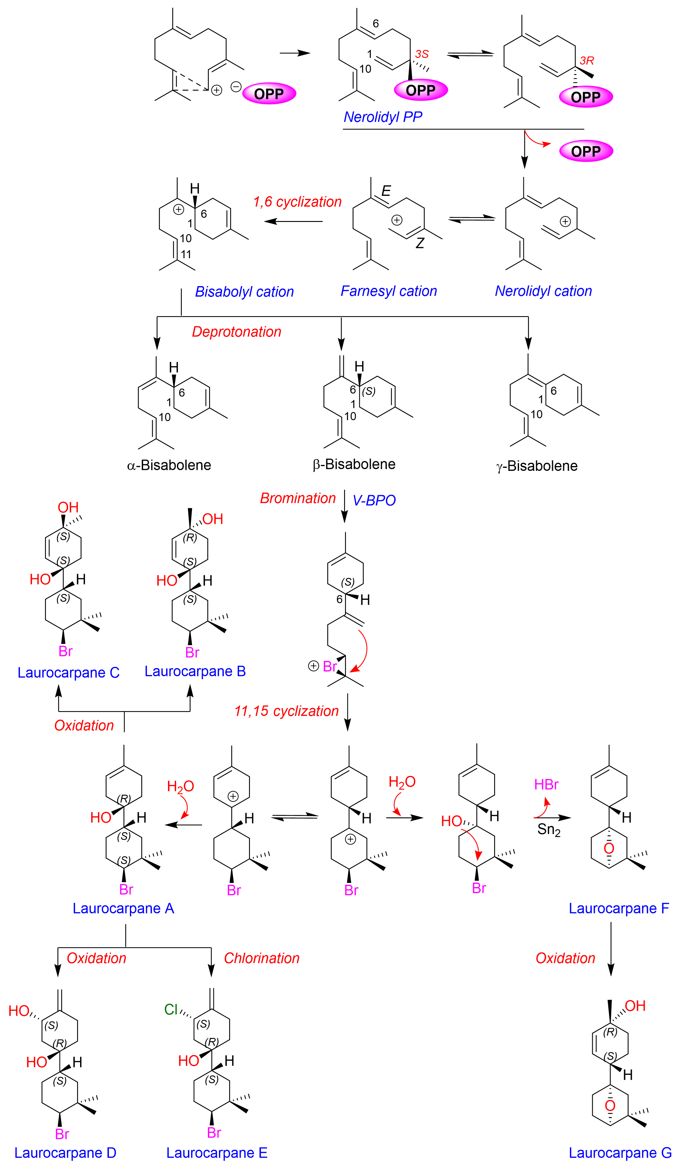
The biogenetic origin of the macrocarpane skeleton from the 2Z,6E-nerolidyl cation via a key monocyclic intermediate, (S)-β-bisabolene, has been described in detail by Cool [2] and experimentally supported by Köllner and colleagues [15]. Based on these data, we propose a biogenetic model for the formation of the metabolites isolated from L. microcladia (Figure 4).
The hypothesis posits that the formation of a bromonium ion on the olefin between C-10 and C-11, via vanadium-dependent bromoperoxidase (V-BPO), could induce the formation of the macrocarpane cation at C-7. The reaction of water with the carbocation could result in the subsequent formation of an ether bridge with C-10 on the opposite side from the bromine loss, leading to the formation of laurocarpanes F and G (67). On the other hand, hydride migration from C-6 to C-7 could generate the cation at C-6, which upon the subsequent addition of water would result in laurocarpane A. The action of peroxidases or chloroperoxidases would result in the formation of laurocarpanes B–E (25).

2.3. Computational Analysis

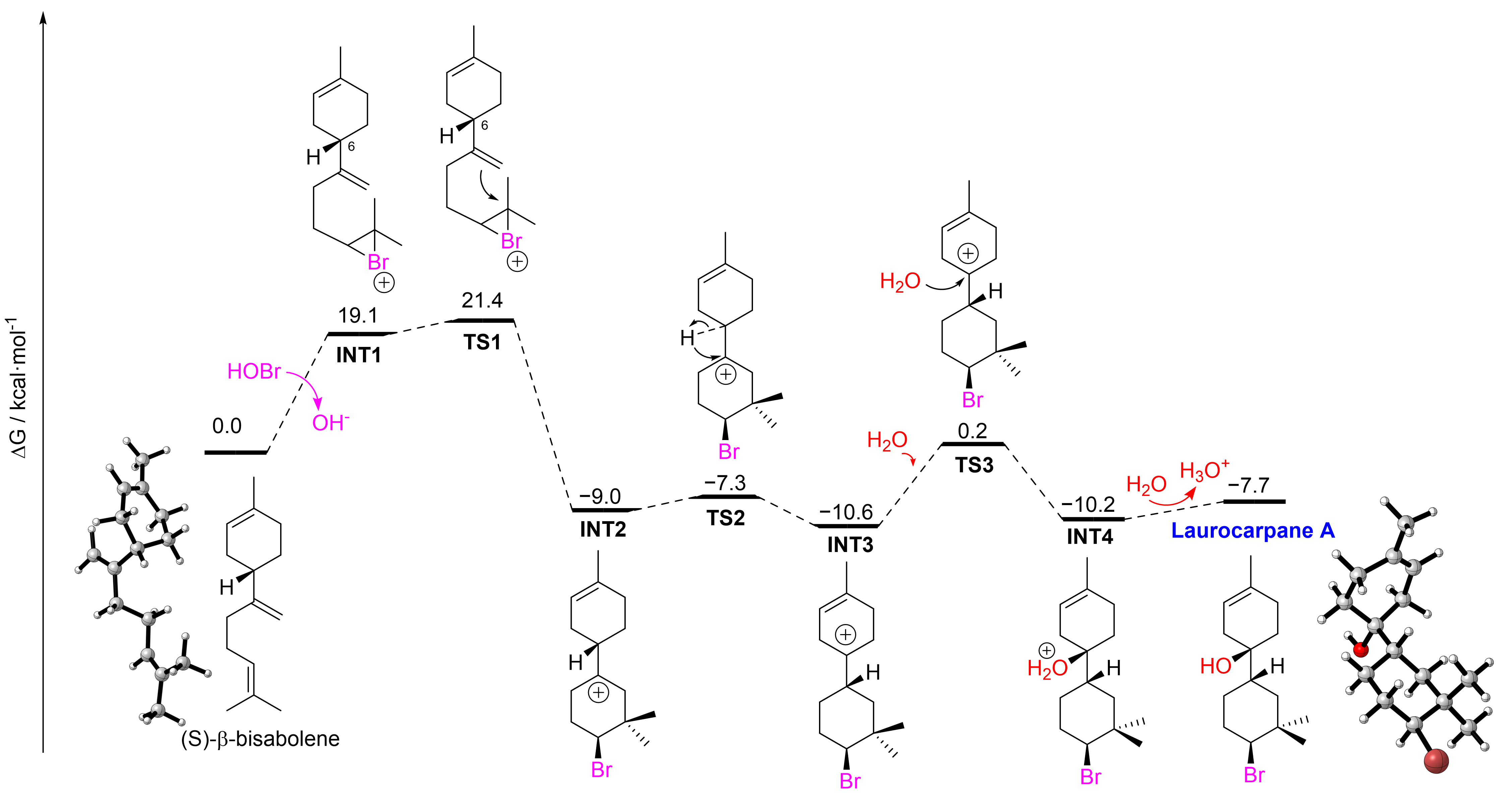
To assess the plausibility of the proposed biogenetic formation of laurocarpanes A–G (17), Density Functional Theory (DFT) calculations were performed on the mechanism shown in Figure 4 (see ESI for computational details). Firstly, we focused on the formation of laurocarpane A (1), hypothesized to be the key biosynthetic precursor to laurocarpanes B–E (25). The computed reaction profile (Figure 5) begins with electrophilic bromination of (S)-β-bisabolene to generate the bromonium cation INT1 in a markedly endergonic step (ΔG = 19.1 kcal/mol). In line with previous studies on related red algae [16], we propose that this energetically demanding activation is mediated by a vanadium-dependent bromoperoxidase, which catalyzes the oxidation of bromide by hydrogen peroxide to generate an electrophilic brominating species, commonly formulated as HOBr or an equivalent Br+-donating species, capable of initiating substrate bromination [16,17].
Once INT1 is formed, the subsequent cyclization leading to the characteristic bicyclic laurocarpane scaffold proceeds through a remarkably low-energy transition state (TS1). This step requires an activation barrier of only 2.3 kcal/mol relative to the bromonium intermediate and remains accessible at physiological temperature from the initial bisabolene substrate with an overall barrier of 21.4 kcal/mol. TS1 affords the bicyclic cation INT2 in a strongly exergonic process relative to the starting bisabolene (ΔG = −9.0 kcal/mol). These results indicate that, although bromination is the most energetically demanding step, the subsequent ring-closing event is highly facile and provides a strong driving force for the rapid assembly of the bicyclic skeleton.
Next, we suggest that INT2 is in equilibrium with INT3 through a 1,2-hydride shift. Consistent with this hypothesis, we located the corresponding Meerwein–Wagner rearrangement transition state, TS2, which proceeds with a very low activation barrier in both directions: 1.7 kcal/mol from INT2 to INT3 and 3.3 kcal/mol for the reverse process.
The subsequent addition of water to INT3 occurs through TS3 (ΔG = 10.8 kcal/mol), yielding INT4 in a nearly isoenergetic step relative to INT3, although this transformation remains strongly exergonic with respect to the initial bisabolene (ΔG = −10.2 kcal/mol). Finally, deprotonation of INT4 yields laurocarpane A in an overall exergonic process (ΔG = −7.7 kcal/mol). The accessible computed activation barriers, together with the overall exergonic character of the sequence, support the plausibility of the proposed biogenetic formation of laurocarpane A and are consistent with its proposed role as a common precursor to laurocarpanes B–E.
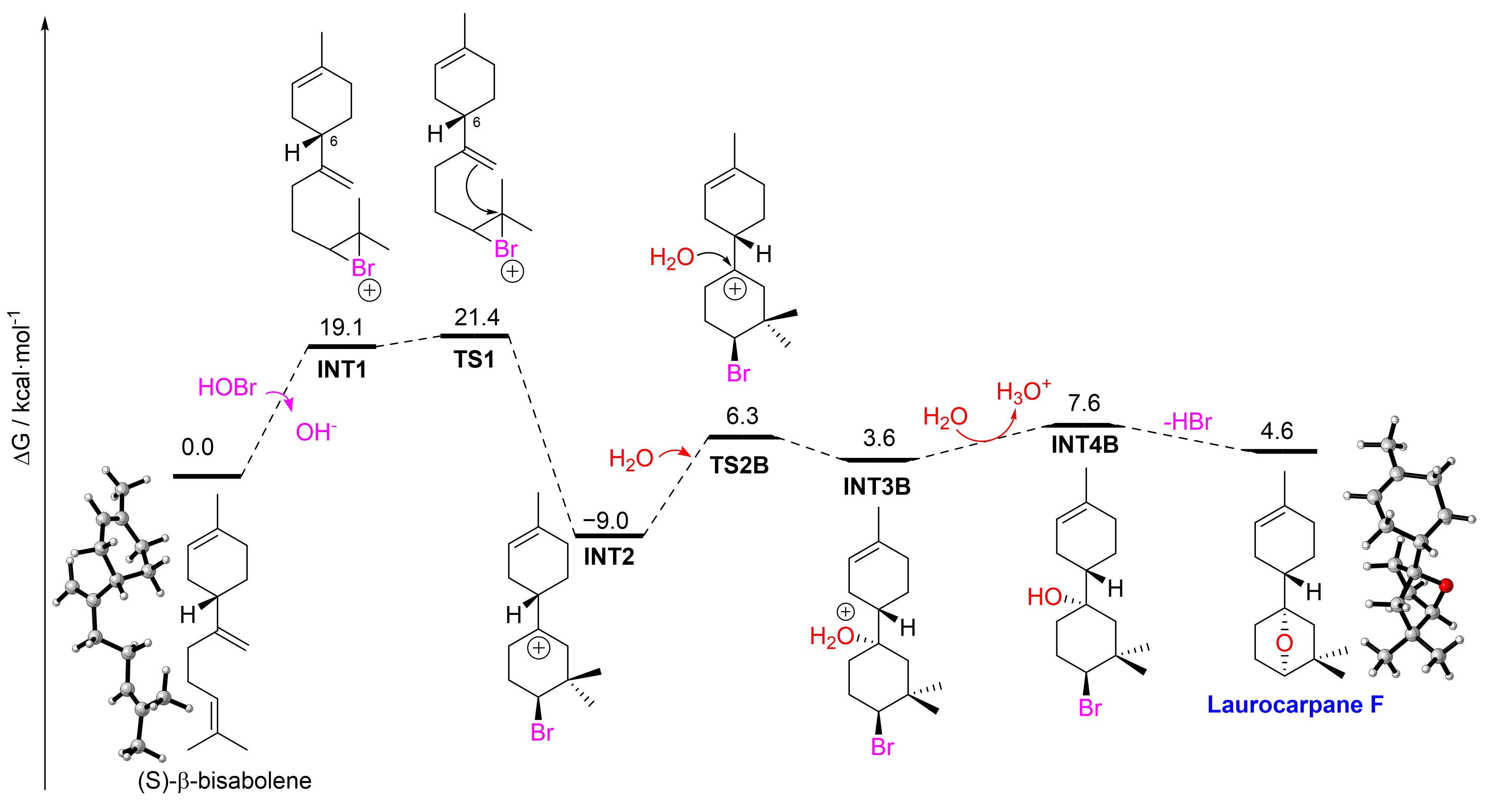
We then investigated by DFT the mechanism for the formation of laurocarpane F (Figure 6). Laurocarpane F is hypothesized to be the biosynthetic precursor of laurocarpane G, as both compounds share the same cyclic ether motif.
The computed mechanism shown in Figure 6 indicates that the pathway leading to laurocarpane F is identical to that proposed for laurocarpane A up to the formation of the bicyclic carbocation INT2. From this point onward, however, the reaction diverges. Instead of undergoing a 1,2-hydride shift, INT2 is trapped by water through the transition state TS2B, with an associated activation free energy of 15.3 kcal/mol, to give INT3B. This intermediate lies slightly higher in energy than the initial bisabolene substrate (ΔG = 3.6 kcal/mol). From INT3B, subsequent deprotonation and intramolecular cyclic ether formation accompanied by HBr elimination afford laurocarpane F in a process that is overall slightly endergonic relative to the initial bisabolene (ΔG = 4.6 kcal/mol). Despite this modest thermodynamic penalty, the relatively low computed barrier for water trapping of INT2 indicates that this competing pathway is kinetically accessible under biosynthetic conditions. Therefore, the DFT results support the feasibility of laurocarpane F formation and its plausible role as a precursor to laurocarpane G.

2.4. Preliminary Bioactivity Assay

The apoptosis-inducing activity of the major compound, laurocarpane A (1), was investigated against the human hepatocellular carcinoma cell line HepG2 (HB-8065). Interestingly, laurocarpane A induced apoptosis in more than 98% of HepG2 cells at a concentration of 50 μM. Unfortunately, the limited availability and stability of the isolated compounds prevented the completion of a comprehensive bioactivity study. Nevertheless, these preliminary results suggest that laurocarpane A has potential as an apoptosis-inducing agent.

3. Materials and Methods

3.1. General Experimental Procedures

The optical rotations were obtained using a Perkin–Elmer 241 polarimeter (Waltham, MA, USA) equipped with a sodium lamp. UV spectra were acquired on a Jasco V-560 spectrophotometer (Easton, MD, USA). The infrared spectra were collected on a Bruker IFS55 spectrometer (Ettlingen, Germany). NMR spectra were recorded on a Bruker AVANCE 600 MHz instrument (Bruker, Karlsruhe, Germany) equipped with a 5 mm TCI (Triple Resonance Inverse) detection cryo-probe. Chemical shifts were referenced to the CDCl3 signals at 300 K (δH 7.26 ppm, δC 77.0 ppm). The 2D NMR experiments (COSY, HSQC, HMBC, and ROESY) were performed using standard pulse sequences. 3JH,H values were measured from 1D 1H NMR. The J-HMBC pulse sequence was used to measure long-range heteronuclear coupling constants. A J-scale factor of 52 was used, and the experiment was optimized for long-range couplings of 2 Hz. Mass spectra were recorded on a LCT Premier XE Micromass spectrometer (Waters, Milford, CT, USA) using electrospray ionization. HPLC separations were carried out with a preparative silica column (10 μm, 19 × 150 mm) and Waters system (Waters, Milford, CT, USA) equipped with a Binary HPLC Pump 1525 and Photodiode Array Detector 2996. TLC (thin-layer chromatography) (Merck, Darmstadt, Germany) plates were visualized by spraying with phosphomolybdic acid reagent (10% in EtOH) and heating.

3.2. Biological Material

Specimens of Laurencia microcladia Kützing were collected by hand at a depth of 2 m at El Cotillo (Fuerteventura, the Canary Islands) (GPS coordinates 28°42′41.7″ N, 14°00′27.2″ W). A voucher specimen was deposited at the Department of Biología Vegetal, Botánica, University of La Laguna, Tenerife (TFC Phyc 14446).

3.3. Extraction and Isolation

Fresh alga (2.0 kg) was extracted with CHCl3:MeOH (1:1, v/v, 3×) at room temperature and the solvent removed in vacuo to give a dark-green viscous oil. The resulting extract (14.1 g) was subjected to gel filtration using Sephadex LH-20 (70 × 600 mm) and eluted with CHCl3:MeOH (1:1) to obtain five fractions. Fraction LM-4 (4.78 g) was rechromatographed on a medium-pressure normal-phase Lobar LiChroprep Si 60 column (25 × 310 mm) using a step gradient elution of increasing polarity (n-hexane to EtOAc) to yield five fractions. Final purifications of fractions LM-41 (eluted with 10% EtOAc in n-Hex) and LM-42 (30% EtOAc in n-Hex) were achieved on a μ-Porasil HPLC column (10 μm, 19 × 150 mm) using n-hexane:EtOAc (9:1 and 7:3, respectively), yielding compounds 1 (5.0 mg), 2 (1.4 mg), 3 (1.0 mg), 4 (2.8 mg), 5 (2.0 mg), 6 (1.8 mg), and 7 (2.5 mg).
Laurocarpane A (1): colorless oil; [α]25D +5 (c 0.57, CHCl3); UV (MeOH) λmax (log ε) 203 (3.11) nm; IR (CHCl3) νmax 3416, 2950, 2871, 1706, 1665, 1453, 1369, 1265, 1217, 963 cm−1; 1H and 13C NMR data (CDCl3), see Table 1; ESI-FTICR m/z 301.1410, 303.1390 [M + H]+ (100:82) (calcd for C15H2679BrO, 301.1167; C15H2681BrO, 303.1147).
Laurocarpane B (2): colorless oil; [α]25D + 1 (c 0.02, CHCl3); UV (MeOH) λmax (log ε) 204 (3.53) nm; IR (CHCl3) νmax 3402, 2930, 2859, 1714, 1666, 1456, 1372, 1264, 1021, 964, 865 cm−1; 1H and 13C NMR data (CDCl3), see Table 1; ESI-HRMS m/z 339.0911, 341.0928 [M + Na]+ (100:80) (calcd for C15H2579BrO2Na, 339.0936; C15H2581BrO2Na, 341.0915).
Laurocarpane C (3): colorless oil; [α]25D −4 (c 0.20, CHCl3); UV (MeOH) λmax (log ε) 204 (3.14) nm; IR (CHCl3) νmax 3400, 2930, 2868, 1729, 1665, 1455, 1367, 1253, 1021, 984, 763 cm−1; 1H and 13C NMR data (CDCl3), see Table S1; ESI-HRMS m/z 339.0940, 341.0922 [M + Na]+ (100:76) (calcd for C15H2579BrO2Na, 339.0936; C15H2581BrO2Na, 341.0915).
Laurocarpane D (4): colorless oil; [α]25D + 4 (c 0.08, CHCl3); UV (MeOH) λmax (log ε) 203 (3.67) nm; IR (CHCl3) νmax 3393, 2926, 2855, 1650, 1456, 1368, 1260, 1080, 1023, 963, 898 cm−1; 1H and 13C NMR data (CDCl3), see Table 1; ESI-FTICR m/z 315.0954, 317.0934 [M]+ (100:97) (calcd for C15H2479BrO2, 315.0960; C15H2481BrO2, 317.0939).
Laurocarpane E (5): white amorphous solid; 1H and 13C NMR data (CDCl3), see Table S1; ESI-HRMS m/z 357.2040, 359.2204 [M + Na]+ (100:96) (calcd for C15H2479Br35ClONa, 357.0597; C15H2481Br35ClONa, 359.0576). Complementary spectroscopic data are not available due to the rapid degradation of the sample.
Laurocarpane F (6): white amorphous solid; [α]25D + 16 (c 0.03, CHCl3); UV (MeOH) λmax (log ε) 202 (3.38) nm; IR (CHCl3) νmax 2955, 2928, 2867, 1729, 1450, 1367, 1240, 1253, 1161, 988, 751 cm−1; 1H and 13C NMR data (CDCl3), see Table 2; ESI-FTICR m/z 259.1743 [M + K]+ (calcd for C15H24OK, 259.1464).
Laurocarpane G (7): white amorphous solid; [α]25D + 16 (c 0.03, CHCl3); UV (MeOH) λmax (log ε) 202 (3.14) nm; IR (CHCl3) νmax 3400, 2955, 2928, 2867, 1729, 1450, 1368, 1253, 1195, 1161, 988, 751 cm−1; 1H and 13C NMR data (CDCl3), see Table 2; ESI-HRMS m/z 236.1766, [M]+ (calcd for C15H24O2, 236.1776).

3.4. Density Functional Theory (DFT) Calculations

All geometry optimizations were performed without imposing symmetry or geometrical constraints using the Gaussian 16 quantum chemistry package, Revision B.01 [18]. The PBE0 [19] hybrid density functional was employed in combination with Grimme’s D3 empirical dispersion correction with Becke–Johnson damping [20,21] together with the def2-SVP [22] basis set for all atoms. Frequency calculations were carried out at the same level of theory to characterize the stationary points, confirming that all minima exhibited no imaginary frequencies, whereas each transition state showed exactly one imaginary frequency. The connectivity of the transition states with their corresponding minima was verified by intrinsic reaction coordinate (IRC) [23] calculations. Single-point energy refinements were subsequently carried out on the optimized geometries using the larger def2-TZVPP [22] basis set. Solvent effects were included in these single-point calculations through the SMD implicit solvation model [24], using water as the solvent. Accordingly, the overall computational protocol is denoted as SMD(water)-PBE0-D3(BJ)/def2-TZVPP//PBE0-D3(BJ)/def2-SVP. Figures of the 3D molecular geometries were generated using CYLview, version 1.0 [25].

3.5. Apoptosis-Inducing Activity

The human hepatocellular carcinoma cell line HepG2 (ATCC HB-8065) was obtained from the American Type Culture Collection (ATCC) and maintained in DMEM supplemented with 10% FBS, penicillin/streptomycin (100 U/mL and 100 μg/mL, respectively), and glutamine (2 mM) at 37 °C in a humidified atmosphere containing 5% CO2. Laurocarpane A (1) was dissolved in DMSO and added to the culture medium at the appropriate concentration. The final DMSO concentration did not exceed 0.25% (v/v) in either treated or control cultures.
Apoptosis was assessed by phosphatidylserine externalization using annexin V-FITC, while cell membrane integrity was evaluated by propidium iodide (PI) staining. Briefly, HepG2 cells were cultured in 96-well plates and treated with laurocarpane A (1) or vehicle. Following treatment, adherent and non-adherent cells were collected and analyzed by capillary flow cytometry using a Guava EasyCyte Plus cytometer (Millipore/Merck). At least 2000 cells were analyzed per sample using InCyte software version 2.7 (Guava/Millipore/Merck, CA, USA). Annexin V-FITC and PI staining were performed according to the manufacturers’ instructions (ImmunoTools and Miltenyi Biotec, respectively).

4. Conclusions

In conclusion, this study reports the isolation and structural elucidation of seven previously undescribed sesquiterpenes, named laurocarpanes A–G (17), from fresh specimens of the red marine alga Laurencia microcladia collected in Fuerteventura, the Canary Islands. These metabolites possess a highly uncommon macrocarpane bicyclic framework, expanding the limited family of macrocarpane metabolites currently documented from marine and terrestrial sources. Although sesquiterpenes represent one of the most structurally diverse classes of secondary metabolites in Rhodophyta, their carbon skeletons are not evenly distributed in nature. The high prevalence of the chamigrane, cuparane, and bisabolane families contrasts sharply with the macrocarpane framework, which has been reported only sporadically.
From chemical and chemotaxonomic perspectives, the identification of these macrocarpane-type sesquiterpenes within L. microcladia is of particular interest. Because terpenoid profiles serve as valuable markers for assessing relationships among algal taxa, the identification of these compounds within the investigated species provides deeper insights into its distinctive, highly specialized metabolic capabilities and clarifies the distribution of this uncommon structural class within marine red algae.
Furthermore, a plausible biosynthetic model was proposed linking the assembly of the macrocarpane skeleton directly to an (S)-β-bisabolene monocyclic precursor. The pathway is initiated via vanadium-dependent bromoperoxidase (V-BPO) mediation, generating a transient bromonium ion intermediate. To evaluate the chemical and energetic validity of this hypothesis, Density Functional Theory (DFT) calculations were performed. The computational results confirmed that, while the initial enzyme-assisted bromination step demands significant energetic input, the subsequent intramolecular ring-closing events to form the bicyclic framework proceed via highly accessible, low-energy transition states.
Preliminary bioactivity evaluation showed that laurocarpane A (1) induced apoptosis in more than 98% of HepG2 cells at 50 μM, suggesting promising biological potential.
Ultimately, the successful characterization of laurocarpanes A–G (1–7) substantially enriches our current understanding of the structural and biosynthetic diversity of marine natural products and serves as a crucial baseline for future investigations into the ecological roles and potential biological activities of these rare macrocarpane frameworks.

Supplementary Materials

The following supporting information can be downloaded at: https://www.mdpi.com/article/10.3390/md24090316/s1. Table S1: 1H NMR (600 MHz) and 13C NMR (150 MHz) data of 3 and 5 in CDCl3; Figure S1: Key 2D NMR spectra for laurocarpane A (1): (A) HSQC and key COSY/HMBC correlations, and (B) COSY spectrum; Figures S2–S9: NMR spectra (1H, 13C, 1H-1H COSY, HSQC, HMBC, J-HMBC, TROESY) and ESI-FTICR mass spectrum of laurocarpane A (1); Figures S10–S16: NMR spectra (1H, 13C, 1H-1H COSY, HSQC, HMBC, TROESY) and ESI- HRMS mass spectrum of laurocarpane B (2); Figures S17–S22: NMR spectra (1H, 13C, 1H-1H COSY, HSQC, HMBC) and ESI- HRMS mass spectrum of laurocarpane C (3); Figures S23–S29: NMR spectra (1H, 13C, 1H-1H COSY, HSQC, HMBC, TROESY) and ESI-FTICR mass spectrum of laurocarpane D (4); Figures S30–S34: NMR spectra (1H, 13C, 1H-1H COSY, HSQC, HMBC) and ESI- HRMS mass spectrum of laurocarpane E (5); Figures S35–S40: NMR spectra (1H, 13C, 1H-1H COSY, HSQC, HMBC) and ESI-FTICR mass spectrum of laurocarpane F (6); Figures S41–S47: NMR spectra (1H, 13C, 1H-1H COSY, HSQC, HMBC) and ESI-FTICR mass spectrum of laurocarpane G (7); computational details; Cartesian coordinates and energies in au of DFT-optimized structures.

Author Contributions

M.L.S. and J.J.F. conceived and designed the experiments; A.G.-C. and M.L.S. collected the alga; A.G.-C. prepared the extracts and carried out the isolation and purification; A.G.-C., J.J.F., A.H.-D., and M.L.S. achieved structural elucidation; J.J.C.-T. designed and performed the Density Functional Theory (DFT) calculations. M.N. and M.L.S. drafted the manuscript. All authors have read and agreed to the published version of the manuscript.

Funding

This research was funded by Gobierno de Canarias (ProID2024010033, BIMICMAR); the EU Interreg Atlantic Area 2021–2027 Program (RESILCROPS Project, EAPA_0100/2024); and Biodiversa+, the European Biodiversity Partnership under the 2024–2025 BiodivTransform joint call for research proposals, co-funded by the European Commission (GA No. 101052342) and Fundación Biodiversidad.

Institutional Review Board Statement

Not applicable.

Data Availability Statement

The original contributions presented in this study are included in the article/supplementary material. Further inquiries can be directed to the corresponding authors.

Acknowledgments

The authors thank Julián Lucio and Sheila E. Hernández-Hernández for their invaluable technical assistance. The authors also gratefully acknowledge Christian D. Muller (Laboratoire d’Innovation Thérapeutique, Université de Strasbourg, France) for conducting the preliminary apoptosis assays.

Conflicts of Interest

The authors declare no conflicts of interest.

References

  1. Hong, Y.J.; Tantillo, D.J. Branching out from the bisabolyl cation. Unifying mechanistic pathways to barbatene, bazzanene, chamigrene, chamipinene, cumacrene, cuprenene, dunniene, isobazzanene, iso-γ-bisabolene, isochamigrene, laurene, microbiotene, sesquithujene, sesquisabinene, thujopsene, trichodiene, and widdradiene sesquiterpenes. J. Am. Chem. Soc. 2014, 136, 2450–2463. [Google Scholar] [CrossRef] [Scilit] [PubMed]
  2. Cool, L.G. Sesquiterpenes from Cupressus macrocarpa foliage. Phytochemistry 2005, 66, 249–260. [Google Scholar] [CrossRef] [Scilit] [PubMed]
  3. Zhao, M.; Wu, E.; Yang, Y.; Sun, L.; Shi, Z.; Zhao, Y.; Li, J.; Wang, D. Three new macrocarpene-type sesquiterpenes and three new ent-kaurane-type diterpenoids isolated from the styles and stigmas of Zea mays L. Tetrahedron Lett. 2024, 153, 155359. [Google Scholar] [CrossRef] [Scilit]
  4. Erickson, K.L.; Beutler, J.A.; Gray, G.N.; Cardellina, J.H., II; Boyd, M. Majapolene A, a cytotoxic peroxide, and related sesquiterpenes from the red alga Laurencia majuscula. J. Nat. Prod. 1995, 58, 1848–1860. [Google Scholar] [CrossRef] [Scilit] [PubMed]
  5. Vairappan, C.S.; Suzuki, M.; Ishii, T.; Okino, T.; Abe, T.; Masuda, M. Antibacterial activity of halogenated sesquiterpenes from Malaysian Laurencia spp. Phytochemistry 2008, 69, 2490–2494. [Google Scholar] [CrossRef] [Scilit] [PubMed]
  6. González, A.G.; Martín, J.D.; Norte, M.; Pérez, R.; Rivera, P.; Ruano, J.Z.; Rodríguez, M.L.; Fayos, J.; Perales, A. X-ray structure determination of new brominated metabolites isolated from the red seaweed Laurencia obtusa. Tetrahedron Lett. 1983, 24, 4143–4146. [Google Scholar] [CrossRef] [Scilit]
  7. Pereira, R.C.; Nocchi, N.; Souto, M.L.; Fernández, J.J.; Norte, M.; Duarte, H.M. The sea-hare Aplysia brasiliana promotes induction in chemical defense in the seaweed Laurencia dendroidea and in their congeneric neighbors. Plant Physiol. Biochem. 2020, 154, 295–303. [Google Scholar] [PubMed]
  8. Cen-Pacheco, F.; Pérez Manríquez, C.; Norte, M.; Souto, M.L.; Fernández, J.J.; Hernández-Daranas, A. Marine longilenes, oxasqualenoids with Ser-Thr protein phosphatase 2A inhibition activity. Mar. Drugs 2018, 16, 131. [Google Scholar] [CrossRef] [Scilit] [PubMed]
  9. Morales-Amador, A.; de Vera, C.R.; Márquez-Fernández, O.; Hernández Daranas, A.; Padrón, J.M.; Fernández, J.J.; Souto, M.L.; Norte, M. Pinnatifidenyne-derived ethynyl oxirane acetogenins from Laurencia viridis. Mar. Drugs 2018, 16, 5. [Google Scholar] [CrossRef] [Scilit] [PubMed]
  10. Gutiérrez-Cepeda, A.; Fernández, J.J.; Norte, M.; López-Rodríguez, M.; Brito, I.; Muller, C.D.; Souto, M.L. Additional insights into the obtusallene family: Components of Laurencia marilzae. J. Nat. Prod. 2016, 79, 1184–1188. [Google Scholar] [CrossRef] [Scilit] [PubMed]
  11. Gutiérrez-Cepeda, A.; Hernández Daranas, A.; Fernández, J.J.; Norte, M.; Souto, M.L. Stereochemical determination of five-membered cyclic ether acetogenins using a spin-spin coupling constant approach and DFT calculations. Mar. Drugs 2014, 12, 4031–4044. [Google Scholar] [CrossRef] [Scilit] [PubMed]
  12. Gutiérrez-Cepeda, A.; Fernández, J.J.; Gil, L.V.; López- Rodríguez, M.; Norte, M.; Souto, M.L. Nonterpenoid C15 acetogenins from Laurencia marilzae. J. Nat. Prod. 2011, 74, 444–448. [Google Scholar] [CrossRef] [Scilit] [PubMed]
  13. Gutiérrez-Cepeda, A.; Fernández, J.J.; Norte, M.; Souto, M.L. New bicyclotridecane C15 nonterpenoid bromoallenes from Laurencia marilzae. Org. Lett. 2011, 13, 2690–2693. [Google Scholar] [CrossRef] [Scilit] [PubMed]
  14. Bifulco, G.; Dambruoso, P.; Gómez-Paloma, L.; Riccio, R. Determination of relative configuration in organic compounds by NMR spectroscopy and computational methods. Chem. Rev. 2007, 107, 3744–3779. [Google Scholar] [CrossRef] [Scilit] [PubMed]
  15. Köllner, T.G.; Schnee, C.; Li, S.; Svatos, A.; Schneider, B.; Gershenzon, J.; Degenhardt, J. Protonation of a neutral (S)-β-bisabolene intermediate is involved in (S)-β-macrocarpene formation by the maize sesquiterpene synthases TPS6 and TPS11. J. Biol. Chem. 2008, 283, 20779–20788. [Google Scholar] [CrossRef] [Scilit] [PubMed]
  16. Ishikawa, T.; Washio, K.; Kaneko, K.; Tang, X.R.; Morikawa, M.; Okino, T. Characterization of vanadium-dependent bromoperoxidases involved in the production of brominated sesquiterpenes by the red alga Laurencia okamurae. Appl. Phycol. 2022, 3, 120–131. [Google Scholar] [CrossRef] [Scilit]
  17. Wever, R.; van de Horst, M.A. The role of vanadium haloperoxidases in the formation of volatile brominated compounds and their impact on the environment. Dalton Trans. 2013, 42, 11778–11786. [Google Scholar] [CrossRef] [Scilit] [PubMed]
  18. Frisch, M.J.; Trucks, G.W.; Schlegel, H.B.; Scuseria, G.E.; Robb, M.A.; Cheeseman, J.R.; Scalmani, G.; Barone, V.; Petersson, G.A.; Nakatsuji, H.; et al. Gaussian 16; Gaussian Inc.: Wallingford, CT, USA, 2016. [Google Scholar]
  19. Adamo, C.; Barone, V. Toward reliable density functional methods without adjustable parameters: The PBE0 model. J. Chem. Phys. 1999, 110, 6158–6170. [Google Scholar] [CrossRef] [Scilit]
  20. Grimme, S.; Antony, J.; Ehrlich, S.; Krieg, H. A consistent and accurate ab initio parametrization of density functional dispersion correction (DFT-D) for the 94 elements H-Pu. J. Phys. Chem. 2010, 132, 154104. [Google Scholar]
  21. Grimme, S.; Ehrlich, S.; Goerigk, L. Effect of the damping function in dispersion corrected density functional theory. J. Comput. Chem. 2011, 32, 1456–1465. [Google Scholar] [CrossRef] [Scilit] [PubMed]
  22. Weigend, F.; Ahlrichs, R. Balanced basis sets of split valence, triple zeta valence and quadruple zeta valence quality for H to Rn: Design and assessment of accuracy. Phys. Chem. Chem. Phys. 2005, 7, 3297–3305. [Google Scholar] [CrossRef] [Scilit] [PubMed]
  23. Gonzalez, C.; Schlegel, H.B. Reaction path following in mass-weighted internal coordinates. J. Phys. Chem. 1990, 94, 5523–5527. [Google Scholar] [CrossRef] [Scilit]
  24. Marenich, A.V.; Cramer, C.J.; Truhlar, D.G. Universal solvation model based on solute electron density and on a continuum model of the solvent defined by the bulk dielectric constant and atomic surface tensions. J. Phys. Chem. B 2009, 113, 6378–6396. [Google Scholar] [CrossRef] [Scilit] [PubMed]
  25. Legault, C.Y. CYLview, Université de Sherbrooke: Sherbrooke, QC, Canada, 2009; version 1.0b. Available online: http://www.cylview.org (accessed on 7 July 2026).
Figure 1. Structures of new sesquiterpenes isolated from the red alga Laurencia microcladia.
Marinedrugs 24 00316 g001
Figure 2. COSY and selected HMBC correlations of compounds 1, 2, 4, 6, and 7.
Marinedrugs 24 00316 g002
Figure 3. Determination of the relative stereochemistry of the C-7/C-6 fragment of laurocarpane A (1): J-HMBC experiment (600 MHz) in CDCl3 and the rotamers from coupling constant values and key ROE/NOE correlations.
Marinedrugs 24 00316 g003
Figure 4. Biogenetic proposal for the formation of laurocarpanes A–G (17).
Marinedrugs 24 00316 g004
Figure 5. Computed Gibbs free energy profile for the proposed biogenetic formation of laurocarpane A from (S)-β-bisabolene. Relative free energies (ΔG) are given with respect to the initial substrate in kcal/mol. All data were computed at the SMD(water)-PBE0-D3BJ/def2TZVPP//PBE0-D3BJ/def2SVP.
Marinedrugs 24 00316 g005
Figure 6. Computed Gibbs free energy profile for the proposed biogenetic formation of laurocarpane F from β-bisabolene. Relative free energies (ΔG) are given with respect to the initial substrate in kcal/mol. All data were computed at the SMD(water)-PBE0-D3BJ/def2TZVPP//PBE0-D3BJ/def2SVP.
Marinedrugs 24 00316 g006
Table 1. 1H NMR (600 MHz) and 13C NMR (150 MHz) data of 1, 2, and 4 in CDCl3.
PositionLaurocarpane A (1)Laurocarpane B (2)Laurocarpane D (4)
δCδH, mult. (J in Hz)δCδH, mult. (J in Hz)δCδH, mult. (J in Hz)
135.2α 2.14, dd (3.2, 15.5)136.85.77, d (10.1)40.0α 2.10, dd (4.6, 12.1)
β 1.89, br d (15.5)β 1.42, dd (11.3, 12.1)
2118.15.27, br s131.35.67, d (10.1)82.74.73, dd (4.6, 11.3)
3134.0 78.8 146.4
426.92.13, m 28.2β 2.08, m 29.5α 2.40, ddd (4.1, 13.2, 13.7)
1.94, mα 1.69, mβ 2.26, ddd (3.6, 3.8, 13.7)
531.0α 1.66, ddd (2.9, 5.9, 13.0) 28.0α 1.93, ddd (3.7, 12.8, 13.7) 35.6α 1.64, ddd (3.6, 4.1, 13.4)
β 1.51, ddd (6.1, 11.5, 13.0)β 1.51, ddd (3.7, 4.8, 13.7)β 1.47, ddd (3.8, 13.2, 13.4)
671.0 70.9 74.9
741.31.61, dddd (3.2, 3.4, 12.6, 12.6)42.21.71, dddd (2.8, 3.2, 13.1, 13.3)43.21.58, dddd (2.9, 3.1, 12.3, 13.2)
828.1α 1.78, dddd (3.4, 3.9, 4.2, 13.1)27.8β 1.88, dddd (2.8, 3.1, 4.0, 13.2)28.2β 1.84, dddd (3.1, 3.7, 3.9, 13.0)
β 1.19, dddd (3.5, 12.6, 13.1, 13.1)α 1.20, dddd (4.4, 13.0, 13.2, 13.3)α 1.16, dddd (3.7, 13.0, 13.2, 13.5)
934.2α 2.20, dddd (3.5, 3.9, 4.4, 13.4)34.1α 2.20, dddd (3.1, 4.1, 4.4, 13.4)34.0α 2.21, dddd (3.7, 3.7, 4.0, 13.3)
β 2.00, dddd (4.2, 12.8, 13.1, 13.4)β 1.99, dddd (4.0, 12.7, 13.0, 13.4)β 1.99, dddd (3.9, 12.7, 13.3, 13.5)
1066.53.94, dd (4.4, 12.8)66.13.91, dd (4.1, 12.7)65.73.90, dd (4.0, 12.7)
1136.6 36.7 36.6
1231.91.07, s (3H)31.91.06, s (3H)31.91.07, s (3H)
1320.41.04, s (3H)20.61.04, s (3H)20.51.03, s (3H)
1440.3β 1.82, ddd (2.8, 3.2, 13.4)40.9β 1.64, ddd (2.9, 3.2, 13.3)40.4β 1.75, ddd (2.7, 2.9, 13.1)
α 1.21, ddd (2.8, 12.6, 13.4)α 1.15, dd (13.1, 13.3)α 1.17, br dd (12.3, 13.1)
1523.31.68, s (3H)23.41.37, s (3H)105.74.93, s
4.85, s
Table 2. 1H NMR (600 MHz) and 13C NMR (150 MHz) data of 6 and 7 in CDCl3.
PositionLaurocarpane F (6)Laurocarpane G (7)
δCδH, mult. (J in Hz)δCδH, mult. (J in Hz)
127.32.03, m
1.81, m
134.96.07, d (10.2)
2120.35.39, br s129.45.65, d (10.2)
3134.0 78.9
430.32.04, m
1.92, m
31.62.17, m
1.43, m
524.61.82, m
1.29, m
21.51.63, m
1.51, m
639.31.90, m41.62.42, m
790.5 89.6
831.21.50, ddd (3.5, 3.5, 11.9)
1.44, m
33.41.52, m
1.45, m
925.91.92, m
1.62, m
25.91.94, ddd (4.0, 9.0, 12.7)
1.67, m
1084.93.84, br d (5.2)84.93.86, br d (5.3)
1140.9 41.0
1230.11.03, s (3H)29.41.03, s (3H)
1325.01.03, s (3H)24.91.02, s (3H)
1448.61.34, dd (2.3, 11.6)
1.20, br d (11.6)
46.61.32, br d (11.8)
1.16, br d (11.8)
1523.61.65, s (3H)24.81.35, s (3H)
Disclaimer/Publisher’s Note: The statements, opinions and data contained in all publications are solely those of the individual author(s) and contributor(s) and not of MDPI and/or the editor(s). MDPI and/or the editor(s) disclaim responsibility for any injury to people or property resulting from any ideas, methods, instructions or products referred to in the content.