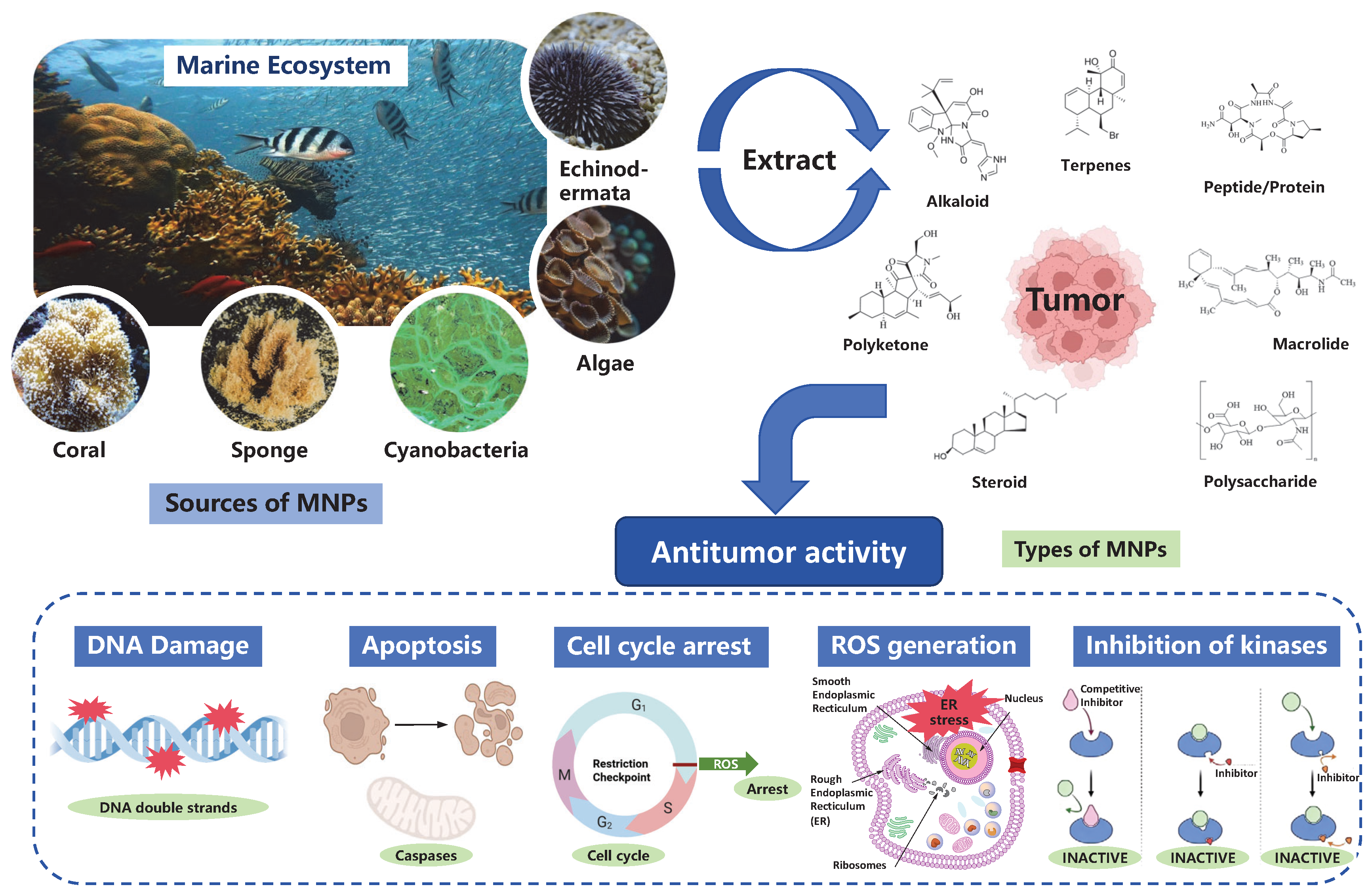
2.1. Marine-Derived Terpenoids
Marine-derived terpenoids are characterized by isoprene units as basic skeleton, and structurally cover multiple levels including monoterpenes, sesquiterpenes, diterpenes, triterpenes and higher terpenoids. Terpenoids are widely distributed in the metabolic systems of sponges, algae, fungi and their symbiotic microorganisms, presenting complex oxidation states, cyclization patterns and stereoconfiguration diversity. The unique marine environment drives their chemical evolution toward highly oxidized and structurally novel scaffolds, establishing terpenoids as a prolific source of anticancer drug leads [
34].
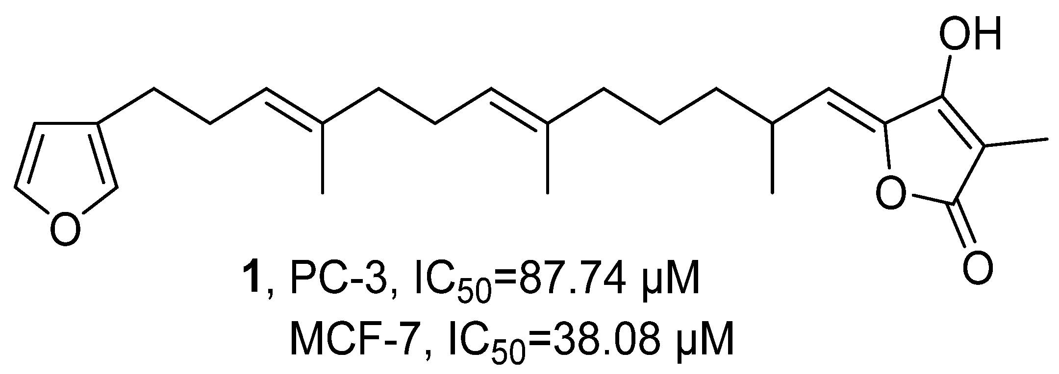
In 2020, Beukes’ research group isolated the terpene component Variabilin (
1) from the South African sponge
Ircinia sp. through silica gel column chromatography and HPLC purification (
Figure 2) [
35].
1 was determined to be C
25H
34O
4 using HRMS, and its structure was confirmed by
1H NMR and
2D NMR. Using the WST-1 method, the activity assessment showed that
1 had a significant inhibitory effect on prostate cancer cells PC-3, with an IC
50 value of 87.74 µM, but its efficacy was inferior to Ceramide which refers to a class of waxy lipid compounds formed by the combination of sphingosine and fatty acids through amide bonds (IC
50 = 4.81 µM); moreover,
1 also exhibited significant inhibitory activity against breast cancer cells MCF-7 (IC
50 = 38.08 µM), which was comparable to the standard compound Ceramide (IC
50 = 33.61 µM).
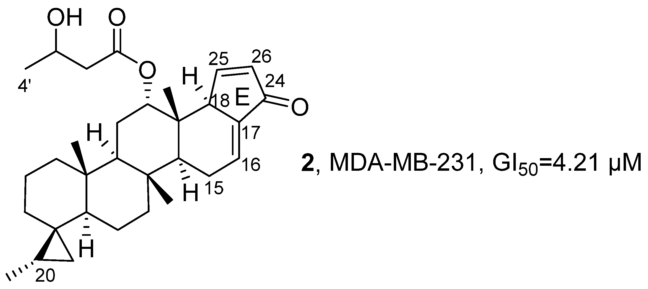
In 2021, Lee’s research group isolated 15 new scalarane-type sesterterpenoids from the marine sponge
Dysidea sp. collected from Bohol Island using methanol and dichloromethane extracts (
Figure 3) [
36]. Taking
2 as a reference, its molecular formula was determined to be C
31H
44O
4 using HRMS, and the core cyclopentenone E-ring skeleton was inferred based on the correlation signals in the HMBC spectrum between H-25 and C-17/C-18/C-24, and between H-26 and C-17/C-24/C-25, which was further confirmed by the H-18-H-25-H-26 coupling chain observed in the
1H−
1H COSY spectrum. Using CCK-8 assay, the activity assessment showed that, compared with other compounds,
2 exhibited the strongest cytotoxic activity against MDA-MB-231 cells, with a GI
50 value of 4.21 μM. SAR studies indicated that the Δ
15,16 double bond generally reduced activity, the methylene elongation at the C-4′ position helped enhance activity. However, SAR analysis also suggested that oxidation at C-20 reduced cytotoxic potency relative to the non-oxidized analogs, indicating that the hydrophobic substitution pattern at this position is important for activity.
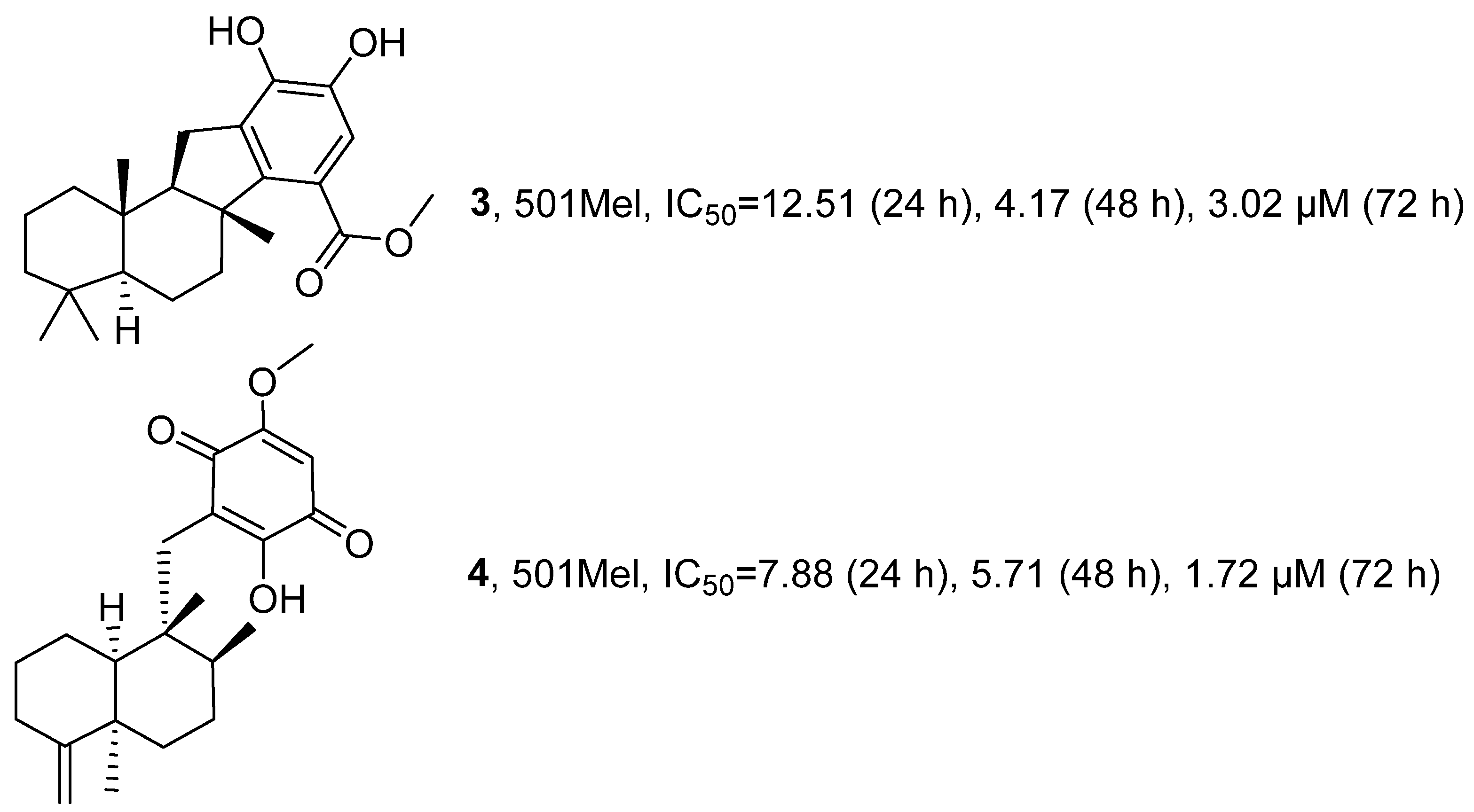
In 2022, Carpi’s research group conducted in-depth investigations on two existing terpenoids, Pelorol (PEL) (
3) and 5-epi-Ilimaquinone (EPI) (
4), isolated from the methanol exudates of the marine sponge
Dactylospongia elegans, building upon previous explorations (
Figure 4) [
37]. Activity evaluation results showed that when tested on human 501 Mel melanoma cells, the average IC
50 values for
3 after 24, 48, and 72 h of administration were 12.51 µM, 4.17 µM and 3.02 µM, respectively, whereas the average IC
50 values for
4 during the same time periods were 7.88 µM, 5.71 µM and 1.72 µM, respectively.
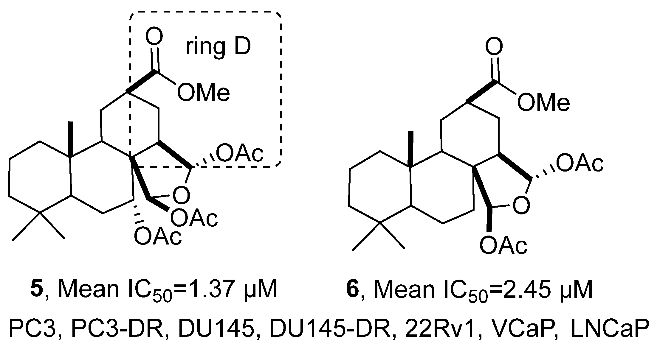
In 2022, Dyshlovoy’s research group isolated six classes of spongian-type diterpenoids from the methanol extract of the marine sponge
Spongionella sp., including five known compounds (
Figure 5) [
38]. The molecular formula of the new compound
5 was determined by HRMS to be C
27H
40O
9, and
1H NMR spectrum revealed the presence of three sets of methyl groups, three sets of acetyl methyl groups, one methoxycarbonyl group, and two sets of acetal protons. The HMBC spectrum confirmed the attachment of an additional acetoxy group to the C-7 position, and further information from HSQC, COSY, and NOESY spectra was integrated to deduce its relative configuration and the structural feature of the opened D-ring. Cell viability was examined by MTT assay. Compared with the positive control drug cisplatin (average IC
50 = 8.40 μM), compounds
5 and
6 exhibited stronger cytotoxic activity against multiple prostate cancer cell lines (PC3, PC3-DR, DU145, DU145-DR, 22Rv1, VCaP, LNCaP), with average IC
50 values of 1.37 μM and 2.45 μM, respectively. SAR studies indicated that the opening of the D-ring modification plays a crucial role in enhancing the cytotoxicity and selectivity of these compounds.
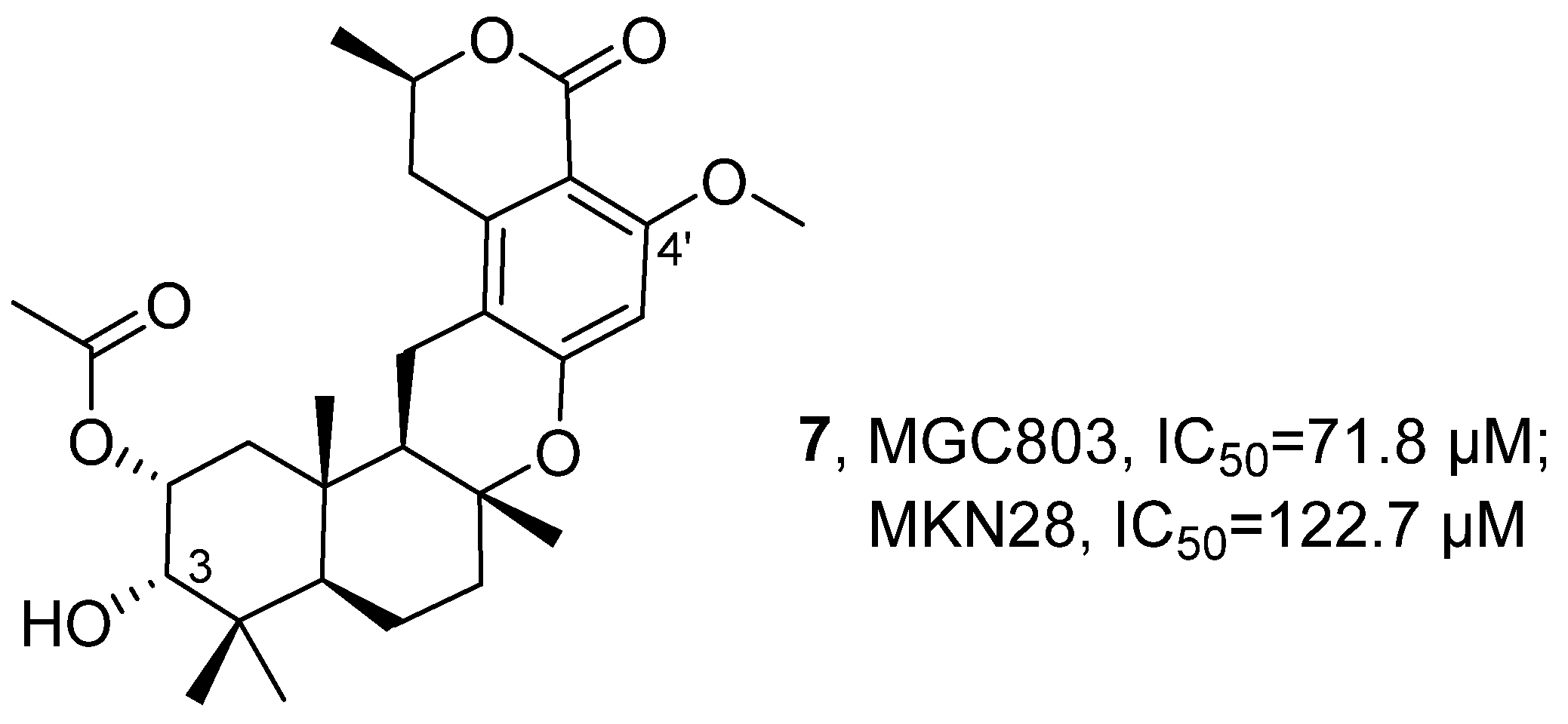
In 2022, Wang’s research group isolated a novel methylenediterpene compound, Taladrimanin A (
7), and 11 related known compounds from the marine-derived fungus
Talaromyces sp. HM6-1-1 (
Figure 6) [
39]. For
7, its structure was determined by various spectroscopic techniques: HRMS indicated its molecular formula as C
28H
38O
7 with an unsaturation degree of 10; NMR (including
1H NMR,
13C NMR, DEPT, and HSQC) revealed that it contained 7 methyl groups, 6 methylene groups, and 10 non-protonated carbons; COSY correlation signals identified four independent spin systems; HMBC spectra further revealed a drimane-type sesquiterpene unit, which was connected to an ester carbonyl and two oxygenated sp
2 hybridized quaternary carbons. Combining the sesquiterpene and isochromenone skeletons, as well as the substitution at C-3 and C-4′ positions,
7 was ultimately determined to be a hybrid compound of a pentaketide-peptide and a terpene. Bioactivity was examined by CCK8 assay. Compared with the positive control drug cisplatin (IC
50 = 6.2 μΜ, 19.7 μΜ), compound 7 exhibited weaker anti-tumor activity against gastric cancer cells MGC803 and MKN28, with IC
50 values of 71.8 μM and 122.7 μM, respectively. SAR analysis indicated that the activity of
7 originated from the hybridization of the unique drimane-type sesquiterpene unit and an isochromenone unit derived from polyketides.
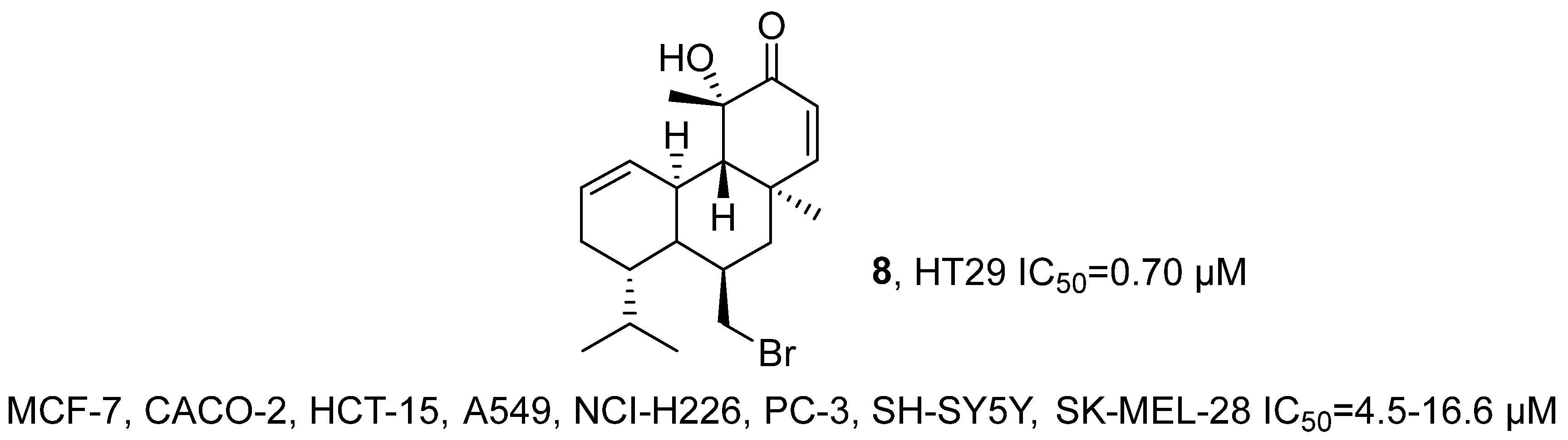
In 2022, Pedrosa’s research group isolated the brominated diterpene compound sphaerococcenol A (
8) from the red alga
Sphaerococcus coronopifolius Stackhouse 1797 (
Figure 7) [
40]. After extraction, the structure of the compound was characterized by MS and NMR techniques and confirmed by comparison with the reports by Rodrigues [
41]. The results of the activity evaluation indicated that
8 exhibited broad-spectrum cytotoxic activity against 8 tumor cell lines (MCF-7, CACO-2, HCT-15, A549, NCI-H226, PC-3, SH-SY5Y, SK-MEL-28) with IC
50 values ranging from 4.5 to 16.6 μM. Notably, it demonstrated significant anti-tumor activity against colon cancer stem cell HT29 spheres with an IC
50 of 0.70 μM.
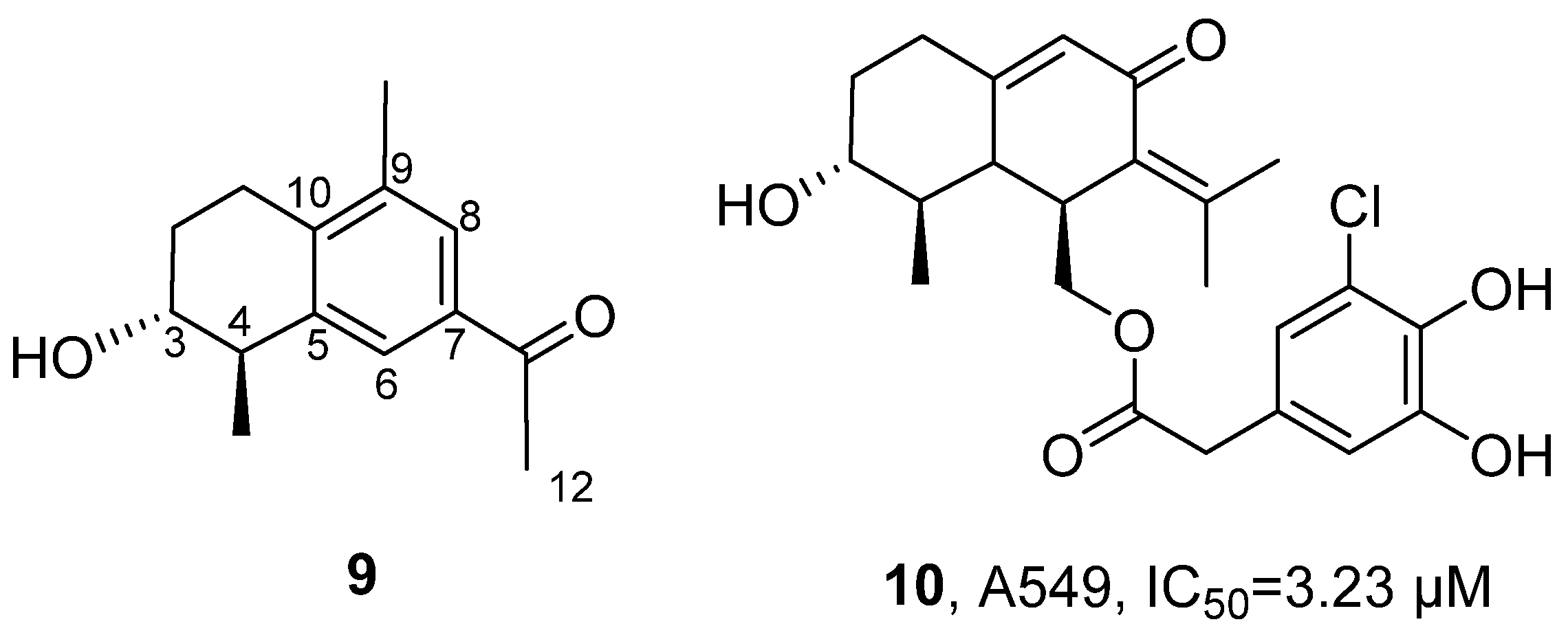
In 2022, Lin’s research group isolated 12 new compounds from the ethyl acetate extract of the yellow-green
Penicillium copticola fungus that lives symbiotically with marine sponges (
Figure 8) [
42]. Among them, 10 compounds were identified as furostanol sesquiterpenes, and the other two compounds were confirmed as glucosides. For Copteremophilane A (
9), molecular formula was determined as C
14H
18O
2 by HRMS;
13C NMR and DEPT spectra showed 14 carbon signals, including six aromatic carbons, one ketone carbonyl carbon, and seven alkyl carbons. Through two meta coupling aromatic proton signals (H-6 and H-8), a tetrasubstituted aromatic ring was identified; further combined with HMBC and COSY correlation signals, the presence of a methyl group at C-9, acetoxy group at C-7, methyl group at C-4, and hydroxyl group at C-3 was determined, and it was inferred that the C-5 and C-10 units fused with the aromatic ring at the C-5 and C-10 positions to form a cyclohexene ring; NOE correlation signals indicated that H
3-15 was oriented towards OH-3. Comparison of the experimental ECD data to those calculated for (3
R, 4
R, 5
S)-8 established 3
R, 4
R and 5
S configurations for
9. The activity evaluation was examined by MTT assay. Results showed that Copteremophilane H (
10) had selective inhibitory activity against A549 cells, with an IC
50 value of 3.23 μM. The SAR analysis indicated that the introduction of the benzene acetic acid unit significantly enhanced the anti-tumor effect of furostanol compounds, and the position of substitution and the composition of the acyl group had a significant impact on their selective inhibitory activity.
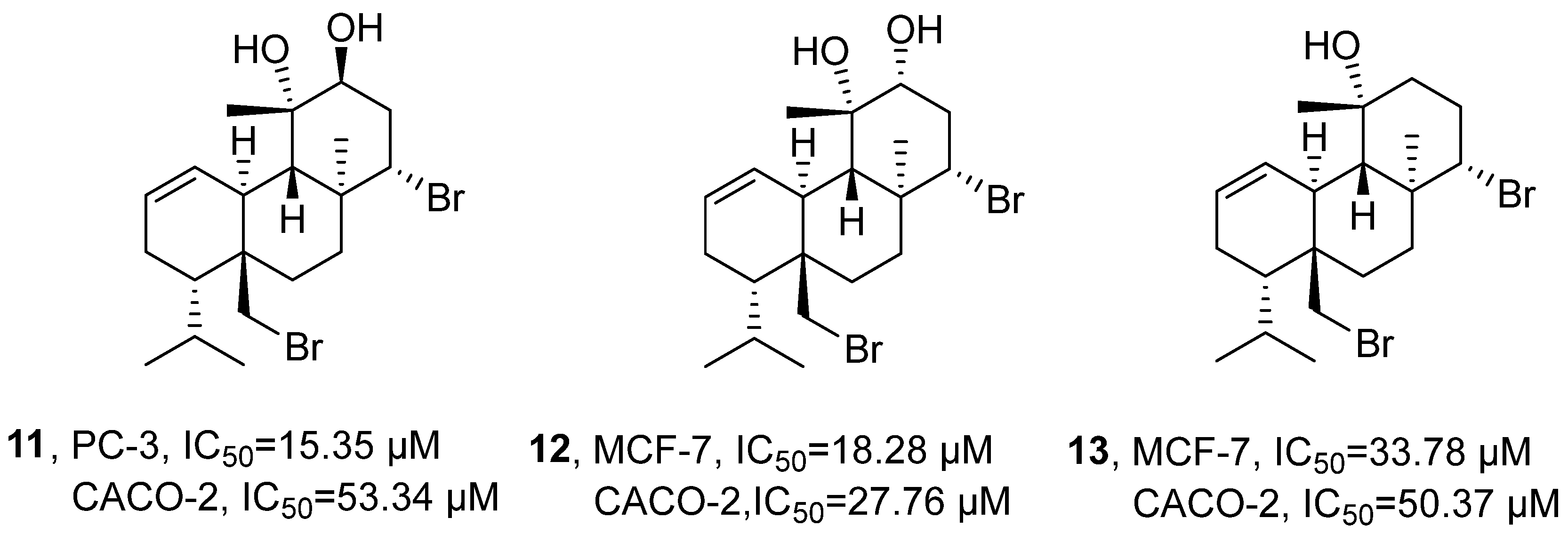
In 2022, Pedrosa’s research group isolated three brominated diterpenoids (12
S-hydroxy-bromosphaerol, 12
R-hydroxy-bromosphaerol, and bromosphaerol) (
11–13) from the dichloromethane extract of
Sphaerococcus coronopifolius (
Figure 9) [
43]. Through the comprehensive application of NMR (
1H and
13C NMR, COSY, HMBC, and HSQC) techniques, their chemical structures were finally determined. Activity evaluation results indicated that
12 exhibited significant anti-tumor activity, with an IC
50 of 18.28 µM in MCF-7 cells and 27.76 µM in Caco-2 cells. For
11, the IC
50 was 15.35 µM in PC-3 cells and 53.34 µM in Caco-2 cells. In comparison,
13 showed IC
50 values of 33.78 µM in MCF-7 cells and 50.37 µM in Caco-2 cells.
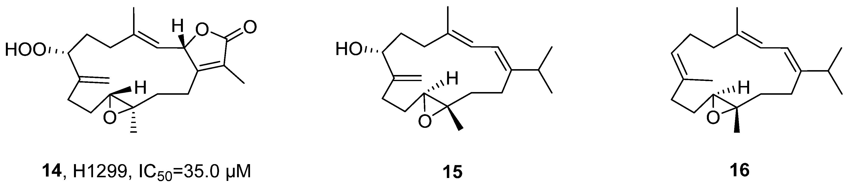
In 2023, Wang’s research group isolated two undescribed cembranoids, Sarcoboettgerol D (
15) and Sarcoboettgerol E (
16), along with four known related analogs, from the acetone extract of
Sarcophyton boettgeri (
Figure 10) [
44]. The structural elucidation, exemplified by
15, began with the determination of its molecular formula as C
20H
32O
2 by HRMS. The characteristic functional groups, including a conjugated diene and an exocyclic double bond, as well as the planar structure were established through comprehensive NMR analysis (
1H,
13C, COSY, and HMBC). Subsequently, the relative and absolute configurations were unambiguously assigned using combined QM-NMR and TDDFT-ECD calculations. In vitro activity evaluation indicated that Sarcomililatin B (
14) exhibited weak cytotoxicity against H1299 lung cancer cells, with an IC
50 value of 35.0 μM.
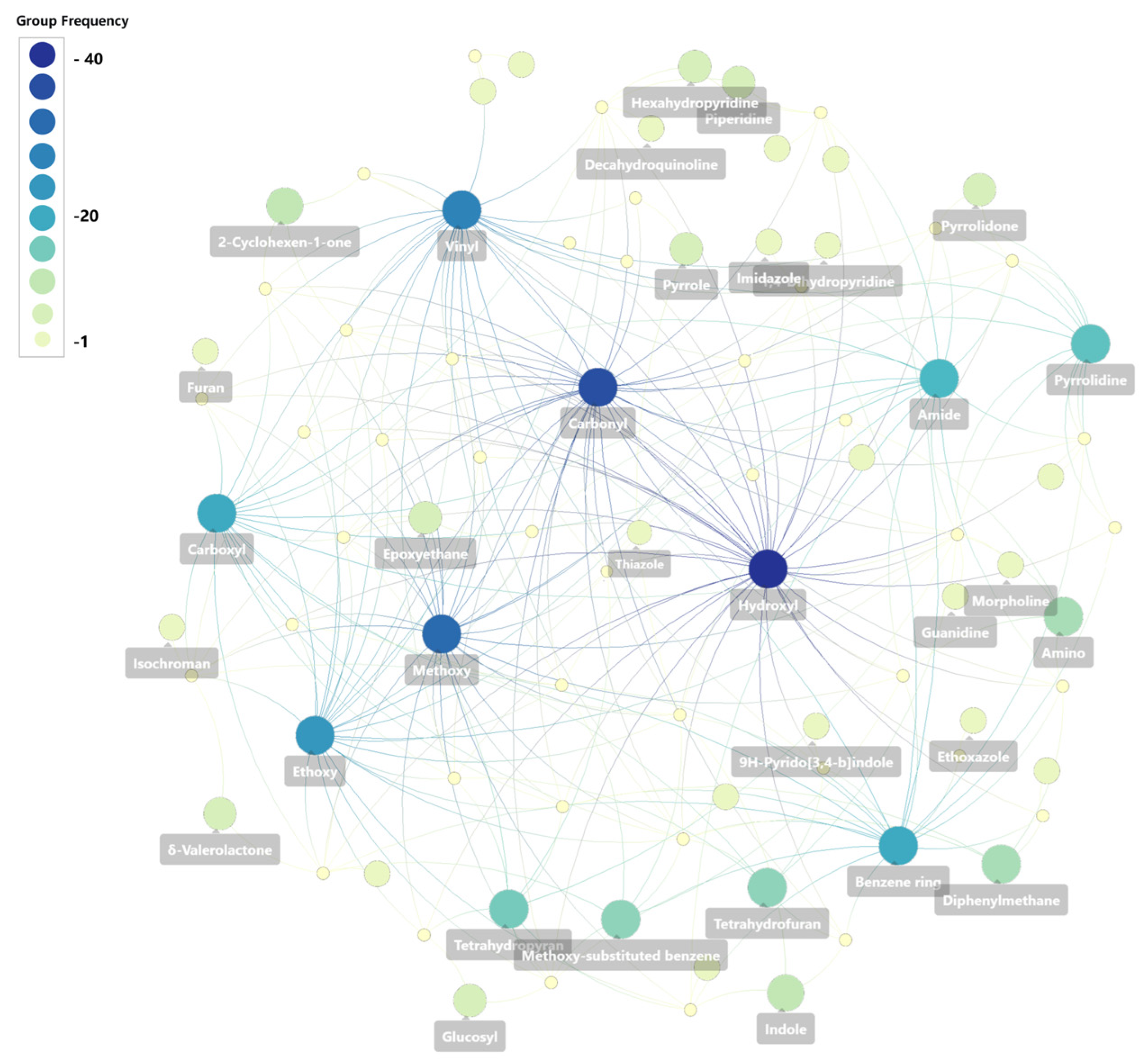
In terpenoids, hydroxyl, ketone, double bond, and ester groups are the most frequent motifs, forming a highly oxidized core. The most active terpenoids originate mainly from sponges (e.g., Ircinia, Dysidea, Spongionella) and red alga (Sphaerococcus coronopifolius). D-ring opening and bromination greatly enhance selectivity and cytotoxicity. Trends from 2020 to 2024 include that detailed SAR studies (e.g., Δ15,16 double bond reduces activity, C-4′ methylene elongation increases activity), absolute configuration determination by QM-NMR and ECD calculations.
2.2. Marine-Derived Alkaloids
Marine-derived alkaloids are a class of natural products with nitrogen-containing heterocycles as the basic framework. Their structural types are diverse, including indoles, quinolines, quinazolines, and polycyclic amines, etc. Marine-derived alkaloids often exhibit complex characteristics such as halogen substitution, high oxidation states, and multi-center chirality, which give them remarkable marine chemical uniqueness. They are mainly discovered from sponges and their symbiotic microbial systems and are an important source of anticancer drug lead compounds.
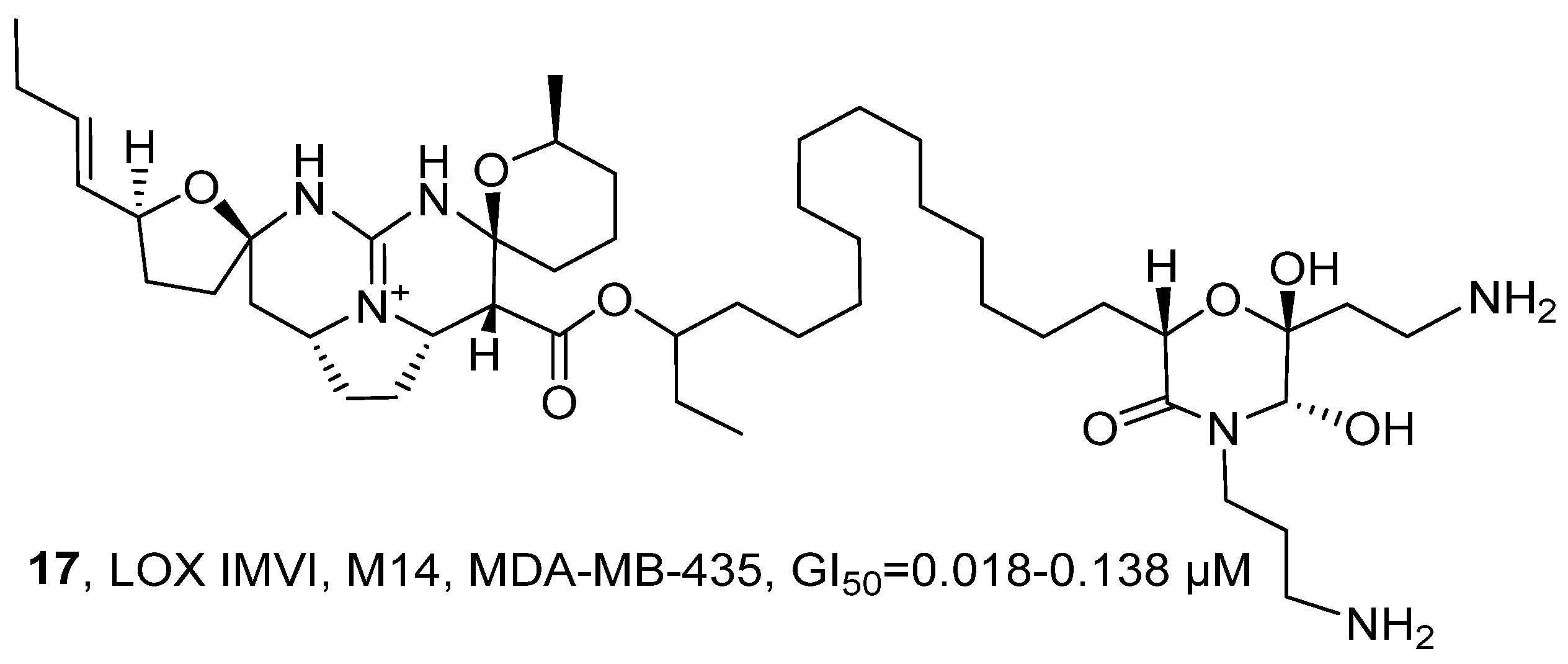
In 2020, T. Hamann’s research group isolated a biogenic alkaloid named Monanchocidin A (
17) from the ethanol extract of the
Monanchora genus sponges in the subarctic waters (
Figure 11) [
45]. The structure of
17 was determined by comparing it with its known standard data set (including
1H NMR,
13C NMR and HRMS). Compared with the positive control drug paclitaxel,
17 exhibited stronger cytotoxic activity against LOX IMVI, M14, and MDA-MB-435 cells, with GI
50 values ranging from 0.018 to 0.138 µM. By comparing the SAR with the structurally similar guanidine-based alkaloid Ptilomycalin A, it was found that they had a high Pearson correlation (0.638), indicating that the pentacyclic guanidine core skeleton and the spermine fragment at the end are crucial for its anti-tumor activity.
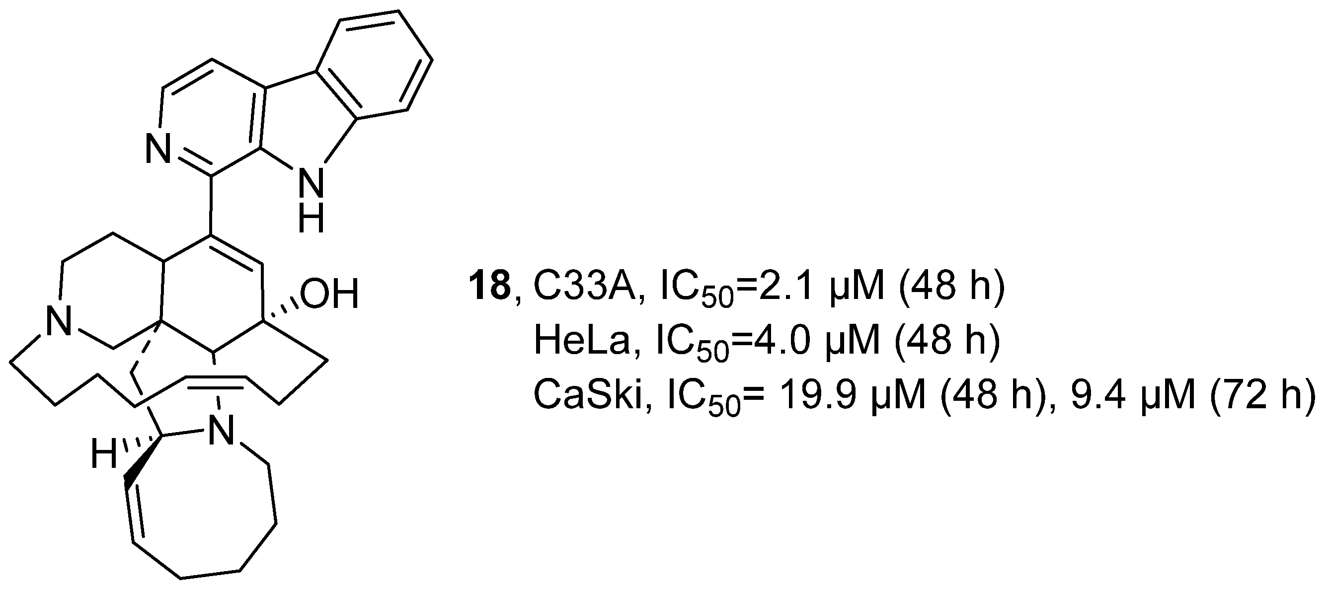
In 2020, T. Hamann’s research group isolated Manzamine A (
18) from the Indonesian sponge
Acanthostrongylophora (
Figure 12) [
46].
18 belongs to the
β-cycline alkaloids with complex polycyclic structures. Through techniques such as HRMS, LCMS/MS and NMR, the main components and secondary components of
18 were identified. Biological activity was evaluated by CellTiter-Glo cell viability assay.
18 demonstrated potent inhibitory activity against C33A and HeLa cell lines, with respective 48 h IC
50 values of 2.1 μM and 4.0 μM. Against CaSki cells, its inhibitory effect was further characterized by IC
50 values of 19.9 μM at 48 h and 9.4 μM at 72 h.
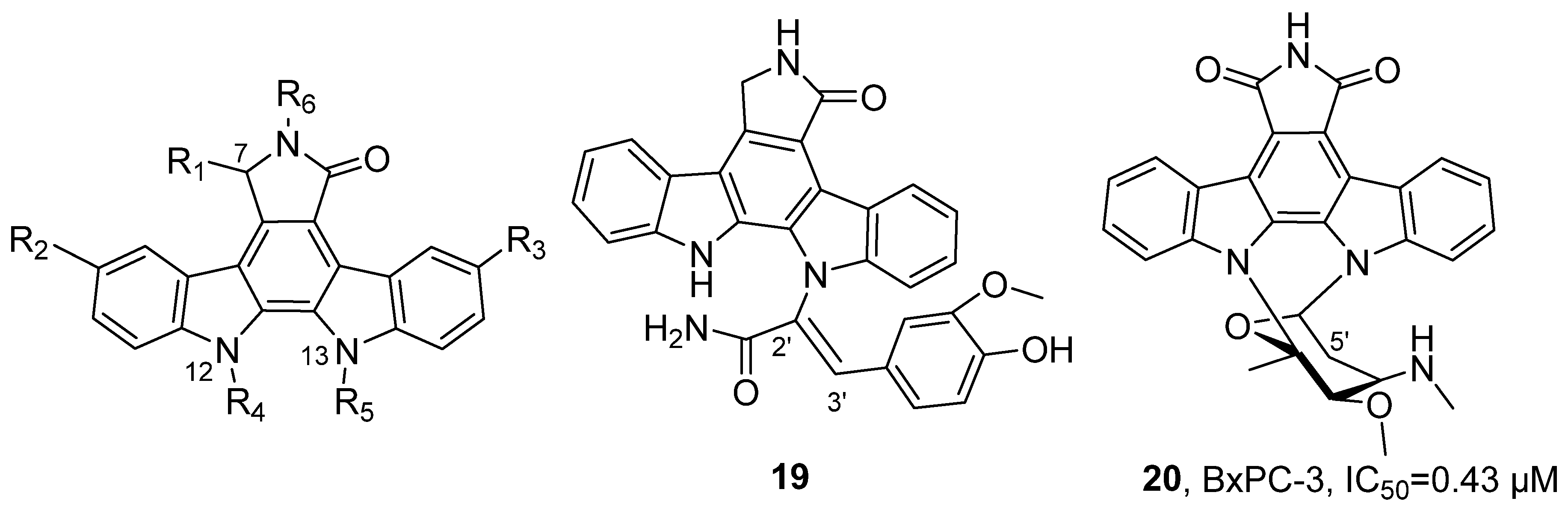
In 2020, Chen’s research group isolated and identified 69 natural indolocarbazole alkaloids from the ethyl acetate extracts of four marine-derived
Streptomyces strains, including 15 novel compounds (
Figure 13) [
47]. For
19, molecular formula was determined as C
30H
22N
4O
4 with 22 degrees of unsaturation by HRMS. Further combined with NMR data, correlation spectroscopy (COSY, HMBC), and single-crystal X-ray diffraction analysis, the connectivity of its fragment structures was confirmed, including the geometry of the C2′-C3′ double bond. Activity evaluation results showed that
20 exhibited inhibitory activity against nine human tumor cell lines, with the highest sensitivity observed in pancreatic cancer cells BxPC-3 (IC
50 = 0.43 μM). In mouse xenograft model experiments,
19 significantly inhibited tumor growth (tumor growth inhibition rate TGI = 49.6%). SAR analysis revealed that the presence of an aglycone is a key factor for maintaining activity; compounds with a disaccharide linkage at N-12 and N-13 and no substitution at the C-5′ position demonstrated stronger activity; the configuration at the C-7 position influences activity; for example, the (
S)-methoxy substituted compound showed lower activity than its epimer; additionally, the chair conformation exhibited higher activity compared to the boat conformation.
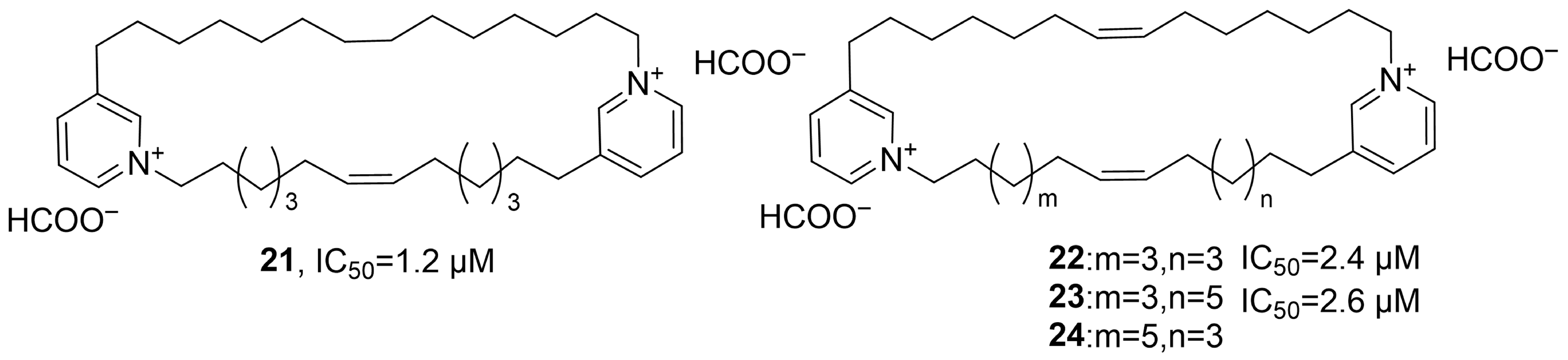
In 2021, Tsukamoto’s research group isolated four new pyridine alkaloids, neopetrosidines A–D (
21–
24), from the ethanol extract of the marine sponge
Neopetrosia chaliniformis (
Figure 14) [
48]. Taking the structure analysis of neopetrosidine A as an example, HRMS determined its molecular formula to be C
38H
62N
2; the
1H NMR spectrum showed aromatic proton signals and alkene proton signals; through COSY, HMBC and HSQC spectroscopic analysis, it was confirmed that the structure contained two 1,3-disubstituted pyridine units; combined with the
13C NMR chemical shift information, the double bond configuration was further determined to be the Z form. Using MTT assay, activity studies showed that
21–
24 could significantly inhibit the growth of HeLa/Fucci2 cells, with IC
50 values of 1.2, 2.4 and 2.6 μM, respectively.
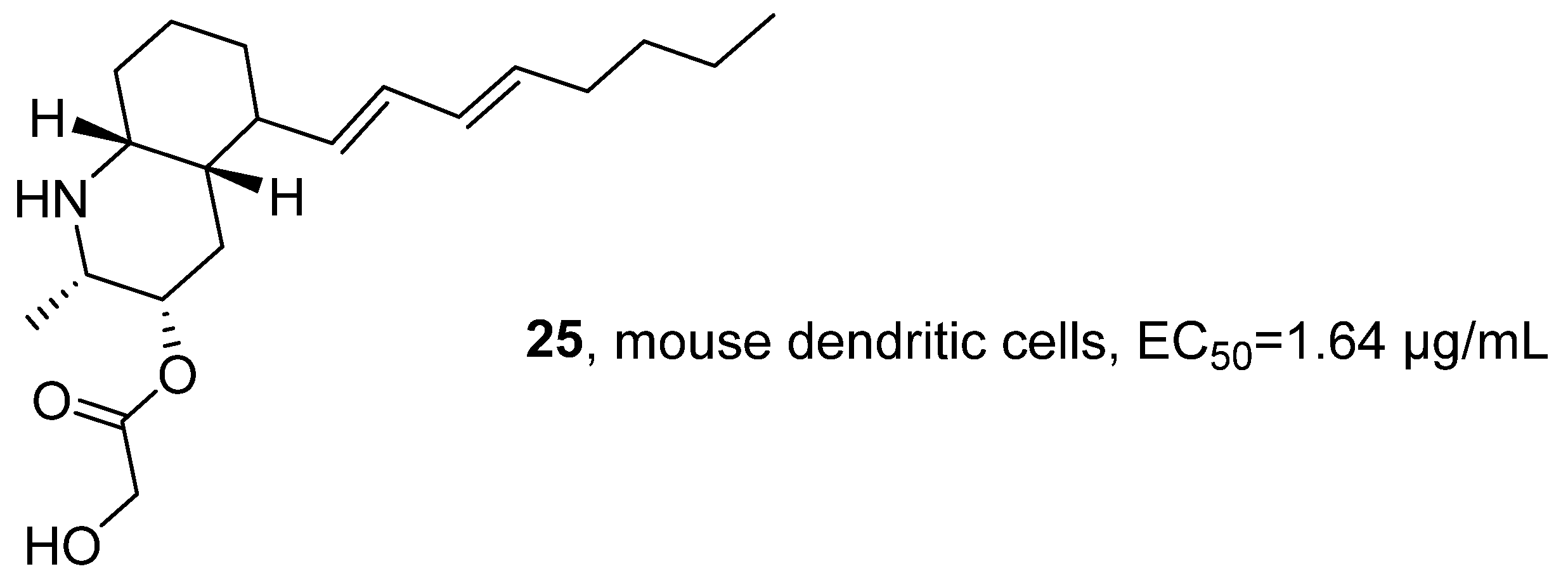
In 2021, Fontana’s research group isolated the marine alkaloid Lepadin A (
25) from the methanol extract of
Clavelina lepadiformis sp. B collected from the Tyrrhenian Sea (
Figure 15) [
49]. In terms of structural identification, the molecular formula was determined to be C
20H
34NO
3 by HRMS, and the
1H NMR spectral signals confirmed the presence of the butadienyl system and two methyl groups. The activity evaluation results showed that
25 had cytotoxic effects on various cancer cell lines (such as lung cancer cells CALU-1, CALU-3, HCC827, melanoma cells MALME-3M, A375, A2058, and multiple myeloma cells KMS-12, RPMI 8226, and JJN-3); the test on D1 cells confirmed its immunomodulatory activity, with an EC
50 of 1.64 μg/mL, while its cytotoxic activity was 4.20 μg/mL in the same cell line.
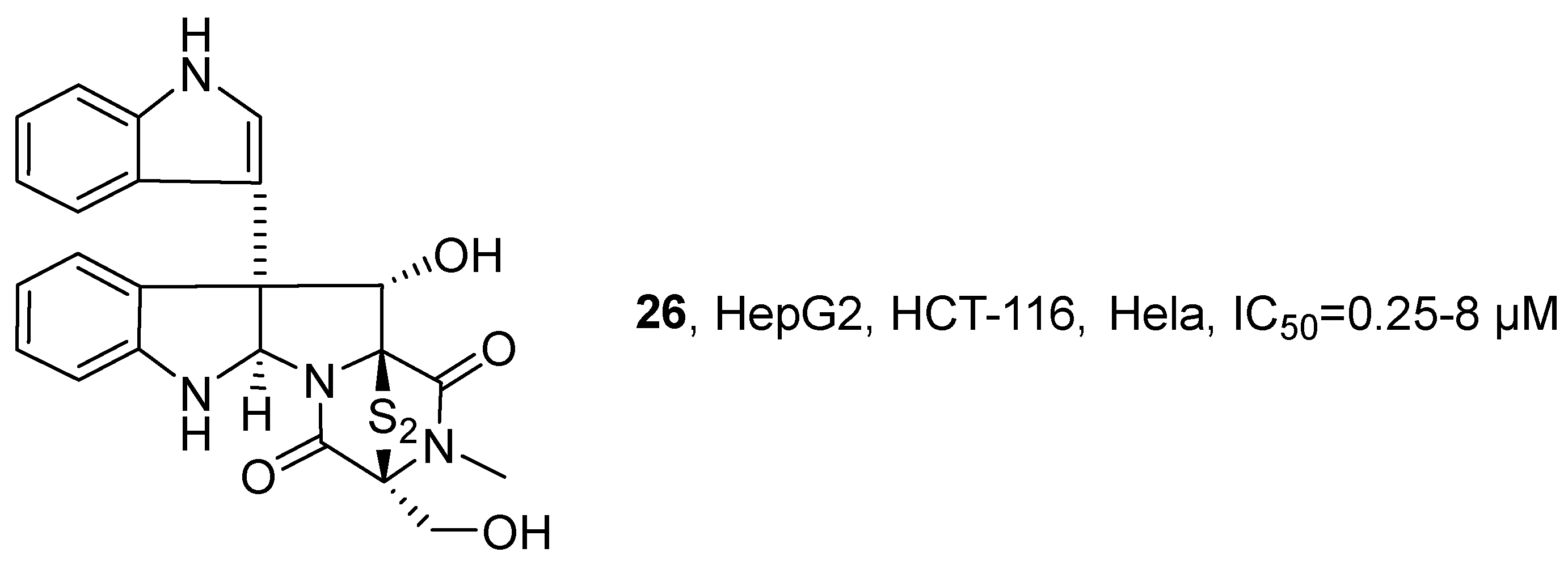
In 2021, Li’s research group isolated epipolythiodiketopiperazines (ETPs) compounds from the MNP,
Tilachidium sp., an endophytic fungus found in mangroves. GQQ-792 (
26), identified from the MNPs, also belongs to the ETPs class of alkaloids (
Figure 16) [
50]. The activity evaluation results showed that
26 exhibited IC
50 values for HepG2, HCT-116, and Hela cell lines within the range of 0.25–8 μM. The SAR study indicated that the disulfide group is the key pharmacophore, and the absence of this group would significantly reduce the inhibitory activity.
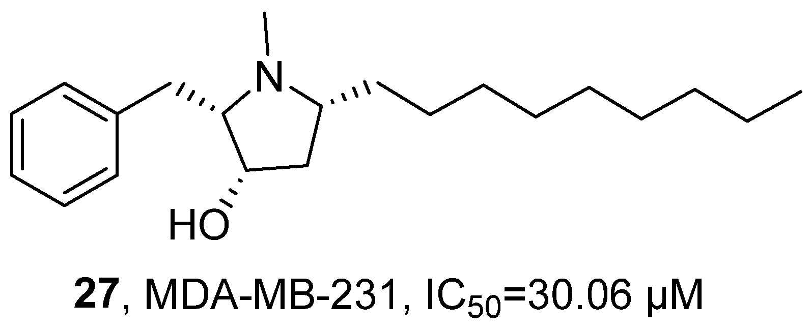
In 2023, Rocha’s research group isolated the alkaloid Preussin (
27) from the sponge-associated fungus
Aspergillus candidus KUFA 0062 (
Figure 17) [
51]. Based on previous studies [
52,
53,
54,
55,
56,
57,
58], they further systematically evaluated its anti-tumor activity. The activity test results showed that
27 exhibited anti-tumor activity against MDA-MB-231 cells, with an IC
50 value of 30.06 μM.
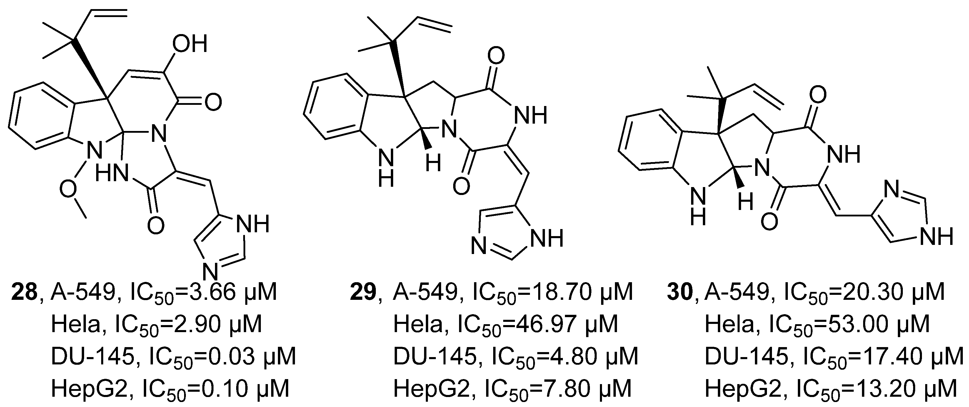
In 2023, A. Bogari’s research group isolated three indole-type alkaloids from the secondary metabolites of the marine fungus
Penicillium chrysogenum S003 sourced from the Red Sea using silica gel column chromatography: meleagrin (MEL,
28), roquefortine C (ROC,
29), and isoroquefortine C (ISO,
30) (
Figure 18) [
59]. Taking
28 as an example for compound structure confirmation,
1H-NMR and
13C-NMR analyses determined its relevant functional groups, and 2D NMR was used to verify its tetracyclic skeleton and the relative configuration of the substituents. In the assessment of anti-tumor activity,
28 demonstrated significant inhibitory effects across four tumor cell lines. The IC
50 values of
28 were determined as 3.66 μM for the lung cancer cell line A549, 2.90 μM for the cervical cancer cell line HeLa, 0.03 μM for the prostate cancer cell line DU-145, and 0.10 μM for the liver cancer cell line HepG2. In comparison, the positive control doxorubicin yielded IC
50 values of 0.01 μM, 0.05 μM, 0.34 μM, and 0.92 μM against the same respective cell lines. Furthermore,
29 and
30 exhibited notably weaker activities, with IC
50 ranges of 18.70–46.97 μM and 13.20–53.00 μM, respectively, across the tested cell lines. Notably,
28 displayed its strongest inhibitory potency against the prostate cancer cell line DU-145, with an IC
50 of merely 0.03 μM, which was substantially lower than that of doxorubicin (0.34 μM), highlighting its promising anti-tumor potential. The SAR analysis indicated that the extended tetracyclic skeleton system in
28′s structure and its terminal aromatic imidazole side chain facilitated its binding to multiple targets, while the introduction of polar functional groups enhanced its affinity and selectivity with the targets.
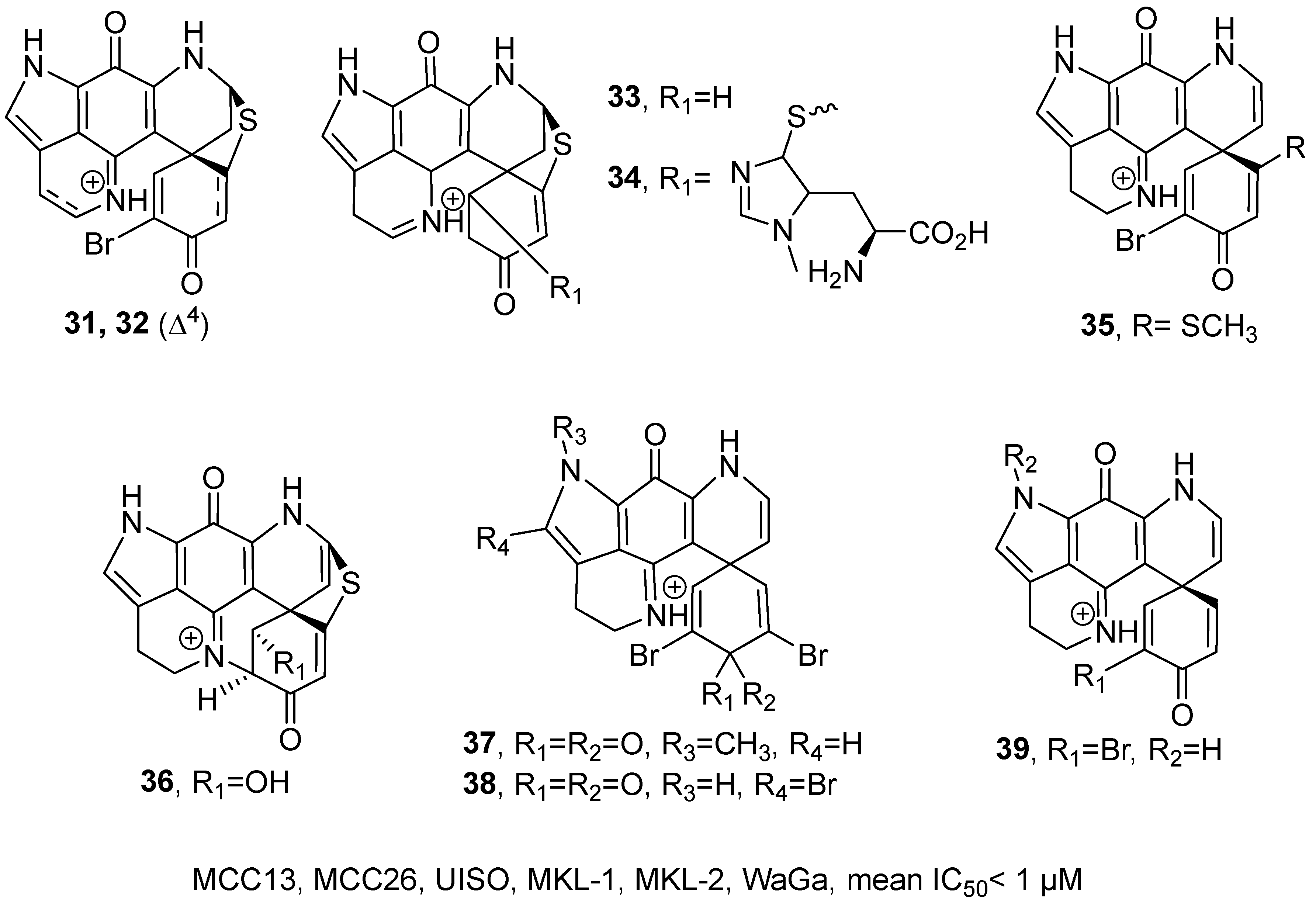
In 2023, J. Henrich’s research group isolated 18 discorhabdin-type compounds from marine sponges and synthesized six derivatives through semi-synthesis (
Figure 19) [
60]. All the compounds belong to alkaloids. Taking the structural elucidation of
34 as an example: its molecular formula was established as C
26H
25N
6O
4S
2 by HRMS, and the
1H NMR spectrum revealed the presence of additional methyl signals, and correlations observed in the
1H and
13C HMBC spectrum. Ultimately, the absolute configuration of
34 was unequivocally assigned based on ECD analysis. Activity assays revealed that discorhabdin derivatives A (
31), B (
32), N-13-demethyl U (
35), P (
37), 14-Br-discorhabdin C (
38), L (
36), E (
39), and G/I (
33) exhibited potent cytotoxicity, with mean IC
50 values below 1 μM across six tested cell lines, including MCC13, MCC26, UISO, MKL-1, MKL-2, and WaGa. SAR analysis identified the α-bromoalkenone group as the essential pharmacophore. Activity decreased upon modification of lactone ring E or insertion of a C-2-N-14 bridge. Methylation of pyrrole ring A and the C-5-C-8 thioether bridge enhanced potency, while saturation of the C-4-C-5 double bond improved activity but reduced selectivity. Introducing an alkenyl group between C-7 and C-8 or a bulky substituent at C-5 negatively impacted efficacy.
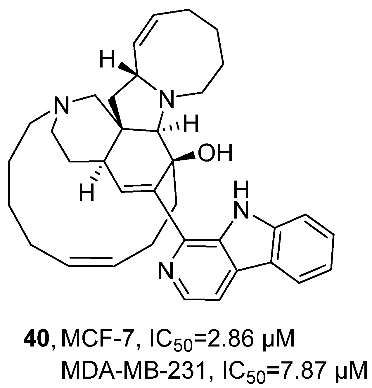
In 2023, Han’s research group conducted an activity assessment on the
β-cycline alkaloid Manzamine A (MA,
40), which was previously isolated from marine sponges (
Figure 20) [
61]. Using MTT assay, activity evaluation demonstrated that, compared with the positive control drug paclitaxel, compound
40 exhibited weaker cytotoxic activity against MCF-7 and MDA-MB-231 breast cancer cells, with IC
50 values of 2.86 μM and 7.87 μM, respectively, while paclitaxel showed IC
50 values of 0.0157 μM and 0.0017 μM, respectively.
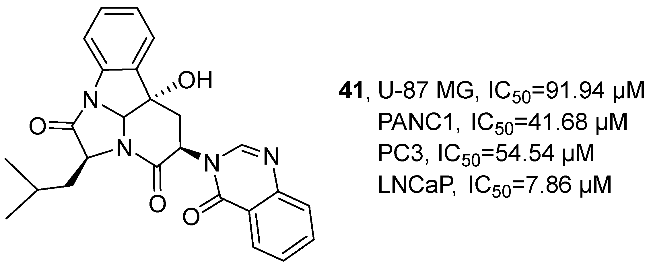
In 2024, Avci’s research group isolated a pyrimidine alkaloid, isopropylchactominine (
41), from the ethyl acetate extract of the symbiotic fungus
Aspergillus carneus (
Figure 21) [
62]. Its molecular formula was determined to be C
25H
25N
4O
4 by HRMS, and the specific structure was verified by
1H NMR and
13C NMR. The activity study showed that the IC
50 values of
41 in U-87 MG, PANC1, PC3 and LNCaP cells were 91.94 μM, 41.68 μM, 54.54 μM and 7.86 μM, respectively.
In alkaloids, nitrogen-containing heterocycles (pyridine, indole, pyrrolidine, guanidine), amide, and aromatic rings are most frequent. The most potent alkaloids come from sponges (Monanchora, Acanthostrongylophora, Neopetrosia) and marine fungi (Aspergillus, Penicillium). Among them, Meleagrin (25) shows an IC50 as low as 0.03 μM against DU-145 prostate cancer cells, far better than doxorubicin; Monanchocidin A (17) exhibits GI50 values of 0.018–0.138 μM against melanoma lines, surpassing paclitaxel. Pentacyclic guanidine core or epidithiodiketopiperazine (ETP)-containing alkaloids display the best activity. Trends from 2020 to 2024 include that structural elucidation by HRMS, 2D-NMR, and X-ray diffraction; identification of pharmacophores (e.g., α-bromoalkenone, disulfide); semi-synthetic optimization; and target prediction revealing multi-target actions on kinases and proteasomes.
2.3. Marine-Derived Steroids
Marine-derived sterols, encompassing sulfated sterols, sterol derivatives, and related congeners, are primarily isolated from sponges and other benthic invertebrates. The fundamental sterol scaffold in these compounds frequently undergoes unique structural modifications, including specialized side-chain substitutions, sulfation, activation, or glycosylation, which impart distinct molecular architectures and physicochemical properties relative to terrestrial sterols. This structural diversification, along with their biosynthetic pathways, reflects a pronounced marine chemical specificity, establishing them as a key resource for the discovery of novel anti-tumor lead compounds.
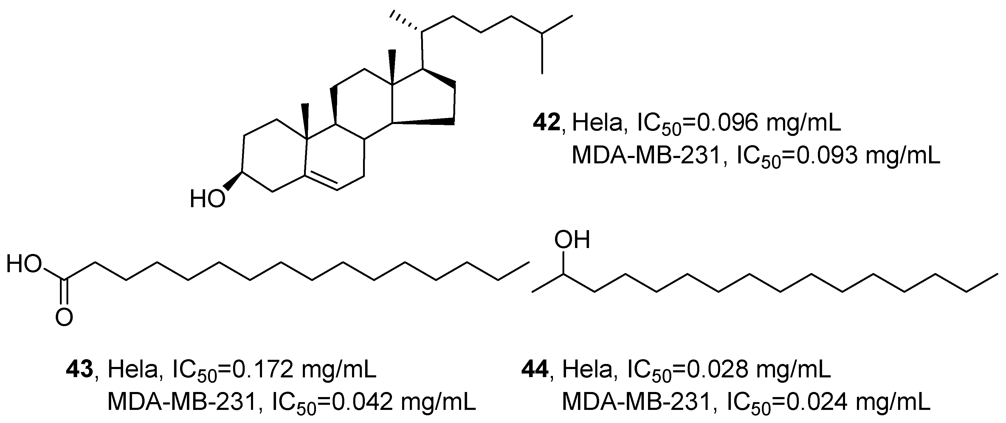
In 2020, Niknejad’s research group isolated three known compounds from the ethyl acetate extract of
Virgularia gustaviana: a steroidal constituent, (3
β)-cholest-5-en-3-ol (
42); a fatty acid, hexadecanoic acid (
43); and a fatty alcohol, 2-hexadecanol (
44) (
Figure 22) [
63]. Taking the structural identification of
42 as an example, by comprehensively applying
1H NMR,
13C NMR, 2D NMR (including
1H−
1H COSY, HMQC, HMBC), MS and UV analysis techniques, and analyzing the absorption at 270 nm, the data were compared with the former reports to confirm that this compound was
42. Bioactivity evaluation revealed that
42 displayed significant cytotoxicity against HeLa and MDA-MB-231 cells, with IC
50 values of 0.096 mg/mL and 0.093 mg/mL, respectively.
43 showed weaker activity against HeLa cells (IC
50 = 0.172 mg/mL) but enhanced potency against MDA-MB-231 cells (IC
50 = 0.042 mg/mL). Notably,
44 exhibited the strongest inhibitory effect on both cell lines, demonstrating IC
50 values of 0.028 mg/mL (HeLa) and 0.024 mg/mL (MDA-MB-231).
In 2021, Zaki’s research group isolated multiple components from a dichloromethane extract of the sea squirt
Eudistoma kaverium, which is from the waters of India (
Figure 23) [
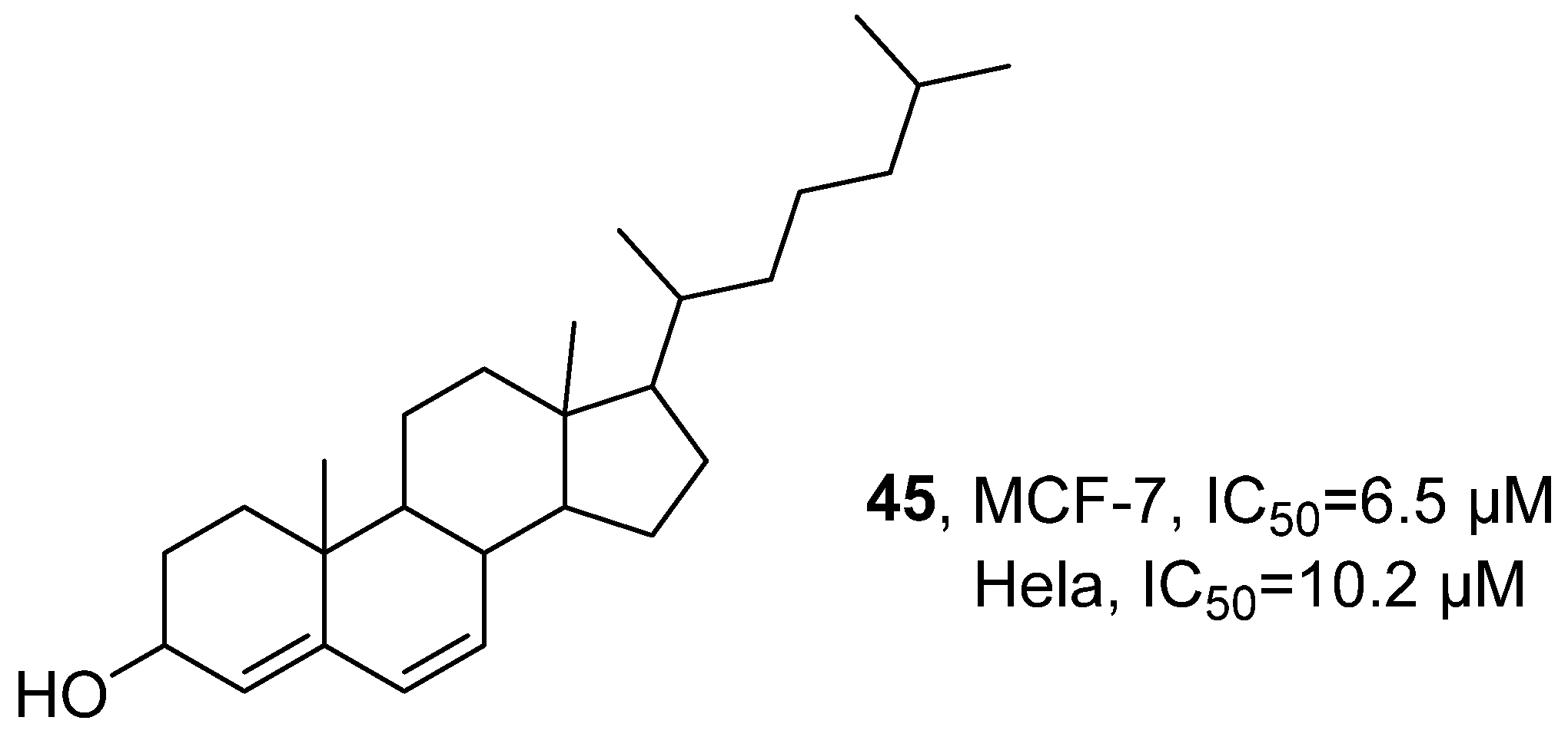
64]. These isolates included steroidal constituents such as cholesta-4,6-dien-3-ol (EK-7,
45). Taking
45 as an example, a comprehensive structural analysis was conducted using techniques such as HPLC, GC-MS, and HRMS to infer its molecular structure and determine the molecular formula as C
27H
44O. Cytotoxicity assessment revealed that
45 demonstrated potent activity against both the breast cancer cell line MCF-7 and the cervical cancer cell line HeLa, with respective IC
50 values of 6.5 μM and 10.2 μM.
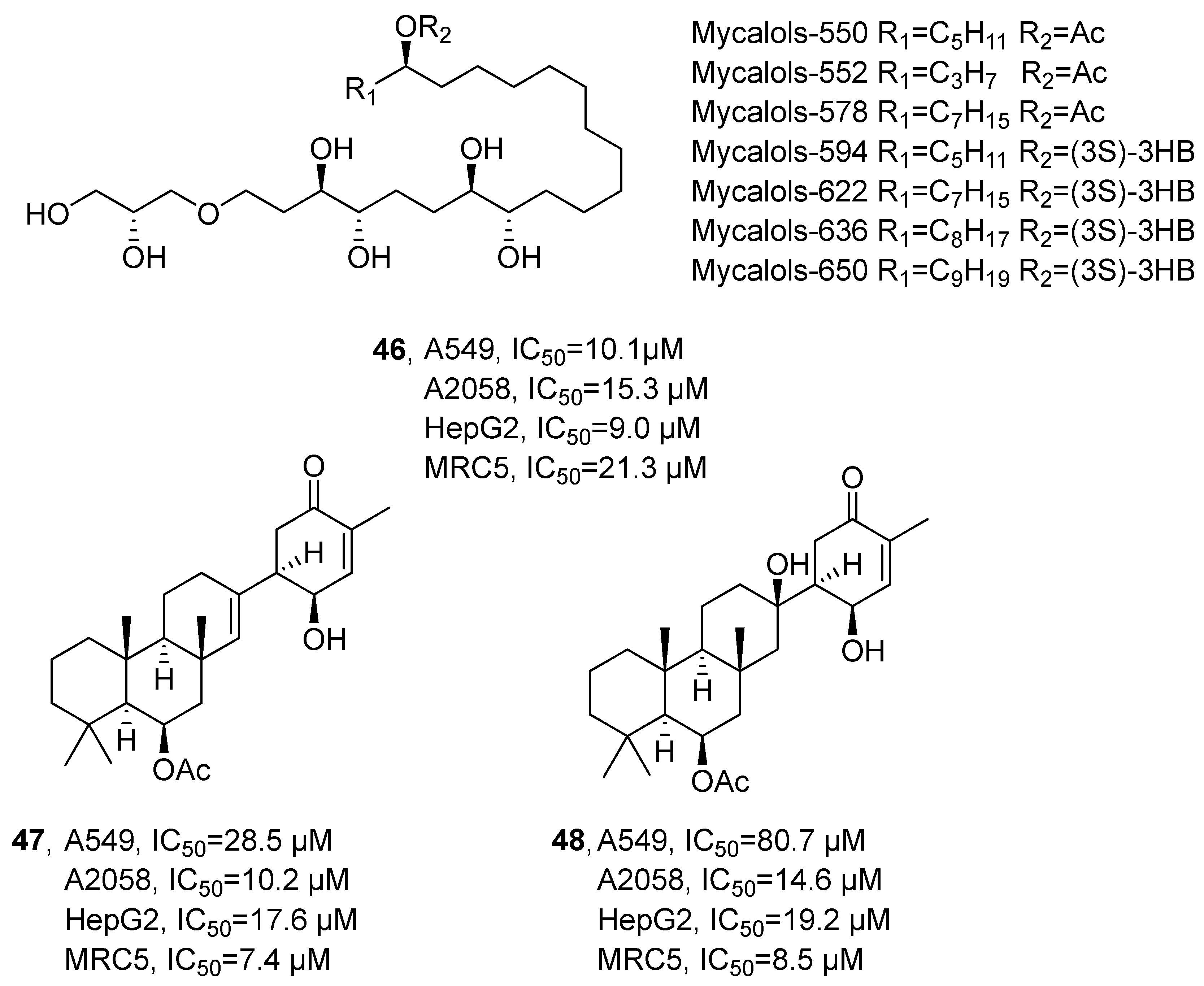
In 2021, Lauritano’s research group isolated mycalols (
46), suberitenones A (
47) and B (
48) from the methanol extracts of the sponges
Hemimycale topsenti and
Haliclona (Rhizoniera) dancoi (
Figure 24) [
65]. The compounds were structurally characterized as an alkyl ether and a terpenoid, respectively, based on NMR and HRMS analyses. Cytotoxicity evaluation revealed that
46 exhibited potent anti-tumor activity against A549, A2058, HepG2, and MRC5 cell lines, with IC
50 values of 10.1, 15.3, 9.0, and 21.3 μM, respectively. In comparison,
47 showed IC
50 values of 28.5, 10.2, 17.6, and 7.4 μM, while
48 displayed IC
50 values of 80.7, 14.6, 19.2, and 8.5 μM against the same panel of cell lines. SAR analysis indicated that cytotoxic potency correlates with alkyl chain length and the nature of substituents.
2.4. Marine-Derived Polyketides
Marine-derived polyketides are precisely synthesized by the modular mechanism of polyketide synthases, feature diverse polycyclic, aromatic, fatty chain, and heterocyclic frameworks with high structural complexity in oxidation states, substitution patterns, and chiral centers. Leveraging the biosynthetic capacity of marine microorganisms and their symbionts, these metabolites greatly expand the accessible natural product chemical space. Their structural complexity and scaffold diversity make them a key class in natural product-based lead discovery for developing novel anticancer therapeutics.
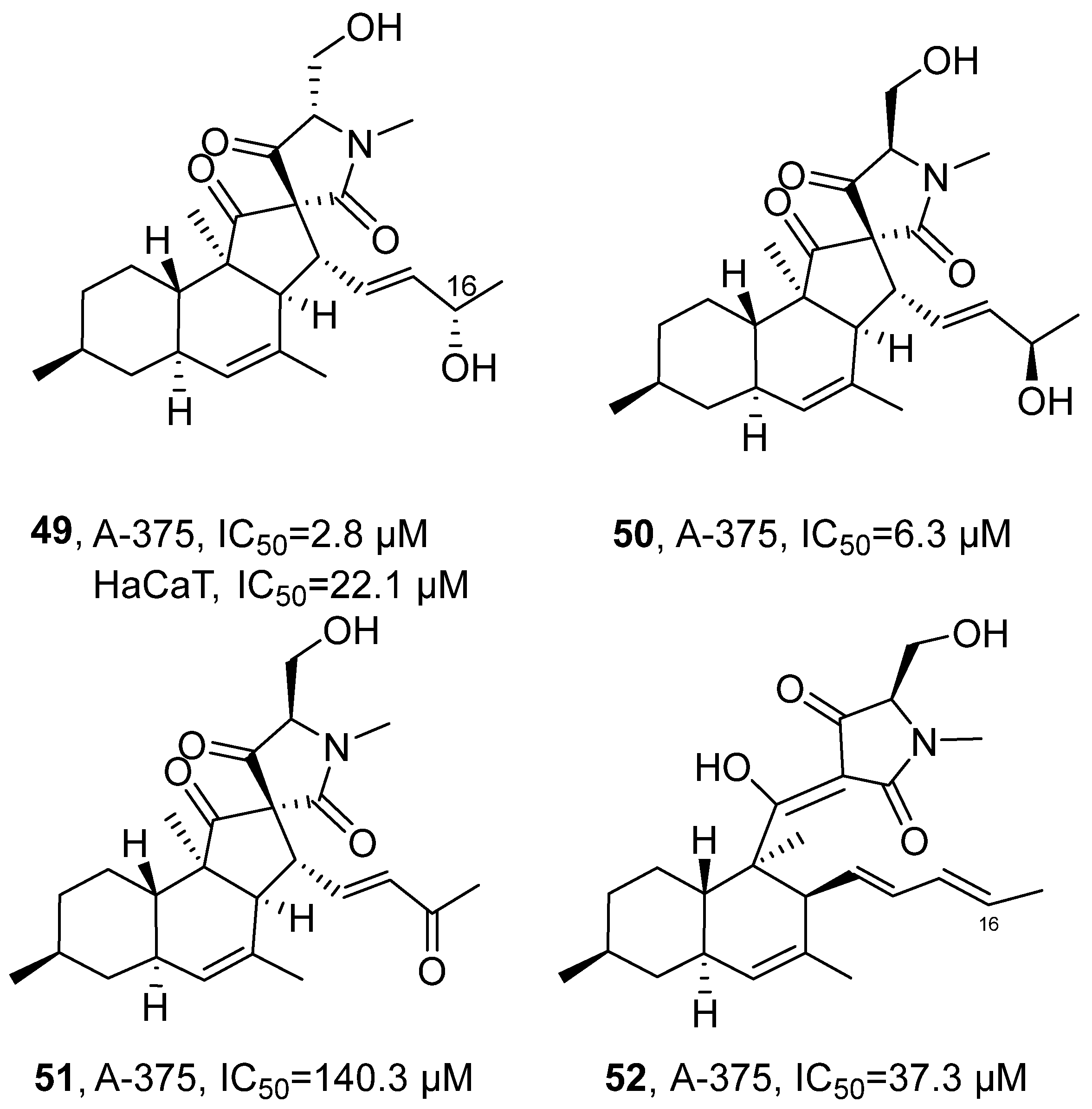
In 2020, Tasdemir’s research group isolated three new decalinoylspirotetramic acid derivatives, pyrenosetins A-C (
49–51), from the ethyl acetate extract of the endophytic fungus
Pyrenochaetopsis sp. FVE-001 of the brown alga
Fucus vesiculosus (
Figure 25) [
66], along with the known compound phomasetin (
52), all belonging to the polyketide class. Taking the structural analysis of
49 as an example, its molecular formula was determined by HRMS as C
25H
35NO
5, corresponding to nine degrees of unsaturation. FT-IR spectrum indicated the presence of hydroxyl and carbonyl groups in the structure. The
1H NMR and DEPT-HSQC spectra displayed five methyl signals including N-methyl. The
13C NMR spectrum further revealed three carbonyl carbon signals. Through COSY correlation signals, three distinct spin systems were identified, while HMBC correlation substantiated the connectivity between the decalin and spirocyclic ring systems. Finally, application of the Mosher ester method established the absolute configuration at C-16 as
S. Compared with the positive control drug doxorubicin (IC
50 = 0.6 μM against A-375 and 22.1 μM against HaCaT), compound 49 exhibited weaker activity against A-375 cells (IC
50 = 2.8 μM) but stronger activity against HaCaT cells (IC
50 = 4.2 μM); compound 50 showed weaker activity against A-375 cells (IC
50 = 6.3 μM); and compounds 51 and 52 exhibited considerably weaker activity against A-375 cells (IC
50 = 140.3 μM and 37.3 μM), respectively. SAR analysis indicated that both the stereochemical configuration and the oxidation state at C-16 critically influence biological activity. The presence of a hydroxyl group at C-16 was associated with enhanced potency; however, variations in stereoconfiguration at this position markedly affected compound selectivity and toxicity, compounds
49 with 16
S configuration and compound
50 with 16
R configuration exhibit higher anti-tumor activity, while compounds
51 with a planar carbonyl structure at C-16 and compounds
52 with a planar carbon–carbon double bond have lower anti-tumor activity; the overall toxicity of the isolated compounds was evaluated using the human keratinocyte cell line HaCaT, and 16
R configuration can effectively reduce the toxicity of the compounds. Conversely, oxidation of the C-16 hydroxyl to a ketone group resulted in a significant reduction in anti-proliferative activity.
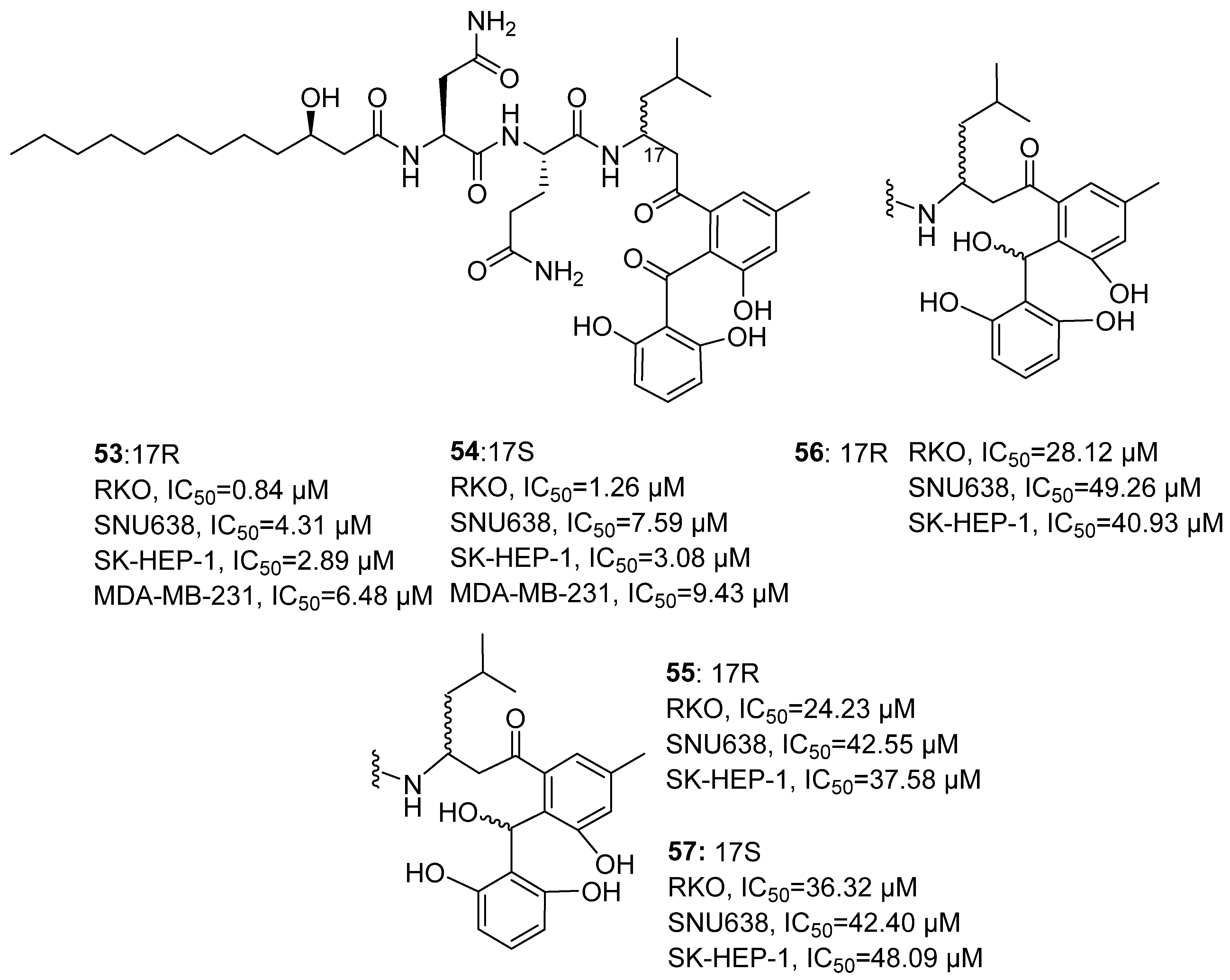
In 2020, Lee’s research group isolated Asperphenin A (
53) from the dimethylformamide extract of marine-derived
Aspergillus sp. (
Figure 26) [
67].
53 is a structurally novel lipopeptidyl phenol ketone-type natural product. The research group had previously synthesized 10 derivatives of
53. Taking
55 as an example, its structure was confirmed through nuclear magnetic resonance
1H NMR and
13C NMR. Combined with the MTPA esterification method, coupling constants (J) analysis, and ECD calculation, the absolute configuration of the trichothecene and its synthetic derivatives at all chiral centers was characterized. Evaluation of in vitro anti-tumor activity showed that
53 exhibited the most significant activity, with IC
50 values of 0.84, 4.31, 2.89, and 6.48 μM against four human tumor cell lines (RKO, SNU638, SK-HEP-1, MDA-MB-231). Asperphenin B (
54) showed slightly lower activity than
53, with corresponding IC
50 values of 1.26, 7.59, 3.08, and 9.43 μM. The positive control drug Etoposide, with IC
50 values of 3.86, 0.30, 0.49, and 10.72 μM, showing pronounced activity in SNU638 cells. The hydroxylated derivatives (
55–57) generally exhibited diminished inhibitory activity, with IC
50 values ranging from 24.23 to 49.26 μM. SAR analysis revealed that the aryl ketone group at the C-7 position is a crucial pharmacophore for maintaining activity. Reduction of this group to a hydroxyl or alkene led to loss of activity, while other structural modifications also resulted in a significant decrease in potency.
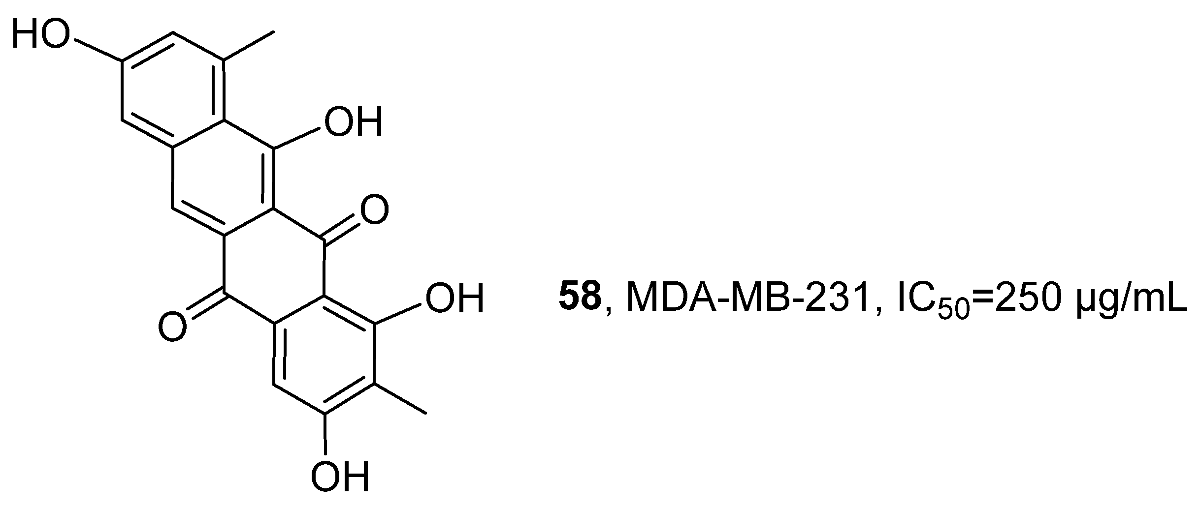
In 2020, Metsä-Ketelä’s research group isolated a new compound, Persiamycin A (
58), from the ethyl acetate and methanol extracts of the halophilic actinobacterium
Streptomonospora sp. PA3 (
Figure 27) [
68]. This compound belongs to the aromatic polyketide class. Its structure was confirmed through the following methods: HRMS determined its molecular formula to be C
20H
13O
6; the
1H NMR spectrum revealed four aromatic proton signals, two methyl signals, and three partially interchangeable hydroxyl signals; HMBC analysis further confirmed the correlation between the methyl group and the phenolic carbon, indicating the presence of methyl and hydroxyl substitution on the aromatic ring. The activity evaluation results showed that Persiamycin A exhibited relatively weak anti-proliferative activity against the human breast cancer cell line MDA-MB-231 in vitro, with an IC
50 value of 250 µg/mL.
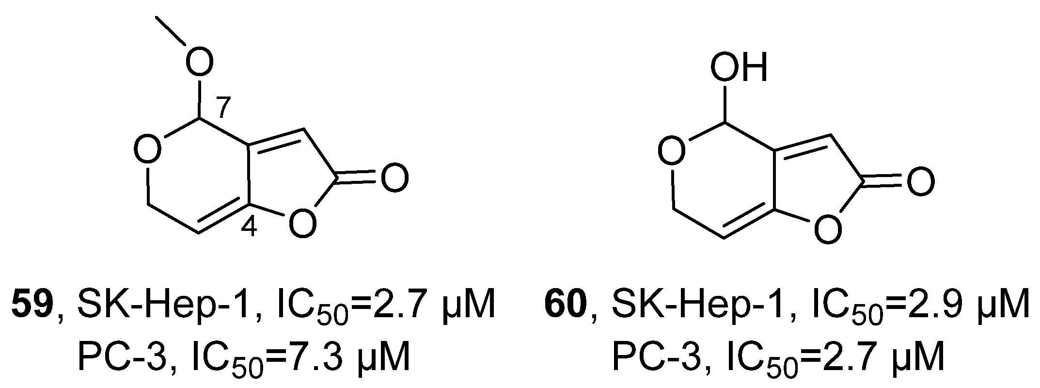
In 2020, Lee’s research group isolated aspergilsmins A-G from the ethyl acetate extract of the
Aspergillus giganteus NTU967 fungus, which were all identified as polyketide compounds (
Figure 28) [
69]. Among them, aspergilsmin C (
59) was confirmed to be a methyl derivative of patulin (
60). Taking aspergilsmin C as an example for structure elucidation, its molecular formula was determined to be C
8H
8O
4, with five unsaturations. NMR- and HMBC-related signals further confirmed that the methyl and ethyl groups were connected at the C-7 position. The activity evaluation results showed that compound 3 had an IC
50 value of 2.7 μM in SK-Hep-1 liver cancer cells and 7.3 μM in PC-3 prostate cancer cells. And compared with the positive control drug paclitaxel, patulin exhibited weaker cytotoxic activity against SK-Hep-1 and PC-3 cells, with IC
50 values of 2.9 μM and 2.7 μM, respectively, while paclitaxel showed IC
50 values of 0.011 μM and 0.013 μM, respectively. The SAR analysis revealed that the size of the substituent at the C-7 position and the olefin structure at the C-4 position are crucial for maintaining the anti-tumor activity of this class of compounds.
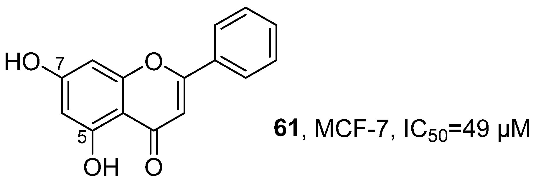
In 2020, Jayabaskaran’s research team isolated Chrysin (5,7-dihydroxyflavone) (
61) from the ethyl acetate extract of the marine endophytic fungus
Chaetomiumglobosum (
Figure 29) [
70]. This compound belongs to the polyketide class. Its structure was confirmed through various spectroscopic techniques: The IR spectrum revealed the presence of O-H, C = O, C-CO-C and C-O-C functional groups; LC-MS analysis established its molecular formula to be C
15H
10O
4; and the NMR spectrum further clarified that the hydroxyl groups were located at C-5 and C-7 positions. Using MTT and resazurin reduction assay, the activity evaluation results showed that Chrysin (5,7-dihydroxyflavone) exhibits significant anti-tumor activity, with an IC
50 value of approximately 49 µM against human breast cancer cells MCF-7.
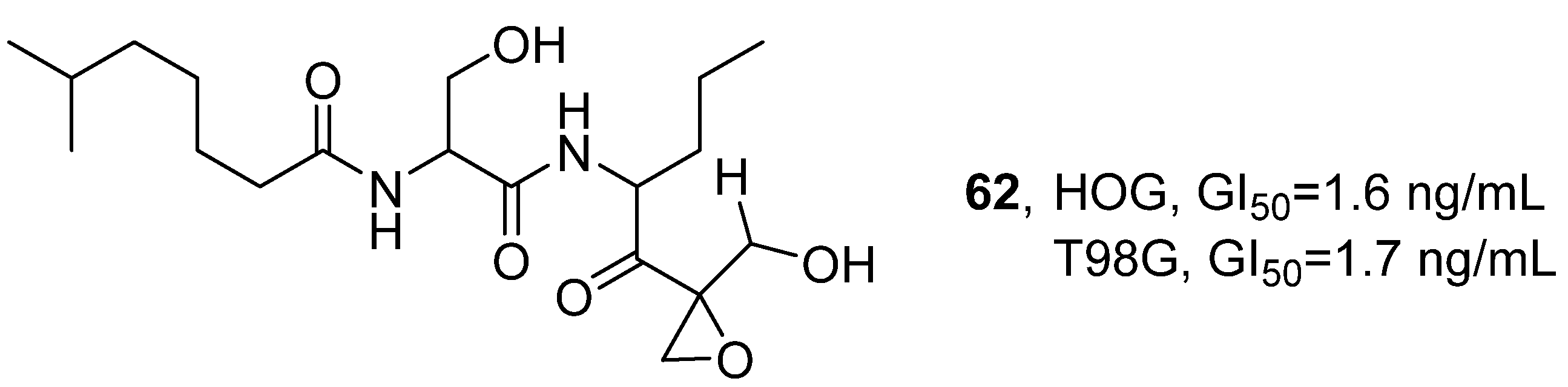
In 2021, V. Costa-Lotufo’s research group isolated a variety of polyketide cyclic compounds, including dihydroeponemycin (DHE,
62), from the marine actinomycete BRA-346 that coexists with the Brazilian endemic ascidian
Euherdmania sp. Through extraction and separation with ethyl acetate, a total of 13 eponemycin analogs were obtained (
Figure 30) [
71]. Taking DHE as an example for structure confirmation, the ion peak
m/
z 401.26 was detected through HPLC-MS/MS analysis, which was consistent with the known structure. The activity evaluation results showed that when the incubation time of DHE and the BRA-346 containing epoxy ketone components was extended from 24 h to 48 h, the growth inhibitory activity increased 10-fold; however, further extension to 72 h did not lead to additional enhancement. Both cell lines exhibited similar sensitivity to DHE, with the GI
50 values at 48 h being 1.6 ng/mL and 1.7 ng/mL in HOG and T98G cells, respectively. In contrast, T98G cells were slightly less sensitive to the epoxyketone-containing fraction of BRA-346, with a GI
50 of 28.2 ng/mL, while that of HOG cells was 17.6 ng/mL. The SAR analysis indicated that the epoxy ketone structure was the key pharmacophore for inhibiting proteasome activity; moreover, synergistic effects among the various structural analogs present in the BRA-346 fraction may further enhance its biological activity.
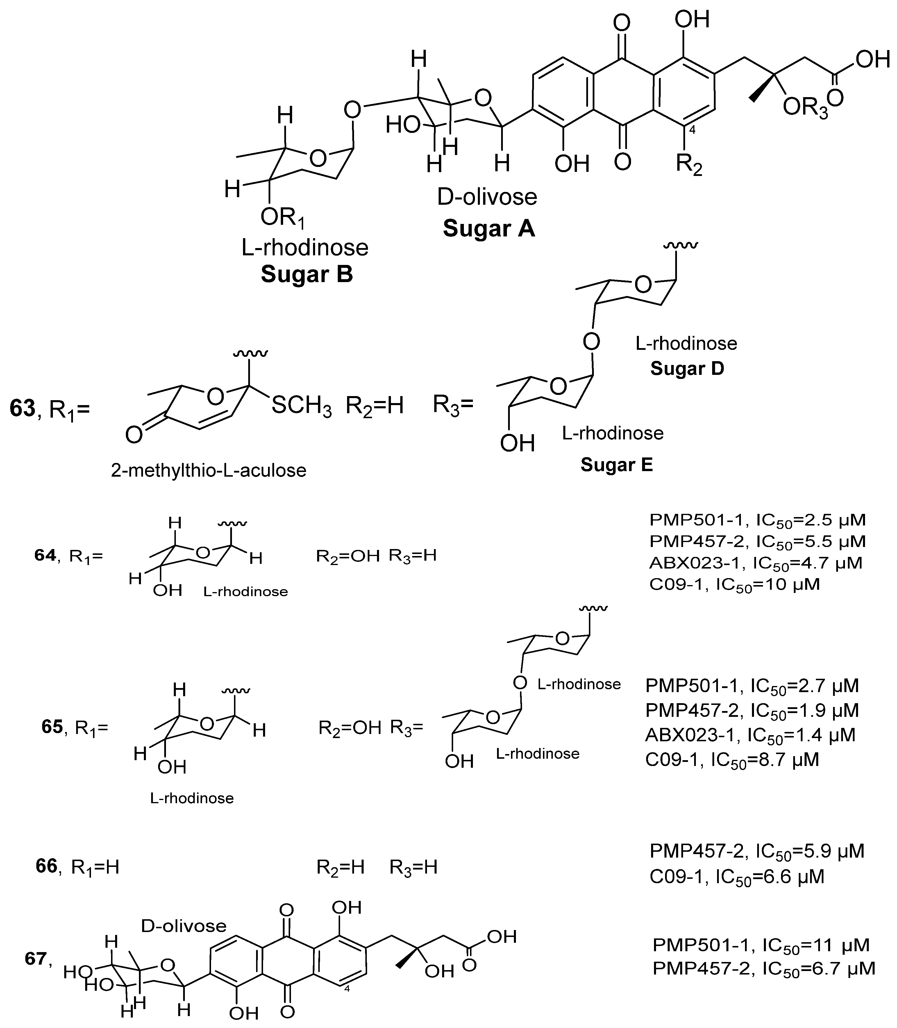
In 2021, Li’s research group isolated a series of rearranged angucycline compounds Grincamycins P-T from the ethyl acetate extract of
Streptomyces sp. CNZ-748 (
Figure 31) [
72], which originated from marine sediments. These compounds belong to the polyketide family. Taking Grincamycin R (
63) as an example for structure identification: HRMS showed its molecular formula as C
50H
64O
18S and NMR data further confirmed its planar structure. Combined with HMBC and COSY correlation signals, a novel
α-L-methylthio-akulose residue was inferred in its structure. The stereochemistry was ultimately determined by integrating NOE correlation data and biosynthetic pathway analysis. The activity evaluation results showed that compound
65 exhibited the optimal inhibitory activity in four tested cell lines, with IC
50 values of 2.7, 1.9, 1.4 and 8.7 μM for PMP501-1, PMP457-2, ABX023-1 and C09-1, respectively. Its activity was superior to the positive control drug 5-fluorouracil in three cell lines. Compound
64 also displayed broad-spectrum inhibitory activity, with IC
50 values ranging from 2.5 to 10 μM. Additionally, compounds
66 and
67 showed moderate activity for some cell lines, with IC
50 values ranging from 5.9 to 11 μM. The SAR analysis suggested that the hydroxylation at the C-4 position and the introduction of a methylthio group in the terminal trisaccharide chain might significantly enhance cytotoxicity, potentially by improving solubility or strengthening interactions with target sites.
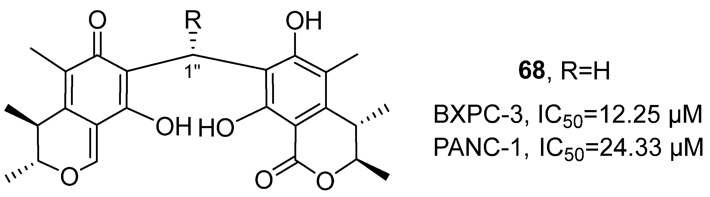
In 2022, Zhang’s research group isolated four novel carbon-linked citrinin dimeric compounds from the ethyl acetate extract of the symbiotic fungus
Penicillium sp. GGF16-1-2 of sea stars (
Figure 32) [
73], all of which belong to the polyketide structural class. Taking the structure of compound
68 as an example: Through HRMS, its molecular formula was determined as C
25H
28O
7. HMBC and COSY correlation signals confirmed that the two structural fragments were connected by a carbon bridge (C-1″). The relative configuration was deduced by NOESY spectroscopy, and the absolute configuration (3
R, 4
S, 3′
R, 4′
S) was confirmed by comparison of experimental and calculated ECD spectra. Based on
1H NMR and
13C NMR data, it can be inferred that its structure contains a ketone carbonyl and an ester carbonyl. The in vitro cell activity evaluation results showed that, compared with the positive control drug doxorubicin (IC
50 = 18.24 µM for BXPC-3 and 24.00 µM for PANC-1), compound 66 exhibited stronger cytotoxic activity against BXPC-3 and PANC-1 cells, with IC
50 values of 12.25 µM and 24.33 µM, respectively.
In 2022, Tasdemir’s research team isolated two endophytic fungi,
Pyrenochaetopsis sp. FVE-001 and FVE-087, from the brown alga Fucus vesiculosus (
Figure 33) [
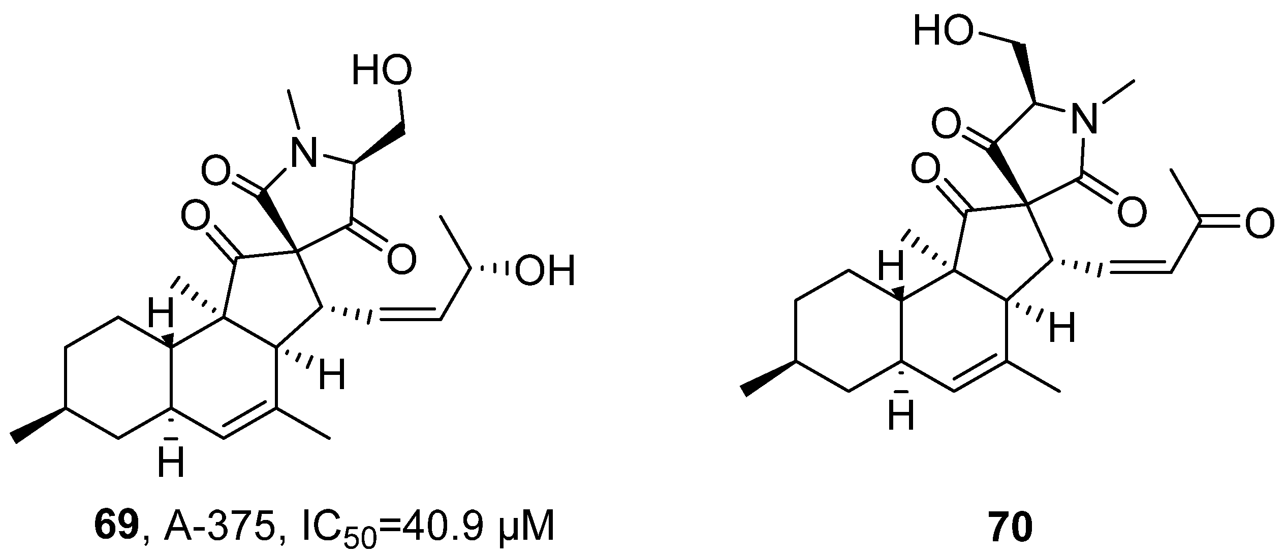
74]. After extraction with ethyl acetate, tenhydronaphthalenyl lactone tetracyclic acid derivatives Pyrenosetin E (
69) and Pyrenosetin F (
70) belonging to polyketide compounds were obtained. Taking Pyrenosetin E as an example, its molecular formula was determined as C
25H
35NO
5 by HRMS. The structure was confirmed by FT-IR to contain carbonyl and hydroxyl groups. Further analysis using NMR (
1H-NMR,
13C-NMR) revealed that the compound has four alkenes, three carbonyls, and five unsaturation degrees, indicating a tetracyclic structure. Finally, combined with 2D NMR data such as COSY, DEPT-HSQC, and HMBC, the chemical structure was further verified and determined. The activity evaluation results showed that Pyrenosetin E has inhibitory activity against human malignant melanoma cell line A-375, with an IC
50 value of 40.9 μM.
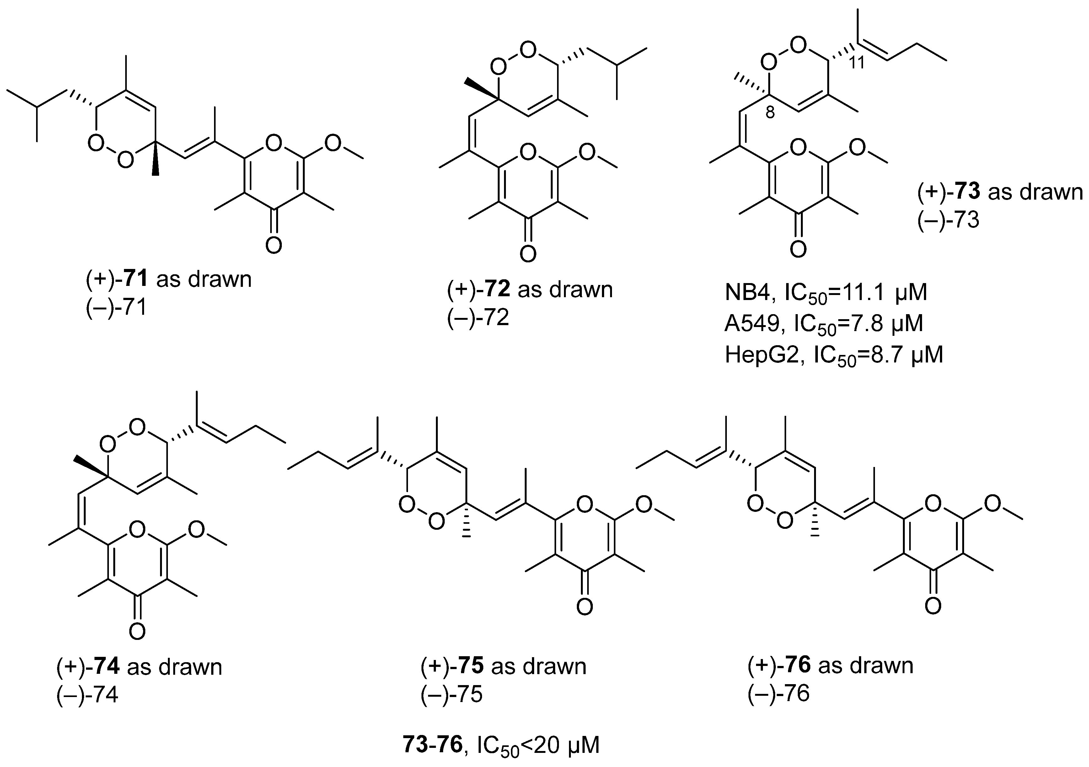
In 2022, Guo’s research group isolated six new pairs of γ-pyrone polypropionate enantiomers with an unusual peroxyl bridge at the side chain, from the MeOH-CH
2Cl
2 extract of the
Placobranchus ocellatus, namely (±)-Ocellatuperoxides A-F (
71–76) (
Figure 34) [
75]. Structural elucidation of (±)-Ocellatuperoxide C was determined by HRMS, with molecular formula C
22H
30O
5. The unsaturated carbonyl group was determined by infrared spectroscopy, and the existence of an endoperoxide bridge between C-8 and C-11 was confirmed by NMR, COSY, and HMBC, forming a 1,2-dioxane ring connection between the fragments. In vitro activity tests showed that the compound (±)-Ocellatuperoxides C-F had inhibitory activity against cancer cells, with IC
50 values all within the range of 20 μM. Among them, (±)-Ocellatuperoxides C had the best activity, with IC
50 values of 11.1, 7.8, and 8.7 μM for NB4, A549, and HepG2 cells respectively. The preliminary SAR indicated that the activity was weaker when the side-chain terminal was isobutyl, while compounds with a 1-methyl-1-butenyl side chain had stronger activity, and the stereoconfiguration had a significant impact on activity; (±)-compound
73 was evaluated on the A549 cell line. Interestingly, the results indicated that only (−)-compound
73 (IC
50 = 8.7 μM) exhibited activity, while (+)-compound
73 (IC
50 > 100 μM) was inactive.
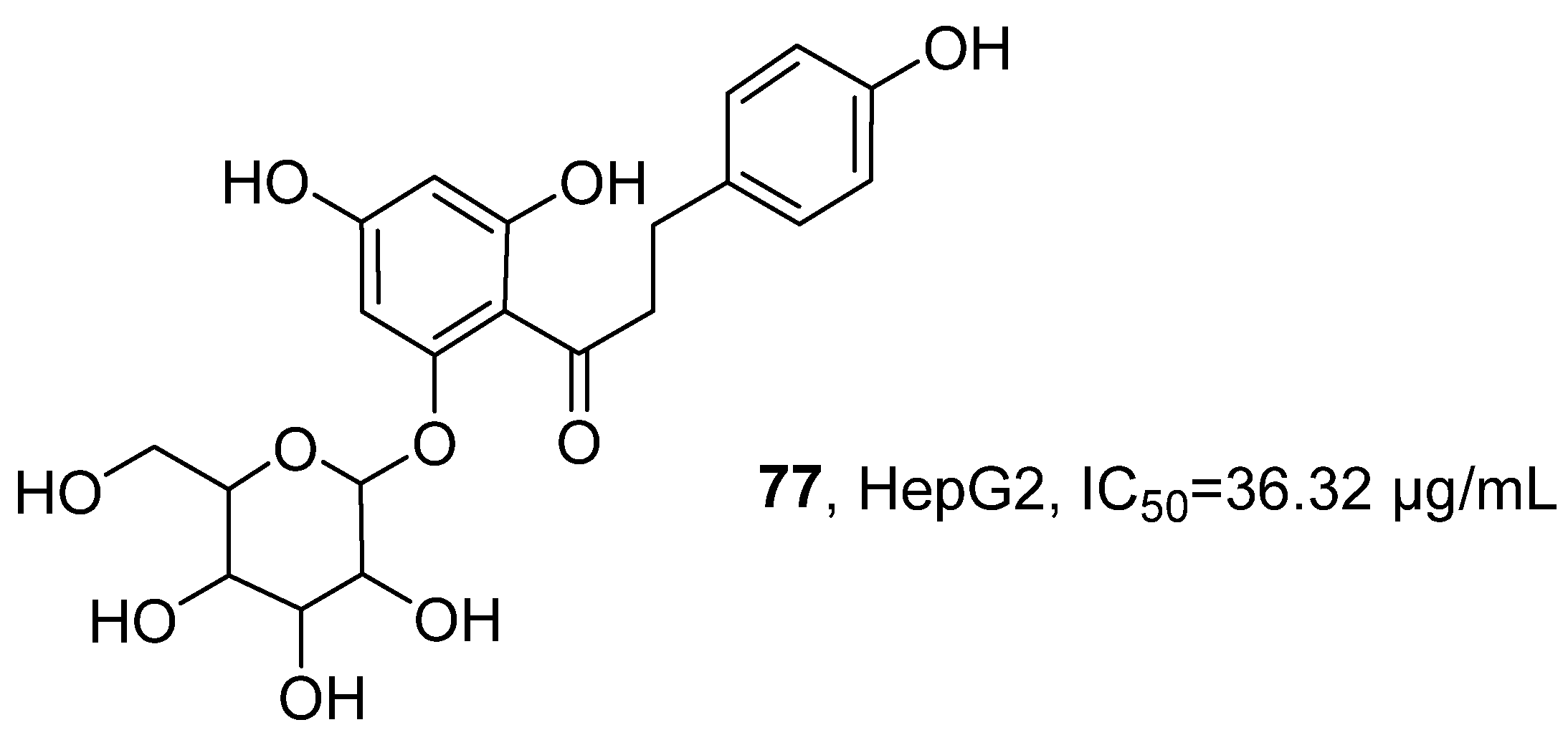
In 2023, Sanniyasi’s research group successfully isolated the known dihydrochalcone glycoside compound phloridzin (
77) from the ethanol–water extract of the sea grass
Syringodium isoetifolium (
Figure 35) [
76]. The molecular formula was determined to be C
21H
24O
10 by MS. Further analysis using various spectroscopic techniques such as FT-IR,
1H NMR, and
13C NMR confirmed its chemical structure. The activity evaluation results showed that, compared with the positive control drug doxorubicin, compound
77 exhibited weaker cytotoxic activity against HepG2 hepatocellular carcinoma cells, with an IC
50 value of 36.32 μg/mL, while doxorubicin showed an IC
50 value of 10.19 μg/mL.
In polyketides, aromatic rings, lactone rings, epoxyketone, methoxy, and glycosyl groups are most frequent. The most potent polyketides derive from marine fungi (Pyrenochaetopsis, Aspergillus, Penicillium) and actinomycetes (Streptomyces). Asperphenin A (53) shows an IC50 of 0.84 μM against RKO colon cancer cells, comparable to etoposide; grincamycin derivative (65) exhibits IC50 as low as 1.4–2.7 μM against rare cancer lines. Spirotetramic acid-, epoxide-, or carbon-bridged dimer-containing polyketides display the best activity. Trends from 2020 to 2024 include that targeted isolation via feature-based molecular networking; decisive role of absolute configuration (e.g., only (−)-ocellataperoxide C is active); identification of key pharmacophores (C-7 aryl ketone, epoxyketone); and development of polyketides as payloads for antibody-drug conjugates (ADCs).
2.5. Marine-Derived Peptides and Proteins
Marine-derived peptides and proteins constitute a highly diverse family in terms of sequence, spatial conformation, and modification patterns. These molecules are often rich in disulfide bonds, undergo glycosylation, or carry other complex post-translational modifications. These molecules are widely distributed in sponges, marine microorganisms, and their symbiotic systems, and frequently contain characteristic motifs such as non-canonical amino acids and unique cyclic backbones. This unique structure endows them with outstanding advantages in molecular recognition ability, conformational stability, and plasticity, making marine peptides a highly promising source of lead compounds in medicinal chemistry and providing a rich array of molecular scaffolds for the development of novel anticancer drugs.
In 2020, R. O’Keefe’s research group conducted an activity substance screening on the marine sponge
Axinella sp. and discovered a new type of cyclic peptide named Recifin A, which is rich in cysteine [
77]. This peptide consists of 42 amino acid residues and belongs to the structurally novel Tyr-lock family. The research team determined its primary sequence through MS/MS and automatic Edman degradation, and elucidated its three-dimensional structure using NMR technology. Based on 425 distance constraints and 75 dihedral angle constraints, the core structure was defined as a four-chain
β-sheet flanked by two helical turns; the disulfide bonds formed a typical cysteine knot, locking Tyr6 into a specific spatial conformation. Further partial reduction and alkylation experiments clarified that the disulfide connectivity was Cys I–III, II–V, and IV–VI. The activity study showed that Recifin A could effectively inhibit tyrosine-DNA phosphodiesterase 1 (TDP1), with an IC
50 value of 190 nM.
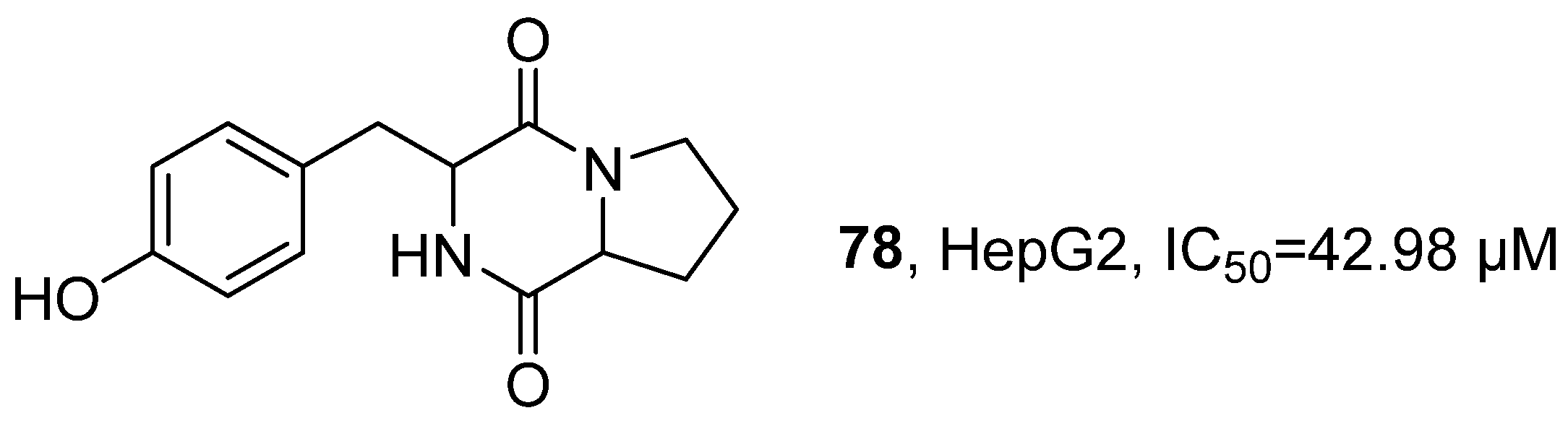
In 2020, Karanam’s research group isolated 11 components from the ethyl acetate extract of
Bacillus pumilus co-associated with marine sponges (
Figure 36) [
78]. Through preliminary TLC screening, component F2 (Cyclo(-Pro-Tyr),
78) showed high biological activity and belonged to a polypeptide compound. Using techniques such as LC-MS/MS, FT-IR, and NMR, the structure was systematically analyzed: HRMS showed its parent ion peak at
m/
z 261.07 [M + H]
+, suggesting its molecular formula as C
14H
16N
2O
3; IR confirmed the presence of amide carbonyl (C = O), N-H, and O-H stretching vibrations in the structure; NMR spectrum further revealed signals corresponding to a benzene ring and two carbonyl groups. Using MTT assay, the activity evaluation results indicated that
78 had significant cytotoxicity against HepG2 human hepatocellular carcinoma cells, with an IC
50 value of 42.98 μM.
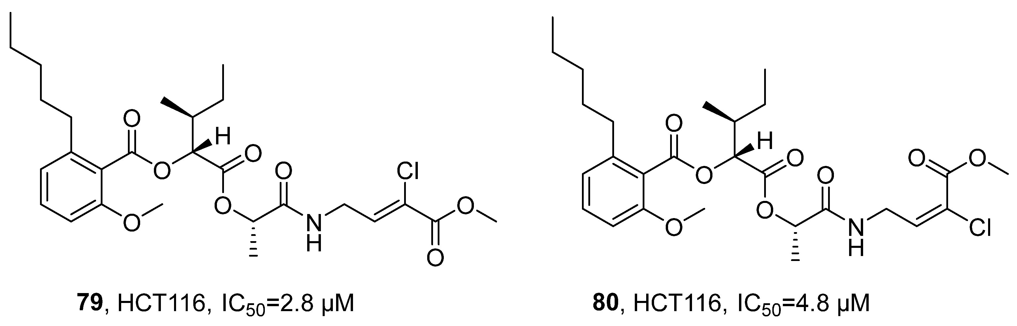
In 2020, Luesch’s research group isolated a new class of peptide natural products anaenamides A (
79) and B (
80) from the marine cyanobacterium
Hormoscilla sp. (
Figure 37) [
79]. Taking anaenamide A as an example, its molecular formula was determined to be C
27H
39NO
8Cl by HRMS. The structure was analyzed using multi-dimensional NMR techniques: the HSQC spectrum indicated the presence of six methines, three methylenes, four methyl groups, three consecutive aromatic proton signals, one isolated olefin proton, and two methoxy signals; the
13C NMR spectrum displayed the presence of four unprotonated sp
2 hybrid carbons and four carbonyl carbon signals. By further combining COSY and HMBC correlation spectra, three structural fragments, alkyl salicylate, 2-hydroxy-3-methylpentanoic acid, and lactate, were identified. Their planar structures and relative configurations were ultimately confirmed, with the assistance of HMBC and NOESY spectra. The activity study showed that anaenamides A and B exhibit significant inhibitory activity against human colon cancer cells HCT116, with IC
50 values of 2.8 μM and 4.8 μM, respectively. Preliminary SAR analysis suggested that the halogenated
α,
β-unsaturated ester unit acts as a Michael acceptor, as its removal or saturation led to a significant reduction in cytotoxicity, indicating that this moiety is the key pharmacophore responsible for the cytotoxic activity.
In 2020, Giovine’s research group isolated and purified a new protein from the extruded crude extract of the sponge
Chondrosia reniformis through a series of steps including 10 kDa filtration dialysis, ammonium sulfate precipitation, and high-performance liquid chromatography size exclusion chromatography [
80]. MS analysis revealed that the protein precursor consisted of 199 amino acids, including a 21-amino acid signal peptide and a 178-amino acid mature peptide, with a theoretical molecular weight of 19,611.12 Da and a theoretical isoelectric point was 5.11. Sequence alignment results indicated that this protein exhibits structural homology with the N-terminal region of the ryanodine receptor and defensin-like proteins. The research further completed the three-dimensional structural modeling of the protein using Phyre2. The activity evaluation results showed that the purified components Chondrosin P4 and P5 exhibited differential activities on various cell lines: the EC
50 for RAW264.7 cells was 1.99 μg/mL and 1.12 μg/mL, respectively; for L929 cells, the EC
50 was 3.56 μg/mL and 4.90 μg/mL, respectively; for MDA-MB-468 cells, the EC
50 of P4 was 59.88 μg/mL; for HeLa cells, the EC
50 of P4 was 17.7 μg/mL; and for normal human dermal fibroblasts, the EC
50 of P5 and P4 were 14.6 μg/mL and 17.8 μg/mL, respectively.
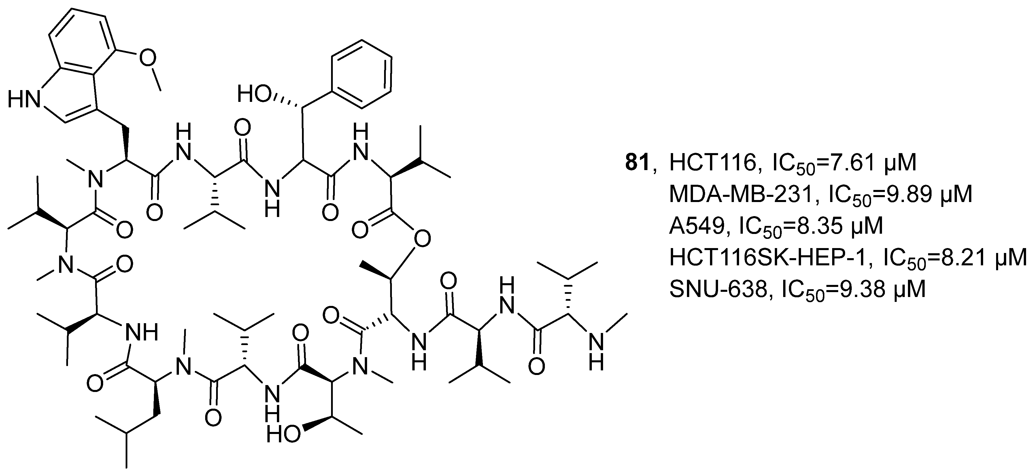
In 2020, building upon previous studies, Lee’s research group conducted further research on a novel cyclic peptide named Ohmyungsamycin A (
81), which was isolated from the marine bacterium SNJ042 of
Streptomyces genus (
Figure 38) [
81]. The activity evaluation results showed that, compared with the positive control drug etoposide, compound
81 exhibited weaker cytotoxic activity against MDA-MB-231, HCT116, A549, SK-HEP-1, and SNU-638 cells, with IC
50 values of 9.89, 7.61, 8.35, 8.21, and 9.38 μM, respectively, while etoposide showed IC
50 values of 3.85, 0.52, 0.48, 0.78, and 0.82 μM, respectively.
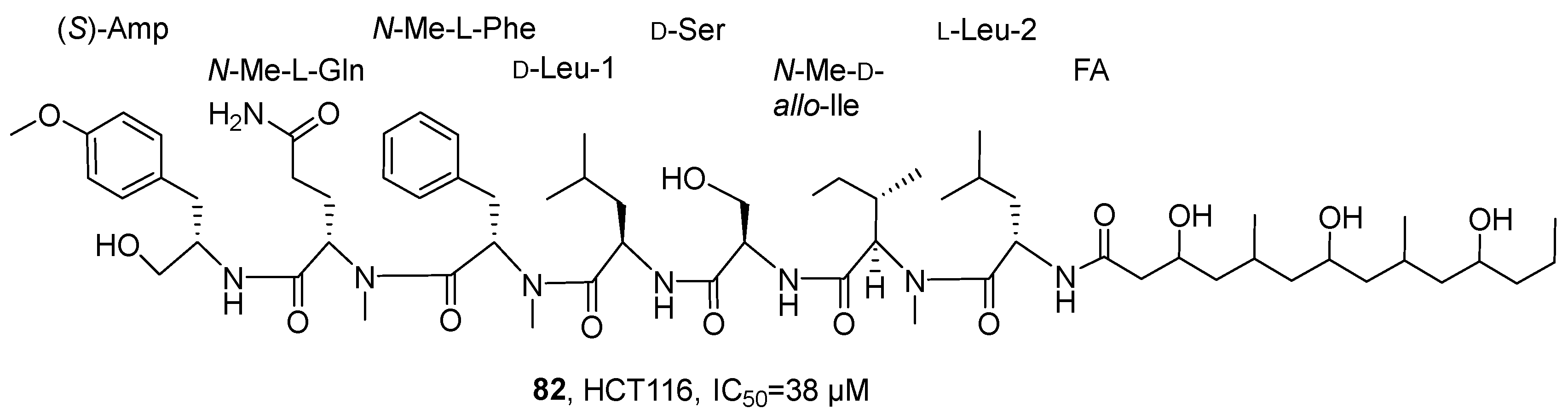
In 2021, Luzzatto-Knaan’s research group isolated and identified a lipopeptide compound, wenchangamide A (
82), from the dichloromethane and methanol extracts of cf.
Neolyngbya cyanobacteria from the South China Sea (
Figure 39) [
82]. For the structural analysis of wenchangamide A, the study initially characterized it using LC-MS/MS technology and further determined the absolute configuration of its amino acid residues by the Marfey method. In vitro activity evaluation showed that this compound could concentration-dependently induce apoptosis in HCT116 human colon cancer cells, with a 24 h IC
50 value of 38 μM. The SAR analysis revealed that the core peptide backbone, such as the N-methylphenylalanine residue, might constitute the key pharmacophore, while fatty acid chain length, such as the extended segment in wenchangamide B, might have a regulatory effect on its activity intensity and solubility; the structural differences from mimamide A further indicated that specific domains might have a significant impact on the specificity of the compound’s activity.
In 2021, Zhang’s research group extracted sea cucumber intestinal peptides (SCIP) from the intestinal tissues of sea cucumbers through alkaline protease hydrolysis [
83]. HPLC analysis showed that SCIP consisted of a series of oligopeptides. Further analysis revealed that 98.41% of SCIP has a relative molecular mass of less than 2000 Da, and approximately 95.36% of SCIP is distributed within a molecular weight range of less than 1041 Da. The amino acid composition analysis results indicated that SCIP is rich in hydrophobic amino acids and branched amino acids. In vivo zebrafish experiments demonstrated that SCIP reduced MCF-7 fluorescence intensity in a dose-dependent manner when administered via a water-soluble route. The results of in vitro experiments showed that SCIP could dose-dependently inhibit the proliferation of MCF-7 cells and promote their apoptosis at concentrations of 27.8, 83.3, and 250 μg/mL.
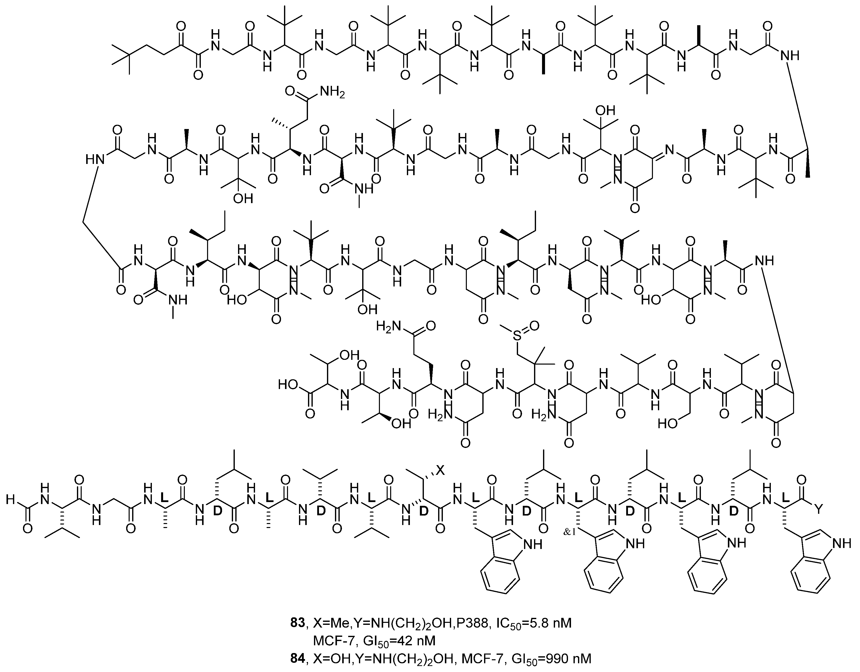
In 2022, Inoue’s research group further explored the mechanism of action of Gramicidin A (
83), a natural ion channel-forming peptide, based on previous studies by others (
Figure 40) [
84]. The research team synthesized derivatives 2–4, which are all polypeptides, and their structures were confirmed through key steps in the synthesis route and standard purification methods. The activity detection results showed that, compared with the inactive control drug (
84), compound
83 exhibited stronger cytotoxic activity against P388 murine leukemia cells, with a GI
50 value of 42 nM, while the inactive control compound showed a GI
50 value of 990 nM. The SAR analysis revealed that analog 2, due to a minor structural modification at residue 8 (Me→OH), led to a significant reduction in biological activity (GI
50 = 990 nM), thereby affecting its ion channel depolarization capacity.
In 2022, Yu’s research group used the hemichordate
Arca inflata Reeve’s hemolymph as the research subject [
85]. Through multi-step of separation and purification combined with an activity-oriented screening strategy, a novel lysine-rich polypeptide P6 was obtained, with a molecular weight of 2794.8 Da. The complete amino acid sequence of this peptide was analyzed using tandem mass spectrometry as WYIRKIRRFFKWLKKKLKK. Circular dichroism spectroscopy analysis revealed that the secondary structure of this peptide segment was mainly α-helix. Three-dimensional structure simulation further indicated that P6 has typical amphiphilic characteristics, forming a positively charged hydrophilic surface and a hydrophobic region. In vitro anti-tumor activity evaluation results showed that the IC
50 values of P6 against colorectal cancer cells DLD-1, HT-29, and HCT116 were 2.14, 4.43, and 10.88 μg/mL, respectively; although its activity was lower than the positive control cisplatin (with corresponding IC
50 values were 1.06, 1.90, and 1.38 μg/mL), it still exhibited significant inhibitory effects. In the HT-29 xenograft model, compound P6 demonstrated tumor inhibition rates of 72.66% at a dosage of 30 mg/kg and 49.46% at 15 mg/kg, both superior to the positive control 5-fluorouracil (25 mg/kg, with an inhibition rate of 67.28%), indicating its promising in vivo anti-tumor potential.
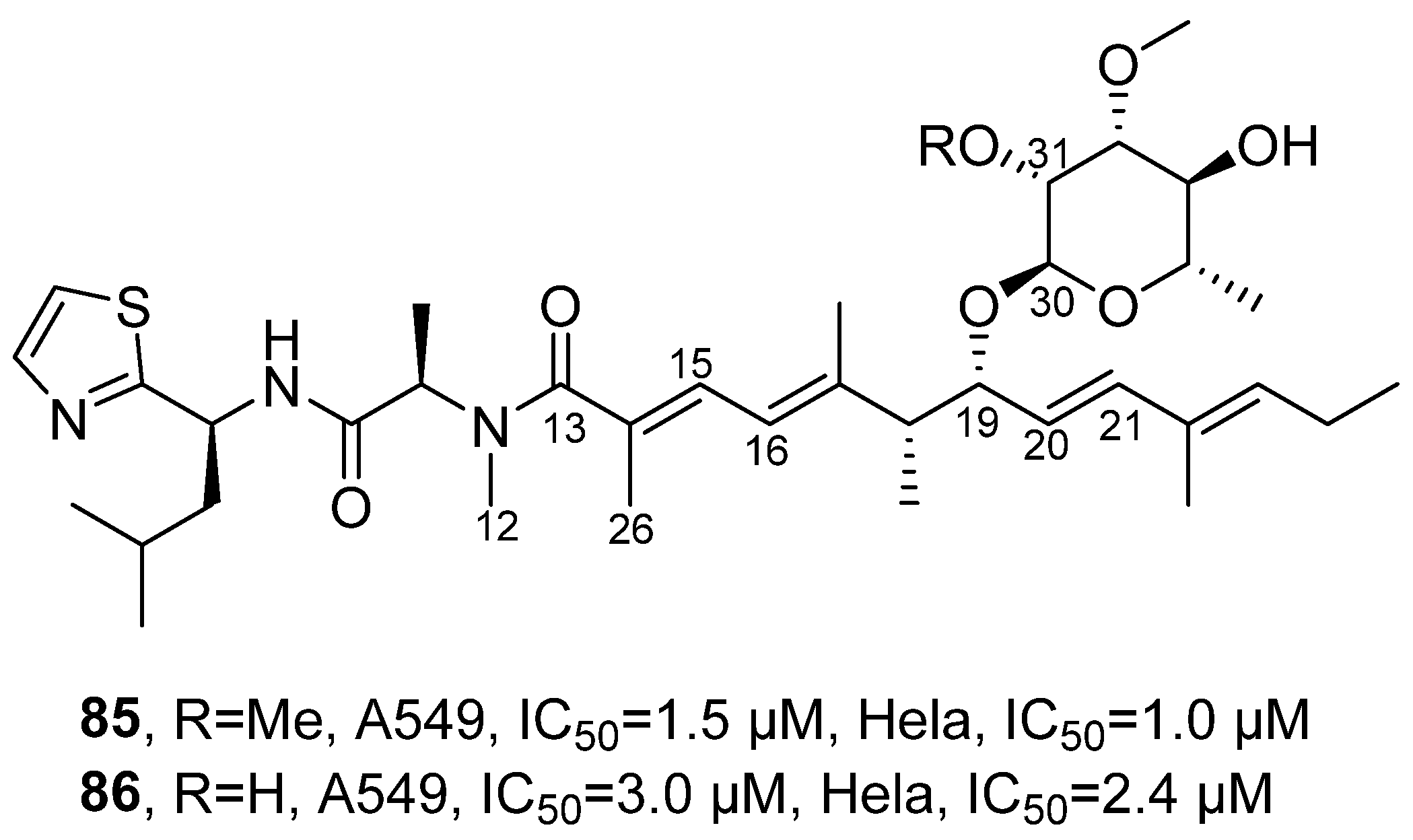
In 2023, Luesch’s research group isolated jezoside (
85) and its novel derivative jezoside B (
86) from the cyanobacterial
assemblage collected in Florida (
Figure 41) [
86]. Both are polypeptide compounds. For the structural analysis of jezoside B, HRMS determined its molecular formula as C
36H
57N
3O
7S, identifying it as a similar compound to jezoside with demethylation at C-31. Through COSY and HMBC related signals, its tripeptide unit was determined to be Thz-Leu-N-Me-Ala, where the NMR signals of the N-Me-Ala residue were broadened due to slow conformational exchange. The polyketide unit contained a trans double bond (H-20/H-21) and a conjugated diene (H-15/H-16). With the aid of HMBC related signals, it was further revealed that the tripeptide unit and the polyketide unit were connected through H-26 and H-12 with C-13; the sugar unit was 2,3-O-dimethyl-methylglucoside, and it was connected to the polyketide unit through H-30. Based on J coupling constants, its configuration was determined as s-trans. Activity detection results showed that jezoside and its novel derivative jezoside B had IC
50 values of 1.5/3.0 µM and 1.0/2.4 µM against A549 and HeLa cells. SAR analysis indicated that the methoxy group at the C-31 position of the sugar moiety enhanced the activity but was not an essential structural unit. Removal of this group only caused a slight reduction in activity, suggesting that this site has a certain degree of modification tolerance and that the activity possesses tunable potential.
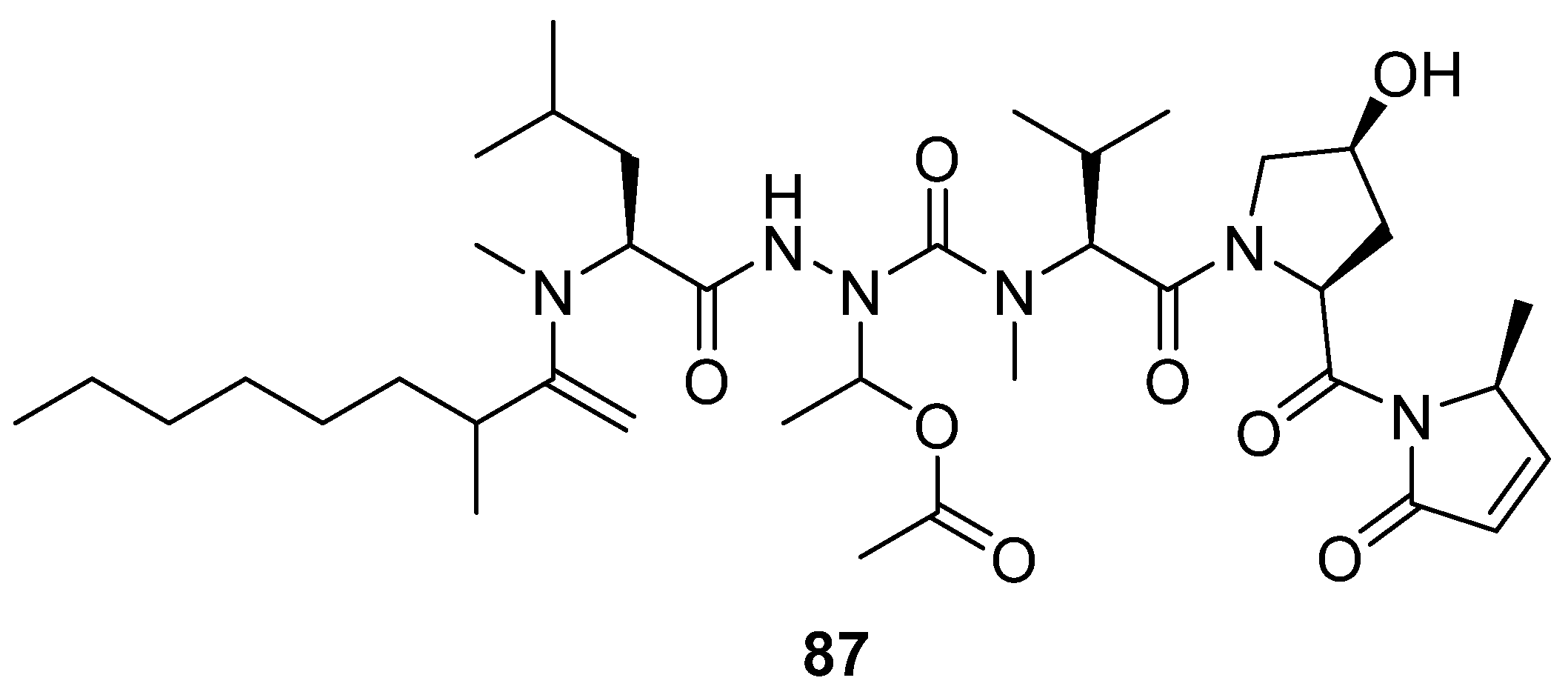
In 2023, Wang’s research group synthesized the lipopeptide natural product microcolin H (
87) derived from the marine organism
Moorea producens using a total synthesis method (
Figure 42) [
87]. The activity evaluation results showed that, when assessing its anti-tumor effects using the HGC-27 cell model, the tumor growth inhibition rate (TGI) reached 74.2% in the group treated with 10 mg/kg microcolin H, which was significantly higher than the TGI value of the positive control drug paclitaxel (8 mg/kg). The SAR analysis further revealed that the length of the fatty acid chain is the key structural factor affecting its activity.
In 2024, Wen’s research group conducted in-depth studies [
88] based on the marine antibacterial peptide Piscidin-1, which can be isolated from the mast cells of the hybrid striped bass [
89]. The activity test results showed that the marine antibacterial peptide Piscidin-1 could inhibit the viability of oral squamous cell carcinoma OC2 and SCC4 cells in a concentration- and time-dependent manner. Within 24 to 72 h, the IC
50 values of Piscidin-1 for the OC2 and SCC4 cell lines ranged from 10.82 to 13.77 μM and 16.94–19.20 μM respectively, demonstrating a significant anti-proliferative effect.
In 2024, Hao’s research group performed tryptic hydrolysis of phycocyanin, followed by separation, purification and identification of the resulting peptides using ultrafiltration, HPLC, and MS [
90]. They identified three predicted peptide segments of phycobilin with anti-tumor activity, namely PCP1 (AGDASVLEDR), PCP2 (ADSLLSGLR), and PCP3 (MFDAFTK). Using CCK-8 assay, anti-tumor activity assays showed that after 24 h of treatment at a concentration of 50 μg/mL, PCP1 reduced the viability of A549 cells to 0.77 μg/mL, H1299 cells to 0.78 μg/mL, and LTEP-a-2 cells to 0.80 μg/mL. PCP2 also showed inhibitory effects at the same concentration, with viability rates of A549, H1299, and LTEP-a-2 cells being 0.76, 0.73, and 0.77 μg/mL, respectively. In contrast, PCP3 at 100 μg/mL reduced the viability of these three cell lines to 0.69, 0.72, and 0.71 μg/mL, respectively. The SAR analysis indicated that acidic amino acid residues could form hydrogen bonds with basic amino acids in EGFR, thereby enhancing the binding ability, while hydrophobic amino acid residues help strengthen hydrophobic interactions with the active pocket of EGFR, further improving the binding stability.
In 2024, Wen’s research group conducted further studies based on the antibacterial peptide Tilapia piscidin 4 (TP4) identified from the Nile tilapia (
Oreochromis niloticus) by previous researchers [
91]. Using MTT assays, anti-tumor activity assays showed that TP4 exhibited IC
50 values of 6.71 μM against J82 cells and 6.14 μM against T24 cells. In the colony formation experiment, the number of colonies formed by cells treated with TP4 was significantly reduced.
In peptides, amide bonds, hydrophobic amino acid residues (Leu, Phe, Trp), disulfide bridges, and fatty acid chains are most frequent. Major sources are marine cyanobacteria (Hormoscilla, Moorea), sponges (Chondrosia), and mollusks (Arca inflata). Gramicidin A (83) shows the highest activity with an IC50 of 5.8 nM against P388 leukemia cells; Recifin A (78) selectively inhibits TDP1 (IC50 = 190 nM) via an allosteric mechanism; P6 peptide achieves 72.66% tumor inhibition in an HT-29 xenograft model, better than 5-fluorouracil. Ion-channel-forming linear peptides or cyclic peptides targeting protein–protein interactions exhibit the best selectivity. Trends from 2020 to 2024 include shifting from extraction to total synthesis and derivative design (e.g., microcolin H); mechanistic studies on membrane depolarization and ion channels; determination of amino acid configuration by Marfey’s method and MS/MS; and repurposing marine antimicrobial peptides as anticancer agents (piscidin-1, TP4).
2.6. Marine-Derived Polysaccharides
Marine-derived polysaccharides are structurally diverse macromolecules from seaweeds, fungi, and microorganisms, composed of monosaccharides with varied linkages, sulfation, and branching. Adapted to extreme marine environments, such as high salinity and high pressure, they possess unique chemical and structural features, making them valuable for natural macromolecular drug development and anticancer agent discovery.
In 2022, M. Saied’s research group isolated and purified a novel sulfated polysaccharide EPSR4 from marine
Bacillus subtilis AC4 sourced from the Red Sea sediment [
92]. Structural characterization revealed that the polysaccharide has a molecular weight of 1.48 × 10
4 g/mol as determined by gel permeation chromatography, and its monosaccharide composition consists of glucose, rhamnose, and arabinose, with a molar ratio of 5:1:3. The FT-IR spectrum further confirmed it as a sulfated polysaccharide with
β-glycosidic linkages, exhibiting a sulfation degree of 48%. The activity evaluation showed that, compared with the positive control drug cisplatin, compound EPSR4 exhibited weaker cytotoxic activity against bladder cancer T-24 cells (IC
50 = 244 μg/mL) but stronger cytotoxic activity against lung cancer A549 cells (IC
50 = 4.08 μg/mL) and hepatocellular carcinoma Hep-G2 cells (IC
50 = 1.29 μg/mL). Based on existing research, it is speculated that its anti-tumor activity may be closely related to structural features such as the high degree of sulfation,
β-glycosidic bond configuration, and high crystallinity.
In 2022, S. Ali’s research group isolated an extracellular polysaccharide component, EPSR3, from
Bacillus cereus AG3 [
93]. Structural characterization revealed that this polysaccharide is a non-sulfated acidic heteropolysaccharide with a uronic acid content of 28.7%. The monosaccharide composition analysis indicated that it was composed of glucose, galacturonic acid, and arabinose in a molar ratio of 2.0:0.8:1.0. Gel permeation chromatography determined its weight-average molecular weight to be 1.66 × 10
4 g/mol, its number-average molecular weight to be 1.37 × 10
4 g/mol, and its polydispersity index to be 1.2. Infrared spectroscopy analysis revealed characteristic absorption peaks at wave numbers of 3420.14 cm
−1 (O-H stretching vibration), 1670.05 cm
−1 (carboxylic acid C = O vibration), 1126.22 cm
−1 (sugar ring C-O-C vibration), and 832.13 cm
−1 (α-configuration glycosidic bond). The anti-tumor activity evaluation indicated that EPSR3 exhibited significant inhibitory activity against bladder cancer T-24 cells, breast cancer MCF-7 cells, and prostate cancer PC-3 cells, with IC
50 values of 121 μg/mL, 55.7 μg/mL, and 61.4 μg/mL, respectively.
In 2023, Ghareeb’s research group isolated an exopolysaccharide designated, EPSF6, from
Bacillus velezensis AG6 [
94]. The structural analysis results indicated that this compound was a non-sulfated acidic heteropolysaccharide, with a uronic acid content of 43.8%. Its monosaccharide composition included xylose, galactose, and galacturonic acid, with a molar ratio of 2.0:0.5:2.0. Gel permeation chromatography determined its weight-average molecular weight (M
w) to be 2.7 × 10
4 g/mol, its number-average molecular weight to be 2.6 × 10
4 g/mol, and the polydispersity index to be 1.1. Fourier transform infrared spectroscopy showed characteristic absorption peaks at 3443.28 cm
−1 (O-H stretching vibration), 1647.87 cm
−1 (carboxylic acid C = O stretching vibration), and 864 cm
−1 (α-glycosidic bond configuration) in the vicinity, further confirming its polysaccharide structure. Activity studies demonstrated that EPSF6 exhibited broad-spectrum inhibitory activity against various tumor cells, with IC
50 values for HepG2, A-549, HCT-116, MCF-7, Hep-2, and PC-3 cells being 471.88, 532.81, 1089, 483.54, 1586.22, and 450.45 μg/mL, respectively. Although its activity was lower than that of the positive control cisplatin (the percentages of activity calculated based on the IC
50 of the positive drug cisplatin were 0.273%, 0.766%, 0.217%, 0.705%, 0.265%, and 0.841%), it still showed potential as a natural anti-tumor lead compound.
In 2024, Li’s research group extracted from brown algae
Fucus vesiculosus and purified it using Q-Sepharose fast-flow ion exchange chromatography, obtaining a new type of brown alginate, MF4 [
95]. This substance was identified as a sulfated polysaccharide with an average molecular weight of 67.7 kDa and a sulfate content of 21.5%. The monosaccharide composition included fucosyl, xylose, galactose, glucose, and mannose. IR analysis showed characteristic absorption peaks at 1256.74 cm
−1, 846.81 cm
−1, and 1027.92 cm
−1. Activity evaluation results indicated that in the LLC tumor-bearing mouse model, the in vivo tumor inhibition rate of MF4 was 39.50%, while that of positive control carboplatin-treated group was 64.13%.
2.7. Marine-Derived Macrolides
Marine-derived macrolides are characterized by a large polycyclic core scaffold, often featuring complex structural attributes such as highly oxidized modifications, fused-ring systems, and multi-substituted side chains. They exhibit rich stereoscopic chemical diversity and unique marine biosynthetic pathways. Compared to those derived from land sources, these compounds demonstrate greater structural novelty and molecular diversity. As a result, they hold significant importance in the field of natural product chemistry and drug development, continuing to serve as a valuable source of novel lead structures for anticancer drug development.
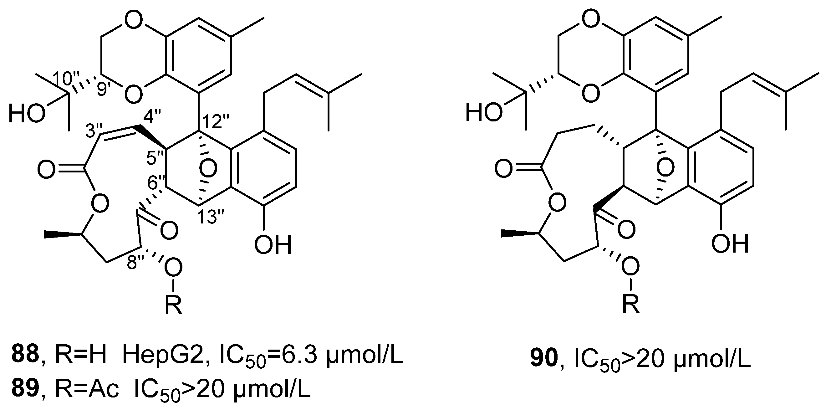
In 2021, Zhang’s research group isolated three new lithocarpin analogs from the deep-sea fungus
Phomopsis lithocarpus FS508, and named them Lithocarpins E-G (
88–90) (
Figure 43) [
96]. These compounds belong to the rare class of highly oxidized tenellone–macrolide heterodimers in nature, featuring a unique 9,14-epoxy naphtho [2,3-
b]oxacycloundecin-3(2H)-one skeleton. Taking Lithocarpins E as an example for structural confirmation: its molecular formula was determined to be C
35H
40O
9 with an unsaturation of 16; the presence of a tetrasubstituted benzene ring and an oxygenated methine group was deduced from the
1H NMR data. Based on the key HMBC correlation signals, the tenellone unit was confirmed to be connected to the macrocyclic lactone skeleton. Through the key spatial interaction of NOESY, the partial relative configuration was determined, such as H-6″, H-8″ and H-13″ being located on the same side of the macrocyclic lactone ring; the ECD spectrum matched the experimental spectrum, thus determining the absolute configuration of compound 1 as 9′
S, 5″
R, 6″
R, 8″
R, 10″
R, 12″
R, 13″
S. The in vitro activity test results showed that Lithocarpin E exhibited the strongest activity, with an IC
50 value of 6.3 μM for HepG2 cells, which was comparable to that of the positive drug cisplatin (2.4 μM). In contrast, Lithocarpins F and G showed significantly reduced activity, with the IC
50 values for various cell lines being greater than 20 μM. By comparing the activities of Lithocarpins E-G, the SAR revealed that the hydroxyl group at C-8″ and the double bond between C-3″/C-4″ were crucial for activity. Lithocarpins F, which was the acetylated product of the hydroxyl group at C-8″ of 1, displayed decreased activity; Lithocarpins G, the double bond reduction product, showed further diminished activity, indicating that both functional groups were essential structural elements for maintaining high activity.
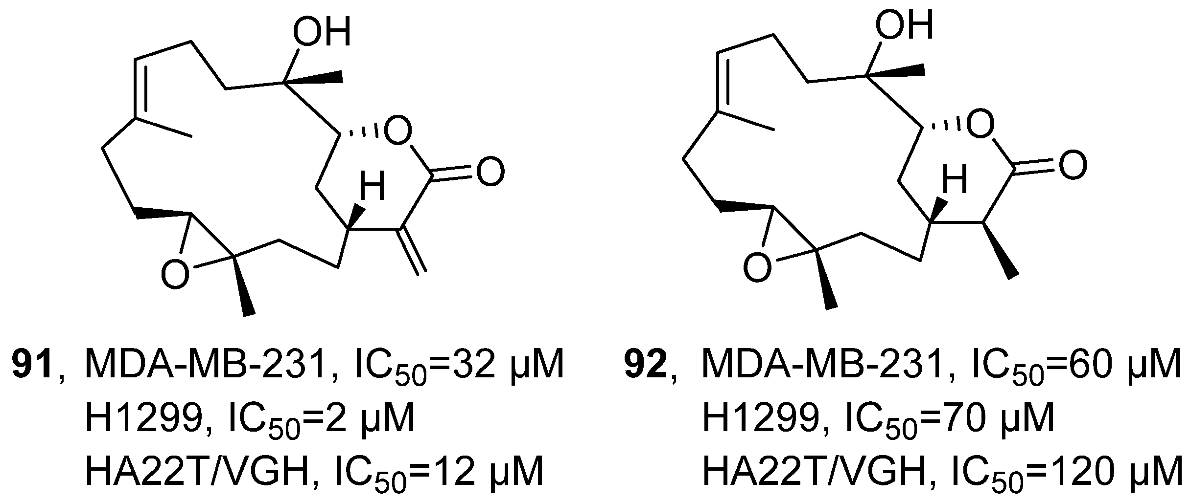
In 2021, Chang’s research group isolated Sinularin (
91) and Dihydrosinularin (
92) from the ethyl acetate extracts of the soft corals
Sinularia manaarensis and
S. flexibilis (
Figure 44) [
97]. Both compounds belong to the macrolide class, and their structures were elucidated using NMR spectroscopy. The activity evaluation results showed that the IC
50 values of Sinularin in MDA-MB-231, H1299 and HA22T/VGH cells were 32 µM, 2 µM and 12 µM respectively; while the IC
50 values of Dihydrosinularin were 60 µM, 70 µM and 120 µM respectively. The SAR analysis indicated that the differences in antioxidant activity between Sinularin and Dihydrosinularin might be attributed to the presence or absence of conjugated double bonds in their molecular structures.
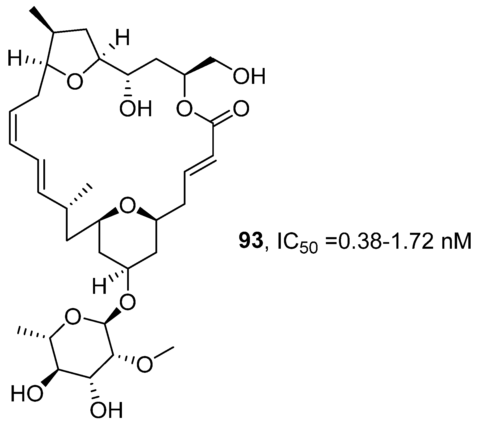
In 2022, E. Ishmael’s research group, based on previous work [
98], systematically evaluated the anti-tumor activity of the polyketide macrolide natural product Mandelalides (A-L) series compounds (
Figure 45) [
99]. The study obtained Mandelalide A (
93) via total synthesis and confirmed its structure by comparing NMR and LC-MS data with those reported in the literature. The activity evaluation results showed that the 48 h EC
50 value of
93 for wild-type mouse embryonic fibroblast cells (MEF) was 37.1 nM, while its sensitivity to AMPKα-deficient MEF was significantly enhanced (EC
50 = 13.5 nM). In non-small cell lung cancer cells, regardless of the LKB1 expression status (such as H292, PC-9 being LKB1 positive; H460, 11–18 being LKB1 deficient), the GI
50 values ranged between 2.0 and 4.7 nM. After extended exposure,
93 showed complete cytotoxicity to all tested glioblastoma cells, with IC
50 values ranging from 0.38 to 1.72 nM. The SAR analysis further revealed that the type A (featuring a simple lactone linkage) and type B (containing butenolide structure) macrolides could inhibit ATP synthase and activate AMPK, thus showing significant anti-tumor activity. In contrast, Type C macrocycles, which contain a 23-hydroxybutyrolactone structure, had no effect on mitochondrial function and exhibited relatively weak activity. Moreover,
93 displayed stronger cytotoxicity and AMPK activation ability, while the activity of non-glycosylated type C was significantly reduced, indicating that glycosylation is a key structural element for maintaining the strong anti-tumor activity in this class of compounds.
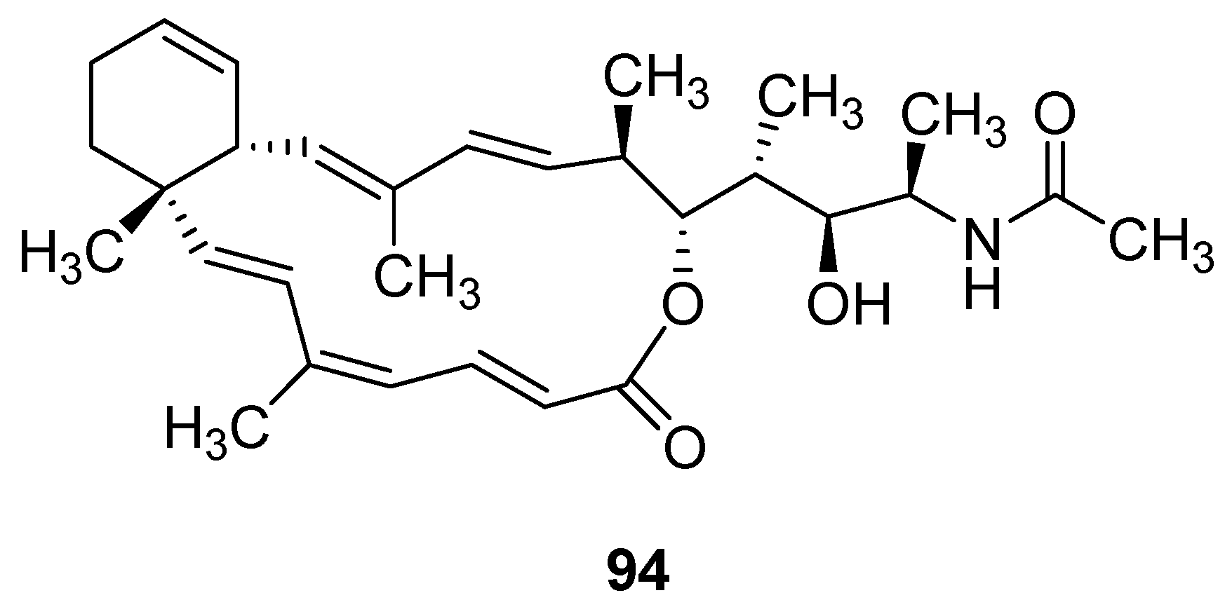
In 2023, Liu’s research group further explored the activities of macrocyclic laccoid MNP Superstolide A, based on its analog ZJ-101 (
94) (
Figure 46) [
100]. The activity evaluation results showed that
94 exhibited a typical cell growth inhibition effect, with the growth rate inhibition index (GR value) always greater than 0. After 72 h treatment in MDA-MB-231 cells, the GR value reached 0.5 at the highest dose. The EC
50 measured by the GR method was 29–96 nM. Using triptolide and flavopiridol as cytotoxic control compounds, both of which showed GR values below 0, further supported that
94 primarily functions through growth inhibition. Additionally,
94 showed great activity in 3D tumor sphere models: the EC
50 for inhibiting tumor sphere formation was approximately 1 nM, while the EC
50 for disrupting preformed tumor spheroids was about 5 nM, indicating its mechanism of action may be closely related to interfering with the cell adhesion process. In terms of the SAR, as a simplified analog of Superstolide A,
94 not only successfully retained the core biological activity of the parent compound but even enhanced it in some respects, thereby verifying the effectiveness of the structural simplification strategy.
In macrolides, macrocyclic lactone ring, glycosyl groups (especially 2,3-O-dimethylglucose), conjugated dienes, and hydroxyl groups are most frequent. The most active macrolides come from deep-sea fungi (Phomopsis lithocarpus), soft corals (Sinularia, Sarcophyton), and sponge-associated microbes. Mandelalide A (93) exhibits a GI50 as low as 2.0–4.7 nM against non-small cell lung cancer cells, among the highest in the literature; Superstolide analog (94) shows an EC50 of 1 nM for inhibiting 3D tumor sphere formation. Glycosylated macrolides with rigid macrocyclic scaffolds and conjugated enone structures display the best activity and selectivity. Trends from 2020 to 2024 include that total synthesis to overcome supply issues; glycosylation identified as critical for activity (deglycosylation drastically reduces potency); use of GR values to distinguish cytostasis from cytotoxicity; discovery of new mechanisms targeting ATP synthase and the AMPK pathway; and design of simplified analogs (e.g., ZJ-101 (94)) that retain core activity while improving synthetic accessibility.




 ,
,