Article

OSMAC-Driven Discovery of Six New Alkaloids from the Cold-Seep-Derived Fungus Talaromyces amestolkiae HDN21-0307

1
Key Laboratory of Marine Drugs Ministry of Education, School of Medicine and Pharmacy, Ocean University of China, Qingdao 266003, China
2
Qingdao Institute of Innovation, East China University of Science and Technology, Qingdao 266003, China
3
Laboratory for Marine Drugs and Bioproducts & Laboratory for Marine Biology and Biotechnology, Qingdao Marine Science and Technology Center, Qingdao 266237, China
4
Sanya Oceanographic Institute, Ocean University of China, Sanya 572025, China
*
Authors to whom correspondence should be addressed.
These authors contributed equally to this work.
Mar. Drugs 2025, 23(9), 337; https://doi.org/10.3390/md23090337
Submission received: 3 August 2025 / Revised: 21 August 2025 / Accepted: 22 August 2025 / Published: 25 August 2025
(This article belongs to the Section Structural Studies on Marine Natural Products)

Abstract

Six new alkaloid compounds, including two rare aromatic nitrile compounds talaronitriles A–B (12), a novel oxime-functionalized azadiphilone analogue talarooxime A (3), a new phenylhydrazone alkaloid talarohydrazone E (4), and two new dipeptide compounds talarodipeptides A–B (56), were isolated from the deep-sea cold-seep-derived fungus Talaromyces amestolkiae HDN21-0307 via OSMAC approach. Compound 1 is the first natural naphthalene compound with cyano groups. Compound 3 represents the first natural product containing an oxime-functionalized azadiphilone scaffold. Their structures and absolute configurations were elucidated through spectroscopic data analysis and quantum chemical calculations. Notably, compound 3 demonstrated moderate DPPH free-radical-scavenging activity, with an IC50 value of 29.41 μM.

1. Introduction

Natural alkaloids are nitrogen-containing alkaline substances found in living organisms and represent one of the most significant classes of secondary metabolites produced by marine fungi [1]. These compounds typically possess structurally complex nitrogenous heterocycles. Owing to their unique chemical scaffolds and diverse biological activities—such as antitumor, antibacterial, antiviral, and anti-inflammatory effects—natural alkaloids have garnered increasing interest in drug research and natural product chemistry [1,2].
Talaromyces fungi are widely distributed across various environments, including marine habitats, soil, and plants. Species within this genus produce an extensive array of secondary metabolites, among which alkaloids represent a major class. To date, numerous alkaloids have been isolated from Talaromyces fungi and can be roughly classified into amide alkaloids, indole alkaloids, terpene alkaloids, and quinoline alkaloids according to their structural types [3,4,5]. Notably, many alkaloids from Talaromyces, such as chaetominine B [3] and ditalaromylectones A–B [4], possess not only novel structures but also promising biological activities, underscoring the remarkable metabolic capacity of Talaromyces fungi for producing novel alkaloids.
In our previous study, the fungal strain Talaromyces amestolkiae HDN21-0307 was isolated from deep-sea cold seep sediments in the South China Sea, yielding four novel phenylhydrazone alkaloids (talarohydrazones A–D) with notable antimicrobial and cytotoxic activities [6]. However, similar to most marine microorganisms, T. amestolkiae HDN21-0307 still harbors numerous biosynthetic gene clusters that remain silent under monoculture conditions, limiting the discovery of novel bioactive metabolites [7]. The OSMAC (One Strain Many Compounds) strategy, which involves altering medium composition and cultivation conditions, has been widely employed to activate these silent gene clusters [8]. To date, this approach remains the most convenient and efficient strategy for mining microbial metabolites [9].
Consequently, the OSMAC strategy was also applied to further explore the metabolites of T. amestolkiae HDN21-0307. Six new alkaloids (16) were isolated from T. amestolkiae HDN21-0307 cultivated under two distinct conditions: (1) shaking conditions in Modified Glucose Minerals Salts 2# liquid medium and (2) static conditions in rice solid media with the addition of NaBr. Notably, these included the first reported naphthalene-fused nitrile natural product (1) and the inaugural oxime-functionalized azaphenanthrenone alkaloid (3). Additionally, compound 3 showed moderate DPPH free-radical-scavenging activity, with an IC50 value of 29.41 μM. Herein, details of the isolation, structure elucidation, and bioactivity evaluation of these compounds are presented.

2. Results

The OSMAC strategy was applied to comprehensively explore the metabolic potential of T. amestolkiae HDN21-0307 by employing six different culture media under shaking or static cultivation conditions. As a result, this strain exhibited distinct UV absorption profiles compared with those of previously known compounds from T. amestolkiae HDN21-0307 [6] under the following conditions: (1) modified Glucose Mineral Salts 2# liquid medium under shaking conditions and (2) rice solid medium supplemented with NaBr under static conditions (Figure S1). To elucidate the structures corresponding to these characteristic peaks, large-scale fermentations were conducted under both conditions, followed by ethyl acetate and methanol extraction. The resulting extracts were subjected to extensive column chromatography over silica gel, ODS, and Sephadex LH-20, ultimately yielding six new alkaloids (16) (Figure 1).
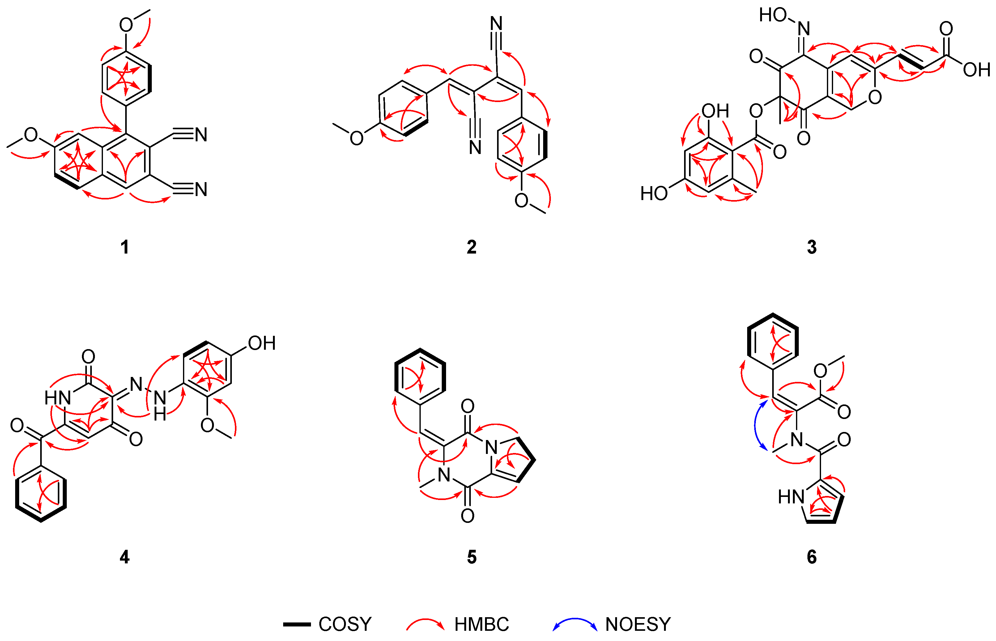
Compound (1), obtained as a colorless powder, has a molecular formula of C20H14N2O2 based on HRESIMS (m/z 315.1128, [M + H]+ calcd. for C20H15N2O2 315.1124), indicating fifteen degrees of unsaturation. Analysis of 1D NMR and HSQC (Table 1) indicated the existence of eighteen aromatic carbons, including ten non-hydrogenated carbons (δC 108.5, 111.7, 117.0, 117.7, 128.9, 130.5, 136.5, 148.2, 162.2, and 162.5), and two oxymethyl carbons (δC 55.9 and 56.0). The characteristic hydrogen signals H-2′/H-6′ (δH 7.42, d, J = 8.7 Hz) and H-3′/H-5′ (δH 7.18, d, J = 8.7 Hz) indicated the presence of a para-substituted benzene ring. Among them, the substituent at the 4′-position of the benzene ring was unambiguously identified as a methoxy group (C-7′), as confirmed by its characteristic chemical shift (C-4′, δC 162.2; C-7′, δC 55.9) and key HMBC correlation from H-7′ (δH 3.93, s) to C-4′. Additionally, a trisubstituted benzene ring with C-7 oxygen methyl group was determined through characteristic signals of H-5 (δH 8.07, d, J = 9.0 Hz), H-6 (δH 7.46, dd, J = 9.0, 2.5 Hz), and H-8 (δH 7.06, d, J = 2.5 Hz) and the HMBC correlations from H-11 (δH 3.77, s) to C-7 (δC 162.9), from H-5 to C-7/C-8a (δC 163.0), from H-6 to C-4a (δC 130.5), and from H-8 to C-4a/C-6 (δC 123.8)/C-7.
The established structural fragments account for eight degrees of unsaturation, leaving seven remaining unsaturation to be assigned. Analysis of the NMR data indicated only six aromatic carbons (δC 108.5, 111.7, 117.0, 117.7, 136.2, and 148.2) and two nitrogen atoms remained unassigned. To satisfy the high degree of unsaturation, compound 1 likely contained an additional aromatic ring. The key HMBC correlations of H-8/C-1, H-5/C-4 (δC 163.2), and H-4/C-2/C-5/C-8a confirmed the pentasubstituted aromatic ring as part of a poly-substituted naphthalene scaffold. Furthermore, the remaining two non-protonated aromatic carbons (C-9, δC 117.0; C-10, δC 117.7) and two nitrogen atoms needed to account for four additional degrees of unsaturation, leading to the identification of two cyano groups in compound 1. Finally, the para-substituted benzene ring was determined to be connected to the C-1 of the naphthalene scaffold through the correlations between H-2′/H-6′ and C-1 (δC 148.2), while the remaining two cyanide groups were connected to C-2 (δC 111.7) and C-3 (δC 108.5), respectively. In addition, the presence of a cyano group was also confirmed by its characteristic infrared absorption band at 2227 cm−1. The structure of 1 was conclusively established and named talaronitrile A [10].
The cyano group is widely used in the pharmaceutical industry for its ability to enhance binding affinity, improve pharmacokinetic properties, and reduce drug resistance [11,12]. Naturally occurring nitriles, mostly existing as cyanogenic glycosides, are widely distributed across various organisms including plants, fungi, and marine species [11,12]. Aromatic nitrile-containing natural products are relatively rare [11,12]. Notably, compound 1 represents the first natural product featuring a naphthalene-linked nitrile scaffold.
Compound (2) was obtained as a pale-yellow powder with a molecular formula of C20H16N2O2, based on HRESIMS data (m/z 317.1294 [M + H]+ calcd. for C20H17N2O2, 317.1285). Comparative analysis of the 1D NMR and HSQC spectra (Table 1) revealed the existence of sixteen aromatic carbons, including eight non-hydrogenated carbons (δC 100.8, 106.1, 116.1, 119.1, 125.4, 125.4, 161.9, and 162.7) and two oxymethyl carbons (δC 55.6 and 55.7). Compound 2 and epurpurin C shared the similar nitrile skeleton, as evidenced by the HMBC correlations from H-7′ (δH 7.35, s) to C-1 (δC 119.1)/C-3/C-2′, from H-2′/6′ (δH 7.41, d, J = 8.9 Hz) to C-1′/C-4′ (δC 161.9)/C-7′, from H-3′/5′ (δH 6.93, d, J = 8.9 Hz) to C-1′/C-4′, from H-7″ (δH 7.46, s) to C-2/C-4 (δC 116.1)/C-2″, from H-2″/H-6″ (δH 7.87, d, J = 8.9 Hz) to C-1″/C-4″ (δC 162.7)/C-7″, and from H-3″/5″ (δH 6.98, d, J = 8.9 Hz) to C-1″/C-4″ (Figure 2) [13]. The only difference between 2 and epurpurin C was that the absolute configuration of Δ3,7″ in 2 was E, supported by the existence of asymmetrical peaks in 1D NMR. Additionally, the C-H coupling constants (3JC-4,H-7″ = 14.6 Hz) further corroborated the double-bond configurations of Δ3,7″ (Figure S18) [14]. Hence, the structure of 2 was established and named talaronitrile B.
Compound (3), obtained as a bright yellow powder, has a molecular formula of C21H17NO10, based on HRESIMS data (m/z 444.0927 [M + H]+ calcd. for C21H18NO10, 444.0925). The 1D NMR and HSQC spectra (Table 2) revealed the presence of seventeen aromatic carbons, including thirteen non-hydrogenated carbons (δC 86.9, 104.3, 120.0, 141.3, 142.4, 145.0, 158.8, 164.4, 166.6, 166.9, 170.5, 185.4, and 189.0), five methine carbons (δC 101.8, 104.9, 113.0, 124.4, and 137.3), one methylene carbon (δC 64.6), and two methyl carbons (δC 21.9, 24.3). The 3JH-9,H-10 (15.5 Hz) unambiguously assigned the E configuration of the double bond Δ9,10. Comparative NMR data revealed that compound 3 and talarohydrazone B share an essentially identical core structure (Figure 2). The key structural differences include the absence of a phenylhydrazone moiety in compound 3, along with the presence of an additional nitrogen atom and hydroxyl group. The remaining degree of unsaturation, combined with the characteristic downfield shift of C-4 (δC 142.4), suggested the presence of an oxime moiety at C-4 in 3. Consequently, the planar structure of 3 was established and designated as talarooxime A.
Due to the lack of diagnostic NOESY correlations, the configuration of the Δ4,N−13 double bond could not be determined spectroscopically. To resolve this, the time-dependent density-functional theory (TDDFT) calculations were performed on both possible diastereomers 4E-3 (3a) and 4Z-3 (3b) (Figure S55). DP4+ probability analysis demonstrated excellent agreement (99.94%) between 3b and the experimental NMR data, establishing the Z-configuration at Δ4,N−13 double bond (Figure S56). The C-2 configuration was assigned as consistent with talarohydrazone B based on biosynthetic considerations. Furthermore, (2S)-3 showed remarkable concordance between experimental and calculated ECD spectra (Figure 3), thereby confirming the absolute configuration at C-2. Thus, the absolute configuration of compound 3 was determined to be (2S, 4Z, 9E). Structural diversity among currently known azaphilones is predominantly concentrated on chain extension at the C-2/C-6 positions of the compound 3 core scaffold, with modifications at the C-4 position being rare. Furthermore, the substituents introduced are predominantly aliphatic chains or aryl/heteroaryl groups [15]. In contrast, compound 3 represents the first reported oxime-containing azaphilone derivative featuring a substitution at the C-4 position.
Compound (4) was isolated as a red amorphous powder. The molecular formula of 4 was determined to be C19H15N3O5 based on the HRESIMS date (m/z 364.0937 [M − H] calcd. for C19H14N3O5, 364.0939), indicating fourteen degrees of unsaturation. Spectroscopic analysis of the 1H NMR data of 4 showed signals for three active hydrogen protons (δH 10.30, 11.15, and 17.30), nine olefinic protons, and one methoxy group. Analysis of the 13C NMR and HSQC spectra (Table 2) indicated three carbonyl carbons (δC 161.5, 178.9, and 180.5), fifteen olefinic carbons, including six non-hydrogenated carbons, and one oxymethyl (δC 56.1) carbon. The aforementioned information accounted for eleven out of the fourteen degrees of unsaturation, suggesting that 4 was a tricyclic compound. The NMR data of 4 was highly similar to that of talarohydrazone A (Figure 2) [6], with the only difference being the presence of a carbonyl group at C-7 in 4 instead of a methylene group at C-7 in talarohydrazone A. The HMBC correlations from H-5/H-9/H-13 to C-7 (δC 180.5) confirmed this difference between them. Thus, the planar structure of 4 was determined, named talarohydrazone E. The configuration of the double bond Δ3,14 could not be determined due to the absence of NOESY correlations. TDDFT was used to calculate NMR data of the two possible configurations 3E-4 (4a) and 3Z-4 (4b). The improved DP4+ probability analysis showed that 4a was in agreement with the NMR experimental data with a probability of 100% (Figure S58 and Table S3), confirming the configuration of the double bond Δ3,14 as E configuration.
Compound (5) was purified as a pale-yellow oil with the molecular formula as C15H14N2O2 based on the HRESIMS data (m/z 255.1126, [M + H]+ calcd. for C15H15N2O2, 255.1128). The 13C NMR and HSQC spectra of 5 indicated the presence of fifteen carbon atoms (Table 3), including two carbonyl carbons (δC 156.79 and 156.84,), ten olefinic carbons, two methylenes (δC 28.3 and 46.0,), and one methyl (δC 36.3). The NMR data of 5 closely resembled that of cyclo Pro-1-N-methyl-9,11-en-Phe [16], indicating that they possessed the same cyclic dipeptide skeleton. The only structural difference between them lies in the bond between C-3 and C-4: a double bond in 5 versus a single bond in cyclo Pro-1-N-methyl-9,11-en-Phe. Based on the COSY correlations of H-4 (δH 6.26)/H-5, and the HMBC correlations from H-5 to C-3 (δC 133.5) and from H-6 to C-4 (δC 120.3), the double bond Δ3 in 5 was established (Figure 2). Thus, the planar structure of 5 could be determined, named talarodipeptide A. Due to the absence of NOESY correlations of H-10/H-11, the configuration of the double bond Δ9,11 was determined by HSQMBC (Figure S44). The C-8 to H-11 (JC,H = 9.6 Hz) obtained from the HSQMBC spectrum unambiguously assigned the E configuration of the double bond Δ9,11 [14].
Compound (6) was isolated as a pale-yellow oil. The molecular formula, C16H16N2O3, was deduced from its HRESIMS data (m/z 285.1237, [M + H]+ calcd. for C16H17N2O3, 285.1234), indicating 10 degrees of unsaturation. The 13C NMR and HSQC spectra of 6 indicated (Table 3) two carbonyl carbons (δC 164.8 and 167.1), twelve olefinic carbons, including three non-hydrogenated carbons, and two methyls (δC 35.9 and 53.2,). The NMR data of 6 closely resembled that of Pyrrole-Phe-OMe ester [17], indicating that they had the same skeleton. The difference was that the 3-NH of 6 was replaced by a methyl group (C-3, δC 35.9), and C-2 (δC 134.1) and C-11 (δC 137.9) were double-bonded rather than single-bonded, confirmed by the HMBC correlations from H-10 (δH 3.06, s) to C-2/C-4, and from H-2′ to C-11 (Figure 2). The NOESY correlation of H-10/H-11 indicated the E-configuration of the double bond Δ2,11. Thus, the structure of 6 was determined, named talarodipeptide B.
Antibacterial, antitumor, and the free-radical-scavenging activity tests were conducted on compounds 16. Only compound 3 exhibited strong activity (IC50 = 29.41 μM) in the free-radical scavenging. Vitamin C (IC50 = 14.8 μM) was used as the positive control.

3. Materials and Methods

3.1. General Experimental Procedures

Ultraviolet (UV) spectroscopy was recorded on a Hitachi 5430 spectrophotometer (Hitachi, Tokyo, Japan). Circular dichroism (CD) spectra and optical rotations were measured by JASCO P-1020 digital polarimeter (JASCO Corporation, Tokyo, Japan), respectively. Infrared (IR) spectra were obtained on a Bruker Tensor-27 spectrophotometer (Bruker, Saarbruecken, Germany) with KBr pellets. High-resolution electrospray ionization mass spectrometry (HRESIMS) data were acquired on a Thermo Scientific LTQ Orbitrap XL mass spectrometer (Thermo Fisher Scientific, Waltham, MA, USA). Nuclear magnetic resonance (NMR) spectra were recorded on a JEOL JNM-ECP 600 MHz spectrometer (JEOL, Beijing, China) and an Agilent 500 MHz DD2 spectrometer (Agilent, Beijing, China), with tetramethylsilane (TMS) as an internal standard. Column chromatography was performed using Sephadex LH-20 (Amersham Biosciences, Uppsala, Sweden) and silica gel (Qingdao Marine Chemical Factory, Qingdao, China). Preparative high-performance liquid chromatography (HPLC) was carried out on a YMC-Pack ODS-A column (10 × 250 mm, 5 μm, 3 mL/min; YMC Co., Ltd., Kyoto, Japan).

3.2. Fungal Material and Fermentation

T. amestolkiae HDN21-0307 (GenBank No. OQ954837) was isolated from deep-sea cold seep sediments in the South China Sea (119°17′005.887″ E, 22°07′02.589″ N). This strain is a facultative anaerobe. Its mycelium, initially off-white, developed a green pigmentation after 4–5 days of incubation. Additionally, the strain produced a diffusible red pigment over time. This strain has been deposited in the Marine Medicinal Bioresources Center, School of Medicine and Pharmacy, Ocean University of China, Qingdao, China.

3.3. OSMAC Research, Fermentation, and Extraction

Based on the OSMAC strategy, T. amestolkiae HDN21-0307 was cultured in two different ways—shaking condition and static condition.
Shaking conditions: Using five distinct liquid media formulations: Modified Fungus 2# liquid media (Table S1. N1), PDB liquid media (Table S1. N2), SDA liquid media (Table S1. N3), Modified Glucose Minerals Salts 1# liquid media (Table S1. N4), and Modified Glucose Minerals Salts 2# liquid media (Table S1. N5). The products of T. amestolkiae HDN21-0307 cultured in MGMS (Table S1. N5) liquid media 2# under shaking conditions displayed unusual UV absorptions, which was the reason for selecting as the optimal conditions for large-scale fermentation. T. amestolkiae HDN21-0307 was cultured in 500 mL Erlenmeyer flasks, containing 150 mL of GMS liquid media for 9 days under shaking conditions (180 rpm, 28 °C, pH 7.0) in seawater (collected from JiaoZhou Bay, Qingdao, China). A total of 45 L of broth was extracted using EtOAc (3 × 45 L) to obtain crude extract (45.0 g).
Static conditions: Using two distinct solid media formulations: Rice solid media (Table S1. N6), NaBr rice solid media (Table S1. N7). The HPLC analysis of extracts revealed the chromatogram of the extract from the strain HDN21-0307 cultured in rice media with the addition of NaBr exhibited different peaks compared to rice media with no addition. Therefore, rice media with the addition of NaBr was selected as the optimal condition for large-scale fermentation. The fungus was cultured in 500 mL Erlenmeyer flasks each containing 80 g rice, 1 g NaBr and 120 mL freshwater at 28 °C for 30 days. A total of 12 L of broth was extracted using methanol (3 × 12 L) to obtain crude extract (100.0 g).

3.4. Separation and Purification of Compounds

The shaking crude extract was fractionated into five subcomponents (Fr.A-Fr.E) through VLC column chromatography, utilizing a stepped gradient elution protocol with MeOH-CH2Cl2. Fr.C was separated by ODS column to obtain five subfractions (Fr.C.1-Fr.C.5). Fr. C.2 was purified by Sephadex LH-20 and semi-preparative HPLC (YMC-pack ODS, 10 × 250 mm, 3.0 mL/min) to afford 3 (3.2 mg, tR = 25 min). Fr.C.4 was purified by semi-preparative HPLC to afford 1 (3.2 mg, tR = 31 min). Fr.C.4 was purified by semi-preparative HPLC to afford 2 (2.1 mg, tR = 29 min).
The static crude extract also used the same steps to separate the extract into the five subfractions (Fr.A-E). Fr. C was separated by ODS column to obtain four subfractions (Fr.C.1-Fr.C.4). Fr. C.1 was purified by semi-preparative HPLC to afford 5 (5.0 mg, tR = 24 min). Fr.C.2 was purified by semi-preparative HPLC to afford 6 (6.0 mg, tR = 24 min). Fr.C.3 was purified by semi-preparative HPLC to afford 4 (2.0 mg, tR = 25 min).
Talaronitrile A (1): white powder, UV (MeOH) λmax 223 (0.3), 258 (1.0) nm; IR (KBr) νmax 2925, 2227, 1677, 1614, 1206 cm−1; 1H and 13C NMR data, Table 1; HRESIMS m/z 315.1124 [M + H]+ (calcd. for C20H15N2O2, 315.1128).
Talaronitrile B (2): faint yellow powder, UV (MeOH) λmax 241 (0.3), 311 (0.4) nm, 360 (0.5) nm; IR (KBr) νmax 3419, 2921, 2221, 1602, 1510, 1263, 1178, cm−1; 1H and 13C NMR data, Table 1; HRESIMS m/z 317.1294 [M + H]+ (calcd. for C20H17N2O2, 317.1285).
Talarooxime A (3): green yellow powder, [ α ] D 25 150 (MeOH); UV (MeOH) λmax 215 (1.0), 265 (1.0) nm, 406 (0.4) nm; IR (KBr) νmax 1646, 1536, 1324, 1267, 1172 cm-1; 1H and 13C NMR data, Table 2; HRESIMS m/z 444.0927 [M + H]+ (calcd. for C21H18NO10, 444.0925).
Talarohydrazone E (4): red amorphous powder; UV (MeOH) λmax 206 (1.86), 258 (0.81), 510 (1.39) nm; IR (KBr) νmax 3446, 2923, 1626, 1417, 1307, 1284, 1024 cm−1; 1H and 13C NMR data, Table 2; HRESIMS m/z 364.0937 [M − H] (calcd. for C19H14N3O5, 364.0939).
Talarodipeptide A (5): pale-yellow oil; UV (MeOH) λmax 225 (0.52), 308 (1.25) nm; IR (KBr) νmax 3436, 1682, 1435, 1136, 760 cm−1; 1H and 13C NMR data, Table 3; HRESIMS m/z 255.1126 [M + H]+ (calcd. for C15H15N2O2, 255.1128).
Talarodipeptide B (6): pale-yellow oil; UV (MeOH) λmax 220 (0.65), 274 (1.02) nm; IR (KBr) νmax 3279, 1717, 1612, 1544, 1437, 762 cm−1; 1H and 13C NMR data, Table 3; HRESIMS m/z 285.1237 [M + H]+ (calcd. for C16H17N2O3, 285.1234).

3.5. Computation Section

The details of ECD calculation for 3 and the NMR calculation for 34 are available in the Supporting Information (Figures S54–S58 and Tables S2 and S3).

3.6. Assay of Activity

DPPH activity. The free-radical clearance experiment is based on the Sharma method [18] and has been modified. Compound 13 and vitamin C are dissolved in anhydrous ethanol and diluted into 6 gradients. Mix the vortex of these samples evenly, avoid light at room temperature for 30 min, and read the absorption at 515 nm as a positive control.
The ability to scavenge the DPPH was calculated according to the equation:
DPPH free-radical-scavenging rate DVC% = [(Ablank − Apostive control) ÷ Ablank] × 100%.
DPPH free-radical-scavenging rate Dsample% = [[Ablank − (Asample − Acontrol)] ÷ Ablank] × 100%.
Ablank: the absorbance of the DPPH solution;
Apostive control: the absorbance of the solution with VC;
Acontrol: absorbance of the sample.
Antibacterial activity. Antibacterial activities of compounds 16 were evaluated against Bacillus cereus ATCC 4342, MRCNS, MRSA, Staphylococcus aureus ATCC 29213, Klebsiella pneumoniae ATCC 43816, Escherichia coli ATCC 25922, Enterococcus faecalis ATCC 29212, Pseudomonas aeruginosa ATCC 27853, and Bacillus subtilis ATCC 6051. Ciprofloxacin was used as a positive control. The detailed methodologies for biological testing have been described in a previous report [19].
Cytotoxicity Assay. Cytotoxic activities of compounds 1, 46 were evaluated against K562 (using the MTT method), MDA-MB-231, NCI-H446, NCI-H446/EP, and ASPC-1 (using the SRB method) cell lines. Adriamycin was used as a positive control. The detailed methodologies for biological testing have been described in previous reports [20,21].

4. Conclusions

In summary, the application of the OSMAC strategy successfully activated the secondary metabolic potential of T. amestolkiae HDN21-0307, leading to the isolation of six new alkaloids. Among these, compounds (1) and (2), isolated for the first time from the genus Talaromyces, represent cyano-containing natural products. Notably, compound (1) constitutes the first naturally occurring cyano-substituted naphthalene derivative, while compound (3) is the first natural product featuring an azaphilone scaffold bearing an oxime functional group. This result emphasizes the significance of the OSMAC approach in unlocking the metabolic potential of microorganisms and further demonstrates the substantial potential for discovery within the secondary metabolites of fungi originating from extreme habitats such as cold springs.

Supplementary Materials

The following supporting information can be downloaded at: https://www.mdpi.com/article/10.3390/md23090337/s1. Figure S1: OSMAC cultivation strategy for T. amestolkiae HDN21-0307; Table S1: OSMAC consists of media for T. amestolkiae HDN21-0307; Figure S2: The 16S rRNA sequences data of T. amestolkiae HDN21-0307; Figure S3: HRESIMS spectrum of compound 1; Figure S4: 1H NMR spectrum (400 MHz, CD3OD) of compound 1; Figure S5: 13C NMR spectrum (125 MHz, CD3OD) of compound 1; Figure S6: HSQC spectrum of compound 1; Figure S7: HMBC spectrum of compound 1; Figure S8: COSY spectrum of compound 1; Figure S9: IR spectrum of compound 1; Figure S10: HRESIMS spectrum of compound 2; Figure S11: 1H NMR spectrum (400 MHz, CDCl3) of compound 2; Figure S12: 13C NMR spectrum (125 MHz, CDCl3) of compound 2; Figure S13: HSQC spectrum of compound 2; Figure S14: HMBC spectrum of compound 2; Figure S15: COSY spectrum of compound 2; Figure S16: NOESY spectrum of compound 2; Figure S17: HSQMBC spectrum of compound 2; Figure S18: 3JC-H values obtained from the HSQMBC spectrum (500 MHz, CDCl3) of 2; Figure S19: IR spectrum of compound 2; Figure S20: HRESIMS spectrum of compound 3; Figure S21: 1H NMR spectrum (500 MHz, Acetone-d6) of compound 3; Figure S22: 13C NMR spectrum (125 MHz, Acetone-d6) of compound 3; Figure S23: HSQC spectrum of compound 3; Figure S24: HMBC spectrum of compound 3; Figure S25: COSY spectrum of compound 3; Figure S26: NOESY spectrum of compound 3; Figure S27: IR spectrum of compound 3; Figure S28: HRESIMS spectrum of compound 4; Figure S29: 1H NMR spectrum (500 MHz, DMSO-d6) of compound 4; Figure S30: 13C NMR spectrum (125 MHz, DMSO-d6) of compound 4; Figure S31: HSQC spectrum of compound 4; Figure S32: HMBC spectrum of compound 4; Figure S33: COSY spectrum of compound 4; Figure S34: NOESY spectrum of compound 4; Figure S35: IR spectrum of compound 4; Figure S36: HRESIMS spectrum of compound 5; Figure S37: 1H NMR spectrum (400 MHz, CDCl3) of compound 5; Figure S38: 13C NMR spectrum (100 MHz, CDCl3) of compound 5; Figure S39: HSQC spectrum of compound 5; Figure S40: HMBC spectrum of compound 5; Figure S41: COSY spectrum of compound 5; Figure S42: NOESY spectrum of compound 5; Figure S43: HSQMBC spectrum of compound 5; Figure S44: 3JC-H values obtained from the HSQMBC spectrum (400 MHz, CDCl3) of 5. Figure S45: IR spectrum of compound 5; Figure S46: HRESIMS spectrum of compound 6; Figure S47: 1H NMR spectrum (100 MHz, CD3OD) of compound 6; Figure S48: 13C NMR spectrum (100 MHz, CD3OD) of compound 6; Figure S49: HSQC spectrum of compound 6; Figure S50: HMBC spectrum of compound 6; Figure S51: COSY spectrum of compound 6; Figure S52: NOESY spectrum of compound 6; Figure S53: IR spectrum of compound 6; Figure S54: ECD: B3LYP/6-31G(d) optimized lowest energy conformers for 3 [22,23]; Figure S55: B3LYP/6-31G(d) optimized lowest energy conformers for 3; Figure S56: NMR calculations with DP4+ probability analysis for compounds 3; Table S2. DP4+ evaluation of theoretical and experimental one of 3 [24]; Figure S57: B3LYP/6-31G(d) optimized lowest energy conformers for 4; Figure S58: NMR calculations with DP4+ probability analysis for compounds 4; Table S3. DP4+ evaluation of theoretical and experimental one of 4.

Author Contributions

X.H. and J.W. drafted the work and performed isolation and structural elucidation of the extract. L.Z. also carried out structural elucidation and software analysis. X.H. and J.W. performed amplification fermentation and isolation of the strain. X.H. and Z.W. evaluated biologically. L.C. also performed amplification fermentation. D.L. provided the necessary strains and guidance of fermentation process for the experiment. W.W. and Q.C. provided guidance for structural analysis. T.Z. designed this project and contributed to the critical reading of the manuscript. Q.C., D.L. and T.Z. checked the procedure of this work. All authors have read and agreed to the published version of the manuscript. X.H. and J.W. contributed equally to this paper.

Funding

This work was funded by the National Key R&D Program of China (2024YFC2816004), the Major Basic Research Programs of Natural Science Foundation of Shandong Province (ZR2019ZD18), the Qingdao Marine Science and Technology Center (2022QNLM030003-1), the Key R&D Program of Hainan Province (ZDYF2023SHFZ144), the Fundamental Research Funds for the Central Universities (202172002, 202172009, and 202262015), the Taishan Scholar Distinguished Expert Program in Shandong Province (tstp20240504), and the Research on the Development Strategy of Qingdao’s Synthetic Biology Future Industry (25-1-4-zlyj-16-zhc).

Institutional Review Board Statement

Not applicable.

Data Availability Statement

The original data presented in the study are included in the article/Supplementary Material; further inquiries can be directed to the corresponding author.

Conflicts of Interest

The authors declare no conflicts of interest.

References

  1. Xu, L.; Meng, W.; Cao, C.; Wang, J.; Shan, W.; Wang, Q. Antibacterial and Antifungal Compounds from Marine Fungi. Mar. Drugs 2015, 13, 3479–3513. [Google Scholar] [CrossRef]
  2. Yin, Q.; Liu, X.; Zhang, Z.; Lei, H.; Wu, B. Chemistry and Bioactivities of Alkaloids Isolated from Marine Fungi (Covering 2016–2022). Fitoterapia 2023, 164, 105377. [Google Scholar] [CrossRef] [PubMed]
  3. Cong, M.; Zhang, Y.; Feng, X.; Pang, X.; Liu, Y.; Zhang, X.; Yang, Z.; Wang, J. Anti-Inflammatory Alkaloids from the Cold-Seep-Derived Fungus Talaromyces helicus SCSIO41311. 3 Biotech 2022, 12, 161. [Google Scholar] [CrossRef] [PubMed]
  4. Lei, L.-R.; Gong, L.-Q.; Jin, M.-Y.; Wang, R.; Liu, R.; Gao, J.; Liu, M.-D.; Huang, L.; Wang, G.-Z.; Wang, D.; et al. Research Advances in the Structures and Biological Activities of Secondary Metabolites from Talaromyces. Front. Microbiol. 2022, 13, 984801. [Google Scholar] [CrossRef] [PubMed]
  5. An, T.; Tan, H.; Guo, R.; Ma, X.; Wen, H. Research progress on the structure and activity of secondary metabolites of Talaromyces sp. Chin. J. Antibiot. 2024, 49, 961–985. [Google Scholar]
  6. Wu, J.; Wang, W.; Yang, Y.; Shah, M.; Peng, J.; Zhou, L.; Zhang, G.; Che, Q.; Li, J.; Zhu, T.; et al. Phenylhydrazone Alkaloids from the Deep-Sea Cold Seep Derived Fungus Talaromyces amestolkiae HDN21-0307. J. Nat. Prod. 2024, 87, 1407–1415. [Google Scholar] [CrossRef]
  7. Paoli, L.; Ruscheweyh, H.-J.; Forneris, C.C.; Hubrich, F.; Kautsar, S.; Bhushan, A.; Lotti, A.; Clayssen, Q.; Salazar, G.; Milanese, A.; et al. Biosynthetic Potential of the Global Ocean Microbiome. Nature 2022, 607, 111–118. [Google Scholar] [CrossRef]
  8. Bode, H.B.; Bethe, B.; Höfs, R.; Zeeck, A. Big Effects from Small Changes: Possible Ways to Explore Nature’s Chemical Diversity. ChemBioChem 2002, 3, 619–627. [Google Scholar] [CrossRef]
  9. Jin, J.; Fang, M.; Yin, K.; Jiang, M.; Wang, Y. Research progress of secondary metabolites of marine fungi by OSMAC strategy. Chin. J. Antibiot. 2023, 48, 481–491. [Google Scholar]
  10. Takahashi, H.; Nozawa, K.; Kawai, K.I. Isolation and Structures of Dicyanide Derivatives, Epurpurins A to C, from Emericella purpurea. Chem. Pharm. Bull. 1996, 44, 2227–2230. [Google Scholar] [CrossRef]
  11. Scotti, C.; Barlow, J.W. Natural Products Containing the Nitrile Functional Group and Their Biological Activities. Nat. Prod. Commun. 2022, 17, 1–24. [Google Scholar] [CrossRef]
  12. Liu, M.; Li, S. Nitrile Biosynthesis in Nature: How and Why? Nat. Prod. Rep. 2024, 41, 649–671. [Google Scholar] [CrossRef] [PubMed]
  13. Komai, S.; Hosoe, T.; Itabashi, T.; Nozawa, K.; Okada, K.; De Campos Takaki, G.M.; Chikamori, M.; Yaguchi, T.; Fukushima, K.; Miyaji, M.; et al. A New Funicone Derivative Isolated from Talaromyces flavus IFM52668. Mycotoxins 2004, 54, 15–19. [Google Scholar] [CrossRef]
  14. Wang, W.; Yang, J.; Liao, Y.-Y.; Cheng, G.; Chen, J.; Mo, S.; Yuan, L.; Cheng, X.-D.; Qin, J.-J.; Shao, Z. Aspeterreurone A, a Cytotoxic Dihydrobenzofuran–Phenyl Acrylate Hybrid from the Deep-Sea-Derived Fungus Aspergillus terreus CC-S06-18. J. Nat. Prod. 2020, 83, 1998–2003. [Google Scholar] [CrossRef] [PubMed]
  15. Liu, L.; Wang, Z. Azaphilone Alkaloids: Prospective Source of Natural Food Pigments. Appl. Microbiol. Biotechnol. 2022, 106, 469–484. [Google Scholar] [CrossRef]
  16. Jin, S.; Liebscher, J. Optically Active Precursors for Quaternary Amino Acids by Addition of N-Heteroaromatics to 3-Alkylidene-2,5-Diketopiperazines. Synlett 1999, 1999, 459–461. [Google Scholar] [CrossRef]
  17. Tian, H.; Ermolenko, L.; Gabant, M.; Vergne, C.; Moriou, C.; Retailleau, P.; Al-Mourabit, A. Pyrrole-Assisted and Easy Oxidation of Cyclic α-Amino Acid- Derived Diketopiperazines under Mild Conditions. Adv. Synth. Catal. 2011, 353, 1525–1533. [Google Scholar] [CrossRef]
  18. Sharma, O.P.; Bhat, T.K. DPPH Antioxidant Assay Revisited. Food Chem. 2009, 113, 1202–1205. [Google Scholar] [CrossRef]
  19. Chang, Y.; Zhou, L.; Hou, X.; Zhu, T.; Pfeifer, B.A.; Li, D.; He, X.; Zhang, G.; Che, Q. Microbial Dimerization and Chlorination of Isoflavones by a Takla Makan Desert-Derived Streptomyces sp. HDN154127. J. Nat. Prod. 2023, 86, 34–44. [Google Scholar] [CrossRef]
  20. Skehan, P.; Storeng, R.; Scudiero, D.; Monks, A.; McMahon, J.; Vistica, D.; Warren, J.T.; Bokesch, H.; Kenney, S.; Boyd, M.R. New Colorimetric Cytotoxicity Assay for Anticancer-Drug Screening. J. Natl. Cancer Inst. 1990, 82, 1107–1112. [Google Scholar] [CrossRef]
  21. Mosmann, T. Rapid Colorimetric Assay for Cellular Growth and Survival: Application to Proliferation and Cytotoxicity Assays. J. Immunol. Methods 1983, 65, 55–63. [Google Scholar] [CrossRef]
  22. Frisch, M.J.; Trucks, G.; Schlegel, H.B.; Scuseria, G.E.; Robb, M.A.; Cheeseman, J.; Scalmani, G.; Barone, V.; Mennucci, B.; Petersson, G.A.; et al. Gaussian 09 Revision A.1; Gaussian Inc.: Wallingford, CT, USA, 2009. [Google Scholar]
  23. Bruhn, T.; Schaumlöffel, A.; Hemberger, Y.; Bringmann, G. SpecDis: Quantifying the Comparison of Calculated and Experimental Electronic Circular Dichroism Spectra. Chirality 2013, 25, 243–249. [Google Scholar] [CrossRef]
  24. Grimblat, N.; Zanardi, M.M.; Sarotti, A.M. Beyond DP4: An Improved Probability for the Stereochemical Assignment of Isomeric Compounds Using Quantum Chemical Calculations of NMR Shifts. J. Org. Chem. 2015, 80, 12526–12534. [Google Scholar] [CrossRef]
Figure 1. Structures of the isolated compounds 16.
Marinedrugs 23 00337 g001
Figure 2. Key COSY, key HMBC, and key NOESY correlations of compounds 16.
Marinedrugs 23 00337 g002
Figure 3. Experimental and calculated ECD spectra of compound 3.
Marinedrugs 23 00337 g003
Table 1. 1H NMR (400 MHz) and 13C NMR (125 MHz) data of 1 in methanol-d4 and 2 in chloroform-d (δ in ppm, J in Hz).
No.1No.2
δC, TypeδH (J in Hz)δC, TypeδH (J in Hz)
1148.2, C 1119.1, C
2111.7, C 2106.1, C
3108.5, C 3100.8, C
4136.2, CH8.50, s4116.1, C
4a130.5, C 1′125.4, C
5132.2, C8.07, d (9.0)2′132.5, CH7.41, d (8.9)
6123.8, CH7.46, dd (9.0, 2.5)3′114.4, CH6.93, d (8.9)
7162.9, CH 4′161.9, C
8107.1, CH7.06, d (2.5)5′114.4, CH6.93, d (8.9)
8a136.5, C 6′132.5, CH7.41, d (8.9)
9117.0, C 7′146.1, CH7.35, s
10117.7, C 8′55.6, CH33.88, s
1156.0, CH33.77, s1″125.4, C
1′128.9, C 2″132.1, CH7.87, d (8.9)
2′132.3, CH7.42, d (8.7)3″114.8, CH6.98, d (8.9)
3′115.5, CH7.18, d (8.7)4″162.7, C
4′162.2, C 5″114.8, CH6.98, d (8.9)
5′115.5, CH7.18, d (8.7)6″132.1, CH7.87, d (8.9)
6′132.3, CH7.42, d (8.7)7″149.0, CH7.46, s
7′55.9, CH33.93, s8″55.7, CH33.85, s
Table 2. 1H NMR (500 MHz) and 13C NMR (125 MHz) data of 3 in acetone-d6 and 4 in DMSO-d6 (δ in ppm, J in Hz).
No.3No.4
δC, TypeδH (J in Hz)δC, TypeδH (J in Hz)
1189.0, C 1
286.9, C 2160.9, C
3185.4, C 3127.9, C
4142.4, C 4178.2, C
4a120.0, C 5108.6, CH5.91, s
5104.9, CH6.56, s6145.1, C
6158.8, C 7180.5, C
864.6, CH25.14, m8134.6, C
8a141.3, C 9129.9, CH7.91, m a
9137.3, CH7.23, d (15.5)10129.0, CH7.61, m a
10124.4, CH6.42, d (15.5)11134.4, CH7.76, m a
11166.9, CH 12128.6, CH7.61, m a
1221.9, CH31.77, s13129.1, CH7.91, m a
1″170.5, C 16122.6, C
2″104.3, C 17151.6, C
3″166.6, C 1899.7, CH6.62, d (2.3)
4″101.8, CH6.25, d (2.4)19159.8, C
5″164.4, C 20109.3, CH6.57, dd (8.8, 2.1)
6″113.0, CH6.38, d (2.1)21117.9, CH7.69, d (8.3)
7″145.0, C 2256.3, CH33.92, s
8″24.3, CH32.62, s1-NH11.15, s
3″-OH10.68, s15-NH17.30, s
19-OH10.30, s
a Signals were overlapped.
Table 3. 1H NMR (400 MHz) and 13C NMR (100 MHz) data of 5 in chloroform-d and 6 in methanol-d4 (δ in ppm, J in Hz).
No.5No.6
δC, TypeδH (J in Hz)δC, TypeδH (J in Hz)
1 1167.1, C
2156.8, C 2134.1, C
3133.5, C 3
4120.3, CH6.26, t (3.1)4164.8, C
528.3, CH22.84, td (9.1, 3.1)5126.1, C
646.0, CH24.13, t (9.1)6114.7, CH6.53, dd (3.8, 1.1)
7 7110.5, CH6.03, dd (3.8, 2.6)
8156.79, C 8123.6, CH6.87, dd (2.4, 1.2)
9131.7, C 9
1056.3, CH32.91, s1035.9, CH33.06, s
11119.6, CH7.30, s11137.9, CH7.63, s
12 1253.2, CH33.68, s
1′134.7, C 1′134.0, C
2′129.4, CH7.22, t (7.5)2′131.2, CH7.61, ma
3′128.3, CH7.33, ma3′130.2, CH7.40, m a
4′128.2, CH7.33, ma4′131.9, CH7.40, m a
5′128.3, CH7.33, ma5′130.2, CH7.40, m a
6′128.3, CH7.22, t (7.5)6′131.2, CH7.61, m a
a Signals were overlapped.
Disclaimer/Publisher’s Note: The statements, opinions and data contained in all publications are solely those of the individual author(s) and contributor(s) and not of MDPI and/or the editor(s). MDPI and/or the editor(s) disclaim responsibility for any injury to people or property resulting from any ideas, methods, instructions or products referred to in the content.