Review

Neoxanthin: A Promising Medicinal and Nutritional Carotenoid

1
College of Food Sciences and Engineering, Ningbo University, Ningbo 315211, China
2
School of Marine Sciences, Ningbo University, Ningbo 315211, China
*
Author to whom correspondence should be addressed.
These authors contributed equally to this work.
Mar. Drugs 2025, 23(8), 317; https://doi.org/10.3390/md23080317 (registering DOI)
Submission received: 2 July 2025 / Revised: 20 July 2025 / Accepted: 28 July 2025 / Published: 1 August 2025
(This article belongs to the Special Issue Marine Carotenoids and Potential Therapeutic Benefits)

Abstract

Neoxanthin is a xanthophyll carotenoid with high-value nutritional functions for human health due to its anti-cancer, anti-oxidative, and anti-obesity activities. In this present work, we systematically reviewed the structure, source, and biosynthetic pathways of neoxanthin, and discussed the advantages and disadvantages of the prevailing extraction methods of neoxanthin. Meanwhile, this review described the latest research progress on the pharmacological activities of neoxanthin. Finally, we concluded with a discussion on the main challenges of neoxanthin production from microalgae, and proposed some future development prospects and potential solutions.

1. Introduction

Carotenoids are a class of natural isoprene pigments with high-value nutritional functions for human health [1]. The study of carotenoids has a history of around 200 years [2]. The earliest record about the presence of natural carotenoid was published in 1831, when Professor Heinrich Wilhelm Ferdinand Wackenroder discovered the existence of natural carotenoid in carrot juice and its benefits [3]. In 1929, Professor Harry Steenbock proposed the possibility about a relationship between vitamin A activity and carotenoid pigmentation [4]. The possibility has caused great attention to natural carotenoids and their biological activities. In 1931, the chemical structure of β-carotene was described in detail by Professor Paul Karrer, who won the Nobel Prize in Chemistry due to his discovery [5]. Currently, the established carotenoids database could provide the structure, chemical fingerprints, distribution, and source information on 1204 natural carotenoids, as well as their biological functions (See: http://carotenoiddb.jp, accessed on 29 July 2025). Carotenoids have been widely applied in food, pharmaceutics, and cosmetics fields [6,7]. At present, some natural carotenoids, mainly including astaxanthin, β-carotene, fucoxanthin, lutein, lycopene, and zeaxanthin, are the main carotenoids possessing high added value with the largest market share [8].
Neoxanthin, a C40 epoxycarotenoid, has some remarkable bioactivities, including anti-cancer and anti-bacterial (See: http://carotenoiddb.jp, accessed on 29 July 2025). Currently, neoxanthin has been found in a variety of high plants, algae, and animals. In diverse high plants and algae, neoxanthin is one of the key photosynthetic xanthophylls [9], Notably, many microalgae are regarded as potential source species due to their rich content of neoxanthin, especially diverse microalgae species of the Chlorophyta phylum and Chlorophyta class, including Scenedesmus obliquus [10], Chlorella vulgaris [11], and Chaetomorpha antennia [12]. Neoxanthin has some remarkable bioactivities, including anti-cancer, anti-oxidant, and anti-obesity [13]. Therefore, neoxanthin is regarded as a high-added-value carotenoid from high plants and microalgae, which has significant potential for applications in the food, pharmaceutical, and cosmetic industries [14].
In this paper, we present a systematic review of neoxanthin focusing on its source, biosynthetic pathways, prevailing extraction methods, and biological activities. Furthermore, the review discussed the challenges and opportunities facing the future research field of neoxanthin.

2. Structure of Neoxanthin

The study of neoxanthin has a history of around 100 years. The earliest report was in 1938, when Professor Strain first discovered the presence of neoxanthin and named it, which was a common carotenoid distributed in barley leaves. Together with lutein and violaxanthin, neoxanthin is regarded as one of the three major xanthophylls of leaves [15,16]. As a potent light-harvesting carotenoid, neoxanthin is a major xanthophyll carotenoid in dark green leafy vegetables [17], additionally, it is a precursor of the plant hormone abscisic acid in these vegetables [18]. The molecular formula of neoxanthin is C40H56O4, with a molecular weight of 600.87 Da. It is insoluble in water but soluble in organic solvents such as ethanol, methanol, and acetone [19]. The structure of neoxanthin contains an unusual allenic bond, 8 conjugated double bonds, some oxygenic functional groups, including epoxy and hydroxyl moieties, and other functional groups [20]. Like other carotenoids, neoxanthin is chemically unstable, which makes it sensitive to heat and light that can impair its structure due to the conjugated polyenes in its chemical structure [21] (Figure 1).
In nature, neoxanthin mainly exists in two forms of geometrical isomer: all-trans neoxanthin and 9′-cis neoxanthin [22]. Their chemical structures are shown in Figure 1. The two isomers can be distinguished using ultraviolet absorption spectra. In ethanol, all-trans neoxanthin presents the main absorption peaks at 418, 442, and 471 nm [23]. But the 9′-cis neoxanthin presents the main absorption peaks at 413, 437, and 466 nm [24]. In higher plants, two forms of neoxanthin are found in different organs. 9′-cis neoxanthin is found in leaves, which can accomplish photosynthesis, while all-trans neoxanthin is found in some organs like petals and fruits that cannot carry out photosynthesis [18]. As far as algae are concerned, 9′-cis neoxanthin is found in nearly all microalgae species of the Chlorophyta phylum [10]. However, all-trans neoxanthin is the characteristic isomer found in microalgae species of Mesostigmatophyceae [25].

3. Neoxanthin Source

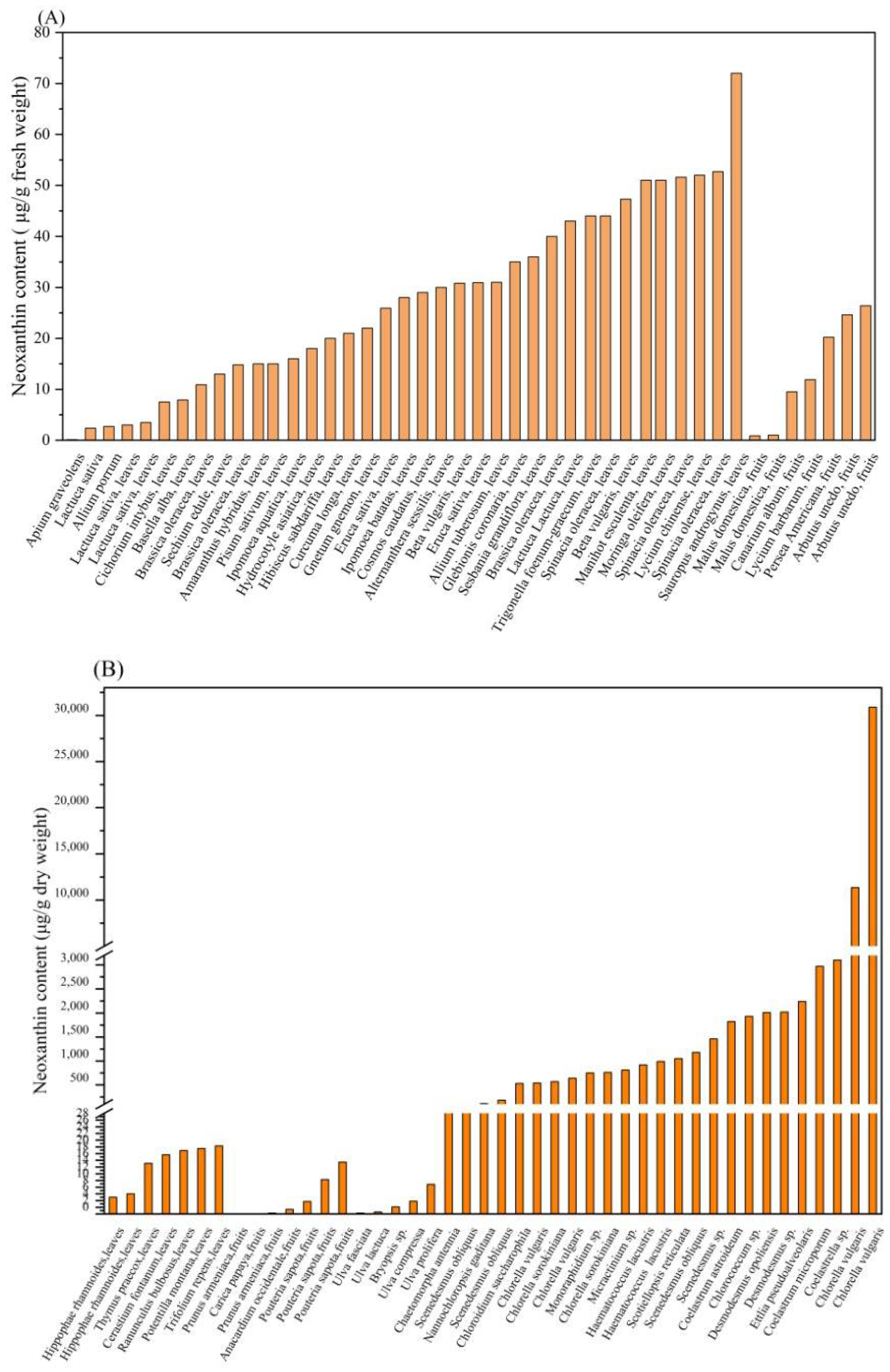
Neoxanthin has been found in a variety of higher plants and microalgae. High plants are one of the main sources of neoxanthin [9]. The neoxanthin content in the leaves and fruits of the high plants is shown in Table 1 and Table 2, while that in microalgal species is shown in Table 3. Additionally, the neoxanthin content in high plants and microalgal species is displayed in Figure 2.
Table 1. Neoxanthin content in the leaves of high plants.
High PlantContent of NeoxanthinReference
Allium porrum2.7 ± 0.9 a[26]
Allium tuberosum31 ± 2 a[26]
Alternanthera sessilis30 ± 3 a[26]
Amaranthus hybridus15 ± 5 a[26]
Apium graveolens0.11 ± 0.09 a[26]
Basella alba7.9 ± 0.7 a[26]
Beta vulgaris30.8 a[27]
Beta vulgaris47.3 a[27]
Brassica oleracea40 ± 10 a[26]
Brassica oleracea14.8 a[28]
Brassica oleracea10.9 a[28]
Cerastium fontanum17.6 ± 0.860 b[29]
Cichorium intybus7.5 a[28]
Cosmos caudatus29 ± 7 a[26]
Curcuma longa21 ± 1 a[26]
Eruca sativa30.9 a[27]
Eruca sativa25.9 a[27]
Glebionis coronaria35 ± 6 a[26]
Gnetum gnemon22 ± 2 a[26]
Hibiscus sabdariffa20 ± 2 a[26]
Hippophae rhamnoides5.0 ± 0.0 b[30]
Hippophae rhamnoides6.0 ± 0.0 b[30]
Hydrocotyle asiatica18 ± 1 a[26]
Ipomoea aquatica16 ± 1 a[26]
Ipomoea batatas28 ± 3 a[26]
Lactuca sativa var. capitata3 ± 1 a[26]
Lactuca sativa var. Longifolia43 ± 1 a[26]
Lactuca sativa var. Longifolia2.36 a[27]
Lactuca sativa var. Longifolia3.49 a[27]
Lycium chinense52 ± 8 a[26]
Manihot esculenta51 ± 5 a[26]
Moringa oleifera51 ± 12 a[26]
Pisum sativum15 ± 2 a[26]
Potentilla montana19.5 ± 0.787 b[29]
Ranunculus bulbosus18.9 ± 0.179 b[29]
Sauropus androgynus72 ± 6 a[26]
Sesbania grandiflora36 ± 4 a[26]
Sechium edule13 ± 6 a[26]
Spinacia oleracea44 ± 9 a[26]
Spinacia oleracea52.7 a[27]
Spinacia oleracea51.6 a[27]
Thymus praecox15.1 ± 0.0803 b[29]
Trifolium repens20.3 ± 0.556 b[29]
Trigonella foenum-graecum44 ± 7 a[26]
a Neoxanthin content profile expressed as μg/g fresh weight (FW) of high plants. b Neoxanthin content profile expressed as μg/g dry weight (DW) of high plants.
The experimental studies have demonstrated that the neoxanthin content in dry leaves of high plants is about 5.00~20.30 μg/g dry weight [29], while that in the fresh leaves is about 0.11~72.00 μg/g fresh weight [26].
Table 2. Neoxanthin content in the fruit of high plants.
High PlantContent of NeoxanthinReference
Arbutus unedo26.4 ± 1.8 a[31]
Arbutus unedo24.6 ± 1.9 a[31]
Anacardium occidentale1.36 b[32]
Canarium album9.5 a[33]
Carica papaya0.007 b[34]
Lycium barbarum11.9 ± 0.0 a[35]
Malus domestica0.99 ± 0.05 a[36]
Malus domestica0.87 ± 0.05 a[36]
Persea Americana20.2 ± 2.80 a[37]
Pouteriasapota15.45 ± 1.32 b[38]
Pouteriasapota10.24 ± 2.63 b[38]
Pouteriasapota3.70 ± 0.99 b[38]
Prunus armeniaca0.005 b[39]
Prunus armeniaca0.257 b[39]
a Neoxanthin content profile expressed as μg/g dry weight (DW) of high plants. b Neoxanthin content profile expressed as μg/g fresh weight (FW) of high plants.
In addition, the neoxanthin content in dry fruit of high plants is about 0.02~51.00 μg/g dry weight [31], while that in the fresh fruit is about 0.005~15.45 μg/g fresh weight [38]. Interestingly, two forms of neoxanthin (all-trans neoxanthin and 9′-cis neoxanthin) are found in the fruits of two high plants, Arbutus unedo and Malus domestica [31,36]. In terms of a promising source of neoxanthin, high plants are difficult to be a competitive neoxanthin feedstock due to some disadvantages, including their low neoxanthin content, and their limited growth dependent on the season [40]. Notably, the content of neoxanthin in the microalgae is about 0.26~30,880 μg/g dry weight [11,12].
Table 3. Neoxanthin content in Chlorophyta microalgal species.
Microalgal SpeciesNeoxanthin ContentReference
Bryopsis sp.2.11 ± 0.10 a[12]
Chaetomorpha antennia33.35 ± 0.23 a[12]
Chloroidium saccharophilum (formerly Chlorella saccharophila)530 ± 40 a[10]
Chlorella sorokiniana570 ± 20 a[10]
Chlorella sorokiniana760 ± 120 a[10]
Chlorella vulgaris540 ± 80 a[10]
Chlorella vulgaris640 ± 50 a[10]
Chlorella vulgaris11,350 ± 17 a[11]
Chlorella vulgaris30,880 ± 426 a[11]
Chlorococcum sp.1930 ± 130 a[10]
Coelastrella sp.3100 ± 220 a[10]
Coelastrum astroideum1820 ± 80 a[10]
Coelastrum microporum2970 ± 170 a[10]
Desmodesmus opoliensis2010 ± 160 a[10]
Desmodesmus sp.990 ± 70 a[10]
Desmodesmus sp.2020 ± 150 a[10]
Ettlia pseudoalveolaris2240 ± 90 a[10]
Haematococcus lacustris (formerly Haematococcus pluvialis)920 ± 20 a[10]
Micractinium sp.810 ± 50 a[10]
Monoraphidium sp.750 ± 70 a[10]
Nannochloropsis gaditana110 ± 20 a[41]
Scenedesmus obliquus1180 ± 30 a[10]
Scenedesmus obliquus55.72 ± 1.72 a[42]
Scenedesmus obliquus180.33 ± 11.23 a[42]
Scenedesmus sp.1460 ± 100 a[10]
Scotiellopsis reticulata1050 ± 110 a[10]
Ulva compressa3.81 ± 0.08 a[12]
Ulva fasciata0.26 ± 0.00 a[12]
Ulva lactuca0.61 ± 0.07 a[12]
Ulva prolifera8.84 ± 0.12 a[12]
a Neoxanthin content profile expressed as μg/g dry weight (DW) of microalgal species.
Among the published reports, the highest recorded neoxanthin content in the microalga Chlorella vulgaris is 30880 μg/g dry weight [11]. Thus, the bioproduction of neoxanthin by microalgae is considered a sustainable and promising option [43]. Generally, microalgae are considered a promising source for diverse carotenoids, such as astaxanthin and fucoxanthin [44]. Microalgae are regarded as an excellent source due to some advantages, including the diversity of microalgal species, their rapid growth rate, the production of target carotenoid under controlled culture conditions, and high content of carotenoid content [45,46]. In conclusion, the bioproduction of neoxanthin is considered a promising and sustainable option.

4. Biosynthetic Pathway of Neoxanthin

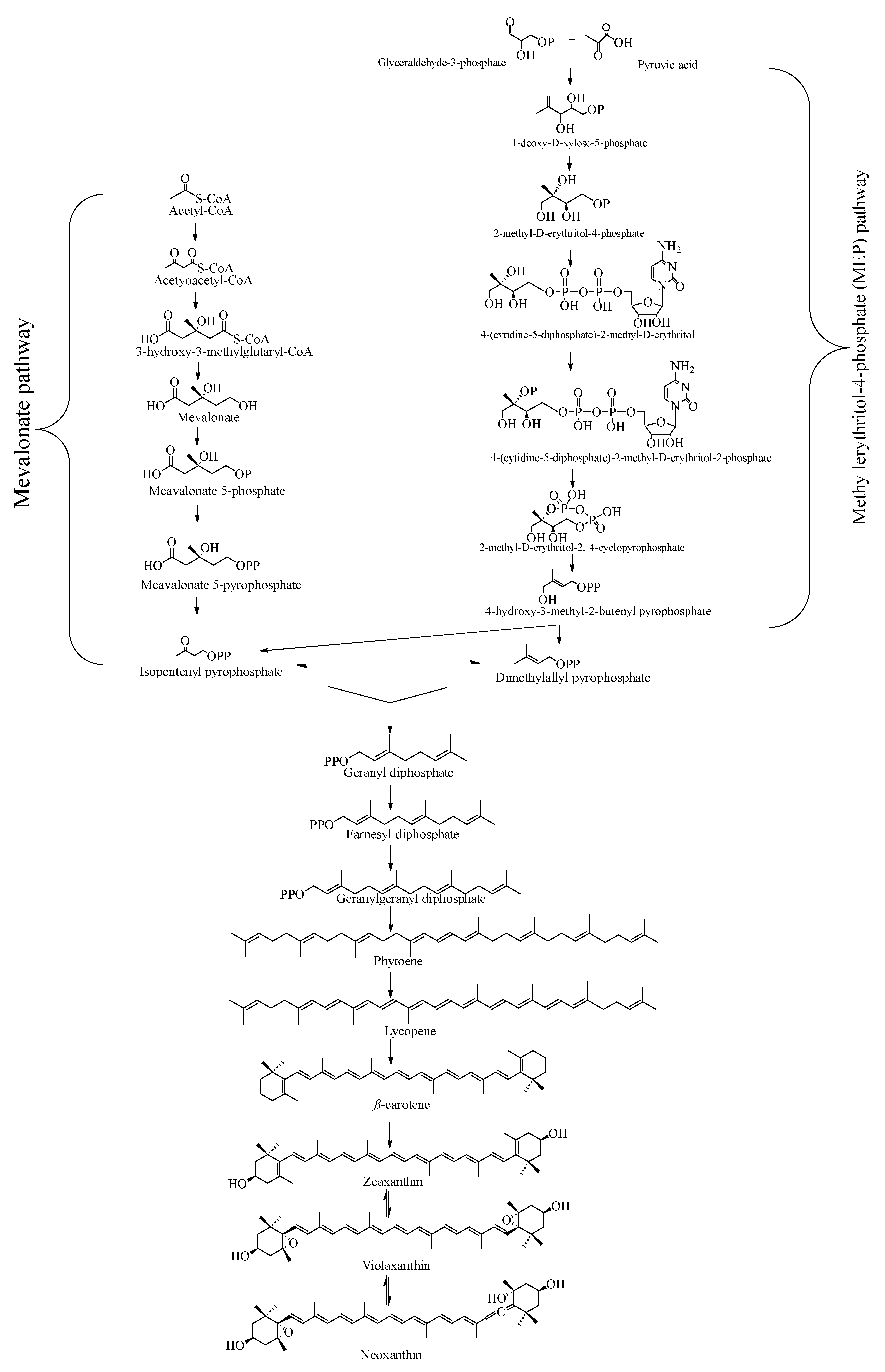
The biosynthetic pathway of neoxanthin is shown in Figure 3. Mevalonate (MVA) pathway mainly occurs in the cytoplasms of some organism including eukaryotes, archaea and plants, while methyl erythritol-4-phosphate (MEP) pathway mainly occurs in the chloroplasts of organisms such as bacteria and plants. Isopentenyl pyrophosphate (IPP) and dimethylallyl pyrophosphate (DMPP) are the precursor metabolites of carotenoid biosynthesis, which are synthesized by the mevalonate (MVA) pathway and the methylerythritol 4-phosphate (MEP) pathway, respectively [47]. Then, Dimethylallyl pyrophosphate(DMAPP) and IPP combine via a three-step condensation reaction to produce geranylgeranyl diphosphate (GGPP), which is the immediate precursor of carotenoid synthesis. Two GGPP molecules are then condensed by the enzymatic action of phytoene synthase (PSY) to produce phytoene (C40) [48,49]. Then phytoene is converted to lycopene by a multi-step process through sequential desaturation and isomerization reactions. Thereafter, lycopene is converted to β-carotene, which is one of key precursors of xanthophyll compounds. β-carotene is converted to zeaxanthin by double hydroxylation reactions catalyzed by β-carotene hydroxylase (BCH). Then zeaxanthin is converted to violaxanthin through two epoxidation reactions in the presence of the zeaxanthin epoxidase (ZEP). Finally, violaxanthin is converted to neoxanthin via the enzyme neoxanthin synthase (NXS) [50,51,52].

5. Extraction of Neoxanthin

Currently, some methods have been explored for the extraction of neoxanthin, including organic solvents extraction [42,53], ionic liquids extraction [54,55], supercritical liquid extraction [56], ultrasound-assisted extraction [57,58], and pressurized liquid extraction [11] (see Table 4).

5.1. Organic Solvents Extraction

Neoxanthin, a lipid-soluble xanthophyll with relatively weak polarity, is easily soluble in polar organic solvents [59]. Generally, some organic solvents, including dimethyl ether, acetone, and ethanol, have been used for the extraction of neoxanthin [60]. Notably, a pioneering study has highlighted the green extraction of liquefied dimethyl ether (DME), which was an efficient solvent for the extraction of neoxanthin from the microalga Chlorococcum humicola [53]. The optimal extraction conditions were determined as follows: liquefied dimethyl ether, 41 °C, 20 min, a liquid-to-solid ratio of 45:1 (w/w), and once extraction process. Under these conditions, the yield of neoxanthin reaches 2.55 mg/g wet sample, and the recovery rate of neoxanthin is 95%. As a medium polarity solvent, liquefied dimethyl ether is regarded as an alternative green solvent for the neoxanthin extraction due to its some advantages, including partial miscibility with water and a low normal boiling point (−24 °C) [61,62].
Generally, organic solvents extraction is a traditional and common technology that has been widely used for the extraction of diverse compounds on a commercial scale due to its advantages [63], including simple operation, wide application, good industrial manufacturing equipment, and rich manufacturing experience on a commercial scale [64]. However, this method possesses some disadvantages, including the toxicity of some organic solvents, high solvent consumption, low selectivity of extraction, and low extraction efficiency [65]. Thus, ethanol is regarded as a suitable solvent for extraction due to its good safety and low cost [66,67]. Ethanol has been approved for the extraction of multiple compounds on a commercial scale [68].

5.2. Ionic Liquids Extraction

In terms of neoxanthin extraction by using ionic liquids technology, some studies have been carried out on diverse microalgae, including Caryocar Brasiliense [55], Chlorella sorokiniana [54], and Scenedesmus obliquus [42]. Generally, ionic liquid is a mixture composed of ionic liquids and/or organic solvents, including 1-hexyl-3-methylimidazolium chloride ([HMIM][Cl]), 1-butyl-3-methylimidazolium tetrafluoroborate ([BMIM][BF4]), ethanol, and chloroform [69]. Interestingly, an essential study has highlighted the extraction of the mixture ([BMIM][BF4]:ethanol = 1:3), which was an efficient ionic liquid solvent for neoxanthin extraction from the microalga Scenedesmus obliquus [42]. The optimum extraction conditions were determined as follows: the mixture ([BMIM][BF4]:ethanol = 1:3), 3 min, a liquid-to-solid ratio of 10:1 (mL/g), and extraction three times. Under the conditions, the yield of neoxanthin reaches 122.66 μg/g dry sample.
Presently, ionic liquids extraction has been used for the extraction of diverse compounds, including neoxanthin, erythromycin A, and astaxanthin [70]. As a new type of green solvents, multiple ionic liquids (ILs) possess some advantages, including high thermal stability, high conductivity, low vapor pressure, and an extensive range of electrochemical properties [71,72]. Generally, ionic liquids (ILs) are regarded as promising sustainable extraction solvents due to their high extraction efficiency, good biocompatibility with multiple compounds, low cost, and low environmental impact [73].

5.3. Supercritical Liquid Extraction

Supercritical liquid extraction has been widely applied for the extraction of diverse non-polar compounds, including neutral oils, essential oils, and various carotenoids [74]. Notably, an interesting study has highlighted the importance of supercritical liquid of a co-solvent composed of CO2 (CO2 density, 830 g L−1) and 10% ethanol during the neoxanthin extraction from Scenedesmus sp. [56]. The optimal extraction conditions were determined as follows: the supercritical liquid of a co-solvent [CO2 (CO2 density, 830 g L−1) and 10% ethanol], 60 °C, 60 min, pressure of 300 bar, and liquid flow of 2 mL min−1. Under the conditions, the yield of neoxanthin reaches 670.8 μg/g dry sample [56].
Generally, supercritical carbon dioxide (SCCO2) has been widely applied for multiple compounds for food application due to its environmentally friendly and high safety [75,76]. However, supercritical liquid extraction is still limited owing to its high cost. Particularly, the extraction using a co-solvent composed of CO2 and ethanol requires a rather high operating pressure in the range of 20~60 MPa [77,78].

5.4. Ultrasound-Assisted Extraction

Generally, ultrasound-assisted extraction is regarded as an efficient method for the extraction of multiple bioactive molecules, including polyphenols, carotenoids, and flavonoids [79]. Interestingly, a study aimed to develop an eco-friendly and high-efficiency method for neoxanthin extraction from Chlorella vulgaris was investigated using ultrasound-assisted extraction. The optimal extraction conditions were determined as follows: Ethanol, 25 °C, 25 min, a liquid-to-solid ratio of 30:1 (mL/g), and amplitude wave of 20%. Under these conditions, the yield of neoxanthin reaches 9.83 mg/g dry weight biomass [11]. Ultrasound-assisted extraction (UAE) has been widely applied for the extraction of diverse natural molecules due to its advantages, including operation simplicity, short extraction duration, low extraction temperature, and ability to retain the original structure of natural products [80,81].

5.5. Pressurized Liquid Extraction

In terms of neoxanthin extraction by using pressurized liquid extraction, an interesting study has been developed from the microalga Chlorella vulgaris [11]. Several pressurized liquid extraction conditions were assessed, including different solvents (water, ethanol, and acetone), different extraction temperatures (50 °C, 100 °C, 150 °C, and 200 °C). The green extraction conditions were determined as follows: ethanol, 50 °C, extraction pressure of 1500 psi, and extraction time of 20 min. Under these conditions, the yield of neoxanthin reaches 11.35 mg/g dry sample [11]. Pressurized liquid extraction is an efficient method because the solvent possesses strong cell-penetrating capacity under suitable temperature and high pressure during the whole extraction procedure [11,82]. In addition, the method possesses some advantages, including green solvents, short extraction duration, and auto operation [83,84].

6. Biological Activities of Neoxanthin

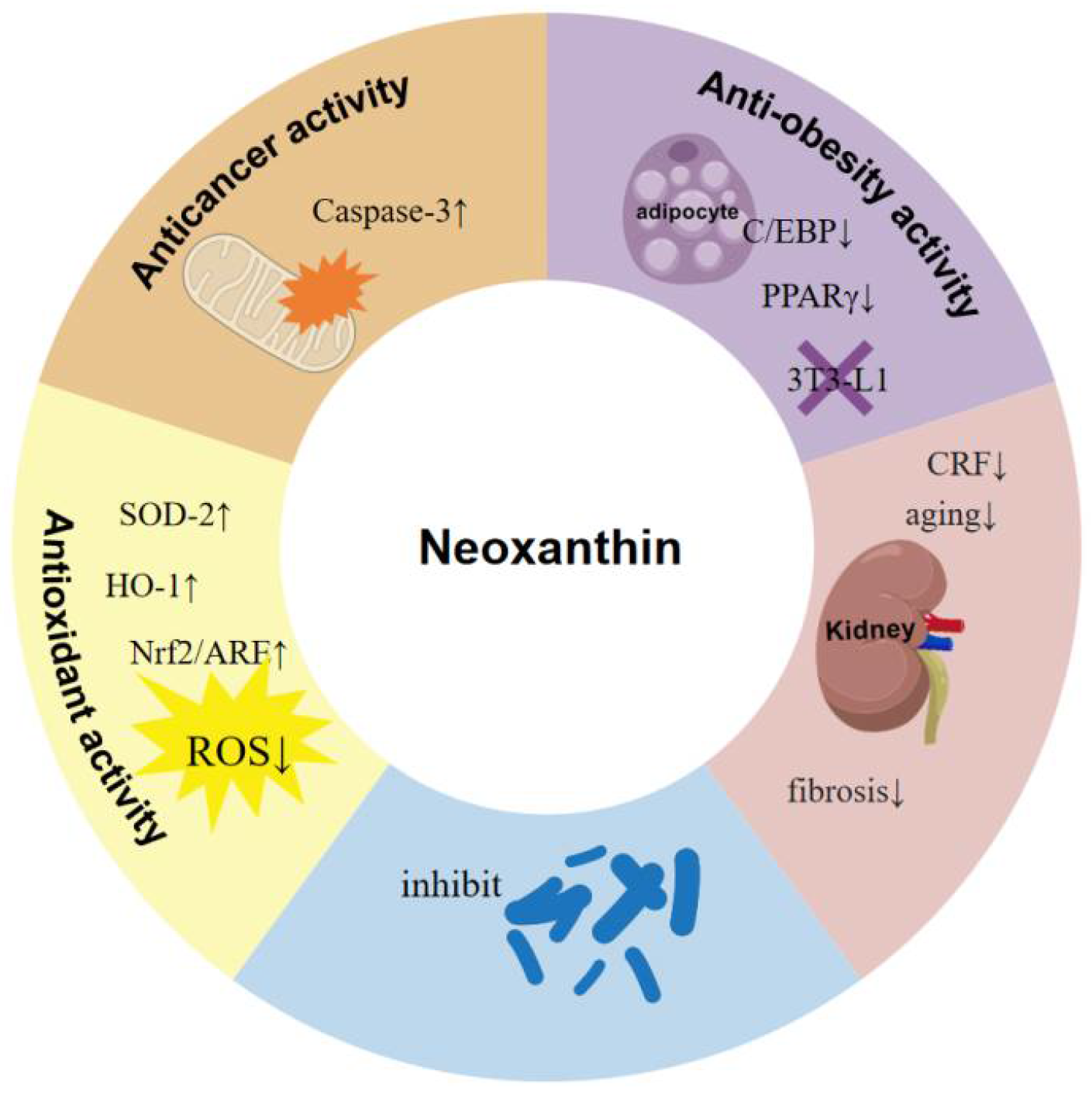
Carotenoids are food functional ingredients with diverse biological activities, including anti-tumor, anti-oxidant, weight loss, and anti-Alzheimer’s effects [85,86]. Some carotenoids are popular in foods, cosmetics, and health products, including astaxanthin and fucoxanthin [1,87,88]. Neoxanthin has shown diverse biological activities, including anti-tumor, anti-oxidant, and anti-obesity [13] (Figure 4).

6.1. Anti-Cancer Activity

The incidence and mortality rates of cancer are increasing rapidly worldwide, which has attracted increasing attention [89]. Dietary modifications are considered one of the most promising lifestyle changes that can reduce cancer risk by around 40% [90]. Carotenoids, distributed in many fruits and vegetables, are a class of natural pigments with anti-tumor activity [91]. Neoxanthin, one of the chief carotenoids occurring in nature, has been found abundantly in diverse higher plants, such as Spinacia oleracea, Citrullus vulgaris, Citrus sinensis, and Solanum lycopersicum [9]. Regarding the anti-tumor activity of neoxanthin, it has shown a multi-target mechanism of action in anti-tumor; additionally, it significantly affects different types of tumor cells [92]. The multi-target mechanism of action of neoxanthin in multiple cancer cell lines is shown in Table 5.
The effects of 9′-cis neoxanthin on the induction of apoptosis in HCT116 human colon cancer cells have been assessed [93]; additionally, the underlying mechanisms for its anti-cancer effects have been elucidated. The results have shown that chromatin condensation, DNA fragmentation, and increased hypodiploid cells have been observed when the HCT116 human colon cancer cells are exposed to 20 μM neoxanthin. In addition, the treatment with neoxanthin at 5 μM significantly inhibits the proliferation of HCT116 cells. Mechanisms underlying the anti-cancer action of 9′-cis neoxanthin can be elucidated as follows. Neoxanthin treatment causes an enhancement in the activities of caspase-3, -8, and -9, as well as an increase in the protein levels of their active subunits, except for caspase-8. In addition, the treatment results in an early-stage reduction in mitochondrial transmembrane potential, followed by the subsequent release of cytochrome c and apoptosis-inducing factor (AIF) from the mitochondria into the cytosol. Notably, the direct exposure of neoxanthin to mitochondria isolated from HCT116 cells results in an enhanced release of cytochrome c and apoptosis-inducing factor (AIF) in a dose-dependent manner. Around 50% of the neoxanthin absorbed by the cells was observed to be accumulated within the mitochondrial fraction. These findings suggest that the accumulation of neoxanthin in the mitochondrial fraction induces a loss of mitochondrial transmembrane potential, subsequently facilitating the release of cytochrome c and AIF, thereby promoting cell apoptosis [93].
The effects of neoxanthin on the induction of apoptosis in PC-3 cells have been investigated [94], which is characterized by morphological alterations, DNA fragmentation, an elevated proportion of hypodiploid cells, and the cleavage of caspase-3 and poly (ADP-ribose) polymerase (PARP) [94]. The results suggest that the proportion of apoptotic cells exceeds 30% after the PC-3 cells are exposed to 20 μM neoxanthin for 48 h. In addition, this treatment causes a reduction in the expression levels of Bax and Bcl-2 proteins, but does not affect Bcl-XL expression. In all, neoxanthin treatment has been observed to induce apoptosis via the activation of caspase-3 in PC-3 human prostate cancer cells [94].
CagA is an oncoprotein that significantly contributes to the progression of gastric cancer through its interaction with phosphatidylserine in the host cell plasma membrane [97]. This protein represents a promising target for cancer therapy [98]. A total of 38 natural compounds from medicinal plants are screened to evaluate their effects on the interaction of CagA protein with host cell membrane phosphatidylserine (PS) through computational methods such as molecular docking and molecular dynamics simulation [99]. The results suggest that neoxanthin has strong and steady interactions between its molecules and CagA [95]. The molecular docking results indicate that neoxanthin can bind tightly to the positively charged helix α18 region (residues 610–639) of CagA protein, which is a key region for the interaction between CagA and PS. Notably, neoxanthin also exhibits the lowest binding energy (PBTOT) with −34.39 kcal/mol. The complexes of neoxanthin with CagA are the most stable; additionally, the binding free energy (PBTOT) values are the lowest. Therefore, the key pharmacophores of neoxanthin are more favorable to the binding interactions within the CagA binding site. In all, the results suggest that neoxanthin is a potentially excellent active compound against the oncoprotein CagA [95].
In oncological research, 9′-cis neoxanthin has demonstrated significant inhibitory effects on the viability of cervical cancer (HeLa) and lung cancer (A549) cells, with IC50 values of 3.8 μM and 7.5 μM, respectively [96]. The treatment with 9′-cis neoxanthin at 10 μM results in a significant reduction in cell viability, with values decreasing to 10.9%, 15.0%, and approximately zero for PC-3, DU 145, and LNCaP cell lines, respectively [96].
The incidence and progression of tumors are prevalent across various cancer types, including colorectal cancer, lung cancer, and others [100]. These cancers often exhibit common biochemical disruptions, such as unchecked cellular proliferation and evasion of apoptosis [101]. Neoxanthin has emerged as a potential compound for the prevention and treatment of these malignancies [102]. Several studies suggest that neoxanthin significantly impacts the proliferation of diverse tumor cell types [94]. The anti-cancer effects of neoxanthin are likely mediated through multiple pathways, including mitochondrial-induced apoptosis, reversal of multidrug resistance, caspase-3-dependent apoptosis, inhibition of oncogenic signaling, and suppression of cell proliferation [13,93,95]. Consequently, neoxanthin represents a promising candidate for the prevention and treatment of various cancers.

6.2. Anti-Oxidant Activity

Oxidative stress (OS) is characterized by an imbalance between the production of reactive oxygen species (ROS) and the capacity of anti-oxidant systems to neutralize these products [103]. Oxidative stress is implicated in the pathogenesis of cancer, inflammatory and hyperpigmentation disorders, neurodegenerative diseases, diabetes, and cardiovascular diseases [104]. Various carotenoids, such as astaxanthin, fucoxanthin, and lutein, are well-known for their notable anti-oxidant properties [105]. They possess the capacity to quench singlet oxygen and scavenge harmful free radicals, thereby preventing or decreasing damage to living cells [106].
Neoxanthin and lutein are purified from the microalga Chlamydomonas reinhardtii; additionally, their protective effects against oxidative stress-induced DNA damage have been evaluated [107]. The results show that 10.4 μM of neoxanthin inhibits about 72% of DNA damage under Fenton reaction-induced oxidative stress, while the concentration of 35 μM lutein reduces only about 55% of DNA damage. These results suggest that neoxanthin exhibits stronger anti-oxidant activity than lutein at lower concentrations, proving to be a better inhibitor of oxidatively induced DNA damage [107].
In a study investigating lipid peroxidation using mouse C3H10T1/2 fibroblasts, the anti-oxidant properties of neoxanthin were validated [13]. Uric acid and superoxide ions are generated from xanthine through the action of xanthine oxidase. Consequently, superoxide anions target the unsaturated fatty acids within the membrane, leading to the production of malondialdehyde, a critical indicator of membrane damage. It has been demonstrated that neoxanthin, like β-carotene, can significantly reduce malondialdehyde formation, exhibiting an activity level one hundred times greater than that of β-carotene [13]. In a separate investigation utilizing HepG2 hepatocyte cells, the cytotoxic effects of H2O2 and the cytoprotective efficacy of neoxanthin are assessed. Compared to β-carotene and lutein, pretreatment with neoxanthin offers superior protection against cell death induced by H2O2. Neoxanthin achieves this by upregulating intracellular anti-oxidant enzymes, such as HO-1 and SOD-2, enhancing the expression of anti-apoptotic proteins, and decreasing the expression of pro-apoptotic proteins [13].
In a study examining the cytotoxic effects of H2O2 on the HepG2 cell model, neoxanthin demonstrates a significant protective effect against H2O2-induced oxidative stress in HepG2 cells [108] (see Table 6). In comparison to β-carotene and lutein, neoxanthin pretreatment markedly enhances cell survival rates of HepG2 cells under H2O2 exposure and demonstrates more potent cytoprotective effects at lower concentrations (0.05 μM and 0.1 μM). The findings indicate that following a 4 h exposure to 0.5 mM H2O2, the cell viability in the neoxanthin pretreatment groups (0.05 μM and 0.1 μM) is significantly greater compared to the group subjected solely to H2O2 treatment. These results suggest that neoxanthin effectively mitigates H2O2-induced cytotoxicity by activating endogenous anti-oxidant signaling pathways within the cells, thereby preserving cell viability [108].
Additionally, neoxanthin can also reduce oxidative stress by directly scavenging ROS produced within cells [109]. Experimental data indicate that treatment with H2O2 significantly elevates the levels of reactive oxygen species (ROS) in HepG2 cells, with an increase of 38% compared to the control group. In contrast, pretreatment with neoxanthin at concentrations of 0.05 μM and 0.1 μM results in a reduction In ROS production by 12% and 24%, respectively. These findings suggest that neoxanthin is effective in decreasing intracellular ROS concentrations, thereby offering protection against oxidative damage [108].
Certain anti-oxidant enzymes, such as heme oxygenase-1 (HO-1) and superoxide dismutase-2 (SOD-2), possess the capability to directly scavenge reactive oxygen species (ROS), thereby decreasing cellular damage induced by oxidative stress [110]. Notably, neoxanthin has been observed to enhance cellular anti-oxidant capacity by upregulating the expression of intracellular anti-oxidant enzymes, specifically HO-1 and SOD-2. Experimental results indicate that H2O2 treatment significantly diminishes the expression of HO-1 and SOD-2 in HepG2 cells. However, pretreatment with neoxanthin markedly restores the expression levels of these enzymes. Specifically, neoxanthin increases the expression of HO-1 by 17% and 22%, and SOD-2 by 21% and 35% at concentrations of 0.05 μM and 0.1 μM, respectively [108].
Neoxanthin also augments the anti-oxidant defenses of HepG2 cells through the modulation of transcription factor expression [108,111]. Numerous studies have demonstrated that the activation of the nuclear factor erythroid 2-related factor 2/anti-oxidant response element (Nrf2/ARE) pathway leads to an increased expression of anti-oxidant enzymes, which play a crucial role in safeguarding cells against oxidative damage [112]. Experimental evidence indicates that treatment with H2O2 significantly suppresses Nrf2 expression, whereas pretreatment with neoxanthin at 0.1 μM markedly restores Nrf2 expression by up to 42%. The activation of Nrf2 through neoxanthin pretreatment facilitates the upregulation of downstream anti-oxidant enzyme genes, such as HO-1 and SOD-2, thereby enhancing the cellular anti-oxidant capacity [108].
Oxidative stress is generally associated with the induction of apoptosis [113,114]. Experimental findings indicate that neoxanthin confers cellular protection by inhibiting the apoptotic signaling pathway. In HepG2 cells, treatment with H2O2 induces oxidative stress, leading to a significant increase (60%) in the expression of the pro-apoptotic protein Bax and a concurrent decrease (50%) in the expression of the anti-apoptotic protein Bcl-2. However, pretreatment with neoxanthin for 3 h effectively mitigates this oxidative stress-induced upregulation of Bax and prevents the reduction in Bcl-2 expression. Notably, the treatment with neoxanthin at 0.1 μM demonstrates the most pronounced reversal effect, restoring approximately 51% and 44% of the expression levels of Bax and Bcl-2, respectively, thereby significantly inhibiting apoptosis induced by oxidative stress [108].
In conclusion, neoxanthin demonstrates its anti-oxidant properties through multiple mechanisms, including the direct scavenging of reactive oxygen species (ROS), preservation of mitochondrial function, activation of endogenous anti-oxidant enzymes, regulation of transcription factor expression, and inhibition of apoptosis [108]. Collectively, these mechanisms render neoxanthin highly effective in safeguarding HepG2 cells against oxidative stress induced by H2O2 [108].
Collectively, neoxanthin has demonstrated good anti-oxidant activity in various assessment of in vitro anti-oxidant activity [13,108]. Oxidative stress is implicated in the pathogenesis of diverse disorders, including cancer, inflammatory and hyperpigmentation disorders, neurodegenerative diseases, diabetes, and cardiovascular diseases [115]. Thus, this shared biochemical mechanism suggests neoxanthin a promising candidate compound for the prevention and/or treatment of the coexistence of these multiple diseases. Further studies can be carried out to validate the anti-oxidant properties of neoxanthin in multiple anti-oxidant activities (in vivo and in vitro); additionally, the anti-oxidant mechanisms are required to be determined for its further application in the food industry [116].

6.3. Anti-Obesity Activity

Presently, the rising prevalence of overweight has become an immense challenge for human health [117,118]. Various carotenoids have exhibited promising anti-obesity effects, including fucoxanthin, lutein, and zeaxanthin [119,120]. Notably, neoxanthin has also shown remarkable prevention effects on fat accumulation in adipocytes [121].
In a study assessing the suppressive effects of 13 natural carotenoids on the adipocyte differentiation of 3T3-L1, treatment with neoxanthin significantly reduces lipid accumulation, as well as glycerol-3-phosphate dehydrogenase activity [122]. Compared to the untreated controls, neoxanthin treatment with 5 μM reduces intracellular lipid accumulation by 32%, while neoxanthin treatment with 20 μM suppresses lipid accumulation to 36% of control levels. The dose-dependent inhibition observed in this study substantiates the capability of neoxanthin to reduce lipid accumulation in adipocytes. Notably, neoxanthin treatment with 20 μM has resulted in a reduction in glycerol-3-phosphate dehydrogenase (GPDH) activity (a key terminal differentiation marker) by around 40% compared to fully differentiated adipocytes, indicating an interference with adipocyte maturation. Additionally, the mRNA expression levels of critical adipogenic transcription factors, C/EBPα and PPARγ, have been significantly downregulated; specifically, 20 μM neoxanthin has reduced C/EBPα expression by 86% and markedly suppressed PPARγ. Given their roles as master regulators of adipogenesis, the coordinated repression of these factors directly decreases differentiation programs. These mechanisms collectively obstruct the differentiation of 3T3-L1 preadipocytes, thereby diminishing adipogenesis and lipid deposition. In addition, the analysis based on the examination of structure and function suggests that neoxanthin has suppressive effects on adipocyte differentiation in 3T3-L1 cells [122].
Fucoxanthinol, a bioactive metabolite of fucoxanthin, is considered the real molecular structure responsible for the anti-obesity effects attributed to fucoxanthin [121,123]. The chemical structures of neoxanthin and fucoxanthinol are very similar, both containing functional groups such as conjugated polyene chains, epoxy groups, and diene groups [20,124]. This molecular similarity implies that neoxanthin may exert its anti-obesity effects through mechanisms similar to those of fucoxanthinol [119]. In particular, the shared structural features present in both compounds are likely to play a crucial role in mediating their biological activity against adiposity [125]. In addition, given the good safety of neoxanthin, it is worthwhile to conduct further research on the weight loss effects and mechanisms of neoxanthin [13].

6.4. Anti-Inflammatory Activity

A study has investigated the impact of neoxanthin on inflammation in renal cells, specifically evaluating its effects on inflammatory molecules. The chronic renal failure (CRF) model group has been established by administering a diet containing 0.2% adenine mixed with powdered food to Wistar rats over a period of five weeks. Western Blot analysis reveals that, relative to the control group, the expression levels of tumor necrosis factor-α (TNF-α), interleukin-6 (IL-6), and interleukin-1β (IL-1β) have been markedly elevated in the model group. Conversely, treatment with neoxanthin results in a significant downregulation of TNF-α, IL-6, and IL-1β mRNA expression. These findings suggest that neoxanthin effectively reduces the expression of inflammatory factors TNF-α, IL-6, and IL-1β, while concurrently downregulating the activity of inflammation-associated signaling pathways, including nuclear factor kappa-B(NF-κB). Further preliminary mechanistic analysis indicates that inflammation-related signaling pathways, such as v-rel reticuloendotheliosis viral oncogene homolog A (p65), nuclear factor kappa B subunit 1(p50) have been significantly downregulated following neoxanthin treatment [126].

6.5. Anti-Bacterial Activity

An academic study has examined the anti-bacterial effects of neoxanthin on a specific strain of Helicobacter pylori (ATCC43504). The micro-dilution broth method is employed to ascertain the minimum inhibitory concentration (MIC). The findings reveal that the MIC50 value for all-trans neoxanthin is 11 μg/mL, indicating potent anti-H. pylori activity. In comparison, the MIC50 value for 9′-cis-neoxanthin is 27 μg/mL, demonstrating slightly lower activity than (all-trans)-neoxanthin, yet still exhibiting a significant anti-bacterial effect. This study suggests that neoxanthin, particularly (all-trans)-neoxanthin, possesses notable anti-bacterial activity against H. pylori, with its MIC50 value being substantially lower than that of metronidazole. The anti-bacterial efficacy of neoxanthin may be attributed to specific functional groups within its molecular structure, such as monofuranoid rings or allenic bonds, which are likely critical to its anti-bacterial properties. As a carotenoid naturally occurring in plants, neoxanthin’s potential application in anti-bacterial technology merits further investigation. Neoxanthin could potentially serve as an alternative or complementary therapy, offering novel approaches for the treatment of Helicobacter pylori infections [127].
Currently, some studies have been investigated neoxanthin absorption in animal models and human. First, to assess the bioavailability of neoxanthin, a dietary intervention study has been conducted involving the consumption of raw young spinach leaves (100 g per day for four weeks) by 14 participants (mean age 36.5 ± 8.0 years; male–female ratio = 9:5). The concentrations of neoxanthin, neochrome, β-carotene, and lutein in both the spinach and the participants’ blood samples (collected pre- and post-intervention) are quantified using HPLC. The results reveal that neither neoxanthin nor neochrome is detectable in the blood samples, while the concentrations of β-carotene and lutein exhibited significant increases, by 1.4-fold and 1.9-fold, respectively, over the course of the study. These findings suggest that the bioavailability of neoxanthin in humans is low, indicating that it is unlikely to exert an inhibitory effect on fat accumulation in vivo, contrary to in vitro results. Although consuming raw leafy vegetables may preserve high levels of neoxanthin, it does not enhance its bioavailability [128].
Two epoxyxanthophylls, neoxanthin and fucoxanthin, have been documented to exert antiproliferative effects on various human cancer cell lines in vitro. A study has estimated the intestinal absorption of neoxanthin and fucoxanthin in humans. The plasma concentrations of epoxyxanthophylls before and after a 1-week dietary intervention involving spinach (Spinacia oleracea) and wakame (Undaria pinnatifida) have been evaluated. The epoxyxanthophylls and their metabolites in plasma extracts are quantified using HPLC following partial purification and concentration via solid-phase extraction cartridges. Despite a 1-week intake of spinach providing 3.0 mg of neoxanthin per day, plasma concentrations of neoxanthin and its metabolites (neochrome stereoisomers) remain exceedingly low (approximately 1 nmol/L), whereas levels of β-carotene and lutein show significant increases. Similarly, the plasma concentration of fucoxanthinol, a gastrointestinal metabolite of fucoxanthin, is less than 1 nmol/L after a 1-week intake of wakame providing 6.1 mg of fucoxanthin per day. These findings suggest that the plasma response to dietary epoxyxanthophylls is minimal in humans, even following a 1-week consumption of diets rich in these compounds [27].
These results suggest that the bioavailability of neoxanthin in humans is extremely low. The metabolites of neoxanthin in humans is unclear. Notably, an investigation has been conducted into the metabolites of neoxanthin in mice. The findings indicate that two hours following the oral administration of neoxanthin at a dosage of 40 nmol per mouse, both neoxanthin and (R/S)-neochrome are detected in the plasma and liver tissues of the mice. The concentrations of neoxanthin, (8R)-neochrome, and (8S)-neochrome are approximately 13.6, 10.3, and 11.3 nmol/L in the plasma, and 7.3, 10.4, and 6.9 pmol/g in the liver, respectively. Additionally, (R/S)-neochrome is identified in the small intestinal contents of the mice administered with neoxanthin [129].
Similarly to fucoxanthin, the bioavailability of neoxanthin is low. These results may to attribute to the similarity of chemical structures of the two compounds. Currently, the metabolites of neoxanthin are not yet well characterized. Hence, more studies are required to develop the metabolites of neoxanthin or advance neoxanthin as a drug precursor to be further activated by in vivo metabolism.

7. Current Challenges and Opportunities

Compared with commercial carotenoids (e.g., astaxanthin, lutein and fucoxanthin), neoxanthin has not yet been industrially produced. Considering the several health benefits of neoxanthin in human health, it is a promising carotenoid for further investigation. To meet the demand of neoxanthin production in commercial scale, some major challenges have been faced, and future research is expected to focus on some aspects as follows.

7.1. The Prospect of Microalgae as a Neoxanthin Source

Compared to high plants, microalgae are regarded as promising neoxanthin sources due to some main advantages including higher neoxanthin content, multiple species of microalgae, higher growth rate, and immune to seasonal variations [130]. The content of neoxanthin in the microalgae is in the range of 0.26~30,880 μg/g dry weight [11,12]. Notably, the neoxanthin content in the dry powders of Chlorella vulgaris is 30.88 mg/g [11]. The complex and limited genetic potential of microalgae constrains pigment production, presenting a significant challenge to their commercial utilization. To overcome the obstacle, the application of genetic and metabolic engineering techniques is regarded to be an effective strategy. Metabolic engineering and synthetic biology strategies have been used to enhance microalgal pigment production, including lutein; β-carotene, fucoxanthin, zeaxanthin and astaxanthin [131]. In addition, an effective and economic photoautotrophic cultivation systems is vital for microalgal neoxanthin production. The future research is expected to focus on some aspects including the suitable photobioreactors, optimal light supply, temperature, effective capture of carbon dioxide, and protection of the cultures from contamination [132]. Currently, tubular photobioreactors are the most common industrial photobioreactors, which has been applied for the production of astaxanthin [133].

7.2. Extraction and Purification of Neoxanthin

Some progress has been made for the extraction of neoxanthin from high plants or microalgae biomass, including organic solvents extraction [42,53], ionic liquids extraction [54,55], supercritical liquid extraction [56], ultrasound-assisted extraction [60,61], and pressurized liquid extraction [11]. Considering the high-polarity of neoxanthin, the use of polar organic solvents including dimethyl ether, acetone, and ethanol, can enhance the extraction efficiency [42]. In addition, ionic liquids technology has been investigated for the extraction of neoxanthin by using some mixture composed of ionic liquids and/or organic solvents [54]. Notably, the choice of a suitable extraction solvent remains a key challenge for sustainable extraction processes in a large-scale. A truly optimal solvent is fully enabled to meet the safety requirement of neoxanthin in the foods and cosmetics fields and facilitate the further purification of neoxanthin. Generally, ethanol is regarded as an optimal solvent due to its green, high-polarity, and safety.
To the best of our knowledge, the purification technology of neoxanthin has not yet been found. The purification of neoxanthin remains a great challenge due to the lack of suitable raw material with high content of neoxanthin, the co-existed compounds with similar polarity, the chemical instability of neoxanthin, the lack of purification technology of neoxanthin with the property of green, efficient and economic feasibility. The development of purification technology of neoxanthin is an essential research aspect for the further application of neoxanthin. The establishment of purification technology can provide necessary high-purity neoxanthin for the further studies of biological activity of neoxanthin. Some xanthophyll carotenoids (such as lutein, fucoxanthin) have been commercial produced. The successful purification of these xanthophyll carotenoids can provide good cases for the further purification of neoxanthin.
Several advanced purification techniques have been documented, including the selective adsorption of lutein on a solid phase [130], as well as reversed-phase HPLC and high-speed counter-current chromatography for obtaining small quantities of high-purity carotenoids [134,135]. Additionally, supercritical anti-solvent precipitation has been shown to produce solid carotenoids within a matter of minutes [136].

7.3. Biological Activity of Neoxanthin

Carotenoids are a class of compounds with diverse bioactivities including anti-cancer [89], anti-oxidant [11], anti-obesity [119], anti-inflammatory [126,137], anti-senile dementia as well as ocular-protective [138]. Presently, neoxanthin exhibits some activities such as anti-cancer, anti-oxidative, and anti-obesity activities [13]. Some new technology such as high throughput screening, can facilitate the efficient studies of neoxanthin. Recent investigations have explored the absorption of neoxanthin in both animal models and humans. Similarly to fucoxanthin, neoxanthin exhibits low bioavailability, which may be attributed to the structural similarities between the two compounds. Currently, the metabolites of neoxanthin remain inadequately characterized. It is known that dietary neoxanthin is partially converted into (R/S)-neochrome due to intragastric acidity before intestinal absorption. Notably, (R/S)-neochrome demonstrates an antiproliferative effect on PC-3 cells without significantly inducing apoptosis. The limited bioavailability of neoxanthin presents a significant challenge for its clinical application. Fortunately, strategies such as combining fucoxanthin with dietary oil or milk, or incorporating it into nanoparticles, have been developed to enhance its bioavailability [139]. These successful cases can provide practical and feasible solutions for improving the bioavailability of neoxanthin.

8. Conclusions

Carotenoids are a promising class of compounds with diverse beneficial effects on human health. Some carotenoids such as lutein, fucoxanthin, and astaxanthin, have been produced on a commercial scale; in additionally, the global carotenoids market has experienced rapid expansion in recent years. In this review, we systematically reviewed the current progress on the structure, source, biosynthetic pathways, extraction methods and bioactivities of neoxanthin. Moreover, this review discussed the major challenges of neoxanthin production from microalgae, and proposed some future development prospects and potential solutions. Overall, this review provided useful information in the future research aspects of neoxanthin.

Author Contributions

J.Z. (Jiarong Zhao): Conceptualization, Methodology, Writing—original draft, Writing; G.Z.: Conceptualization, Formal analysis, Writing—original draft, Writing; J.Z. (Jinrong Zhang): Conceptualization, Writing—review and editing, Supervision. All authors have read and agreed to the published version of the manuscript.

Funding

This work was financially supported by the Science and Technology Program of Zhejiang Province (LGG22D060001, LTGC24D060001), the Key Program of Science and Technology of Ningbo (2023Z118), Ningbo University Li Da San Ye Yao Zhen and Li Benjun Marine Biomedical New Drug Research and Development Special Fund (2025-03), the Ningbo Public Welfare Science and Technology Program (No. 2022S161), One Health Interdisciplinary Research Project, Ningbo University (HY2024010).

Institutional Review Board Statement

Not applicable.

Data Availability Statement

The data presented in this study are available on request from the corresponding author.

Conflicts of Interest

The authors declare no conflicts of interest.

References

  1. Maoka, T. Carotenoids as natural functional pigments. J. Nat. Med. 2020, 74, 1–16. [Google Scholar] [CrossRef]
  2. Britton, G. Carotenoid research: History and new perspectives for chemistry in biological systems. Biochim. Biophys. Acta BBA—Mol. Cell Biol. Lipids 2020, 1865, 158699. [Google Scholar] [CrossRef]
  3. Sourkes, T.L. The discovery and early history of carotene. Hist. Chem 2009, 34, 33. [Google Scholar] [CrossRef]
  4. Steenbock, H.; Coward, K.H. Fat-soluble vitamins. J. Biol. Chem. 1927, 72, 765–779. [Google Scholar] [CrossRef]
  5. Uray, I.P.; Dmitrovsky, E.; Brown, P.H. Retinoids and rexinoids in cancer prevention: From laboratory to clinic. Semin. Oncol. 2016, 43, 49–64. [Google Scholar] [CrossRef] [PubMed]
  6. Kolašinac, S.M.; Dajić Stevanović, Z.; Kilibarda, S.N.; Kostić, A.Ž. Carotenoids: New applications of “old” pigments. Phyton 2021, 90, 1041–1062. [Google Scholar] [CrossRef]
  7. Choi, S.S.; Seo, Y.B.; Nam, S.-W.; Kim, G.-D. Enhanced production of astaxanthin by co-culture of Paracoccus haeundaensis and lactic acid bacteria. Front. Mar. Sci. 2021, 7, 597553. [Google Scholar] [CrossRef]
  8. Liu, C.; Hu, B.; Cheng, Y.; Guo, Y.; Yao, W.; Qian, H. Carotenoids from fungi and microalgae: A review on their recent production, extraction, and developments. Bioresour. Technol. 2021, 337, 125398. [Google Scholar] [CrossRef]
  9. Giossi, C.; Cartaxana, P.; Cruz, S. Photoprotective role of neoxanthin in plants and algae. Molecules 2020, 25, 4617. [Google Scholar] [CrossRef] [PubMed]
  10. León-Vaz, A.; León, R.; Vigara, J.; Funk, C. Exploring nordic microalgae as a potential novel source of antioxidant and bioactive compounds. New Biotechnol. 2023, 73, 1–8. [Google Scholar] [CrossRef]
  11. Plaza, M.; Santoyo, S.; Jaime, L.; Avalo, B.; Cifuentes, A.; Reglero, G.; García-Blairsy Reina, G.; Señoráns, F.J.; Ibáñez, E. Comprehensive characterization of the functional activities of pressurized liquid and ultrasound-assisted extracts from Chlorella vulgaris. LWT—Food Sci. Technol. 2012, 46, 245–253. [Google Scholar] [CrossRef]
  12. Bhat, I.; Haripriya, G.; Jogi, N.; Mamatha, B.S. Carotenoid composition of locally found seaweeds of Dakshina Kannada district in India. Algal Res. 2021, 53, 102154. [Google Scholar] [CrossRef]
  13. Parida, S.; Jena, M.; Behera, A.K.; Mandal, A.K.; Nayak, R.; Patra, S. A novel phytocolorant, neoxanthin, as a potent chemopreventive: Current progress and future prospects. Curr. Med. Chem. 2024, 31, 5149–5164. [Google Scholar] [CrossRef]
  14. Cezare-Gomes, E.A.; Mejia-da-Silva, L.D.C.; Pérez-Mora, L.S.; Matsudo, M.C.; Ferreira-Camargo, L.S.; Singh, A.K.; De Carvalho, J.C.M. Potential of microalgae carotenoids for industrial application. Appl. Biochem. Biotechnol. 2019, 188, 602–634. [Google Scholar] [CrossRef]
  15. Heller, J.; Szarkowska, L.; Michalek, H. Ubiquinone (coenzyme Q) in insects. Nature 1960, 188, 491. [Google Scholar] [CrossRef]
  16. Bungard, R.A.; Ruban, A.V.; Hibberd, J.M.; Press, M.C.; Horton, P.; Scholes, J.D. Unusual carotenoid composition and a new type of xanthophyll cycle in plants. Proc. Natl. Acad. Sci. USA 1999, 96, 1135–1139. [Google Scholar] [CrossRef] [PubMed]
  17. Wang, K.; Tu, W.; Liu, C.; Rao, Y.; Gao, Z.; Yang, C. 9-cis neoxanthin in light harvesting complexes of photosystem II regulates the binding of violaxanthin and xanthophyll cycle. Plant Physiol. 2017, 174, 86–96. [Google Scholar] [CrossRef] [PubMed]
  18. Parry, A.D.; Horgan, R. Carotenoids and abscisic acid (ABA) biosynthesis in higher plants. Physiol. Plant. 1991, 82, 320–326. [Google Scholar] [CrossRef]
  19. Frank, H.A.; Bautista, J.A.; Josue, J.; Pendon, Z.; Hiller, R.G.; Sharples, F.P.; Gosztola, D.; Wasielewski, M.R. Effect of the solvent environment on the spectroscopic properties and dynamics of the lowest excited states of carotenoids. J. Phys. Chem. B 2000, 104, 4569–4577. [Google Scholar] [CrossRef]
  20. Pereira, A.G.; Otero, P.; Echave, J.; Carreira-Casais, A.; Chamorro, F.; Collazo, N.; Jaboui, A.; Lourenço-Lopes, C.; Simal-Gandara, J.; Prieto, M.A. Xanthophylls from the sea: Algae as source of bioactive carotenoids. Mar. Drugs 2021, 19, 188. [Google Scholar] [CrossRef]
  21. Aparicio-Ruiz, R.; Gandul-Rojas, B. Thermal degradation kinetics of neoxanthin, violaxanthin, and antheraxanthin in virgin olive oils. J. Agric. Food Chem. 2012, 60, 5180–5191. [Google Scholar] [CrossRef]
  22. Takaichi, S.; Mirauro, M. Distribution and geometric isomerism of neoxanthin in oxygenic phototrophs: 9’-cis, a sole molecular form. Plant Cell Physiol. 1998, 39, 968–977. [Google Scholar] [CrossRef]
  23. Yoshii, Y.; Takaichi, S.; Maoka, T.; Inouye, I. Photosynthetic pigment composition in the primitive green alga Mesostigma viride (prasinophyceae): Phylogenetic and evolutionary implications. J. Phycol. 2003, 39, 570–576. [Google Scholar] [CrossRef]
  24. Nurafifah, I.; Hardianto, M.A.; Erfianti, T.; Amelia, R.; Suyono, E.A. The effect of acidic ph on chlorophyll, carotenoids, and carotenoid derivatives of Euglena sp. as antioxidants. AACL Bioflux. 2023, 16, 2391–2401. [Google Scholar]
  25. Mc Gee, D.; Gillespie, E. The bioactivity and chemotaxonomy of microalgal carotenoids. In Biodiversity and Chemotaxonomy; Springer: Berlin/Heidelberg, Germany, 2019; pp. 215–237. [Google Scholar]
  26. Wen Lee, H.; Bi, X.; Jeyakumar Henry, C. Carotenoids, tocopherols and phylloquinone content of 26 green leafy vegetables commonly consumed in Southeast Asia. Food Chem. 2022, 385, 132729. [Google Scholar] [CrossRef] [PubMed]
  27. Sekiya, M.; Suzuki, S.; Ushida, Y.; Suganuma, H. Neoxanthin in young vegetable leaves prevents fat accumulation in differentiated adipocytes. Biosci. Biotechnol. Biochem. 2021, 85, 2145–2152. [Google Scholar] [CrossRef] [PubMed]
  28. De Sá, M.C.; Rodriguez-Amaya, D.B. Optimization of hplc quantification of carotenoids in cooked green vegetables—Comparison of analytical and calculated data. J. Food Compos. Anal. 2004, 17, 37–51. [Google Scholar] [CrossRef]
  29. Valdivielso, I.; Bustamante, M.Á.; Ruiz De Gordoa, J.C.; Nájera, A.I.; De Renobales, M.; Barron, L.J.R. Simultaneous analysis of carotenoids and tocopherols in botanical species using one step solid–liquid extraction followed by high performance liquid chromatography. Food Chem. 2015, 173, 709–717. [Google Scholar] [CrossRef]
  30. Pop, R.M.; Weesepoel, Y.; Socaciu, C.; Pintea, A.; Vincken, J.-P.; Gruppen, H. Carotenoid composition of berries and leaves from six romanian sea buckthorn (Hippophae rhamnoides L.) varieties. Food Chem. 2014, 147, 1–9. [Google Scholar] [CrossRef] [PubMed]
  31. Delgado-Pelayo, R.; Gallardo-Guerrero, L.; Hornero-Méndez, D. Carotenoid composition of strawberry tree (Arbutus unedo L.) fruits. Food Chem. 2016, 199, 165–175. [Google Scholar] [CrossRef]
  32. De Abreu, F.P.; Dornier, M.; Dionisio, A.P.; Carail, M.; Caris-Veyrat, C.; Dhuique-Mayer, C. Cashew apple (Anacardium occidentale L.) extract from by-product of juice processing: A focus on carotenoids. Food Chem. 2013, 138, 25–31. [Google Scholar] [CrossRef]
  33. Criado, M.N.; Motilva, M.J.; Goñi, M.; Romero, M.P. Comparative study of the effect of the maturation process of the olive fruit on the chlorophyll and carotenoid fractions of drupes and virgin oils from Arbequina and Farga cultivars. Food Chem. 2007, 100, 748–755. [Google Scholar] [CrossRef]
  34. Barreto, G.P.M.; Fabi, J.P.; De Rosso, V.V.; Cordenunsi, B.R.; Lajolo, F.M.; Do Nascimento, J.R.O.; Mercadante, A.Z. Influence of ethylene on carotenoid biosynthesis during papaya postharvesting ripening. J. Food Compos. Anal. 2011, 24, 620–624. [Google Scholar] [CrossRef]
  35. Inbaraj, B.S.; Lu, H.; Hung, C.F.; Wu, W.B.; Lin, C.L.; Chen, B.H. Determination of carotenoids and their esters in fruits of Lycium barbarum Linnaeus by HPLC–DAD–APCI–MS. J. Pharm. Biomed. Anal. 2008, 47, 812–818. [Google Scholar] [CrossRef] [PubMed]
  36. Delgado-Pelayo, R.; Gallardo-Guerrero, L.; Hornero-Méndez, D. Chlorophyll and carotenoid pigments in the peel and flesh of commercial apple fruit varieties. Food Res. Int. 2014, 65, 272–281. [Google Scholar] [CrossRef]
  37. Hong, H.T.; Takagi, T.; O’Hare, T.J. An optimal saponification and extraction method to determine carotenoids in avocado. Food Chem. 2022, 387, 132923. [Google Scholar] [CrossRef] [PubMed]
  38. Chacón-Ordóñez, T.; Schweiggert, R.M.; Bosy-Westphal, A.; Jiménez, V.M.; Carle, R.; Esquivel, P. Carotenoids and carotenoid esters of orange- and yellow-fleshed mamey sapote (Pouteria sapota (Jacq.) H.E. Moore & Stearn) fruit and their post-prandial absorption in humans. Food Chem. 2017, 221, 673–682. [Google Scholar]
  39. Zhou, W.; Niu, Y.; Ding, X.; Zhao, S.; Li, Y.; Fan, G.; Zhang, S.; Liao, K. Analysis of carotenoid content and diversity in apricots (Prunus armeniaca L.) grown in china. Food Chem. 2020, 330, 127223. [Google Scholar] [CrossRef]
  40. Sun, H.; Wang, Y.; He, Y.; Liu, B.; Mou, H.; Chen, F.; Yang, S. Microalgae-derived pigments for the food industry. Mar. Drugs 2023, 21, 82. [Google Scholar] [CrossRef]
  41. Ryckebosch, E.; Bruneel, C.; Termote-Verhalle, R.; Muylaert, K.; Foubert, I. Influence of extraction solvent system on extractability of lipid components from different microalgae species. Algal Res. 2014, 3, 36–43. [Google Scholar] [CrossRef]
  42. Fernandes, A.S.; Caetano, P.A.; Jacob-Lopes, E.; Zepka, L.Q.; De Rosso, V.V. Alternative green solvents associated with ultrasound-assisted extraction: A green chemistry approach for the extraction of carotenoids and chlorophylls from microalgae. Food Chem. 2024, 455, 139939. [Google Scholar] [CrossRef]
  43. Mapelli-Brahm, P.; Gómez-Villegas, P.; Gonda, M.L.; León-Vaz, A.; León, R.; Mildenberger, J.; Rebours, C.; Saravia, V.; Vero, S.; Vila, E.; et al. Microalgae, seaweeds and aquatic bacteria, archaea, and yeasts: Sources of carotenoids with potential antioxidant and anti-inflammatory health-promoting actions in the sustainability era. Mar. Drugs 2023, 21, 340. [Google Scholar] [PubMed]
  44. Raposo, M.; De Morais, A.; De Morais, R. Carotenoids from marine microalgae: A valuable natural source for the prevention of chronic diseases. Mar. Drugs 2015, 13, 5128–5155. [Google Scholar] [CrossRef]
  45. Amaro, H.M.; Macedo, A.C.; Malcata, F.X. Microalgae: An alternative as sustainable source of biofuels? Energy 2012, 44, 158–166. [Google Scholar] [CrossRef]
  46. Dolganyuk, V.; Belova, D.; Babich, O.; Prosekov, A.; Ivanova, S.; Katserov, D.; Patyukov, N.; Sukhikh, S. Microalgae: A promising source of valuable bioproducts. Biomolecules 2020, 10, 1153. [Google Scholar] [CrossRef]
  47. Sathasivam, R.; Radhakrishnan, R.; Kim, J.K.; Park, S.U. An update on biosynthesis and regulation of carotenoids in plants. South Afr. J. Bot. 2021, 140, 290–302. [Google Scholar] [CrossRef]
  48. Sun, T.; Li, L. Toward the ‘golden’ era: The status in uncovering the regulatory control of carotenoid accumulation in plants. Plant Sci. 2020, 290, 110331. [Google Scholar] [CrossRef] [PubMed]
  49. Miras-Moreno, B.; Pedreño, M.Á.; Romero, L.A. Bioactivity and bioavailability of phytoene and strategies to improve its production. Phytochem. Rev. 2019, 18, 359–376. [Google Scholar] [CrossRef]
  50. Dautermann, O.; Lyska, D.; Andersen-Ranberg, J.; Becker, M.; Fröhlich-Nowoisky, J.; Gartmann, H.; Krämer, L.C.; Mayr, K.; Pieper, D.; Rij, L.M.; et al. An algal enzyme required for biosynthesis of the most abundant marine carotenoids. Sci. Adv. 2020, 6, eaaw9183. [Google Scholar] [CrossRef]
  51. Dautermann, O.; Lohr, M. A functional zeaxanthin epoxidase from red algae shedding light on the evolution of light-harvesting carotenoids and the xanthophyll cycle in photosynthetic eukaryotes. Plant J. 2017, 92, 879–891. [Google Scholar] [CrossRef]
  52. Gómez-García, M.; Ochoa-Alejo, N. Biochemistry and molecular biology of carotenoid biosynthesis in chili peppers (Capsicum spp.). Int. J. Mol. Sci. 2013, 14, 19025–19053. [Google Scholar] [CrossRef] [PubMed]
  53. Eghbali Babadi, F.; Boonnoun, P.; Nootong, K.; Powtongsook, S.; Goto, M.; Shotipruk, A. Identification of carotenoids and chlorophylls from green algae Chlorococcum humicola and extraction by liquefied dimethyl ether. Food Bioprod. Process. 2020, 123, 296–303. [Google Scholar] [CrossRef]
  54. Bianchini, C.B.; Ramos-Souza, C.; Schappo, F.B.; Farina, M.; De Rosso, V.V.; Nunes, I.L. Ionic liquid and ultrasound as a fast and innovative combination for improved extraction of Chlorella sorokiniana-derived carotenoids. Algal Res. 2024, 82, 103650. [Google Scholar] [CrossRef]
  55. Ramos-Souza, C.; Nass, P.; Jacob-Lopes, E.; Zepka, L.Q.; Braga, A.R.C.; De Rosso, V.V. Changing Despicable Me: Potential replacement of azo dye yellow tartrazine for pequi carotenoids employing ionic liquids as high-performance extractors. Food Res. Int. 2023, 174, 113593. [Google Scholar] [CrossRef]
  56. Abrahamsson, V.; Rodriguez-Meizoso, I.; Turner, C. Determination of carotenoids in microalgae using supercritical fluid extraction and chromatography. J. Chromatogr. A 2012, 1250, 63–68. [Google Scholar] [CrossRef]
  57. Fei, T.; Wei, Y.; Xiao, L.; Lin, X.; Wang, L. Green extraction of carotenoids from canistel (Lucuma nervosa) pulps using ultrasound-assisted fatty acid-based deep eutectic solvents: Optimization and mechanism. J. Food Compos. Anal. 2024, 134, 106510. [Google Scholar] [CrossRef]
  58. Song, J.; Yang, Q.; Huang, W.; Xiao, Y.; Li, D.; Liu, C. Optimization of trans lutein from pumpkin (Cucurbita moschata) peel by ultrasound-assisted extraction. Food Bioprod. Process. 2018, 107, 104–112. [Google Scholar] [CrossRef]
  59. Singh, A.; Ahmad, S.; Ahmad, A. Green extraction methods and environmental applications of carotenoids-a review. RSC Adv. 2015, 5, 62358–62393. [Google Scholar] [CrossRef]
  60. Fatima, I.; Munir, M.; Qureshi, R.; Hanif, U.; Gulzar, N.; Sheikh, A.A. Advanced methods of algal pigments extraction: A review. Crit. Rev. Food Sci. Nutr. 2024, 64, 9771–9788. [Google Scholar] [CrossRef] [PubMed]
  61. Eltringham, W.; Catchpole, O.J. Relative permittivity measurements of gaseous, liquid, and supercritical dimethyl ether. J. Chem. Eng. Data 2007, 52, 363–367. [Google Scholar] [CrossRef]
  62. Semelsberger, T.A.; Borup, R.L.; Greene, H.L. Dimethyl ether (DME) as an alternative fuel. J. Power Sources 2006, 156, 497–511. [Google Scholar] [CrossRef]
  63. Clarke, C.J.; Tu, W.-C.; Levers, O.; Bröhl, A.; Hallett, J.P. Green and sustainable solvents in chemical processes. Chem. Rev. 2018, 118, 747–800. [Google Scholar] [CrossRef]
  64. Manzoor, M.F.; Afraz, M.T.; Yılmaz, B.B.; Adil, M.; Arshad, N.; Goksen, G.; Ali, M.; Zeng, X.-A. Recent progress in natural seaweed pigments: Green extraction, health-promoting activities, techno-functional properties and role in intelligent food packaging. J. Agric. Food Res. 2024, 15, 100991. [Google Scholar] [CrossRef]
  65. Li, Z.; Smith, K.H.; Stevens, G.W. The use of environmentally sustainable bio-derived solvents in solvent extraction applications—A review. Chin. J. Chem. Eng. 2016, 24, 215–220. [Google Scholar] [CrossRef]
  66. Adadi, P.; Barakova, N.V.; Krivoshapkina, E.F. Selected methods of extracting carotenoids, characterization, and health concerns: A review. J. Agric. Food Chem. 2018, 66, 5925–5947. [Google Scholar] [CrossRef]
  67. Płotka-Wasylka, J.; Rutkowska, M.; Owczarek, K.; Tobiszewski, M.; Namieśnik, J. Extraction with environmentally friendly solvents. TrAC Trends Anal. Chem. 2017, 91, 12–25. [Google Scholar] [CrossRef]
  68. Rosales-Calderon, O.; Arantes, V. A review on commercial-scale high-value products that can be produced alongside cellulosic ethanol. Biotechnol. Biofuels 2019, 12, 240. [Google Scholar]
  69. Chiappe, C.; Pieraccini, D. Ionic liquids: Solvent properties and organic reactivity. J. Phys. Org. Chem. 2005, 18, 275–297. [Google Scholar] [CrossRef]
  70. Cheng, W.; Yu, Q.; Huang, H.; Hu, K.; Gao, J. Extraction of astaxanthin from Haematococcus pluvialis utilizing acidic deep eutectic solvents: A comparison with ionic liquids and organic solvents. Microchem. J. 2025, 210, 113005. [Google Scholar] [CrossRef]
  71. Flieger, J.; Flieger, M. Ionic liquids toxicity—Benefits and threats. Int. J. Mol. Sci. 2020, 21, 6267. [Google Scholar] [CrossRef]
  72. Tiago, G.A.O.; Matias, I.A.S.; Ribeiro, A.P.C.; Martins, L.M.D.R.S. Application of ionic liquids in electrochemistry—Recent advances. Molecules 2020, 25, 5812. [Google Scholar] [CrossRef] [PubMed]
  73. De Souza Mesquita, L.M.; Ventura, S.P.M.; Braga, A.R.C.; Pisani, L.P.; Dias, A.C.R.V.; De Rosso, V.V. Ionic liquid-high performance extractive approach to recover carotenoids from bactris gasipaes fruits. Green Chem. 2019, 21, 2380–2391. [Google Scholar] [CrossRef]
  74. Khaw, K.-Y.; Parat, M.-O.; Shaw, P.N.; Falconer, J.R. Solvent supercritical fluid technologies to extract bioactive compounds from natural sources: A review. Molecules 2017, 22, 1186. [Google Scholar] [CrossRef] [PubMed]
  75. Zhang, X.; Heinonen, S.; Levänen, E. Applications of supercritical carbon dioxide in materials processing and synthesis. RSC Adv. 2014, 4, 61137–61152. [Google Scholar] [CrossRef]
  76. Wang, W.; Rao, L.; Wu, X.; Wang, Y.; Zhao, L.; Liao, X. Supercritical carbon dioxide applications in food processing. Food Eng. Rev. 2021, 13, 570–591. [Google Scholar] [CrossRef]
  77. Machado, B.A.S.; Barreto, G.D.A.; Costa, A.S.; Costa, S.S.; Silva, R.P.D.; Da Silva, D.F.; Brandão, H.N.; Da Rocha, J.L.C.; Nunes, S.B.; Umsza-Guez, M.A.; et al. Determination of parameters for the supercritical extraction of antioxidant compounds from green propolis using carbon dioxide and ethanol as co-solvent. PLoS ONE 2015, 10, e0134489. [Google Scholar] [CrossRef]
  78. Cooper, D.A. Carotenoids in health and disease: Recent scientific evaluations, research recommendations and the consumer. J. Nutr. 2004, 134, 221S–224S. [Google Scholar] [CrossRef]
  79. Viñas-Ospino, A.; Panić, M.; Bagović, M.; Radošević, K.; Esteve, M.J.; Radojčić Redovniković, I. Green approach to extract bioactive compounds from orange peel employing hydrophilic and hydrophobic deep eutectic solvents. Sustain. Chem. Pharm. 2023, 31, 100942. [Google Scholar] [CrossRef]
  80. Chemat, F.; Rombaut, N.; Sicaire, A.-G.; Meullemiestre, A.; Fabiano-Tixier, A.-S.; Abert-Vian, M. Ultrasound assisted extraction of food and natural products. mechanisms, techniques, combinations, protocols and applications. a review. Ultrason. Sonochem. 2017, 34, 540–560. [Google Scholar] [CrossRef]
  81. Zhou, T.; Xu, D.-P.; Lin, S.-J.; Li, Y.; Zheng, J.; Zhou, Y.; Zhang, J.-J.; Li, H.-B. Ultrasound-assisted extraction and identification of natural antioxidants from the fruit of Melastoma sanguineum Sims. Molecules 2017, 22, 306. [Google Scholar] [CrossRef] [PubMed]
  82. Kim, J.; Lee, G.E.; Kim, S. Optimization of accelerated solvent extraction of zeaxanthin from orange paprika using response surface methodology and an artificial neural network coupled with a genetic algorithm. Food Sci. Biotechnol. 2024, 33, 2521–2531. [Google Scholar] [CrossRef]
  83. Carabias-Martínez, R.; Rodríguez-Gonzalo, E.; Revilla-Ruiz, P.; Hernández-Méndez, J. Pressurized liquid extraction in the analysis of food and biological samples. J. Chromatogr. A 2005, 1089, 1–17. [Google Scholar] [CrossRef]
  84. Mustafa, A.; Turner, C. Pressurized liquid extraction as a green approach in food and herbal plants extraction: A review. Anal. Chim. Acta 2011, 703, 8–18. [Google Scholar] [CrossRef]
  85. Fernández-García, E.; Carvajal-Lérida, I.; Jarén-Galán, M.; Garrido-Fernández, J.; Pérez-Gálvez, A.; Hornero-Méndez, D. Carotenoids bioavailability from foods: From plant pigments to efficient biological activities. Food Res. Int. 2012, 46, 438–450. [Google Scholar] [CrossRef]
  86. Galasso, C.; Corinaldesi, C.; Sansone, C. Carotenoids from marine organisms: Biological functions and industrial applications. Antioxidants 2017, 6, 96. [Google Scholar] [CrossRef]
  87. Berman, J.; Zorrilla-López, U.; Farré, G.; Zhu, C.; Sandmann, G.; Twyman, R.M.; Capell, T.; Christou, P. Nutritionally important carotenoids as consumer products. Phytochem. Rev. 2015, 14, 727–743. [Google Scholar] [CrossRef]
  88. Sathasivam, R.; Ki, J.-S. A review of the biological activities of microalgal carotenoids and their potential use in healthcare and cosmetic industries. Mar. Drugs 2018, 16, 26. [Google Scholar] [CrossRef]
  89. Koklesova, L.; Liskova, A.; Samec, M.; Buhrmann, C.; Samuel, S.M.; Varghese, E.; Ashrafizadeh, M.; Najafi, M.; Shakibaei, M.; Büsselberg, D.; et al. Carotenoids in cancer apoptosis—The road from bench to bedside and back. Cancers 2020, 12, 2425. [Google Scholar] [CrossRef] [PubMed]
  90. Anand, P.; Kunnumakara, A.B.; Sundaram, C.; Harikumar, K.B.; Tharakan, S.T.; Lai, O.S.; Sung, B.; Aggarwal, B.B. Cancer is a preventable disease that requires major lifestyle changes. Pharm. Res. 2008, 25, 2097–2116. [Google Scholar] [CrossRef] [PubMed]
  91. Khoo, H.-E.; Prasad, K.N.; Kong, K.-W.; Jiang, Y.; Ismail, A. Carotenoids and their isomers: Color pigments in fruits and vegetables. Molecules 2011, 16, 1710–1738. [Google Scholar] [CrossRef] [PubMed]
  92. Gagez, A.-L.; Thiery, V.; Pasquet, V.; Cadoret, J.-P.; Picot, L. Epoxycarotenoids and cancer. Review. Curr. Bioact. Compd. 2012, 8, 109–141. [Google Scholar] [CrossRef]
  93. Terasaki, M.; Asai, A.; Zhang, H.; Nagao, A. A highly polar xanthophyll of 9′-cis neoxanthin induces apoptosis in HCT116 human colon cancer cells through mitochondrial dysfunction. Mol. Cell. Biochem. 2007, 300, 227. [Google Scholar] [CrossRef]
  94. Kotake-Nara, E.; Asai, A.; Nagao, A. Neoxanthin and fucoxanthin induce apoptosis in pc-3 human prostate cancer cells. Cancer Lett. 2005, 220, 75–84. [Google Scholar] [CrossRef] [PubMed]
  95. Racha, S.; Wongrattanakamon, P.; Raiwa, A.; Jiranusornkul, S. Discovery of novel potent small natural molecules able to enhance attenuation of the pathobiology of gastric cancer-associated Helicobacter pylori by molecular modeling. Int. J. Pept. Res. Ther. 2019, 25, 881–896. [Google Scholar] [CrossRef]
  96. Saini, R.K.; Moon, S.H.; Gansukh, E.; Keum, Y.-S. An efficient one-step scheme for the purification of major xanthophyll carotenoids from lettuce, and assessment of their comparative anticancer potential. Food Chem. 2018, 266, 56–65. [Google Scholar] [CrossRef] [PubMed]
  97. Murata-Kamiya, N.; Kikuchi, K.; Hayashi, T.; Higashi, H.; Hatakeyama, M. Helicobacter pylori exploits host membrane phosphatidylserine for delivery, localization, and pathophysiological action of the CagA oncoprotein. Cell Host Microbe 2010, 7, 399–411. [Google Scholar] [CrossRef]
  98. Gan, Y.; Li, X.; Han, S.; Liang, Q.; Ma, X.; Rong, P.; Wang, W.; Li, W. The cGAS/STING pathway: A novel target for cancer therapy. Front. Immunol. 2022, 12, 795401. [Google Scholar] [CrossRef]
  99. Kotake-Nara, E.; Miyashita, K.; Nagao, A.; Kushiro, M.; Zhang, H.; Sugawara, T. Carotenoids affect proliferation of human prostate cancer cells. J. Nutr. 2001, 131, 3303–3306. [Google Scholar] [CrossRef]
  100. Arnold, M.; Sierra, M.S.; Laversanne, M.; Soerjomataram, I.; Jemal, A.; Bray, F. Global patterns and trends in colorectal cancer incidence and mortality. Gut 2017, 66, 683–691. [Google Scholar] [CrossRef]
  101. Mareel, M.; Leroy, A. Clinical, cellular, and molecular aspects of cancer invasion. Physiol. Rev. 2003, 83, 337–376. [Google Scholar] [CrossRef]
  102. Martin, L. Fucoxanthin and its metabolite fucoxanthinol in cancer prevention and treatment. Mar. Drugs 2015, 13, 4784–4798. [Google Scholar] [CrossRef] [PubMed]
  103. Pisoschi, A.M.; Pop, A. The role of antioxidants in the chemistry of oxidative stress: A review. Eur. J. Med. Chem. 2015, 97, 55–74. [Google Scholar] [CrossRef]
  104. Galadari, S.; Rahman, A.; Pallichankandy, S.; Thayyullathil, F. Reactive oxygen species and cancer paradox: To promote or to suppress? Free Radic. Biol. Med. 2017, 104, 144–164. [Google Scholar]
  105. Pérez-Gálvez, A.; Sánchez-García, A.; Garrido-Fernández, J.; Ríos, J.J. MS tools for a systematic approach in survey for carotenoids and their common metabolites. Arch. Biochem. Biophys. 2018, 650, 85–92. [Google Scholar] [CrossRef]
  106. Srivastava, R. Physicochemical, antioxidant properties of carotenoids and its optoelectronic and interaction studies with chlorophyll pigments. Sci. Rep. 2021, 11, 18365. [Google Scholar] [CrossRef]
  107. Sahin, S.; Aybastıer, Ö.; Dawbaa, S.; Karkar, B.; Cakmak, T. Study of the ability of lutein and neoxanthin as standards and in the extract of Chlamydomonas reinhardtii to prevent oxidatively induced dna base damage using ultrasensitive GC–MS/MS analysis. Chromatographia 2020, 83, 919–926. [Google Scholar] [CrossRef]
  108. Udayawara Rudresh, D.; Maradagi, T.; Stephen, N.M.; Niraikulam, A.; Nambi Ramudu, K.; Ponesakki, G. Neoxanthin prevents H2O2-induced cytotoxicity in HepG2 cells by activating endogenous anti-oxidant signals and suppressing apoptosis signals. Mol. Biol. Rep. 2021, 48, 6923–6934. [Google Scholar] [CrossRef] [PubMed]
  109. Kaulmann, A.; Bohn, T. Carotenoids, inflammation, and oxidative stress—Implications of cellular signaling pathways and relation to chronic disease prevention. Nutr. Res. 2014, 34, 907–929. [Google Scholar] [CrossRef]
  110. Udomsak, W.; Kucinska, M.; Pospieszna, J.; Dams-Kozlowska, H.; Chatuphonprasert, W.; Murias, M. Antioxidant enzymes in cancer cells: Their role in photodynamic therapy resistance and potential as targets for improved treatment outcomes. Int. J. Mol. Sci. 2024, 25, 3164. [Google Scholar] [CrossRef] [PubMed]
  111. Ferdous, U.T.; Yusof, Z.N.B. Medicinal prospects of antioxidants from algal sources in cancer therapy. Front. Pharmacol. 2021, 12, 593116. [Google Scholar] [CrossRef]
  112. Shivarudrappa, A.H.; Ponesakki, G. Lutein reverses hyperglycemia-mediated blockage of Nrf2 translocation by modulating the activation of intracellular protein kinases in retinal pigment epithelial (ARPE-19) cells. J. Cell Commun. Signal. 2020, 14, 207–221. [Google Scholar] [CrossRef]
  113. Tan, B.L.; Norhaizan, M.E.; Liew, W.-P.-P.; Sulaiman Rahman, H. Antioxidant and oxidative stress: A mutual interplay in age-related diseases. Front. Pharmacol. 2018, 9, 1162. [Google Scholar] [CrossRef] [PubMed]
  114. Slater, A.F.G.; Stefan, C.; Nobel, I.; Van Den Dobbelsteen, D.J.; Orrenius, S. Signalling mechanisms and oxidative stress in apoptosis. Toxicol. Lett. 1995, 82, 149–153. [Google Scholar] [CrossRef]
  115. Teleanu, D.M.; Niculescu, A.-G.; Lungu, I.I.; Radu, C.I.; Vladâcenco, O.; Roza, E.; Costăchescu, B.; Grumezescu, A.M.; Teleanu, R.I. An overview of oxidative stress, neuroinflammation, and neurodegenerative diseases. Int. J. Mol. Sci. 2022, 23, 5938. [Google Scholar] [CrossRef]
  116. Rahman, T.; Hosen, I.; Islam, M.M.T.; Shekhar, H.U. Oxidative stress and human health. Adv. Biosci. Biotechnol. 2012, 03, 997–1019. [Google Scholar] [CrossRef]
  117. Williams, E.P.; Mesidor, M.; Winters, K.; Dubbert, P.M.; Wyatt, S.B. Overweight and obesity: Prevalence, consequences, and causes of a growing public health problem. Curr. Obes. Rep. 2015, 4, 363–370. [Google Scholar] [CrossRef]
  118. James, W.P.T. The epidemiology of obesity: The size of the problem. J. Intern. Med. 2008, 263, 336–352. [Google Scholar] [CrossRef] [PubMed]
  119. Gammone, M.; D’Orazio, N. Anti-obesity activity of the marine carotenoid fucoxanthin. Mar. Drugs 2015, 13, 2196–2214. [Google Scholar] [CrossRef]
  120. Mounien, L.; Tourniaire, F.; Landrier, J.-F. Anti-obesity effect of carotenoids: Direct impact on adipose tissue and adipose tissue-driven indirect effects. Nutrients 2019, 11, 1562. [Google Scholar] [CrossRef] [PubMed]
  121. Miyashita, K.; Beppu, F.; Hosokawa, M.; Liu, X.; Wang, S. Nutraceutical characteristics of the brown seaweed carotenoid fucoxanthin. Arch. Biochem. Biophys. 2020, 686, 108364. [Google Scholar] [CrossRef]
  122. Okada, T.; Nakai, M.; Maeda, H.; Hosokawa, M.; Sashima, T.; Miyashita, K. Suppressive effect of neoxanthin on the differentiation of 3T3-L1 adipose cells. J. Oleo Sci. 2008, 57, 345–351. [Google Scholar] [CrossRef]
  123. Hu, X.; Tao, N.; Wang, X.; Xiao, J.; Wang, M. Marine-derived bioactive compounds with anti-obesity effect: A review. J. Funct. Foods 2016, 21, 372–387. [Google Scholar] [CrossRef]
  124. Mikami, K.; Hosokawa, M. Biosynthetic pathway and health benefits of fucoxanthin, an algae-specific xanthophyll in brown seaweeds. Int. J. Mol. Sci. 2013, 14, 13763–13781. [Google Scholar] [CrossRef]
  125. Zhang, H.; Tang, Y.; Zhang, Y.; Zhang, S.; Qu, J.; Wang, X.; Kong, R.; Han, C.; Liu, Z. Fucoxanthin: A promising medicinal and nutritional ingredient. Evid. Based Complement. Altern. Med. 2015, 2015, 723515. [Google Scholar] [CrossRef]
  126. Fang, J.; Guo, Y.; Yin, W.; Zhang, L.; Li, G.; Ma, J.; Xu, L.; Xiong, Y.; Liu, L.; Zhang, W.; et al. Neoxanthin alleviates the chronic renal failure-induced aging and fibrosis by regulating inflammatory process. Int. Immunopharmacol. 2023, 114, 109429. [Google Scholar] [CrossRef]
  127. Molnár, P.; Deli, J.; Tanaka, T.; Kann, Y.; Tani, S.; Gyémánt, N.; Molnár, J.; Kawase, M. Carotenoids with anti-Helicobacter pylori activity from golden delicious apple. Phytother. Res. 2010, 24, 644–648. [Google Scholar] [CrossRef] [PubMed]
  128. Asai, A.; Yonekura, L.; Nagao, A. Low bioavailability of dietary epoxyxanthophylls in humans. Br. J. Nutr. 2008, 100, 273–277. [Google Scholar] [CrossRef] [PubMed]
  129. Asai, A.; Terasaki, M.; Nagao, A. An epoxide–furanoid rearrangement of spinach neoxanthin occurs in the gastrointestinal tract of mice and in vitro: Formation and cytostatic activity of neochrome stereoisomers. J. Nutr. 2004, 134, 2237–2243. [Google Scholar] [CrossRef]
  130. Fernández-Sevilla, J.M.; Acién Fernández, F.G.; Molina Grima, E. Biotechnological production of lutein and its applications. Appl. Microbiol. Biotechnol. 2010, 86, 27–40. [Google Scholar] [CrossRef] [PubMed]
  131. Cao, K.; Cui, Y.; Sun, F.; Zhang, H.; Fan, J.; Ge, B.; Cao, Y.; Wang, X.; Zhu, X.; Wei, Z.; et al. Metabolic engineering and synthetic biology strategies for producing high-value natural pigments in microalgae. Biotechnol. Adv. 2023, 68, 108236. [Google Scholar] [CrossRef]
  132. Ahmad, S.; Iqbal, K.; Kothari, R.; Singh, H.M.; Sari, A.; Tyagi, V.V. A critical overview of upstream cultivation and downstream processing of algae-based biofuels: Opportunity, technological barriers and future perspective. J. Biotechnol. 2022, 351, 74–98. [Google Scholar] [CrossRef] [PubMed]
  133. Chen, C.-Y.; Yeh, K.-L.; Aisyah, R.; Lee, D.-J.; Chang, J.-S. Cultivation, photobioreactor design and harvesting of microalgae for biodiesel production: A critical review. Bioresour. Technol. 2011, 102, 71–81. [Google Scholar] [CrossRef]
  134. Chen, F.; Li, H.B.; Wong, R.N.S.; Ji, B.; Jiang, Y. Isolation and purification of the bioactive carotenoid zeaxanthin from the microalga Microcystis aeruginosa by high-speed counter-current chromatography. J. Chromatogr. A 2005, 1064, 183–186. [Google Scholar] [CrossRef]
  135. Li, H.B.; Fan, K.W.; Chen, F. Isolation and purification of canthaxanthin from the microalga Chlorella zofingiensis by high-speed counter-current chromatography. J. Sep. Sci. 2006, 29, 699–703. [Google Scholar] [CrossRef] [PubMed]
  136. Shen, C.T.; Chen, P.Y.; Wu, J.J.; Lee, T.M.; Hsu, S.L.; Chang, C.M.J.; Young, C.C.; Shieh, C.-J. Purification of algal anti-tyrosinase zeaxanthin from Nannochloropsis oculata using supercritical anti-solvent precipitation. J. Supercrit. Fluids 2011, 55, 955–962. [Google Scholar] [CrossRef]
  137. Chew, K.W.; Yap, J.Y.; Show, P.L.; Suan, N.H.; Juan, J.C.; Ling, T.C.; Lee, D.-J.; Chang, J.-S. Microalgae biorefinery: High value products perspectives. Bioresour. Technol. 2017, 229, 53–62. [Google Scholar] [CrossRef]
  138. Bungau, S.; Abdel-Daim, M.M.; Tit, D.M.; Ghanem, E.; Sato, S.; Maruyama-Inoue, M.; Yamane, S.; Kadonosono, K. Health benefits of polyphenols and carotenoids in age-related eye diseases. Oxid. Med. Cell. Longev. 2019, 2019, 9783429. [Google Scholar] [CrossRef]
  139. Yang, M.; Xuan, Z.; Wang, Q.; Yan, S.; Zhou, D.; Naman, C.B.; Zhang, J.; He, S.; Yan, X.; Cui, W. Fucoxanthin has potential for therapeutic efficacy in neurodegenerative disorders by acting on multiple targets. Nutr. Neurosci. 2022, 25, 2167–2180. [Google Scholar] [CrossRef] [PubMed]
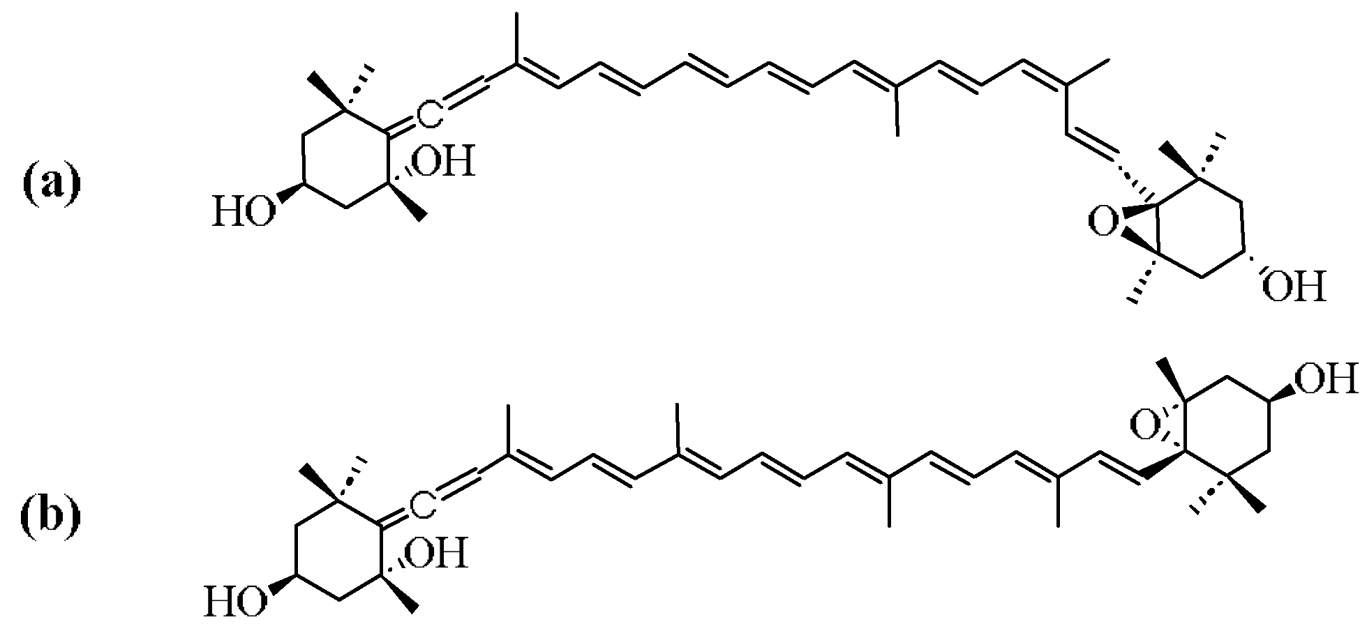
Figure 1. Structure of (a) 9′-cis neoxanthin (b) all-trans neoxanthin.
Marinedrugs 23 00317 g001
Figure 2. Neoxanthin content in various sources. (A) Neoxanthin content expressed in terms of fresh weight (FW) of high plants; (B) Neoxanthin content expressed in terms of dry weight (DW) of high plants and microalgal species.
Marinedrugs 23 00317 g002
Figure 3. Biosynthetic pathway of neoxanthin.
Marinedrugs 23 00317 g003
Figure 4. Biological activities of neoxanthin.
Marinedrugs 23 00317 g004
Table 4. Methods for the extraction of neoxanthin.
Extraction MethodsPlant SpeciesExtraction SolventsExtraction ConditionsNeoxanthin YieldReference
Organic solvents extractionChlorococcum humicolaDimethyl ether45:1 Solid/liquid ratio (w/w),
41 °C,
for 20 min
2.55 mg/g[53]
Caryocar BrasilienseAcetone3 mL/g Solid/liquid ratio,
for 300 s,
three times extraction
0.70 μg/g[55]
Scenedesmus obliquusEthanol10 mL/g Solid/liquid ratio,
for 3 min,
three times extraction
180.33 μg/g[42]
Brazil PouteriaAcetone40–60 °C193 μg/g[55]
Ionic liquids extractionCaryocar Brasiliense1:3 (1-hexyl-3-methylimidazolium chlorid): ethanol (v/v)3 mL/g Solid/liquid ratio,
for 300 s,
three times extraction
1.88 μg/g[55]
Chlorella sorokiniana1:4 (1-hexyl-3-methylimidazolium chloride): ethanol (v/v)10 mL/g Solid/liquid ratio,
for 7.5 min,
two times extraction
0.03 mg/g[54]
Scenedesmus obliquus1:3 (1-butyl-3-methylimidazolium tetrafluoroborate): ethanol (v/v)10 mL/g Solid/liquid ratio,
for 3 min,
three times extraction
48.71 μg/g[42]
Scenedesmus obliquus1:3 (1-butyl-3-methylimidazolium tetrafluoroborate): ethanol (v/v)10 mL/g Solid/liquid ratio,
for 3 min,
three times extraction
122.66 μg/g[42]
Supercritical liquid extractionScenedesmus sp. 9:1 CO2: Ethanol (v/v)Pressure 300 bar,
60 °C,
and CO2
flow rate 2 mL/min,
for 60 min
670.8 μg/g[56]
Ultrasound-assisted extractionCucurbita moschata2:1 Ethanol: Petroleum (v/v)31 mL/g solid/liquid ratio,
203 W,
for 30 min
36.69 μg/g[58]
Chlorella vulgarisEthanol30 mL/g Solid/liquid ratio,
amplitude wave of 20%,
25 °C,
for 25 min
9.83 mg/g[11]
Pressurized liquid extractionChlorella vulgarisEthanolPressure 1500 psi,
50 °C,
for 20 min
11.35 mg/g[11]
Table 5. The multi-target mechanism of action of neoxanthin in multiple cancer cell lines.
No.Type of CancerCell LineTargetMechanism of ActionReference
1Colon cancerHCT116mitochondrial functionCaspase-independent apoptotic pathway via loss of mitochondrial transmembrane potential, involving apoptosis-inducing factor (AIF), cytochrome-C, and endonuclease G (EndoG)[93]
2Prostate cancerPC-3Caspase-3Caspase-3-dependent apoptosis[94]
3Gastric cancermolecular dockingCytotoxin-associated gene A (Cag-A)Inhibits the binding of Cag-A of Helicobacter pylori to phosphatidylserine on host cell membranes[95]
4Lung cancerA549Caspase-3Induce apoptosis through caspase-3 activation; increase ROS clearance and repair activity with IC50 (mg/L) = 7.5 ± 0.6 μM[96]
5Cervical cancerHeLaCaspase-3Modulation of the activity of various transcription factors and responsive elements; Inhibition of the clonal expansion of initiated cells through enhanced gap junctional communication; Immunomodulatory effects by enhancing tumor immunity with IC50 (mg/L) = 3.8 ± 0.2 μM[96]
Table 6. Anti-oxidant mechanisms of neoxanthin in H2O2-treated HepG2 cell model.
No.Mechanism of ActionExperimental Data ResultsReference
1Activating endogenous anti-oxidant signaling pathwaysAt 4 h exposure to 0.5 mM H2O2, the cell viability in the neoxanthin pretreatment groups (0.05 μM and 0.1 μM) is significantly greater compared to the group subjected solely to H2O2 treatment[108]
2Directly removes reactive oxygen species (ROS) produced within cellsTreatment of HepG2 cells with H2O2 resulted in a 38% increase in reactive oxygen species (ROS) levels compared to the control group. Pretreatment with neoxanthin at concentrations of 0.05 μM and 0.1 μM resulted in a 12% and 24% reduction in ROS production[108]
3Upregulates the expression of intracellular anti-oxidant enzymes (HO-1 and SOD-2)H2O2 treatment significantly diminishes the expression of HO-1 and SOD-2, neoxanthin increases the expression of HO-1 by 17% and 22%, and SOD-2 by 21% and 35% at concentrations of 0.05 μM and 0.1 μM[108]
4Regulates transcription factor (Nrf2/ARE) expression Treatment with H2O2 significantly suppresses Nrf2 expression, whereas pretreatment with neoxanthin at 0.1 μM markedly restores Nrf2 expression by up to 42%[108]
5Inhibits the apoptotic signaling pathwayTreatment with H2O2 leading to a significant increase (60%) in the expression of the pro-apoptotic protein Bax and a concurrent decrease (50%) in the expression of the anti-apoptotic protein Bcl-2. Treatment with neoxanthin at 0.1 μM demonstrates the most pronounced reversal effect, restoring approximately 51% and 44% of the expression levels of Bax and Bcl-2[108]
Disclaimer/Publisher’s Note: The statements, opinions and data contained in all publications are solely those of the individual author(s) and contributor(s) and not of MDPI and/or the editor(s). MDPI and/or the editor(s) disclaim responsibility for any injury to people or property resulting from any ideas, methods, instructions or products referred to in the content.