Article

Identification, Expression Profiling, Microbial Binding, and Agglutination Analyses of Two Cathepsin B Genes in Black Rockfish (Sebastes schlegelii)

1
School of Marine Science and Engineering, Qingdao Agricultural University, Qingdao 266109, China
2
Haidu College, Qingdao Agricultural University, Laiyang 265200, China
*
Author to whom correspondence should be addressed.
Mar. Drugs 2025, 23(5), 213; https://doi.org/10.3390/md23050213
Submission received: 25 March 2025 / Revised: 11 May 2025 / Accepted: 16 May 2025 / Published: 18 May 2025
(This article belongs to the Section Marine Biotechnology Related to Drug Discovery or Production)

Abstract

:
As a lysosomal cysteine protease of the papain subfamily, cathepsin B (CTSB) is characterized by its innate immune functions and hydrolytic activity. However, the functions of CTSB in the immune responses of teleosts remain to be clarified. In this study, two CTSB genes in S. schlegelii, SsCTSBa and SsCTSBb, were identified. Both SsCTSBa and SsCTSBb are composed of a 993 bp ORF encoding 330 amino acids. It was found in a phylogeny analysis that both genes form monophyletic clades with their orthologous counterparts of Honeycomb rockfish (Sebastes umbrosus). A synteny analysis indicated that the CTSB homologues were comparatively conserved during vertebrate evolution. Additionally, quantitative real-time PCR revealed the ubiquitous mRNA expression of SsCTSBa and SsCTSBb in all of the examined tissues, and substantially differential expression patterns could be observed following Aeromonas salmonicida infection. A subcellular localization analysis demonstrated that the distribution of SsCTSBa and SsCTSBb was mainly in the cytoplasm. Moreover, rSsCTSBa and rSsCTSBb showed strong binding to Poly(I:C) and exhibited diverse agglutination effects on different bacteria. Overall, these findings suggest that the CTSB genes in black rockfish might show essential functions in the host defense of teleosts against bacterial infections, providing valuable insights for further investigations into the immune mechanism of teleost CTSB.

1. Introduction

Cathepsins are considered to be the primary members of the papain family, and are referred to as proteolytic enzymes that exist in animals and plants [1]. Additionally, cathepsins can be classified into three groups based on their conserved amino acid residues: cysteine proteases (cathepsins B, C, F, H, K, L, O, S, T, U, V, W, X, and Z) [2], serine proteases (cathepsins A and G), and aspartic proteases (cathepsins D and E) [3]. Cysteine proteases are primarily from the C1 peptidase family, otherwise known as the papain family. In addition, cathepsins B, C, F, H, L, O, and Z are universally expressed across various tissues, whereas cathepsins K, W, and X exhibit cell-specific or tissue-specific expression patterns according to their tissue distribution [4]. Moreover, cysteine cathepsins can be further categorized into the B-like subfamily (cathepsins B, C, O, and X) and the L-like subfamily (cathepsins L, V, K, S, W, F, and H), depending on the sequence length, similarity, and structure of the pro-region [5,6].
Cathepsin B belongs to the B-like subfamily of lysosomal proteases, which was initially reported in mice [7]. It has been extensively studied for its involvement in various proteolytic process such as antigen presentation [8], antigen degradation [9], and apoptosis [10]. In mammals, CTSB is significantly expressed in immune cells, including monocytes and macrophages [9]. In addition, CTSB was reported to be involved in numerous physiological processes linked to diseases, including the inflammatory response [11], tumor invasion and metastasis [12], and Alzheimer’s disease [13]. Recently, the roles of CTSB in fish species’ innate immunity have received more attention [14,15,16], and the homologues of numerous fish species have been cloned and characterized, such as miiuy croaker (Miichthys miiuy) [17], channel catfish (Ictalurus punctatus) [18], large yellow croaker (Pseudosciaena crocea) [8], and olive flounder (Paralichthys olivaceus) [19]. In Japanese flounder (Paralichthys olivaceus), CTSB is markedly elevated in the spleen, kidneys, gills, and intestine with LPS and viral infection [20]. In golden pompano (Trachinotus ovatus), CTSB showed an obvious increasing trend in the liver, spleen, and head kidney under challenge by Edwardsiella tarda [21]. Overall, these findings suggest that CTSB might be involved in anti-microbial immune responses. Nevertheless, the specific immunological participation of CTSB in immune responses to bacterial infection remains to be explored.
As the major commercial species in mariculture, black rockfish (S. schlegelii) have been widely cultivated in China, South Korea, and Japan. However, persistent bacterial problems have caused serious losses in the aquaculture industry, particularly with A. salmonicida infection [22]. In addition, although the functions of the genome and virulence factors of A. salmonicida [23,24] have been identified, the interaction between this bacterium and host tissues in infected black rockfish remains to be clarified. Furthermore, a comprehensive investigation into the innate immune responses of black rockfish to pathogen infections is urgently required to develop effective strategies for disease prevention and control in aquaculture. The cathepsin superfamily in the S. schlegelii genome has been systematically analyzed in previous studies, which has indicated the important roles of bacterial infections [25]. However, the immune role of cathepsins requires further investigation. Two homologues of cathepsin B from black rockfish have been systematically characterized in this study, and the expression patterns have been deeply evaluated in multiple tissues of healthy fish and after infection. Additionally, the subcellular localization of these homologues has been investigated in this study. Recombinant proteins were obtained using a prokaryotic expression system, enabling the determination of their binding ability as well as agglutination capacity towards microbial ligands or bacteria in vitro. Overall, this research could provide a foundation for further exploration into the immune functions of CTSB.

2. Results

2.1. Identification of S. schlegelii CTSB

The full-length cDNA of SsCTSBa (GenBank Accession: PQ683368) was composed of a 993 bp ORF that encoded a putative protein consisting of 330 amino acid residues with a predicted molecular mass of 36.06 kDa, a theoretical isoelectric point of 5.38, an aliphatic index of 69.42, and an instability index of 32.27. The GRAVY score was −0.260, with three cysteine protease active sites. Additionally, a total of 37 negatively and 27 positively charged residues were observed in this study (Table 1). In contrast, the full-length cDNA of SsCTSBb (GenBank Accession: PQ683369) contained a 993 bp ORF that encoded 330 amino acid residues with a calculated molecular mass of 36.06 kDa and a theoretical isoelectric point of 5.20, in which the aliphatic and instability indexes were 70.94 and 31.46, respectively. The GRAVY score was −0.309 with three cysteine proteases active sites. In addition, a total of 37 negatively charged and 24 positively charged residues were observed. According to the amino acid sequence alignment, SsCTSBa and SsCTSBb shared overall identities at a relatively high level (97.57% and 99.09%, respectively) with honeycomb rockfish cathepsin B homologues. In contrast, SsCTSBa and SsCTSBb shared relatively low sequence identity (70.18% and 66.77%, respectively) with cathepsin B homologues in humans (Figure S1).

2.2. Characteristics of S. schlegelii CTSB Genes

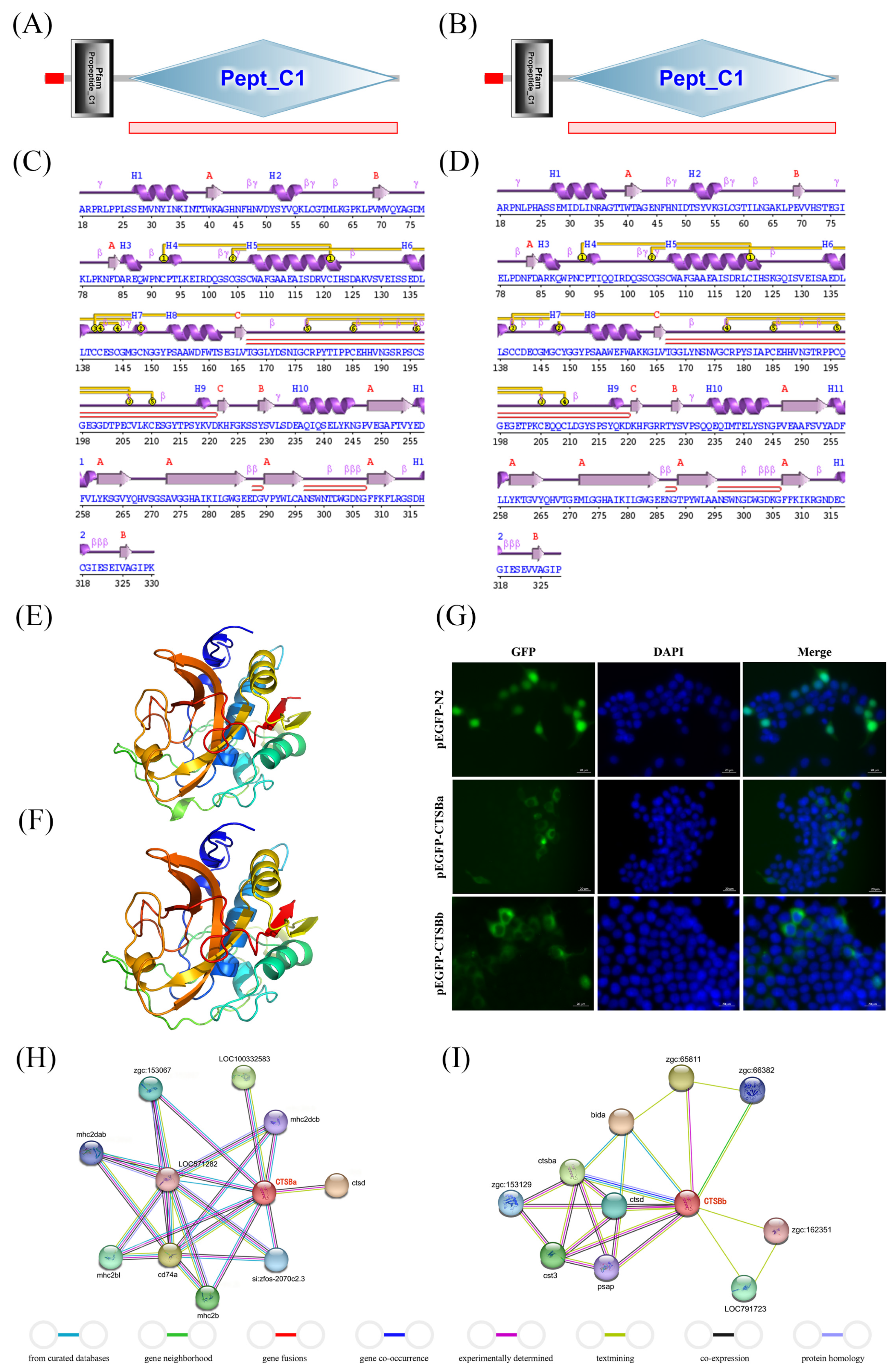
The Pept_C1 domain was present in both SsCTSBa and SsCTSBb as a signal peptide (Figure 1A,B). It could be shown by the secondary structure analysis of SsCTSBa and SsCTSBb that 3 sheets, 3 beta bulges, 3 beta hairpins, 12 strands, and 12 helices were possessed by both of them (Figure 1C,D). Meanwhile, the 3D structure of SsCTSBa was 77% identical to that of c2dcbA, with a confidence of 100%, while that of SsCTSBb was 75% identical to that of c2dcbA, with 100% confidence (Figure 1E,F). Additionally, both SsCTSBa and SsCTSBb might be connected with immune-related signal transductions, which was confirmed by the protein–protein interaction (PPI) network research. As illustrated in Figure 1H, several essential immune-related genes were observed to interact with CTSBa, which were composed of CD74a, mhc2b, mhc2bl, mhc2dab, mhc2dcb, and CTSD. Simultaneously, some of the CTSBb-related genes showed significant roles in host immunity (Figure 1I), including CTSBa, CTSD, CST3, PSAP, and BIDa. In addition, it could be indicated by the subcellular localization assay that SsCTSBa and SsCTSBb were primarily observed within the cytoplasm of HEK293T cells. Moreover, the green fluorescent EGFP protein is primarily localized to the cell nucleus, with some cytoplasmic signal present in the control group (Figure 1G).

2.3. Phylogeny and Synteny Analyses

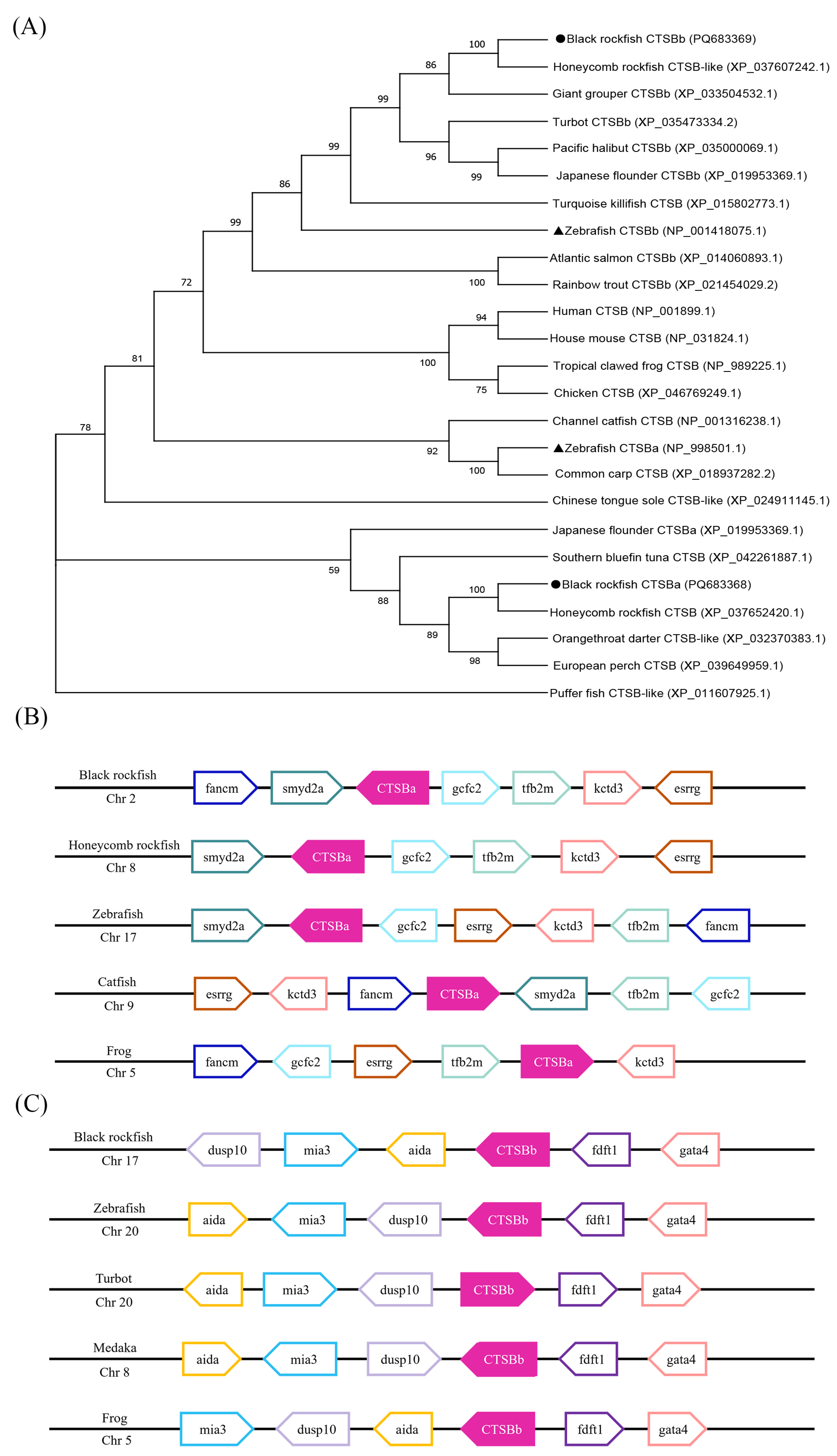
The evolutionary connection of CTSB between various species was analyzed in this study, and a phylogenetic analysis was employed to verify the SsCTSBa and SsCTSBb. It was revealed by the results that the CTSB genes can be assigned into two groups. With the support of bootstrapping, the SsCTSBa was firstly grouped with honeycomb rockfish CTSB, followed by southern bluefin tuna CTSB, Japanese flounder CTSBa, puffer fish CTSB-like, and Chinese tongue sole CTSB-like. Subsequently, it was incorporated into the other clade, which was composed of additional teleosts (channel catfish, zebrafish, and common carp) and tetrapods (human, house mouse, tropical clawed frog, and chicken). In contrast, the highest homology was revealed for SsCTSBb in honeycomb rockfish between the teleost-specific CTSBb gene homologues; then, the SsCTSBb was clustered with giant grouper, turquoise killifish, zebrafish, Atlantic salmon, and rainbow trout, before the development of the CTSBa clade (Figure 2A).
A synteny analysis was performed to verify the identification and orthology results of SsCTSBa and SsCTSBb. Firstly, the results of this analysis could offer compelling evidence contributing to the identification of CTSB genes, which could be beneficial to the deep exploration of the evolutionary connection of CTSB. Additionally, five studied vertebrate species exhibited well-conserved synteny with CTSBa and shared the genes smyd2a, gcfc2, tfb2m, kctd3, and esrrg in their genomic neighborhood, including four teleosts and one tetrapod (Figure 2B). As shown in Figure 2C, CTSBb was conserved among four teleosts and one tetrapod in the analysis of black rockfish, zebrafish, turbot, medaka, and frog with common genes including aida, dusp10, and mia3 upstream, and fdft1 and gata4 downstream.

2.4. Basal Tissue Distribution

qPCR was employed to evaluate the tissue distribution of SsCTSBa and SsCTSBb in all of the tested tissues (kidneys, skin, blood, gills, brain, liver, spleen, intestine, and muscle). Both genes were ubiquitously expressed, with detectable transcript levels in all examined tissues. As shown in Figure 3A, the expression of SsCTSBa in brain showed the lowest level; thus, it was used as the baseline. The highest expression of SsCTSBa could be observed in spleen, followed by muscle, kidneys, gills, blood, skin, liver, and intestine. Notably, the expression of SsCTSBb in the gills showed the lowest level, and it was employed as the baseline. Additionally, the highest expression of SsCTSBb could be observed in the intestine, followed by the liver, skin, muscle, brain, spleen, blood, and kidneys (Figure 3B).

2.5. Expression Profiles of SsCTSBa and SsCTSBb Following A. salmonicida

After infection with A. salmonicida, qPCR was applied in the analysis to evaluate the expression (Figure 3C–F). Overall, the expression level of SsCTSBa within all tissues showed a significant up-regulated trend with the infection of A. salmonicida. In contrast, the expression level of SsCTSBb was up-regulated in gills and liver at all tested time points, while it was down-regulated at 48 and 72 h in kidneys and spleen. In detail, the expression of SsCTSBa in gills was up-regulated at 6 h, 24 h, 48 h, and 72 h, and the peak occurred in 6 h (32.34-fold), as shown in Figure 3C. In contrast, the SsCTSBb showed an up-regulated trend in gills, and the peak occurred at 72 h (92.72-fold). Simultaneously, the expression of SsCTSBa in kidneys and SsCTSBb in liver showed obvious up-regulated trends at each time node (Figure 3D,E). However, although the expression of SsCTSBb firstly up-regulated in the kidneys at 6 and 24 h, it showed an obvious decreasing trend to −3.15-fold at 48 h, and it finally increased to −2.25-fold at 72 h (Figure 3D). In the spleen, the expression level of SsCTSBa was up-regulated at 48 h (2.24-fold) and 72 h (2.78-fold), while that of SsCTSBb was down-regulated at 48 h (−7.15-fold), and sharply dropped at 72 h (−25.47-fold) (Figure 3F). Overall, these findings could further suggest that SsCTSBa and SsCTSBb might be connected with the infection response.

2.6. Binding Abilities of rSsCTSBa and rSsCTSBb to Microbial Ligands and Bacteria

rSsCTSBa and rSsCTSBb were successfully purified with the IPTG-mediated induction. Additionally, the distinct bands for rSsCTSBa and rSsCTSBb were analyzed by the SDS-PAGE, and each of them showed an approximate molecular weight of 36 kDa (Figure 4A). Notably, the antisera showed a distinct reaction with rSsCTSBa and rSsCTSBb with a molecular weight of around 36 kDa, which was in agreement with the prediction as shown in the WB profiles (Figure 4B). In addition, in vitro assays were employed in the assessment of the binding ability of rSsCTSBa and rSsCTSBb in this study. It could be demonstrated by the absorbance results that rSsCTSBa and rSsCTSBb showed dose-dependent binding with Poly(I:C), LPS, PGN, and LTA, as well as with all test strains of bacteria, including Gram-negative bacteria (A. salmonicida, A. hydrophila, and Escherichia coli) and Gram-positive bacteria (Streptococcus agalactiae, Staphylococcus aureus, and Streptococcus iniae) (Figure 4C–H). Additionally, both rSsCTSBa and rSsCTSBb possessed the highest affinity to Poly(I:C) among the four examined ligands. Furthermore, SsCTSBb showed a higher binding ability to the four ligands compared with that of SsCTSBa (Figure 4C,D). In terms of the binding ability to Gram-positive bacteria, rSsCTSBa showed the strongest combination with S. agalactiae followed by S. aureus and S. iniae, whereas rSsCTSBb had the best binding ability with S. aureus followed by S. agalactiae and S. iniae (Figure 4E,G). Regarding the binding ability to Gram-negative bacteria, rSsCTSBa showed the strongest combination with A. salmonicida followed by E. coli and A. hydrophila, while rSsCTSBb had the best binding ability with A. hydrophila followed by A. salmonicida and E. coli (Figure 4F,H).

2.7. Agglutination Assay with Bacteria

To investigate the coagulant capacity of rSsCTSBa and rSsCTSBb with the bacteria, rSsCTSBa and rSsCTSBb were labeled with FITC and coagulated with Gram-negative bacteria (A. salmonicida, A. hydrophila and E. coli) and Gram-positive bacteria (S. agalactiae, S. aureus, and S. iniae) (Figure 5A,B). Simultaneously, bacteria and FITC dye were added as negative controls; alongside, the observation of green fluorescence was observed by an inverted fluorescence microscope. Additionally, SsCTSBb and Gram-positive/negative bacteria incubation displayed oligomerization (speck structures) except for E. coli versus the control groups. In contrast, the Gram-positive bacteria and Gram-negative bacteria were clearly aggregated by SsCTSBa.

3. Discussion

Cathepsin B (CTSB) is referred to as a cysteine protease of the cathepsin family, which features a Cys-His double amino acid group within its active center [26]. Additionally, it can serve as both an exopeptidase and endopeptidase [2] and shows crucial functions in various pathological and physiological processes. Under normal physiological conditions, CTSB can facilitate the degradation of the proteins within the lysosome [27]. However, CTSB engages in physiological processes outside the lysosome under pathological conditions [28]. In mammals, CTSB has been connected to cell death by the mechanisms of triggering apoptotic factors upstream of executioner [29] or directly binding and activating caspases [30,31,32]. Additionally, it has been indicated that CTSB could regulate apoptosis in follicles during serum-starvation in Danio rerio [33]. In Apostichopus japonicus, the down-regulated AjCTSB significantly inhibited coelomocyte apoptosis and influenced the expression of cytochrome c1 and cytochrome c-1/2 genes [34,35]. Due to the involvement in immune-related activities, two CTSB homologues were examined in black rockfish, and the analysis was focused on their immune-related functions and expression profiles in both healthy and bacterially infected fish. Additionally, the abilities to bind and agglutinate microbial ligands and bacteria were characterized in the research. This study could offer a preliminary insight into the immunological participation of CTSB in teleost species, and the results revealed that only one CTSB gene was observed in mammalian species before this study. However, two CTSB homologues can be found in teleosts, including rainbow trout, zebrafish, carp and channel catfish [18,21]. Additionally, CTSBb can only be found in teleosts through lineage-specific tandem gene duplications, which suggests that gene duplication might be the predominant evolutionary mechanism for the adaption of fish to the environment.
The deduced polypeptides of SsCTSBa and SsCTSBb both consisted of 330 amino acids with similar molecular properties (theoretical pI, molecular weight, and instability index) to those in other teleosts [36,37,38]. Moreover, it was revealed by the structural analysis that SsCTSBa and SsCTSBb possessed the typical structural features of the papain family, including a peptidase-C1 domain (Figure 1A,B). Additionally, various active site locations were shown by different species in their amino acid sequences, which were composed of the pacific abalone (Haliotis discus hannai), belonging to gastropods [39], and the large yellow croaker [8] and golden pompano [21], belonging to teleosts. SsCTSBa possessed three active site residues and two N-glycosylation sites (Table 1). Nevertheless, three active site residues and only one N-glycosylation site were observed in SsCTSBb (Table 1). Moreover, the activity of cysteine-like proteases with conserved histidine, cysteine, and asparagine residues could significantly affect the stability of enzyme catalytic sites. It was revealed by Chen et al. [16] that His277 was essential for enzyme activity due to its ability to form an imidazole ring with Asn. Additionally, the N-glycosylation site showed great importance in the development of the organism without relation to the enzyme activity [40,41]. Cathepsins are crucial in the process of lysosomal transport and immune response to pathogen infections [42]. In addition, the duplication of CTSB in black rockfish parallelled the gene duplication that was observed in mammal-specific CC chemokines following the divergence of mammalian and bird ancestral lineages [43,44]. The parallel gene duplication events observed for both CTSB and CC chemokine genes in these distinct lineages likely reflect convergent evolutionary pressures for immune system complexity. Following gene duplication in teleost fish, CTSB may have undergone functional diversification similar to that observed in mammalian CC chemokines.
It could be indicated by the phylogenetic analysis (Figure 2A) and syntenic patterns (Figure 2B,C) of the selected species that the orthology and synteny relationship of the CTSB genes are conserved. Overall, the identification of the CTSB genes of black rockfish could be confirmed by the combined molecular characteristics, numerous species comparisons, phylogenetic analysis, and syntenic analysis, which also showed strong homology with the counterparts in other species. Additionally, the conservation of the structural characteristics of SsCTSBa and SsCTSBb could indicate potential functional similarities with other species.
The interacting genes of SsCTSBa and SsCTSBb were predicted by the PPI network analysis for the deep exploration of their functions in immune-related signaling pathways. The interacting genes of SsCTSBa are composed of CD74a, mhc2b, mhc2bl, mhc2dab, mhc2dcb, and CTSD, while the interacting genes of SsCTSBb include CTSBa, CTSD, CST3, PSAP, and BIDa (Figure 1H,I), among which the four MHC class II molecules (mhc2b, mhc2bl, mhc2dab, and mhc2dcb) are expressed by APCs and exhibit antigen presentation characteristics [45,46,47]. CD74a is referred to as the key bioactive molecule, and it was crucial for the assembly and trafficking of MHC II molecules during antigen presentation [48]. Notably, CTSB/D/E/F/K/L/S have been reported to be connected with the degradation of associated chaperones (invariant chain[li]) and antigen processing in mammals, as demonstrated by vitro studies [3]. Therefore, it was assumed that SsCTSBa and SsCTSBb might be involved in the fish MHC II antigen presentation pathway with CTSD and CD74a, and the precise mechanism of their interaction should be explored. Different from CTSB, which is referred to as a cysteine protease, CTSD is an aspartate protease [49]. It has been proven that PSAP precursors can be divided by CTSD into four small saposins based on PSAP localization [50]. In addition, the lysosomal function and activity of CTSB and CTSD might be associated with saposin-C [51]. It has been revealed by previous studies that autophagic flux and cholesterol metabolism are dramatically impacted by defective endolysosomes with reduced amounts of mature CTSB and CTSD [52]. CST3 was considered to be a cysteine protease inhibitor that controlled lysosomal enzyme activity, and it was confirmed to be linked with neurodegeneration. In vitro, inhibitory effects of CST3 on proteinase are weakened when it is bound to PSAP [53]. Notably, a PPI analysis was conducted according to the D. rerio and C. semilaevis datasets due to the limited identification of black rockfish protein data, and the PPI databases of black rockfish and other fish species need to be established further. On account of the report that CTSB in cytoplasm could result in cell death [54], a chimeric GFP protein was constructed to examine the location of SsCTSBa and SsCTSBb in cells. According to the findings, it was revealed by green fluorescence that SsCTSBa and SsCTSBb were observed in the cytoplasm, and the SsCTSBa and SsCTSBb in the cytoplasm might participate in transportation or degradation processes, which corresponded to the PPI results. It has been demonstrated that the proteolytic enzyme CTSB was essential to antigen processing by breaking down both antigens and B cell receptors (BCRs), which could make it easier for Human Leukocyte Antigen (HLA) class II molecules to present peptide fragments [55].
Ubiquitous expression signatures of SsCTSBa and SsCTSBb were observed, and the widespread expression of CTSB in various tissues was in agreement with previous research [8,41,56,57,58]. Similarly to CTSB in Nile tilapia [41], the expression level of SsCTSBa follows this order: spleen, muscle, kidneys, gills, blood, skin, liver, intestine, and brain (Figure 3A). In contrast, the intestine showed the highest expression of SsCTSBb, which was followed by the liver, skin, muscle, and brain (Figure 3B). Additionally, the gills showed the highest CTSB expression out of the other tissues examined in orange-spotted groupers, followed by the head, kidneys, spleen, and heart [56]. Notably, their potential functions are suggested by the relatively high expression levels in immune-related tissues including the spleen, intestine, muscle, liver, kidneys, skin, and gills. In addition, the spleen is considered to be an important immune organ in fish, and shows significant roles in the immune response. The highest expression level of CTSB in the spleen could be attributed to its involvement in immune processes. In recent years, CTSB and other cathepsins have been discovered to show important effects in both intracellular and extracellular matrix (ECM) degradation [59]. Additionally, the intestine is considered the primary site for digestion and absorption in fish, and it might involve CTSB during the process of food digestion and nutrient absorption within the intestinal tract. In addition, it might be beneficial to the breaking down of protein components in food, contributing to access for absorption by intestinal cells. CTSB was found to be connected with proteins that digest yolk, which benefits the quality and viability of eggs, and also provides an energy source for embryos of fish species [60,61].
It was reported that CTSB might induce the host immune response stimulated by microbial ligands or pathogens [14,16,18,62]. For instance, CTSB could promote TNF-α post-translational processing and contribute to its synthesis in response to LPS stimulation, which results in the host’s immunological response to infections [62]. The expression of CTSBa in channel catfish mucosal tissues was induced following Edwardsiella ictaluri and Flavobacterium columnare challenge [18]. In C. semilaevis, the expression of CTSB in the head kidney was up-regulated to 25-fold at 4 h after Vibrio anguillarum challenge [16]. In Oplegnathus fasciatus, the expression level of CTSB in the spleen was up-regulated by 2.7-fold at 24 h after E. tarda infection [14]. Corresponding to the findings above, the expression of SsCTSBa in our current investigation showed an up-regulated trend after A. salmonicida infection, particularly in the gills of all of the examined tissues at the majority of the time points (Figure 3C). Nevertheless, the expression level of SsCTSBb showed an obvious down-regulation trend in the spleen, while it exhibited up-regulation in the gills and liver (Figure 3C,E,F). It has been revealed by previous research that the overexpression of CTSB could improve the autophagic flux in endothelial cells and reduce cell apoptosis under hypoxic conditions [63]. Although this research mainly focused on hypoxic environments, similar mechanisms could be found in certain bacterial infections. A deficiency of CTSB was confirmed to inhibit intracellular infection with Brucella. With the development of infection, A. salmonicida might inhibit the autophagy process in host cells to down-regulate the expression of SsCTSBb, thereby avoiding the surveillance of the host immune system. Additionally, Ectromelia virus has been shown to inhibit the expression of CTSB in murine conventional dendritic cells, thereby facilitating its replication process [64]. However, in Mycobacterium tuberculosis-infected rabbits, increased CTSB gene expression was observed in the lungs, and increased protein levels were observed in plasma from patients with active tuberculosis [65]. Thus, the changes in the expression levels of CTSB might vary according to the type of pathogen and host response, and it was relatively common for the expression level of cathepsin genes to be induced after bacterial infection. In addition, such general up-regulated expression levels could also be observed for CTSK and CTSS in black rockfish following V. anguillarum infection [38,66]. Notably, the expression of CTSK in the spleen of black rockfish showed an upward trend and peaked at 24 hpi (24.8-fold) [66], while the expression of CTSS in the spleen was also obviously up-regulated, with the maximum induction at 12 hpi (26.5-fold) [38]. The expression levels in the four primary immune-related tissues (gills, kidneys, liver, and spleen) induced by pathogen infection appeared to be different, which indicated the participation of CTSB in the immunological response.
Regarding the deep exploration of the immunological participation of SsCTSBa and SsCTSBb in the host defense against different pathogens, rSsCTSBa and rSsCTSBb were purified, alongside the application of four representative microbial ligands in an in vitro binding experiment. According to the results, both rSsCTSBa and rSsCTSBb were efficiently expressed in BL21 (DE3), and exhibited high binding ability with all the ligands, particularly Poly(I:C) (Figure 4C,D). Additionally, Poly(I:C) is an interferon inducer featuring broad-spectrum antiviral and immunological regulatory properties. And it could indicate the significant participation of SsCTSBa and SsCTSBb in the immune response. Poly(I:C)-induced microglia activation, which might release proinflammatory factors, showed a decreasing trend in BV2 cells with the inhibition of CTSB activity by inhibitor CA-074 after 24 h, and the initial phases of activation-induced microglia death could indicate active functions for cathepsin X [67]. Regarding the flounder embryonic cells, CTSB could be induced by Poly(I:C), LPS, and viruses in vitro and in vivo, and Poly(I:C) is considered to be one of the most effective inducers during the transcription of CTSB [20]. In C2C12 myotubes, CTSB could promote the process of TNF-α post-translation and contribute to its synthesis in response to LPS stimulation, which resulted in a host immunological response to infections [68]. In addition, both rSsCTSBa and rSsCTSBb exhibited strong agglutination effects on S. aureus, S. iniae, S. agalactiae, A. hydrophila, and A. salmonicida (Figure 5A,B). The results above were consistent with their binding abilities to all of the examined bacteria (Figure 4E–H). Additionally, CTSB was confirmed to degrade various extracellular matrix components in vitro according to existing studies, which were composed of glycosaminoglycans, elastin, and collagen. In rheumatoid arthritis and osteoarthritis, CTSB was found to contribute to joint damage by degrading type II collagen and proteoglycans in articular cartilage [59]. Previously, the CTSK and CTSS of black rockfish were observed to agglutinate the bacteria (E. coli, V. anguillarum, and S. aureus) and bound with LPS and PGN [38,66]. In addition, CTSZ showed a high affinity for LPS, PGN, and LTA, and it demonstrated an effective coagulant capacity for Gram-negative and Gram-positive bacteria in turbot [69]. The differences between CTSB and CTSK/CTSS/CTSZ indicated their important immunological participation in the host defense system. Moreover, the black rockfish that were treated with CTSS knockdown showed clearly higher bacteria amounts versus the control after the infection [38], while the fish that underwent the CTSK knockdown exhibited significantly lower bacterial amounts than the control group [66]. Overall, the opposing roles of various cathepsins in innate immunity could be demonstrated by these findings, which could indicate that SsCTSBa and SsCTSBb might further serve as targets within the innate immune response.

4. Materials and Methods

4.1. Sequence Identification

CTSB gene sequences from various vertebrate species were used as queries to search against the black rockfish genome database based on the BLAST program version 2.16 [70,71]. The transcriptome databases were developed in our laboratory and utilized in this study. Subsequently, duplicates were removed from the initial retrieved sequence pool that was recovered by Clustal Omega (https://www.ebi.ac.uk/jdispatcher/msa/clustalo, accessed on 10 August 2024), ensuring the identification of each sequence as one based on its genomic location. Additionally, the sequences mentioned above were initially deduced with the support of the ORF finder application (https://www.ncbi.nlm.nih.gov/orffinder/, accessed on 16 August 2024) and subsequently verified through BLASTP against the NCBI database. In addition, SMART (http://smart.embl-heidelberg.de/, accessed on 24 August 2024) was employed in the operation of the conserved domains and signal peptides.

4.2. Sequence Analyses

A range of bioinformatic analyses were analyzed for the characteristics of CTSB. Additionally, the ExPASy [72] and PROSITE server were used to capture the physiochemical properties and characteristic signatures of CTSB. Generally, the proteins were considered to be stable with an instability index less than 40. In addition, the aliphatic index was determined by the relative volume of aliphatic side chains, and it showed crucial effects in the enhancement of the thermostability of globular proteins. The GRAVY score was calculated by averaging the total hydropathy values of amino acids. The ideational 3D protein structure was generated by the Phyre2 server [73]. Additionally, the protein–protein interaction was analyzed by performing a BLAST search of the amino acid sequences of SsCTSBa and SsCTSBb against Danio rerio or Cynoglossus semilaevis based on STRING software 12.0.

4.3. Subcellular Localization

The HEK293T cell line was obtained from the American Type Culture Collection (ATCC, CRL-3216, RRID:CVCL_0063), located in Manassas, Virginia, USA. HEK293T cells were maintained in Dulbecco’s Modified Eagle Medium (DMEM, Gibco, Grand Island, NY, USA) supplemented with 10% fetal bovine serum (FBS, Gibco) and 1% penicillin/streptomycin (Gibco). The pEGFP-N2-SsCTSBa/b constructs were generated by cloning the full-length SsCTSBa and SsCTSBb coding sequences (amplified from black rockfish cDNA) into the BamHI and Nhel sites of the pEGFP-N2 vector (Clontech, Shiga Prefecture, Kyoto, Japan). The constructs were verified by Sanger sequencing. HEK293T cells were seeded on sterile coverslips in the 24-well plates at a density of 1 × 10⁵ cells per well to locate SsCTSBa and SsCTSBb. After 24 h, cells were transfected with 1 μg of either the pEGFP-N2-empty or pEGFP-N2-SsCTSBa/b plasmid utilizing TurboFect™ transfection reagent. At 48 h post-transfection, the cells were put on slides with Fluoroshield consisting of DAPI (Sigma-Aldrich, Darmstadt, Germany), and examined with an inverted fluorescence microscope (objective 40×). The localization pattern was demonstrated through three independent experiments, confirming the reliability of our conclusion. For each experiment, at least 30 successfully transfected cells (with fluorescence intensity significantly higher than background) were analyzed, with approximately 80% fluorescence-positive cells determined by manual inspection. The pEGFP-N2-empty vector is commercially available (Clontech, Shiga Prefecture, Japan). All custom plasmids (pEGFP-N2-SsCTSBa/b) will be made freely available upon request to the corresponding author.

4.4. Phylogeny and Synteny Analyses

A phylogenetic tree was generated with the application of amino sequences of CTSB in black rockfish and other various species, and the sequences were assessed including human (Homo sapiens), house mouse (Mus musculus), tropical clawed frog (Xenopus tropicalis), chicken (Gallus gallus), honeycomb rockfish, orangethroat darter (Etheostoma spectabile), European perch (Perca fluviatilis), Southern bluefin tuna (Thunnus maccoyii), Japanese flounder (Paralichthys olivaceus), puffer fish (Takifugu rubripes), Chinese tongue sole (Cynoglossus semilaevis), channel catfish, zebrafish, common carp (Cyprinus carpio), Atlantic salmon (Salmo salar), rainbow trout (Oncorhynchus mykiss), turquoise killifish (Nothobranchius furzeri), giant grouper (Epinephelus lanceolatus), turbot (Scophthalmus maximus), and Pacific halibut (Hippoglossus stenolepis). Additionally, the protein sequences were aligned using the ClustalW2 program [74]. Phylogenetic analysis was conducted in MEGA X software to examine the evolutionary connection with the counterpart molecules based on the neighbor-joining method with 1000 bootstrapping replications [75]. Poisson distance was employed for the correction of the analysis, and the gaps were addressed via pairwise deletion.
Syntenic analysis was operated for the orthology analysis of the two CTSB genes in black rockfish, which was based on the comparison between the neighboring genes of SsCTSBa and SsCTSBb and those of zebrafish, frog, catfish, turbot, medaka, and honeycomb rockfish in the FGENESH program [76]. Additionally, the BLASTP was used to annotate the identified protein sequences according to the NCBI database, and the Genomicus and Ensembl database were employed in the evaluation of the conserved syntenic pattern of CTSB genes [77].

4.5. Sample Collection of Healthy Black Rockfish

Samples from nine tissues of healthy black rockfish were obtained to describe the expression profiles of SsCTSBa and SsCTSBb, and the black rockfish were purchased from a black rockfish hatchery in Haiyang (Shandong Province, China), in which the fingerlings showed an average weight of 14.8 g and length of 5.6. The fish were acclimated for at least two weeks in a recirculating seawater system before the collection of tissues. The tissue samples were obtained from 30 healthy black rockfish that had first been anesthetized with seawater containing 100 mg/L of tricaine methane sulfonate (MS-222). All samples were quickly frozen in liquid nitrogen and stored at −80 °C until the RNA preparation.

4.6. Bacterial Challenge and Sample Collection

The bacterium A. salmonicida was selected to perform the bath challenge for the expression of SsCTSBa and SsCTSBb. Additionally, the bacteria were biochemically verified and re-isolated from fish exhibiting symptoms following a pre-challenge before the cultivation. The infection of symptomatic fish with A. salmonicida was checked during the challenge. Additionally, the fish were bathed in the bacterial solution with the concentration of 5 × 10⁶ CFU/mL for 2 h during the experiment, and the gills, kidneys, liver, and spleen tissues were obtained from 15 fish (5 fish per replicate) at intervals of 6 h, 24 h, 48 h, and 72 h after treatment with 100 mg/L MS-222. Samples were promptly frozen by liquid nitrogen and maintained at −80 °C until the RNA preparation.

4.7. Total RNA Extraction and Real-Time PCR Analyses

Trizol® Reagent (Invitrogen, Carlsbad, CA, USA) was employed in the extraction of total RNA by the provided procedure, and 1% agarose gels were used to track RNA contamination and degradation. The concentration value was assessed using the DS-11 Spectrophotometer (Denovix, Wilmington, DE, USA), and the A260/280 ratios for all extracted samples were above 1.8. Then, the expression patterns were determined by qPCR methods. Primer 6 online program was employed in the design of specific primers for the CTSB gene sequences of black rockfish, and the β-actin gene was used as the reference for normalizing the expression levels (Table 2). The synthesis of cDNA was carried out using the PrimeScript RT reagent Kit (TaKaRa, Dalian, China) following the manufacturer’s instructions. Additionally, qPCR was completed using the CFX96 detection system (BioRad Laboratories, Hercules, CA, USA) based on the SYBR ExScript qPCR Kit (Takara, Dalian, China), as instructed. Standard curves were developed in this study to determine the amplification efficiency, threshold values, and the formula for the calculation of relative gene copy numbers. Then, the relative mRNA expression was analyzed in triplicate. All the values were presented as means ± SD, and fold differences were determined with the aid of the Relative Expression Software Tool (REST) to ensure statistical significance (p < 0.05) [78].

4.8. Purification of Recombinant SsCTSBa and SsCTSBb Protein (rSsCTSBa and rSsCTSBb)

The DNA fragment encoding SsCTSBa and SsCTSBb was amplified by the high-fidelity DNA polymerase (TaKaRa, Dalian, China) and specific primers (SsCTSBa-Pr F/R and SsCTSBb-Pr F/R, Table 2). The target PCR product was subcloned into the PET32a-modified HTH expression vector after the sequencing process. Then, E. coli BL21 (DE3) was transformed by the resulting HTH-SsCTSBa and HTH-SsCTSBb plasmids, which expressed histidine-tagged rSsCTSBa and rSsCTSBb. Subsequently, the cells were fostered in the LB medium and induced by 0.5 mM IPTG. Next, the recombinant SsCTSBa and SsCTSBb proteins (rSsCTSBa and rSsCTSBb) were purified with the application of nickel-nitrilotriacetic acid columns (GE Healthcare, Piscataway, NJ, USA). In addition, the screening samples were evaluated by 12% SDS-PAGE. The concentration of the fusion proteins was tested using Bradford’s technique. Moreover, the existence of the purified rSsCTSBa and rSsCTSBb was confirmed by the Western blot analysis. The HTH-SsCTSBa/b constructs were generated by cloning the full-length SsCTSBa and SsCTSBb coding sequences (amplified from black rockfish cDNA) into the BamHI and KpnI sites of the HTH vector. All custom plasmids (HTH-SsCTSBa/b) will be provided upon request to the corresponding author.

4.9. Binding Abilities of rSsCTSBa and rSsCTSBb with Microbial Ligands and Bacteria

Each well of the 96-well microtiter plates (Corning, NY, USA) was covered by 0.25 μg of lipopolysaccharide (LPS), peptidoglycan (PGN), lipoteichoic acid (LTA), and polyinosinic acid–polycytidylic acid (Poly(I:C)) at 4 °C overnight, as well as Gram-negative bacteria (E. coli, A. hydrophila, and A. salmonicida) and Gram-positive bacteria (S. aureus, S. agalactiae, and S. iniae), respectively. Notably, the plates were washed three times by PBST (0.05% Tween-20 in PBS) and then blocked with 5% BSA at 37 °C for 1 h. Subsequently, 100 μL of the control protein alongside the empty HTH or the purified rSsCTSBa and rSsCTSBb with increased concentration was incorporated into the ligand-coated wells, and incubated at 37 °C for 1 h with four replicates for each concentration, followed by five rounds of washing with PBST. Notably, the concentration levels were designed with the values of 5, 10, 20, and 40 μg/mL in the above experiments. The plate was then incubated at 37 °C for 1.5 h with 100 μL mouse anti-His antibody (Affinity Biosciences, Taizhou, China). After washing three times with PBST, horseradish peroxidase-conjugated goat anti-mouse IgG (Affinity Biosciences, Taizhou, China) was incorporated into the plates. An ELISA plate reader was employed in the measurement of OD450 nm after the termination of reactions with 0.5 M sulfate.

4.10. Agglutination of FITC-Labeled rSsCTSBa and rSsCTSBb with Bacteria

Depending on the labeling of rSsCTSBa and rSsCTSBb with fluorescein isothiocyanate (FITC), the agglutination capabilities were investigated in this study. Additionally, S. aureus, S. iniae, S. agalactiae, E. coli, A. hydrophila, and A. salmonicida were cultivated with LB broth in a shaker incubator under the conditions of 180 rpm and 28 °C overnight. After the centrifugation of 5000× g for 10 min, the bacteria were resuspended twice in TBS-Ca and adjusted to a concentration of 2.5 × 10⁹ CFU/mL. In addition, the obtained rSsCTSBa and rSsCTSBb were dialyzed against PBS at 4 °C for 12 h, with the adjustment of concentration to 2 mg/mL. The HOOKTM Dye Labelling Kit (G-Biosciences, St. Louis, MO, USA) was then used to label the rSsCTSBa and rSsCTSBb based on the guidelines. Subsequently, the protein solution was mixed with the computed volume of the newly made Dye Labelling Agent solution. After a quick centrifugation and vortex, the sample was finally obtained from the bottom of the tube, and aluminum foil was wrapped around the tube to block the light. The FITC-labeled rSsCTSBa and rSsCTSBb protein solution were centrifuged at 1000× g for 6 min by SpinOUTTM columns after one hour of incubation under room temperature conditions to remove the unconjugated dye. Following a one-hour incubation period at room temperature, 10 μL of bacteria was combined with 25 μL of FITC-labeled rSsCTSBa and rSsCTSBb. Microbial suspensions (20 μL) were placed on slides and analyzed using an inverted fluorescence microscope. Additionally, the same treatment was conducted on the negative control, and only 10 μL of bacteria and 25 μL of mixed FITC dye were used in the treatment. The agglutination assay was replicated three times with biological replicates, demonstrating consistent and reliable results. For each bacterial agglutination assay, 10 independent microscopic fields per sample were analyzed. Above 90% of the fields showed comparable agglutination patterns across replicates.

4.11. Statistical Analyses

All of the statistical analyses were conducted using SPSS 26.0 software, and a one-way analysis of variance (ANOVA) was employed in the assessments of the study, with p < 0.05 representing statistical significance.

5. Conclusions

Two cysteine cathepsins from black rockfish, SsCTSBa and SsCTSBb, were thoroughly analyzed in this study. The annotation of the SsCTSB genes was performed in this study based on the sequence structure, phylogenetic, and syntenic analysis, which could effectively verify the conservation of these genes among their counterparts in other tetrapod and teleost species. Additionally, SsCTSBa and SsCTSBb were commonly expressed in all of the tissues, and the highest expression was observed in the spleen and intestine, respectively. Furthermore, the significant variation in the expression levels between the immune-related tissues (kidneys, liver, spleen, and gills) after bacterial infection suggests that these tissues are actively involved in immunological responses. Moreover, CTSB genes in black rockfish showed the ability to bind with microbial ligands or bacteria and diverse agglutination effects on Gram-negative/positive bacteria. Overall, this research could provide evidence for the immunological roles of CTSB, and further research is required for the deep exploration of its functions in the immunity defense system.

Supplementary Materials

The following supporting information can be downloaded at https://www.mdpi.com/article/10.3390/md23050213/s1. Figure S1: Alignment of the deduced amino acid sequences of SsCTSBa and SsCTSBb with other species. Table S1: Abbreviations and accession numbers of gene names used in phylogenetic tree. Table S2: Abbreviations of gene names used in synteny analysis. Table S3: Abbreviations and accession numbers of gene names used in PPI. Table S4: Abbreviations of gene names used in PPI. Table S5: Abbreviations of gene names and accession numbers used for sequence identification.

Author Contributions

Writing—original draft preparation, X.Z. (Xinghua Zhuang); methodology, X.X.; data curation, Y.L.; validation, C.C., X.Z. (Xiaoxu Zhang) and P.Z.; formal analysis, F.L.; investigation, X.L.; writing—review and editing, Q.F. and W.L.; funding acquisition, Q.F. and C.L. All authors have read and agreed to the published version of the manuscript.

Funding

This research was funded by the National Natural Science Foundation of China (No. 31802335), Shandong Technical System of Fish Industry (SDAIT-12-02), and the special talent program “One Thing One Decision (Yishi Yiyi)” in Shandong Province, China.

Institutional Review Board Statement

Not applicable.

Data Availability Statement

The original contributions presented in this study are included in the article/Supplementary Materials. Further inquiries can be directed to the corresponding author(s).

Conflicts of Interest

The authors declare no conflicts of interest.

References

  1. Cygler, M.; Sivaraman, J.; Grochulski, P.; Coulombe, R.; Storer, A.C.; Mort, J.S. Structure of rat procathepsin B: Model for inhibition of cysteine protease activity by the proregion. Structure 1996, 4, 405–416. [Google Scholar] [CrossRef] [PubMed]
  2. Chwieralski, C.E.; Welte, T.; Bühling, F. Cathepsin-regulated apoptosis. Apoptosis Int. J. Program. Cell Death 2006, 11, 143–149. [Google Scholar] [CrossRef] [PubMed]
  3. Conus, S.; Simon, H.U. Cathepsins and their involvement in immune responses. Swiss Med. Wkly. 2010, 140, w13042. [Google Scholar] [CrossRef] [PubMed]
  4. Rawlings, N.D.; Barrett, A.J. Families of serine peptidases. Methods Enzymol. 1994, 244, 19–61. [Google Scholar] [CrossRef]
  5. Karrer, K.M.; Peiffer, S.L.; E DiTomas, M. Two distinct gene subfamilies within the family of cysteine protease genes. Proc. Natl. Acad. Sci. USA 1993, 90, 3063–3067. [Google Scholar] [CrossRef]
  6. Rossi, A.; Deveraux, Q.; Turk, B.; Sali, A. Comprehensive search for cysteine cathepsins in the human genome. Biol. Chem. 2004, 385, 363–372. [Google Scholar] [CrossRef]
  7. Takio, K.; Towatari, T.; Katunuma, N.; Teller, D.C.; Titani, K. Homology of amino acid sequences of rat liver cathepsins B and H with that of papain. Proc. Natl. Acad. Sci. USA 1983, 80, 3666–3670. [Google Scholar] [CrossRef]
  8. Li, M.; Li, Q.; Yang, Z.; Hu, G.; Li, T.; Chen, X.; Ao, J. Identification of cathepsin B from large yellow croaker (Pseudosciaena crocea) and its role in the processing of MHC class II-associated invariant chain. Dev. Comp. Immunol. 2014, 45, 313–320. [Google Scholar] [CrossRef]
  9. Hsing, L.C.; Rudensky, A.Y. The lysosomal cysteine proteases in MHC class II antigen presentation. Immunol. Rev. 2005, 207, 229–241. [Google Scholar] [CrossRef]
  10. Wang, Y.; Niu, H.; Hu, Z.; Zhu, M.; Wang, L.; Han, L.; Qian, L.; Tian, K.; Yuan, H.; Lou, H. Targeting the lysosome by an aminomethylated Riccardin D triggers DNA damage through cathepsin B-mediated degradation of BRCA1. J. Cell. Mol. Med. 2018, 23, 1798–1812. [Google Scholar] [CrossRef]
  11. Dong, Z.; Katar, M.; Linebaugh, B.; Sloane, B.; Berk, R. Expression of cathepsins B, D and L in mouse corneas infected with Pseudomonas aeruginosa. Eur. J. Biochem. 2001, 268, 6408–6416. [Google Scholar] [CrossRef] [PubMed]
  12. Peri, P.; Nuutila, K.; Vuorinen, T.; Saukko, P.; Hukkanen, V. Cathepsins are involved in virus-induced cell death in ICP4 and Us3 deletion mutant herpes simplex virus type 1-infected monocytic cells. J. Gen. Virol. 2011, 92, 173–180. [Google Scholar] [CrossRef]
  13. Hook, V.; Funkelstein, L.; Wegrzyn, J.; Bark, S.; Kindy, M.; Hook, G. Cysteine Cathepsins in the secretory vesicle produce active peptides: Cathepsin L generates peptide neurotransmitters and cathepsin B produces beta-amyloid of Alzheimer’s disease. Biochim. Biophys. Acta BBA Proteins Proteom. 2012, 1824, 89–104. [Google Scholar] [CrossRef]
  14. Wei, S.; Huang, Y.; Huang, X.; Cai, J.; Yan, Y.; Guo, C.; Qin, Q. Characterization of cathepsin B gene from orange-spotted grouper, Epinephelus coioides involved in SGIV infection. Fish Shellfish Immunol. 2014, 36, 194–205. [Google Scholar] [CrossRef]
  15. Cha, I.S.; Kwon, J.; Mun, J.Y.; Park, S.B.; Jang, H.B.; Nho, S.W.; del Castillo, C.S.; Hikima, J.; Aoki, T.; Jung, T.S. Cathepsins in the kidney of olive flounder, Paralichthys olivaceus, and their responses to bacterial infection. Dev. Comp. Immunol. 2012, 38, 538–544. [Google Scholar] [CrossRef]
  16. Chen, L.; Sun, L. Cathepsin B of Cynoglossus semilaevis: Identification, expression, and activity analysis. Comp. Biochem. Physiol. Part B Biochem. Mol. Biol. 2012, 161, 54–59. [Google Scholar] [CrossRef]
  17. Che, R.; Wang, R.; Xu, T. Comparative genomic of the teleost cathepsin B and H and involvement in bacterial induced immunity of miiuy croaker. Fish Shellfish Immunol. 2014, 41, 163–171. [Google Scholar] [CrossRef]
  18. Li, C.; Song, L.; Tan, F.; Su, B.; Zhang, D.; Zhao, H.; Peatman, E. Identification and mucosal expression analysis of cathepsin B in channel catfish (Ictalurus punctatus) following bacterial challenge. Fish Shellfish Immunol. 2015, 47, 751–757. [Google Scholar] [CrossRef]
  19. Ahn, S.J.; Bak, H.J.; Park, J.H.; Kim, S.A.; Kim, N.Y.; Lee, J.Y.; Sung, J.H.; Jeon, S.J.; Chung, J.K.; Lee, H.H. Olive flounder (Paralichthys olivaceus) cystatin B: Cloning, tissue distribution, expression and inhibitory profile of piscine cystatin B. Comp. Biochem. Physiol. Part B Biochem. Mol. Biol. 2013, 165, 211–218. [Google Scholar] [CrossRef]
  20. Zhang, F.T.; Zhang, Y.B.; Chen, Y.D.; Zhu, R.; Dong, C.W.; Li, Y.Y.; Zhang, Q.Y.; Gui, J.F. Expressional induction of Paralichthys olivaceus cathepsin B gene in response to virus, poly I:C and lipopolysaccharide. Fish Shellfish Immunol. 2008, 25, 542–549. [Google Scholar] [CrossRef]
  21. Shen, Y.; Zhang, H.; Zhou, Y.; Sun, Y.; Yang, H.; Cao, Z.; Qin, Q.; Liu, C.; Guo, W. Functional characterization of cathepsin B and its role in the antimicrobial immune responses in golden pompano (Trachinotus ovatus). Dev. Comp. Immunol. 2021, 123, 104128. [Google Scholar] [CrossRef] [PubMed]
  22. Han, H.J.; Kim, D.Y.; Kim, W.S.; Kim, C.S.; Jung, S.J.; Oh, M.J.; Kim, D.H. Atypical Aeromonas salmonicida infection in the black rockfish, Sebastes schlegeli Hilgendorf, in Korea. J. Fish Dis. 2011, 34, 47–55. [Google Scholar] [CrossRef] [PubMed]
  23. Nash, J.H.; Findlay, W.A.; Luebbert, C.C.; Mykytczuk, O.L.; Foote, S.J.; Taboada, E.N.; Carrillo, C.D.; Boyd, J.M.; Colquhoun, D.J.; Reith, M.E.; et al. Comparative genomics profiling of clinical isolates of Aeromonas salmonicida using DNA microarrays. BMC Genom. 2006, 7, 43. [Google Scholar] [CrossRef]
  24. Gustafson, C.E.; Thomas, C.J.; Trust, T.J. Detection of Aeromonas salmonicida from fish by using polymerase chain reaction amplification of the virulence surface array protein gene. Appl. Environ. Microbiol. 1992, 58, 3816–3825. [Google Scholar] [CrossRef]
  25. Li, Y.; Li, X.; Zhang, P.; Chen, D.; Tao, X.; Cao, M.; Li, C.; Fu, Q. Genome-Wide Identification, Evolutionary Analysis, and Expression Patterns of Cathepsin Superfamily in Black Rockfish (Sebastes schlegelii) following Aeromonas salmonicida Infection. Mar. Drugs 2022, 20, 504. [Google Scholar] [CrossRef]
  26. Janina, S.; Erik, G.; Reik, L.; Jürgen, B.; Ulrike, B.; Michael, G. Cathepsin B: Active site mapping with peptidic substrates and inhibitors. Bioorg. Med. Chem. 2018, 27, 1–15. [Google Scholar] [CrossRef]
  27. Khaket, T.P.; Kwon, T.K.; Kang, S.C. Cathepsins: Potent regulators in carcinogenesis. Pharmacol. Ther. 2019, 198, 1–19. [Google Scholar] [CrossRef]
  28. Halangk, W.; Lerch, M.M.; Brandt-Nedelev, B.; Roth, W.; Ruthenbuerger, M.; Reinheckel, T.; Domschke, W.; Lippert, H.; Peters, C.; Deussing, J. Role of cathepsin B in intracellular trypsinogen activation and the onset of acute pancreatitis. J. Clin. Investig. 2000, 106, 773–781. [Google Scholar] [CrossRef]
  29. Jose Cazzulo, J.; Stoka, V.; Turk, V. The major cysteine proteinase of Trypanosoma cruzi: A valid target for chemotherapy of Chagas disease. Curr. Pharm. Des. 2001, 7, 1143–1156. [Google Scholar] [CrossRef]
  30. Schotte, P.; Van Criekinge, W.; Van de Craen, M.; Van Loo, G.; Desmedt, M.; Grooten, J.; Cornelissen, M.; De Ridder, L.; Vandekerckhove, J.; Fiers, W.; et al. Cathepsin B-mediated activation of the proinflammatory caspase-11. Biochem. Biophys. Res. Commun. 1998, 251, 379–387. [Google Scholar] [CrossRef]
  31. Vancompernolle, K.; Van Herreweghe, F.; Pynaert, G.; Van de Craen, M.; De Vos, K.; Totty, N.; Sterling, A.; Fiers, W.; Vandenabeele, P.; Grooten, J. Atractyloside-induced release of cathepsin B, a protease with caspase-processing activity. FEBS Lett. 1998, 438, 150–158. [Google Scholar] [CrossRef] [PubMed]
  32. Canu, N.; Tufi, R.; Serafino, A.L.; Amadoro, G.; Ciotti, M.T.; Calissano, P. Role of the autophagic-lysosomal system on low potassium-induced apoptosis in cultured cerebellar granule cells. J. Neurochem. 2005, 92, 1228–1242. [Google Scholar] [CrossRef] [PubMed]
  33. Eykelbosh, A.J.; Van Der Kraak, G. A role for the lysosomal protease cathepsin B in zebrafish follicular apoptosis. Comp. Biochem. Physiol. Part A Mol. Integr. Physiol. 2010, 156, 218–223. [Google Scholar] [CrossRef] [PubMed]
  34. Chen, H.; Lv, M.; Lv, Z.; Li, C.; Xu, W.; Zhang, W.; Zhao, X.; Duan, X.; Jin, C. Molecular cloning and functional characterization of cathepsin B from the sea cucumber Apostichopus japonicus. Fish Shellfish Immunol. 2017, 60, 447–457. [Google Scholar] [CrossRef]
  35. Chen, H.; Lv, M.; Lv, Z.; Li, C.; Zhang, W.; Zhao, X.; Duan, X.; Jin, C.; Xiong, J.; Xu, F.; et al. Divergent roles of three cytochrome c in CTSB-modulating coelomocyte apoptosis in Apostichopus japonicus. Dev. Comp. Immunol. 2017, 76, 65–76. [Google Scholar] [CrossRef]
  36. Kim, N.Y.; Ahn, S.J.; Lee, A.R.; Seo, J.S.; Kim, M.-S.; Kim, J.K.; Chung, J.K.; Lee, H.H. Cloning, expression analysis and enzymatic characterization of cathepsin S from olive flounder (Paralichthys olivaceus). Comp. Biochem. Physiol. Part B Biochem. Mol. Biol. 2010, 157, 238–247. [Google Scholar] [CrossRef]
  37. Sun, B.G.; Chi, H. Cathepsin S of Sciaenops ocellatus: Identification, transcriptional expression and enzymatic activity. Int. J. Biol. Macromol. 2016, 82, 76–82. [Google Scholar] [CrossRef]
  38. Wang, G.H.; He, S.W.; Du, X.; Xie, B.; Gu, Q.Q.; Zhang, M.; Hu, Y.H. Characterization, expression, enzymatic activity, and functional identification of cathepsin S from black rockfish Sebastes schlegelii. Fish Shellfish Immunol. 2019, 93, 623–630. [Google Scholar] [CrossRef]
  39. Qiu, R.; Liu, X.; Hu, Y.-H.; Sun, B.-G. Expression characterization and activity analysis of a cathepsin B from Pacific abalone Haliotis discus hannai. Fish Shellfish Immunol. 2013, 34, 1376–1382. [Google Scholar] [CrossRef]
  40. Zielinska, D.F.; Gnad, F.; Schropp, K.; Wiśniewski, J.R.; Mann, M. Mapping N-glycosylation sites across seven evolutionarily distant species reveals a divergent substrate proteome despite a common core machinery. Mol. Cell 2012, 46, 542–548. [Google Scholar] [CrossRef]
  41. Liang, F.R.; He, H.S.; Zhang, C.W.; Xu, X.M.; Zeng, Z.P.; Yuan, J.P.; Hong, Y.H.; Wang, J.H. Molecular cloning and functional characterization of cathepsin B from Nile tilapia (Oreochromis niloticus). Int. J. Biol. Macromol. 2018, 116, 71–83. [Google Scholar] [CrossRef] [PubMed]
  42. Mellergaard, M.; Skovbakke, S.L.; Schneider, C.L.; Lauridsen, F.; Andresen, L.; Jensen, H.; Skov, S. N-glycosylation of asparagine 8 regulates surface expression of major histocompatibility complex class I chain-related protein A (MICA) alleles dependent on threonine 24. J. Biol. Chem. 2014, 289, 20078–20091. [Google Scholar] [CrossRef]
  43. Fu, Q.; Yang, Y.; Li, C.; Zeng, Q.; Zhou, T.; Li, N.; Liu, Y.; Li, Y.; Wang, X.; Liu, S.; et al. The chemokinome superfamily: II. The 64 CC chemokines in channel catfish and their involvement in disease and hypoxia responses. Dev. Comp. Immunol. 2017, 73, 97–108. [Google Scholar] [CrossRef]
  44. Wang, J.; Adelson, D.; Yilmaz, A.; Sze, S.; Jin, Y.; Zhu, J.J. Genomic organization, annotation, and ligand-receptor inferences of chicken chemokines and chemokine receptor genes based on comparative genomics. BMC Genom. 2005, 6, 45. [Google Scholar] [CrossRef]
  45. Hofmann, U.; Frantz, S. Role of T-cells in myocardial infarction. Eur. Heart J. 2016, 37, 873–879. [Google Scholar] [CrossRef]
  46. Roche, P.A.; Furuta, K. The ins and outs of MHC class II-mediated antigen processing and presentation. Nat. Rev. Immunol. 2015, 15, 203–216. [Google Scholar] [CrossRef]
  47. Unanue, E.R.; Turk, V.; Neefjes, J. Variations in MHC Class II Antigen Processing and Presentation in Health and Disease. Annu. Rev. Immunol. 2016, 34, 265–297. [Google Scholar] [CrossRef]
  48. Schröder, B. The multifaceted roles of the invariant chain CD74–More than just a chaperone. Biochim. Biophys. Acta 2016, 1863, 1269–1281. [Google Scholar] [CrossRef]
  49. Patel, S.; Homaei, A.; El-Seedi, H.R.; Akhtar, N. Cathepsins: Proteases that are vital for survival but can also be fatal. Biomed. Pharmacother. 2018, 105, 526–532. [Google Scholar] [CrossRef]
  50. Kolter, T.; Sandhoff, K. Principles of lysosomal membrane digestion: Stimulation of sphingolipid degradation by sphingolipid activator proteins and anionic lysosomal lipids. Annu. Rev. Cell Dev. Biol. 2005, 21, 81–103. [Google Scholar] [CrossRef]
  51. Tatti, M.; Motta, M.; Di Bartolomeo, S.; Scarpa, S.; Cianfanelli, V.; Cecconi, F.; Salvioli, R. Reduced cathepsins B and D cause impaired autophagic degradation that can be almost completely restored by overexpression of these two proteases in Sap C-deficient fibroblasts. Hum. Mol. Genet. 2012, 21, 5159–5173. [Google Scholar] [CrossRef]
  52. Yaya-Candela, A.P.; Ravagnani, F.G.; Dietrich, N.; Sousa, R.; Baptista, M.S. Specific photodamage on HT-29 cancer cells leads to endolysosomal failure and autophagy blockage by cathepsin depletion. J. Photochem. Photobiol. B Biol. 2024, 255, 112919. [Google Scholar] [CrossRef]
  53. Wada, Y.; Nagai, A.; Sheikh, A.M.; Onoda, K.; Terashima, M.; Shiota, Y.; Araki, A.; Yamaguchi, S. Co-localization of cystatin C and prosaposin in cultured neurons and in anterior horn neurons with amyotrophic lateral sclerosis. J. Neurol. Sci. 2018, 384, 67–74. [Google Scholar] [CrossRef]
  54. Bröker, L.E.; Huisman, C.; Span, S.W.; Rodriguez, J.A.; Kruyt, F.A.; Giaccone, G. Cathepsin B mediates caspase-independent cell death induced by microtubule stabilizing agents in non-small cell lung cancer cells. Cancer Res. 2004, 64, 27–30. [Google Scholar] [CrossRef]
  55. Zhao, K.; Sun, Y.; Zhong, S.; Luo, J.-L. The multifaceted roles of cathepsins in immune and inflammatory responses: Implications for cancer therapy, autoimmune diseases, and infectious diseases. Biomark. Res. 2024, 12. [Google Scholar] [CrossRef]
  56. Deng, J.J.; Xu, S.; Li, Y.W.; Xu, D.D.; Mo, Z.Q.; Li, J.Z.; Dan, X.M.; Luo, X.C. Role of major histocompatibility complex II antigen-presentation pathway genes in orange-spotted grouper infected with Cryptocaryon irritans. J. Fish Dis. 2020, 43, 1541–1552. [Google Scholar] [CrossRef] [PubMed]
  57. Stephens, A.; Rojo, L.; Araujo-Bernal, S.; Garcia-Carreño, F.; Muhlia-Almazan, A. Cathepsin B from the white shrimp Litopenaeus vannamei: cDNA sequence analysis, tissues-specific expression and biological activity. Comp. Biochem. Physiol. Part B Biochem. Mol. Biol. 2012, 161, 32–40. [Google Scholar] [CrossRef]
  58. Yi, P.; Hu, X.; Hu, B.; Wen, C.; Li, Z. Identification and expression of cathepsin B from the freshwater mussel Cristaria plicata. Comp. Biochem. Physiol. Part B Biochem. Mol. Biol. 2018, 225, 21–28. [Google Scholar] [CrossRef]
  59. Fonović, M.; Turk, B. Cysteine cathepsins and extracellular matrix degradation. Biochim. Biophys. Acta BBA Gen. Subj. 2014, 1840, 2560–2570. [Google Scholar] [CrossRef]
  60. Carnevali, O.; Carletta, R.; Cambi, A.; Vita, A.; Bromage, N. Yolk formation and degradation during oocyte maturation in seabream Sparus aurata: Involvement of two lysosomal proteinases. Biol. Reprod. 1999, 60, 140–146. [Google Scholar] [CrossRef]
  61. Raldúa, D.; Fabra, M.; Bozzo, M.G.; Weber, E.; Cerdà, J. Cathepsin B-mediated yolk protein degradation during killifish oocyte maturation is blocked by an H+-ATPase inhibitor: Effects on the hydration mechanism. Am. J. Physiol. Regul. Integr. Comp. Physiol. 2006, 290, R456–R466. [Google Scholar] [CrossRef] [PubMed]
  62. Garcia-Cattaneo, A.; Gobert, F.X.; Müller, M.; Toscano, F.; Flores, M.; Lescure, A.; Del Nery, E.; Benaroch, P. Cleavage of Toll-like receptor 3 by cathepsins B and H is essential for signaling. Proc. Natl. Acad. Sci. USA 2012, 109, 9053–9058. [Google Scholar] [CrossRef] [PubMed]
  63. Bai, H.; Yang, B.; Yu, W.; Xiao, Y.; Yu, D.; Zhang, Q. Cathepsin B links oxidative stress to the activation of NLRP3 inflammasome. Exp. Cell Res. 2018, 362, 180–187. [Google Scholar] [CrossRef] [PubMed]
  64. Bossowska-Nowicka, M.; Mielcarska, M.B.; Romaniewicz, M.; Kaczmarek, M.M.; Gregorczyk-Zboroch, K.P.; Struzik, J.; Grodzik, M.; Gieryńska, M.M.; Toka, F.N.; Szulc-Dąbrowska, L. Ectromelia virus suppresses expression of cathepsins and cystatins in conventional dendritic cells to efficiently execute the replication process. BMC Microbiol. 2019, 19, 92. [Google Scholar] [CrossRef]
  65. Amaral, E.P.; Riteau, N.; Moayeri, M.; Maier, N.; Mayer-Barber, K.D.; Pereira, R.M.; Lage, S.L.; Kubler, A.; Bishai, W.R.; D’Império-Lima, M.R.; et al. Lysosomal Cathepsin Release Is Required for NLRP3-Inflammasome Activation by Mycobacterium tuberculosis in Infected Macrophages. Front. Immunol. 2018, 9, 1427. [Google Scholar] [CrossRef]
  66. He, S.W.; Du, X.; Wang, G.H.; Wang, J.J.; Xie, B.; Gu, Q.Q.; Zhang, M.; Gu, H.J. Identification and characterization of a cathepsin K homologue that interacts with pathogen bacteria in black rockfish, Sebastes schlegelii. Fish Shellfish Immunol. 2020, 98, 499–507. [Google Scholar] [CrossRef]
  67. Pišlar, A.; Nedeljković, B.B.; Perić, M.; Jakoš, T.; Zidar, N.; Kos, J. Cysteine Peptidase Cathepsin X as a Therapeutic Target for Simultaneous TLR3/4-mediated Microglia Activation. Mol. Neurobiol. 2022, 59, 2258–2276. [Google Scholar] [CrossRef]
  68. Ebisui, C.; Tsujinaka, T.; Morimoto, T.; Kan, K.; Iijima, S.; Yano, M.; Kominami, E.; Tanaka, K.; Monden, M. Interleukin-6 induces proteolysis by activating intracellular proteases (cathepsins B and L, proteasome) in C2C12 myotubes. Clin. Sci. (1979) 1995, 89, 431–439. [Google Scholar] [CrossRef]
  69. Cai, X.; Gao, C.; Song, H.; Yang, N.; Fu, Q.; Tan, F.; Li, C. Characterization, expression profiling and functional characterization of cathepsin Z (CTSZ) in turbot (Scophthalmus maximus L.). Fish Shellfish Immunol. 2019, 84, 599–608. [Google Scholar] [CrossRef]
  70. Cao, M.; Zhang, M.; Yang, N.; Fu, Q.; Su, B.; Zhang, X.; Li, Q.; Yan, X.; Thongda, W.; Li, C. Full length transcriptome profiling reveals novel immune-related genes in black rockfish (Sebastes schlegelii). Fish Shellfish Immunol. 2020, 106, 1078–1086. [Google Scholar] [CrossRef]
  71. Zhang, M.; Cao, M.; Xiu, Y.; Fu, Q.; Yang, N.; Su, B.; Li, C. Identification of Antimicrobial Peptide Genes in Black Rockfish Sebastes schlegelii and Their Responsive Mechanisms to Edwardsiella tarda Infection. Biology 2021, 10, 1015. [Google Scholar] [CrossRef] [PubMed]
  72. Gasteiger, E.; Hoogland, C.; Gattiker, A.; Duvaud, S.E.; Wilkins, M.R.; Appel, R.D.; Bairoch, A. Protein Identification and Analysis Tools on the ExPASy Server; Springer: Berlin/Heidelberg, Germany, 2005. [Google Scholar]
  73. Kelley, L.A.; Mezulis, S.; Yates, C.M.; Wass, M.N.; Sternberg, M.J. The Phyre2 web portal for protein modeling, prediction and analysis. Nat. Protoc. 2015, 10, 845–858. [Google Scholar] [CrossRef] [PubMed]
  74. Larkin, M.A.; Blackshields, G.; Brown, N.P.; Chenna, R.; McGettigan, P.A.; McWilliam, H.; Valentin, F.; Wallace, I.M.; Wilm, A.; Lopez, R. Clustal W and Clustal X version 2.0. Bioinformatics 2007, 23, 2947–2948. [Google Scholar] [CrossRef]
  75. Kumar, S.; Stecher, G.; Tamura, K. MEGA7: Molecular evolutionary genetics analysis version 7.0 for bigger datasets. Mol. Biol. Evol. 2016, 33, 1870–1874. [Google Scholar] [CrossRef]
  76. Zhang, S.-L.; Li, D.-F.; Zhang, G.-S.; Wang, J.-W.; Niu, N. The Prediction of Rice Gene by Fgenesh. Agric. Sci. China 2008, 7, 387–394. [Google Scholar] [CrossRef]
  77. Muffato, M.; Louis, A.; Poisnel, C.E.; Roest Crollius, H. Genomicus: A database and a browser to study gene synteny in modern and ancestral genomes. Bioinformatics 2010, 26, 1119–1121. [Google Scholar] [CrossRef]
  78. Pfaffl, M.W.; Horgan, G.W.; Dempfle, L. Relative expression software tool (REST©) for group-wise comparison and statistical analysis of relative expression results in real-time PCR. Nucleic Acids Res. 2002, 30, e36. [Google Scholar] [CrossRef]
Figure 1. Bioinformatics analysis of SsCTSBa and SsCTSBb. (A,B) Sequence analysis of SsCTSBa and SsCTSBb. The amino acid sequences of signal peptide, Pfam Propeptide_c1, and Pept_C1 domain were analyzed by SMART. (C,D) Secondary structures of SsCTSBa and SsCTSBb were predicted using PDBsum Generate. Sec. struc: Marinedrugs 23 00213 i001 Helices labeled H1, H2, … and strands by their sheets A, B, …; motifs: Marinedrugs 23 00213 i002 beta turn Marinedrugs 23 00213 i003 gamma turn Marinedrugs 23 00213 i004 beta hairpin; disulphides: Marinedrugs 23 00213 i005 disulphide bond. (E,F) The 3D structures of SsCTSBa and SsCTSBb were predicted using Phyre2 server. (G) Subcellular localization of SsCTSBa and SsCTSBb in HEK293T cells was analyzed by fluorescence microscopy. HEK293T cells were transfected with either GFP-empty and GFP-SsCTSBa or GFP-SsCTSBb. After 48 h, the cells were fixed and the nuclei stained with DAPI. Green fluorescence and blue fluorescence were visualized in the same field (Merge). Scale bars equal 20 μm. (H,I) Protein–protein interaction analysis of SsCTSBa and SsCTSBb was predicted by STRING 12.0 with the setting of interaction evidence as network edges.
Marinedrugs 23 00213 g001
Figure 2. Evolutionary and genomic comparative analysis of SsCTSBa and SsCTSBb. (A) Phylogeny analysis of SsCTSBa and SsCTSBb. The phylogenetic tree was constructed based on the amino acid sequences of CTSB from other species using the neighbor-joining method in MEGA X. Gaps were removed by pairwise deletion and the phylogenetic tree was evaluated with 1000 bootstrap replications. The bootstrapping values were indicated by numbers beside the internal branches. SsCTSBa and SsCTSBb were underlined with solid circle. Black solid triangle indicated the zebrafish CTSB genes. (B,C) Synteny analysis of SsCTSBa and SsCTSBb with other vertebrates. The CTSB genes were highlighted by red color.
Marinedrugs 23 00213 g002
Figure 3. Expression patterns of the SsCTSBa and SsCTSBb in different tissues of healthy black rockfish and their expression levels in gills, kidneys, liver, and spleen at the time points of 6 h, 24 h, 48 h and 72 h following A. salmonicida infection. (A) The expression levels of SsCTSBa were calibrated against the tissue (brain) that possessed the lowest expression level, and β-actin was used as a reference gene. (B) The expression levels of SsCTSBb were calibrated against the tissue (gills) that possessed the lowest expression level, and β-actin was used as the reference. (CF) Expression profiles of SsCTSBa and SsCTSBb were measured in gills, kidneys, liver, and spleen at the time points of 6 h, 24 h, 48 h, and 72 h following A. salmonicida infection. The fold change was obtained as the expression ratio at a specific time nodes versus the control, and normalized to the expression of the β-actin housekeeping gene. The results are presented as mean ± SD of fold changes and * indicates statistical significance at p < 0.05.
Marinedrugs 23 00213 g003
Figure 4. Purification of rSsCTSBa and rSsCTSBb and analysis of their binding abilities with microbial ligands and bacteria. (A) SDS-PAGE analysis of rSsCTSBa and rSsCTSBb. The rSsCTSBa and rSsCTSBb analyzed by SDS-PAGE and viewed after staining with Coomassie brilliant blue R-250. (B) Western blot analysis of rSsCTSBa and rSsCTSBb. M: Protein marker. (C,D) Results of the vitro binding assay of rSsCTSBa and rSsCTSBb on microbial ligands, including Poly(I:C), PGN, LPS, and LTA by ELISA. (EH) The vitro binding ability of rSsCTSBa and rSsCTSBb with Gram-positive bacteria (S. aureus, S. agalactiae, and S. iniae) or Gram-negative bacteria (E. coli, A. hydrophila, and A. salmonicida) by ELISA.
Marinedrugs 23 00213 g004
Figure 5. Agglutination assay with bacteria were analyzed by fluorescence microscopy. (A) The rSsCTSBa and rSsCTSBb were labeled by FITC and incubated with Gram-positive bacteria (S. aureus, S. iniae and S. agalactiae). (B) The rSsCTSBa and rSsCTSBb were labeled by FITC and incubated with Gram-negative bacteria (E. coli, A. hydrophila, A. salmonicida). A volume of 25 μL of mixed FITC dye and 10 μL of bacteria incubated as the control. Scale bars represented 20 μm.
Marinedrugs 23 00213 g005
Table 1. Primary structural analysis. Properties of SsCTSBa and SsCTSBb were determined by ProtParam.
AnalysesCTSBaCTSBb
mRNA993 bp993 bp
No. of amino acids330 aa330 aa
Molecular weight36.06 kDa36.06 kDa
Theoretical pI5.385.20
FormulaC1599H2412N424O486S22C1590H2411N433O487S21
Instability index32.27 31.46
Aliphatic index69.4270.94
Grand average of hydropathicity (GRAVY)−0.260−0.309
Cysteine proteases active site33
Protein kinase C phosphorylation site44
Casein kinase II phosphorylation site42
N-glycosylation site21
Total number of negatively charged residues (Asp + Glu)3737
Total number of positively charged residues (Arg + Lys)2724
Table 2. Primers used in this study.
PrimerSequence (5′-3′)
CDS clone
SsCTSBa-ORF FCAGTCATTCTCTGTTCTCTGATTCC
SsCTSBa-ORF RACACTCGGCAGGAAATCGTATAAAT
SsCTSBb-ORF FATTTTGACCAGGACAGACACGAT
SsCTSBb-ORF RGCAGATGTAAGATTTATGTGGCAAG
qPCR
SsCTSBa FCACTCCCAGCTACAAAGTAGAC
SsCTSBa RCTACTGGGCCGTTCTTGTATAG
SsCTSBb FTGGGCTGTTATGGTGGTTATC
SsCTSBb RAGCCGACATTGGAGTTATACAG
β-actin FGTGCGTGACATCAAGGAGAAGC
β-actin RTGTTGTAGGTGGTCTCGTGGA
Subcellular localization
EGFP-SsCTSBa FCGTCAGATCCGCTAGCATGTGGCGTGCAGCTTTCCT
EGFP-SsCTSBa RACGGCCGGTGGATCCGTTTGGGAATCCCCGCC
EGFP-SsCTSBb FCGTCAGATCCGCTAGCATGCATCCTCTGGCTCTCGTTT
EGFP-SsCTSBb RACGGCCGGTGGATCCGGTTGAGTGGGATTCCTGCC
Prokaryotic expression
SsCTSBa-Pr FGGTAAAATCGAAGAAGGATCCAGACCCCGCCTCCCACCACTGT
SsCTSBa-Pr RAGAACCGTTACCAGAGGTACCTTTGGGAATCCCCGCCACAATC
SsCTSBb-Pr FGGTAAAATCGAAGAAGGATCCCGGCCTAACCTCCCTCATGCCT
SsCTSBb-Pr RAGAACCGTTACCAGAGGTACCGTTGAGTGGGATTCCTGCCACC
Disclaimer/Publisher’s Note: The statements, opinions and data contained in all publications are solely those of the individual author(s) and contributor(s) and not of MDPI and/or the editor(s). MDPI and/or the editor(s) disclaim responsibility for any injury to people or property resulting from any ideas, methods, instructions or products referred to in the content.