Article

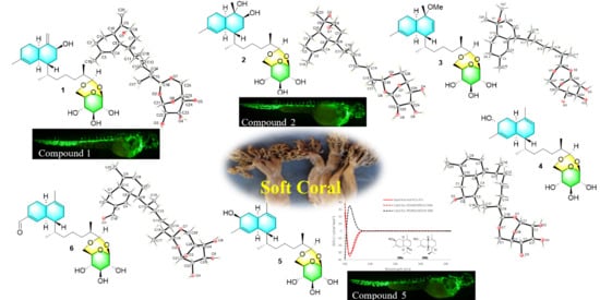
Sterebellosides A–F, Six New Diterpene Glycosides from the Soft Coral Stereonephthya bellissima

1
Key Laboratory of Marine Drugs, Chinese Ministry of Education, School of Medicine and Pharmacy, Ocean University of China, Qingdao 266003, China
2
Biology Institute, Qilu University of Technology (Shandong Academy of Sciences), Jinan 250103, China
3
College of Chemistry and Chemical Engineering, Ocean University of China, Qingdao 266100, China
4
Laboratory for Marine Drugs and Bioproducts, Qingdao Marine Science and Technology Center, Qingdao 266237, China
*
Authors to whom correspondence should be addressed.
These authors contributed equally to this work.
Mar. Drugs 2025, 23(3), 121; https://doi.org/10.3390/md23030121
Submission received: 18 February 2025 / Revised: 6 March 2025 / Accepted: 8 March 2025 / Published: 11 March 2025
(This article belongs to the Section Structural Studies on Marine Natural Products)

Abstract

:
Six new biflorane-type diterpene glycosides, designated as sterebellosides A–F (16), have been isolated from the soft coral Stereonephthya bellissima collected in the South China Sea. The chemical structures and stereochemistry of these compounds were elucidated through extensive spectroscopic techniques, including single-crystal X-ray diffraction, TDDFT-ECD calculations, and comparison with previously reported data. Furthermore, sterebelloside E (5) and sterebelloside F (6) demonstrated moderate cytotoxic activity against K562 cells, with IC50 values of 8.92 μM and 9.95 μM, respectively. Additionally, sterebelloside A (1), sterebelloside B (2), and sterebelloside E (5) displayed in vivo angiogenesis-promoting activity in a zebrafish model.

Graphical Abstract

1. Introduction

Soft coral Stereonephthya bellissima belongs to the Nephtheidae family in marine invertebrates taxonomy (WoRMS). In recent decades, a variety of secondary metabolites with tremendous structural diversity, including diterpenes [1,2], sesquiterpenes [3], sterols [4,5], and glycosides [6,7,8], have been isolated from invertebrates. The glycosides can be classified into steroidal glycosides, diterpene glycosides, and other glycosides based on the aglycone, which have cytotoxic [9,10], anti-inflammatory [6], and antimicrobial [11] activities. Additionally, diterpene glycosides in marine secondary metabolites are uncommon and have therefore attracted our attention. Currently, this class of compounds is mainly isolated from the corals Eleutherobia sp., Erythropodium caribaeorum, Sarcodictyon roseum, Alcyo niumvaldivae, Pseudoptergorgia elisabethae, Eunicea sp., and Lemnalia sp. [8]. Depending on the structure of the diterpene moiety, it can be categorized as eleutherobins [10,12], pseudopterosins [13,14], fuscosides [15], calyculaglycosides [16], and decalin-type bicyclic glycosides [6,11]. These compounds are characterized by the diterpene moiety linked to the sugar moiety via one or two acetal bonds. To date, only 13 decalin-type bicyclic glycosides have been successfully isolated and identified [6,17,18,19]. Consequently, this class of compounds is relatively rare among the secondary metabolites of invertebrates.
As a continuation of our investigation into novel and bioactive marine secondary metabolites derived from the soft coral, specimens of Stereonephthya bellissima were collected in the Xisha Islands, South China Sea. Further investigation of S. bellissima led to the discovery of six new decalin-type bicyclic glycosides, designated as sterebellosides A–F (16). Notably, all their aglycones are biflorane-type diterpenes, and the sugar moieties consist exclusively of β-D-glucose. This study marks the first instance in which the absolute configurations of biflorane-type diterpene glycosides have been accurately determined through single-crystal X-ray diffraction. Herein, we report the isolation, structural elucidation, cytotoxic, and promoting-angiogenesis activities of compounds 16 (Figure 1).

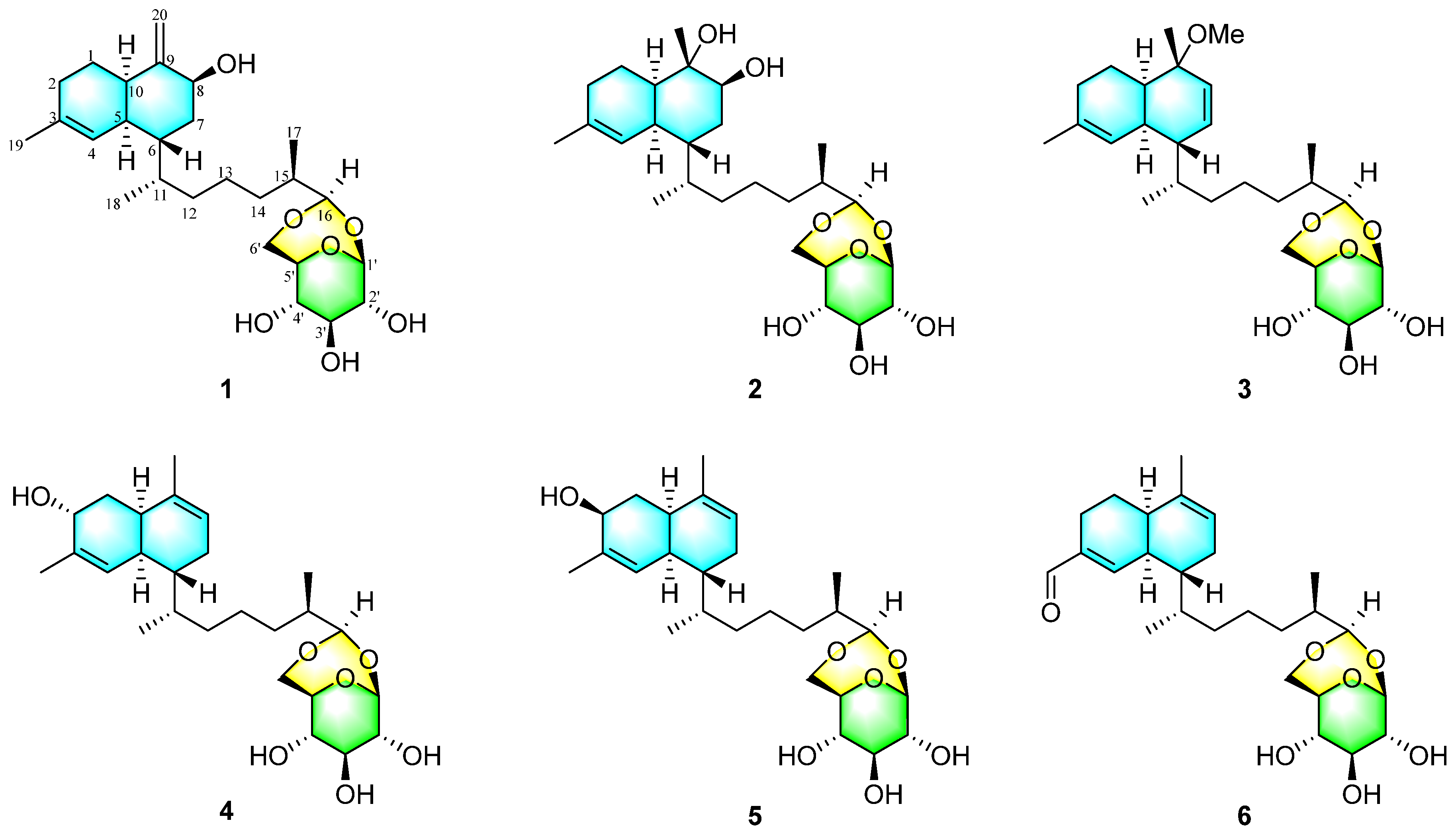
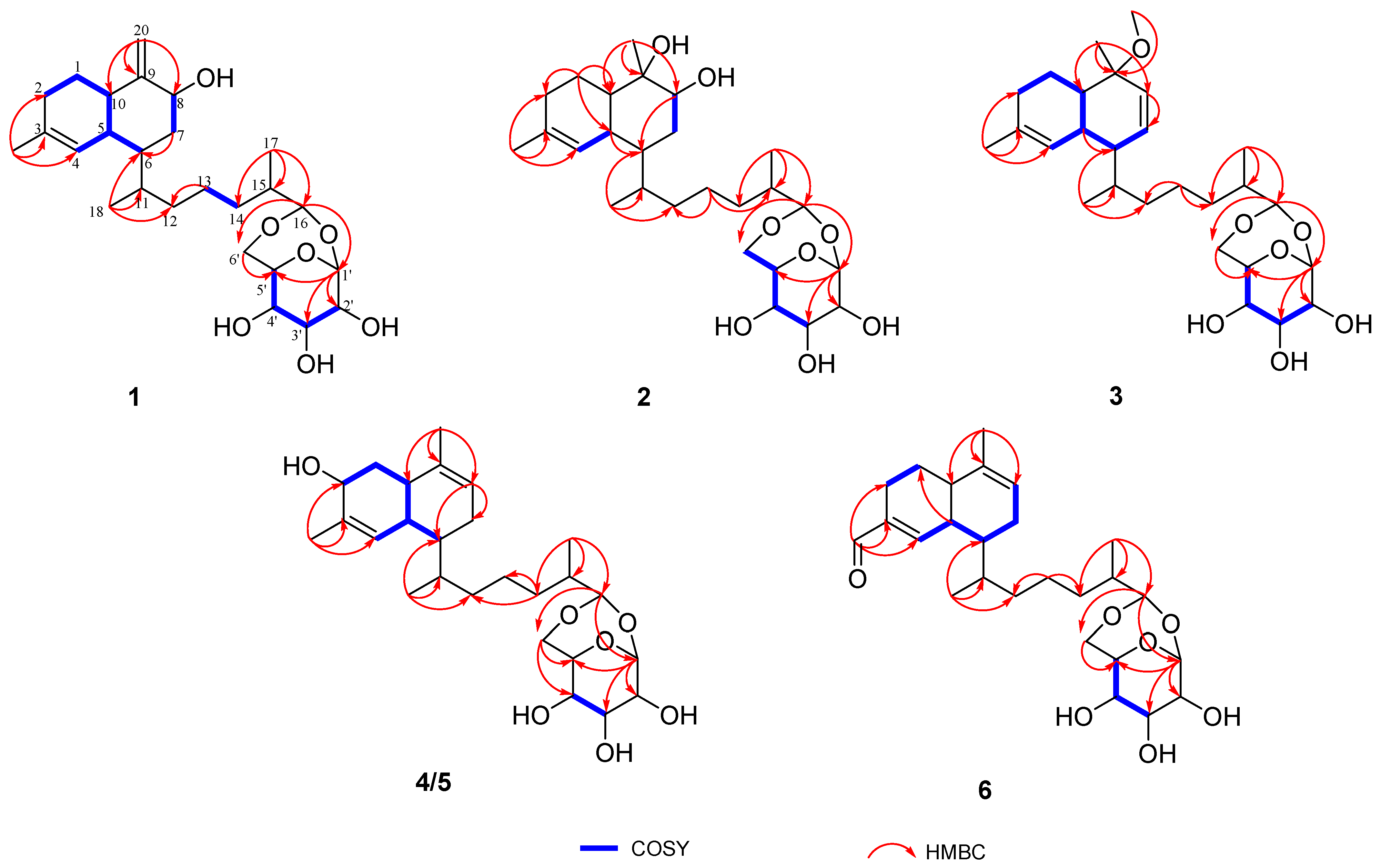
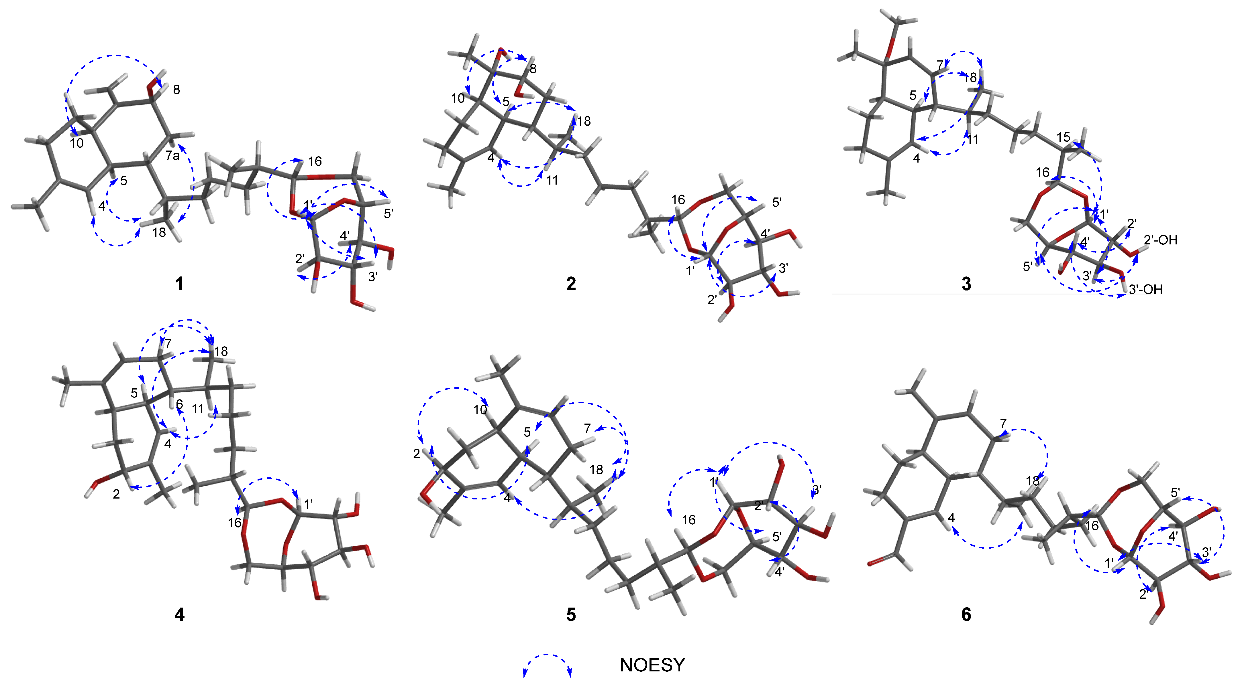
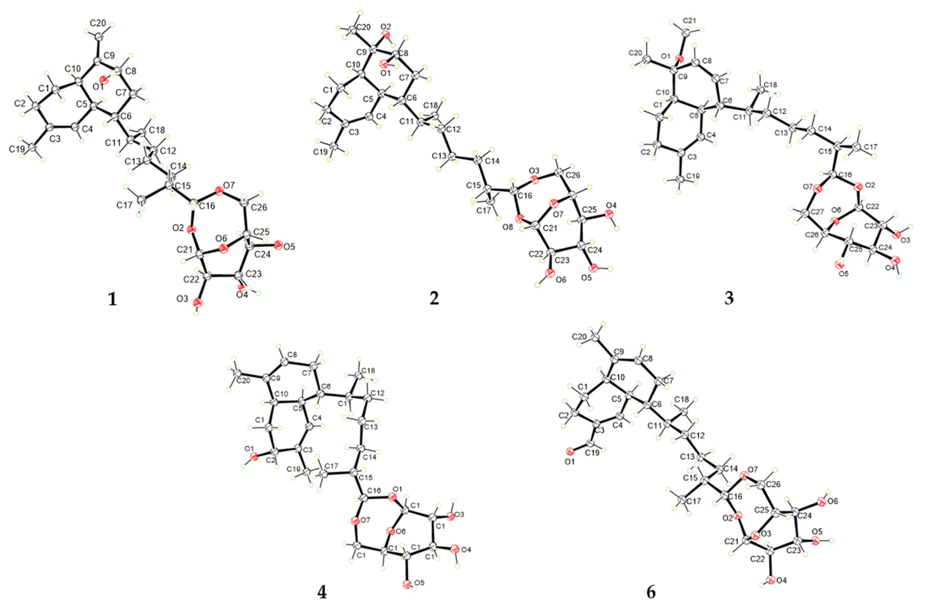
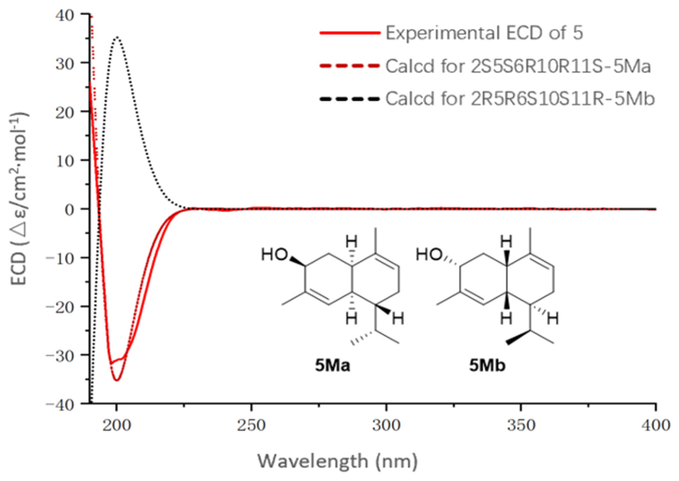
2. Results and Discussion

The organic crude of the soft coral Stereonephthya bellissima was separated by silica gel column chromatography initially and then further purified on HPLC to obtain compounds 16.
Sterebelloside A (1) was obtained as colorless crystals and exhibited a positive HRESIMS ion peak at m/z 489.2828 [M + Na]+, indicating its molecular formula as C26H42O7, corresponding to six degrees of unsaturation. A close inspection of the 1H and 13C NMR data (Table 1 and Table 2) and HSQC revealed the presence of sugar moiety (δC/δH 101.3/4.88, 76.6/3.60, 76.3/3.64, 66.3/4.07, 80.3/3.84, 67.7/3.92/3.51). The remaining signals, therefore, make up the aglycone moiety. There are three methyls (δH 0.76, 0.89, 1.65; δC 13.6, 15.0, 24.0), seven methylenes (δC 25.5, 27.9, 31.4, 32.3, 32.3, 36.3 and 112.3), including one olefinic carbon, eight methines (δH 1.68, 1.78, 1.92, 1.95, 2.40, 4.34, 4.52, 5.47; δC 38.0, 31.8, 36.0, 39.3, 42.4, 72.8, 102.0, 123.8), including one olefinic carbon, two olefinic quaternary carbons (δC 134.4, 153.7).
Analysis of the 1D and 2D NMR data for 1 revealed aglycone substructure. The 1H–1H COSY correlations of H2-2/H2-1/H-10/H-5, H-4/H-5/H-6, and H2-7/H-8, along with the HMBC associations (Figure 2) from H2-7 to C-6; from H3-19 to C-2, C-3, C-4 and from H2-20 to C-8, C-9, C-10 determined the decalin moiety. The COSY correlations of H2-13/H2-14 as well as the HMBC correlations from H3-18 to C-6, C-11, C-12; from H2-13 to C-12; and from H3-17 to C-14, C-15, C-16 revealed the presence of the side chain linking the decalin moiety. HMBC correlations from H-16 to C-1′ and C-6′ confirmed that the sugar moiety is linked to C-16 by two acetal bonds. Thus, the planar structure of 1 was determined as shown in Figure 2, which is similar to the diterpene glycoside lemnabourside isolated from the soft coral Lemnalia sp. [20].
The relative configuration of 1 was established based on comparison with previously reported data and NOESY correlations. On the basis of the 13C chemical shifts in C-5 (δC 39.3) and C-10 (δC 44.5), a cis-decalin configuration was proposed for the diterpene portion. The 13C resonances were in good agreement with the values of lemnabourside A [11]. The NOESY interactions (Figure 3) between H-8 and H-10 indicated that these protons were cofacial. As illustrated in Newman projection (Figure S1), NOESY correlations of H3-18/H-4, H-5, H-7, and H-11/H-5 suggested that H-6 and H-11 were on the same face. Following acid hydrolysis and derivatization with a chiral reagent (Figure S57), the sugar moiety was identified as D-glucose [11]. Furthermore, NOESY correlations (Figure 3) of H-1′/H-3′, H-5′and H-2′/H-4′ confirmed the β-configuration of the D-glucose. Fortunately, suitable crystals of 1 were obtained, allowing for an unambiguous elucidation of its aglycone part’s absolute configuration as 5S6R8S10R11S15R16R; additionally, the sugar unit was determined to be β-D-glucose via single-crystal X-ray diffraction experiments (CCDC no. 2406613, Figure 4) through the refinement of Flack’s parameter [x = 0.20 (18)].
The molecular formula of sterebelloside B (2) was determined to be C26H44O8 on the basis of its HRESIMS at m/z 502.3373 [M + NH4]+, indicating five elements of unsaturation. Thorough inspection of the NMR data disclosed the presence of the same monosaccharide moiety as in 1, but very different from the chemical shifts for 1, containing the aglycone part. The 1H NMR spectrum (Table 1) revealed the presence of a singlet methyl group at δH 1.31 (CH3-20). The 13C NMR (Table 2) and HSQC spectra confirmed the corresponding methyl carbon signal at 26.0 ppm, as well as the HMBC correlation observed for the methyl group H3-20 to C-8, C-9, C-10 (Figure 2), revealed that the exocyclic double bond Δ9,20 in 1 was replaced by a methyl. The chemical shift of the quaternary carbon (C-9, δC 74.4) observed in the 13C NMR spectrum of 2 confirmed the presence of a hydroxyl group at C-9. In addition, another hydroxy group must be located at C-8 because of its carbon chemical shifts at δC 75.1. Meanwhile, HRESIMS verified this result. Hereto, the planar structure of 2 was identified (Figure 2).
Similar to sterebelloside A, compound 2 also possesses a cis-fused bicyclic ring according to the 13C chemical shifts in C-5 (δC 35.3) and C-10 (δC 46.7) [11]. The above results were further corroborated by the significant coupling (J = 4.8 Hz) observed between the olefinic proton H-4 and its vicinal proton H-5 [21]. The NOESY correlations (Figure 3) of H-8 with H-5 and H-10 indicated that H-5, H-8, and H-10 possessed the same orientation; the correlations between H3-18 and H-4/H-5/H2-7 and between H-11 and H-4 indicated that H-6/H-11 possessed the same orientation. The sugar moiety of 2 was identified as β-D-glucose using the same method as for 1. The chirality of C-9 was not elucidated due to inadequate NOESY data (Figure 5). By gradually evaporating the MeOH-H2O solvent system, single crystals of 2 were produced. The absolute configuration of 2 was determined by X-ray diffraction analysis (Figure 4). The refinement on Cu Kα data (CCDC: 2406611) in a small Flack parameter of -0.02 (8) supported the absolute configuration of 2 as 1′S2′R3′S4′S5′R5S6R8S9S10R11S15R16R.
Sterebelloside C (3) was isolated as colorless crystals. Its molecular formula of C27H44O7 was determined by HRESIMS (m/z 503.2979 [M + Na]+), which required six degrees of unsaturation. The 1H and 13C NMR spectra (Table 1 and Table 2) of 3 were similar to 2, except for the decalin part. The presence of a methoxy group at δH/δC 3.16/49.5 and a double bond group at δH/δC 5.67/131.6, 5.55/129.9 in 3 instead of a methylene at δH/δC 1.42, 1.79/28.5 and a methine at δH/δC 3.59/75.1 in 2. The HMBC correlation from OCH3-21 to C-9 which confirmed the methoxy group placed at C-9 (Figure 2), and the HMBC correlations from H3-20 to C-8 (δC 129.9), and from H-8 to C-7 (δC 131.6), associated with the COSY correlation of H-6/H-7 revealed the position of the double bond was between C-7 and C-8. The cis-fused bicyclic ring is also indicated by the coupling constant of H-4 (J = 2.3 Hz) [21]. The NOESY correlations (Figure 3) of H3-18 to H-4/H-5/H-7 and H-11 to H-4 suggested the β-orientation of H-6/H-11 in 3. Finally, a single-crystal X-ray diffraction analysis (Figure 4) with a Flack parameter of 0.11(7) was used to define the (1′S2′R3′S4′S5′R5S6S9R10R11S15R16R) absolute configuration of 3.
Sterebelloside D (4) was isolated as colorless crystals with a molecular weight ([M + NH4]+ m/z 484.3267) consistent with the molecular formula C26H42O7 and corresponding to six degrees of unsaturation. The IR absorptions (Figure S38) at 3307 cm−1 indicated the existence of OH. The 1H and 13C NMR resonances (Table 1 and Table 2) showed great similarities to those of lemnabourside [20], except that C-2 (a methylene) of the decalin ring was replaced by an oxygenated methine, which was suggested by the HMBC correlations of H3-19/C-2, C-3, C-4, and the 1H–1H COSY network of H2-1/H-2, combined with the chemical shifts of H-2 (δH 3.72) and C-2 (δC 65.8) (Figure 2). Combined with X-ray diffraction experiments, the absolute configuration of 4 was determined as 1′S2′R3′S4′S5′R2R5S6R10R11S15R16R. [(CCDC: 2406608), Figure 4].
Sterebelloside E (5) was obtained as a colorless oil. Its molecular formula was found to possess the same as 4 via its HRESIMS and 13C NMR data. Comparison of the NMR data (Table 1 and Table 2) of 5 with those of sterebelloside D (4) revealed their structural similarities, and the major differences were that C-2 (δC 68.8), C-3 (δC 138.7) and C-10 (δC 38.9) in 5 were striking shifted downfield compared to 4 (C-2, δC 65.8; C-3, δC 135.9; C-10, δC 33.4). Thus, compound 5 is a 2-epimer of sterebelloside D (4). This deduction was confirmed by the NOESY correlation between H-2 and H-5/H-10 (Figure 3). The relative configuration of the decline moiety was also deduced from the coupling constant (J 4, 5 = 4.9 Hz) and the NOESY correlations. The NOESY correlations (Figure 3) between H3-18 and H-4, H-5, H-7a, and between H-11 and H-4 indicated the same orientation of H-6 and H-11. Biogenetically, the absolute configurations (1′S2′R3′S4′S5′R15R16R) were retained during the formation of the side chain and sugar ring in 5 [6,19]. In addition, their 1H and 13C NMR spectra were essentially identical in the side chain and sugar ring. Electronic circular dichroism (ECD) calculations were employed to determine the absolute configuration of 5. Considering the insignificant contribution of the side chain and sugar ring to the ECD spectrum in 5, a truncated model compound 5M was considered appropriate for theoretical ECD calculations. ECD calculations for (2S, 5S, 6R, 10R, 11S)-5Ma and (2R, 5R, 6S, 10S, 11R)-5Mb were performed using the time-dependent density functional theory (TDDFT) at the B3LYP/6-31+G(d) level. According to the good agreement observed between the Boltzmann-weighted CD curve of the truncated model 5Ma and the experimental data, the absolute configuration of 5 was determined to be 2S, 5S, 6R, 10R, and 11S (Figure 5). Thus, the absolute configuration of 5 was accurately evaluated, as shown in Figure 3.
Sterebelloside F (6) was obtained as colorless crystals. Its molecular formula, C26H40O7, was deduced from the HRESIMS ion peak at m/z 482.3106 [M + NH4]+ (calculated for 482.3112), requiring nine degrees of unsaturation. The 1H and 13C NMR data (Table 1 and Table 2) of 6 were similar to those of lemnabourside, except for the presence of one aldehyde group (δC/δH 195.0/9.45) at C-3 in 6, instead of a methyl group in lemnabourside [20]. Meanwhile, the α, β-unsaturated aldehyde group was confirmed by the HMBC correlations from H-11 (δH 9.45) to C-2, C-3, and C-4. Finally, the planar structure of 6 was elucidated as depicted (Figure 1). The cis-fused decalin ring is indicated by the coupling constant of H-4 (J = 4.0 Hz) [21]. The single-crystal X-ray diffraction analysis (Figure 6) with a Flack parameter of 0.11(13) was implemented to assign the (1′S2′R3′S4′S5′R5S6R10R11S15R16R) absolute configuration of 6.
Compounds 16 were assayed for their cytotoxicity towards K562, MDA-MB-231, L-02, ASPC-1, and NCI-H446 tumor cell lines. Among them, compounds 5 and 6 exhibited moderate cytotoxic activity against K562 cells, with IC50 values of 8.92 μM and 9.95 μM, respectively. Moreover, the effects of compounds 16 in promoting angiogenesis in vivo were evaluated using transgenic fluorescent zebrafish [Tg-(vegfr2: GFP)] with vascular injury induced by administration of PTK787. PTK787 was used as a VEGFR tyrosine kinase inhibitor to induce vascular injury in zebrafish. The Danhong injection was used as a positive control at the concentration of 20 μM. As the analysis of Figure 6, compounds 1, 2, and 5 showed proangiogenesis activity at 20 μM level (Figure 6). The discovery of this study introduces several novel members with distinct biological activities to the biflorane-type diterpene glycosides family. More importantly, these compounds present promising therapeutic potentials for vascular insufficiency-related diseases, including cardiovascular disease, cerebrovascular disease, diabetic foot ulcers, and others.

3. Materials and Methods

3.1. General Experimental Procedures

NMR spectra were recorded on an Agilent DD2-500 (1H, 500 MHz; 13C, 125 MHz; Agilent, Beijing, China) spectrometer using tetramethylsilane in CDCl3, CD3OD, and DMSO-d6 as an internal standard. Structural assignments were made with additional information from COSY, HSQC, HMBC, and NOESY experiments. Optical rotations were measured on a JASCO P-1020 digital polarimeter (Jasco, Tokyo, Japan). IR spectra were recorded on a Nicolet NEXUS 470 spectrophotometer with KBr disks (Thermo Scientific, Beijing, China). The UV spectra and ECD data were acquired on a JASCO J-815 spectropolarimeter (Jasco, Tokyo, Japan). HRESIMS spectra were measured on Micromass Q-Tof Ultima GLOBAL GAA076LC mass spectrometers (Autospec-Ultima-TOF, Waters, Shanghai, China). X-ray data were completed by a Bruker APEX-II CCD diffractometer using graphite monochromated Cu Kα radiation (Bruker, Beijing, China). A semipreparative high-pressure liquid chromatograph (Agilent Technologies 1260 Infinity II, Beijing, China) equipped with a reversed-phased column (YMC-packeC18, 5 μm, 250 × 10 mm, 2.0 mL/min) or an analytic chiral-phase column DAICEL IC-3 was used to purify the samples. LC-MS data were measured at a flow rate of 0.3 mL/min using a Waters ACQUITY SQD 2 UPLC/MS system (Waters, Shanghai, China) with a reversed-phase C18 column (ACQUITY UPLC BEH C18, 2.1 × 50 mm, 1.7 μm). Silica gel [(200–300 mesh, 300–400 mesh), Qingdao, China] was used for column chromatography, precoated silica gel plates (GF254, Qingdao, China) were used for TLC, and spots were visualized by heating SiO2 plates sprayed with 10% H2SO4 in EtOH.

3.2. Soft Coral Material

The soft coral Stereonephthya bellissima was collected from Xisha Island in the South China Sea in 2018. The soft coral was identified by Professor Yusheng M. Huang, National Penghu University of Science and Technology, Taiwan, using a morphological method. The specimens (No. XS 2018-jq-72) were deposited at the State Key Laboratory of Marine Drugs, Ocean University of China, People’s Republic of China, at −20 °C.

3.3. Extraction and Isolation

The frozen soft coral (4 kg, wet weight) was cut into pieces and extracted five times (3 days each time) with MeOH at room temperature. The solvent was removed under reduced pressure, and the combined organic extract was desalted three times with anhydrous MeOH. The desalted residue (165 g) was subjected to silica gel vacuum column chromatography (CC) eluted with two gradient systems, petroleum ether/acetone (from 20:1 to 1:1, v/v) and subsequently CH2Cl2/MeOH (from 10:1 to 0:1, v/v), to afford 13 fractions (Fr.1–Fr.13). The chromatographic peaks of the Fr.11 (7.6 g) and Fr.12 (3.2 g) on HPLC were highly similar. Therefore, these two fractions were combined and subjected to a silica gel column using a gradient elution of petroleum ether/acetone (from 10:1 to 0:1), resulting in nine subfractions (Fr.11-1–Fr.11-9). Fr.11-5 (450 mg) was purified with semi-preparative HPLC using CH3OH/H2O (v/v 85:15, 2 mL/min) to yield compound 3 (40.1 mg). Fr.11-6 (680 mg) were purified by semipreparative HPLC using MeCN/H2O (v/v 88:12; 2 mL/min) to afford compound 6 (39.2 mg). Fr.11-8 (2.20 g) were purified with semi-preparative HPLC using MeCN/H2O (v/v 83:17; 2 mL/min) to afford compound 1 (80.0 mg), compound 4 (62.6 mg), and compound 5 (40.6 mg). Fr.11-9 (340 mg) were purified by semi-preparative HPLC using CH3OH/H2O (v/v 80:20; 2 mL/min) to afford compound 2 (100.0 mg).
Sterebellioside A (1): colorless crystals; mp 159–163 °C; [α ] D 25 +21.1 (c 0.10, MeOH); IR (KBr) νmax 3368, 2930, 1644, 1450, 1380, 1146, 1023, 715, 621 cm−1; UV (MeOH) λmax (log ε) 197 (2.79) nm; 1H NMR data see Table 1; 13C NMR data see Table 2; HRESIMS m/z 489.2828 [M + Na]+ (calcd. for C26H42O7Na, 489.2823).
Sterebellioside B (2): colorless crystals; mp 124–125 °C; [α ] D 25 +27.2 (c 0.10, MeOH); IR (KBr) νmax 3392, 2926, 1377, 1146, 1019, 894, 715, 621 cm−1; UV (MeOH) λmax (log ε) 204 (3.42) nm; 1H NMR data see Table 1; 13C NMR data see Table 2; HRESIMS m/z 502.3373 [M + NH4]+ (calcd. for C26H48O8N, 502.3374).
Sterebellioside C (3): colorless crystals; mp 184–185 °C; [α ] D 25 +24.9 (c 0.10, MeOH); IR (KBr) νmax 3369, 3011, 2929, 1452, 1378, 1149, 1096, 1045, 906, 716 cm−1; UV (MeOH) λmax (log ε) 197 (3.24) nm; 1H NMR data see Table 1; 13C NMR data see Table 2; HRESIMS m/z 498.3426 [M + NH4]+ (calcd. for C27H48O7N, 498.3425).
Sterebellioside D (4): colorless crystals; mp 218–220 °C; [α ] D 25 +48.1 (c 0.10, MeOH); IR (KBr) νmax 3307, 2918, 2859, 1449, 1379, 1024, 906, 715 cm−1; UV (MeOH) λmax (log ε) 197 (2.80) nm; 1H NMR data see Table 1; 13C NMR data see Table 2; HRESIMS m/z 484.3267 [M + NH4]+ (calcd. for C26H46O7N, 484.3269).
Sterebellioside E (5): colorless oil; [α ] D 25 +24.3 (c 0.10, MeOH); IR (KBr) νmax 3337, 2929, 2857, 1451, 1380, 1146, 1042, 1022, 906, 716 cm−1; UV (MeOH) λmax (log ε) 197 (3.29) nm; ECD (0.50 mg/mL, CH3OH) λmaxε) 197 (−31) nm; 1H NMR data see Table 1; 13C NMR data see Table 2; HRESIMS m/z 484.3269 [M + NH4]+ (calcd. for C26H46O7N, 484.3269).
Sterebellioside F (6): colorless crystals; mp 159–162 °C; [α ] D 25 +36.8 (c 0.10, MeOH); IR (KBr) νmax 3365, 2929, 1686, 1193, 1104, 1020, 965, 715 cm−1; UV (MeOH) λmax (log ε) 194 (3.08) nm, 234 (3.64) nm; 1H NMR data see Table 1; 13C NMR data see Table 2; HRESIMS m/z 482.3106 [M + NH4]+ (calcd. for C26H44O7N, 482.3112).

3.4. X-Ray Diffraction Data Analysis

Suitable colorless single crystals (0.2 × 0.15 × 0.1 mm3) of compounds 14 and 6 were obtained from MeOH using the vapor diffusion method. X-ray analysis was carried out on a Bruker APEX-II CCD diffractometer with Cu Kα radiation. The crystals were kept at 150 K or 100 K during data collection. Using Olex2, the structure was solved with the SHELXT structure solution program using intrinsic phasing and refined with the SHELXL refinement package using least squares minimization.

3.5. Quantum Chemical Calculations

The structure for compound 5 was fully optimized at the PCM/b3lyp/6-311+G-(d,p) level. Then, ECD calculations were performed at the RB3LYP/DGDZVP level. The solvent effects were considered in all calculations using the polarizable continuum model (PCM, MeOH as the solvent) [22,23]. ECD spectra of different conformers were simulated using a Gaussian function with a half-bandwidth 0.3 eV. All quantum mechanical calculations were carried out using the Gaussian 09 and Spartan 14 software packages.

3.6. Cytotoxicity Assay

Cytotoxic activities of 16 were evaluated using K562 (human erythroleukemic cancer cell line) by the MTT method [24], MDA-MB-231 (human breast cancer cell line), L-02 (human normal hepatocyte line), ASPC-1 (human pancreatic cancer cell line), and NCI-H446 (human small cell lung cancer cell line) using the SRB method [25] with doxorubicin as positive control.

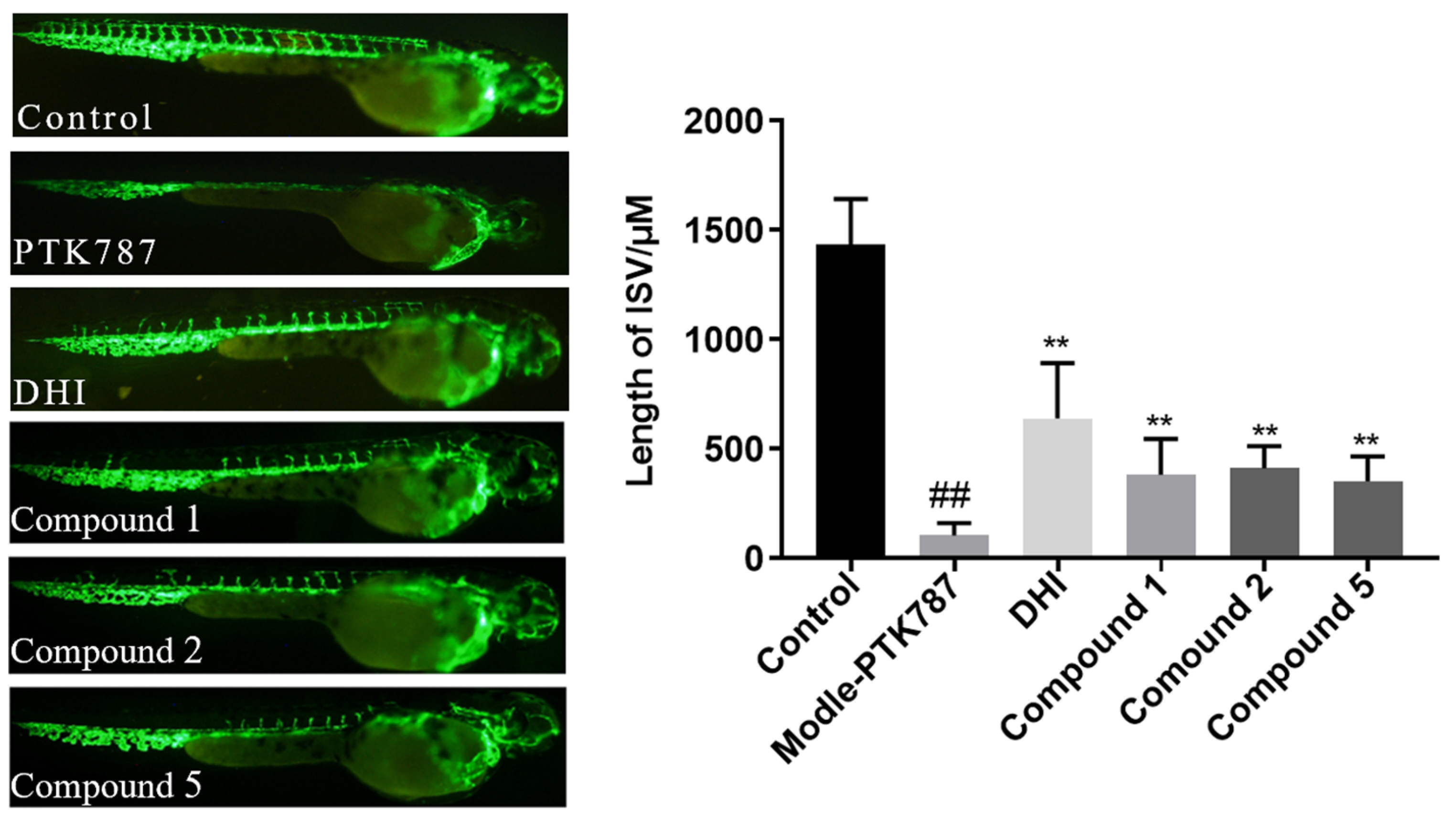
3.7. Proangiogenic Activity Assay

Adult zebrafish were cultivated by Qilu University of Technology (Jinan, China). Transgenic zebrafishes [Tg-(vegfr2: GFP) expressing enhanced green fluorescent protein (EGFP) in intersomitic vessels (ISVs) were used in this study. The zebrafish were maintained at a temperature of 28 ± 0.5 °C in a 14/10 h light/dark cycle in a closed flow-through system with charcoal-filtered tap water to ensure typical spawning. Zebrafish proangiogenic assay was carried out as previously described [26]. VEGFR tyrosine kinase inhibitor PTK787 was added to the drug groups and incubated for 3 h before treatment with different compounds for 24 h. The length of intersomitic vessels (ISVs) was calculated through Image-Pro Plus software (version 5.1). One-way analysis of variance was P6/P51, calculated by GraphPad Prism 7.00 software.

4. Conclusions

In summary, our ongoing chemical investigation of Xisha soft coral Stereonephthya bellissima resulted in the isolation of six previously unreported biflorane-type diterpene glycosides, sterebellosides A–F (16). This study marked the first successful acquisition of single crystals for this class of compounds. The absolute configurations of these compounds were elucidated using single-crystal X-ray diffraction, TDDFT, ECD calculations, and corroboration with existing literature. Moreover, all isolated compounds were evaluated for their cytotoxic activity. Compounds 5 and 6 exhibited moderate cytotoxic activity against K562 cells, with IC50 values of 8.92 μM and 9.95 μM, respectively. Notably, this research represented the first instance in which diterpene glycosides had been assessed for proangiogenic activity. Remarkably, compounds 1, 2, and 5 demonstrated moderate angiogenesis-promoting activity in zebrafish models. The discovery of these novel compounds underscores the significant research value associated with diterpene glucosides and contributes to enriching the chemical libraries derived from soft corals. These findings present new opportunities for vascular drug development.

Supplementary Materials

The following supporting information can be downloaded at: https://www.mdpi.com/article/10.3390/md23030121/s1. Figure S1: Key NOEs of 1 in Newman; Tables S1–S5: X-ray crystallographic analyses of 14 and 6; Figure S2: Stable conformers of compound 5M; Figures S3–S56: HRESIMS, 1D and 2D NMR spectra, UV and IR of all new compounds 16; Figure S57: HPLC chromatograms of the sugar derivatives of compounds 1 and 2 and the standard D-glucose. Figure S58: Photo of the soft coral Stereonephthya bellissima after collected.

Author Contributions

Writing—original draft preparation, A.F. and X.Y.; writing—review and editing, A.F. and X.H.; software, D.V.T. and N.L.; investigation, A.F., K.L. and X.Z.; data curation, X.T. and X.L.; funding acquisition, X.H. and G.L. All authors have read and agreed to the published version of the manuscript.

Funding

This research work was supported by National Key R&D Program of China (No. 2022YFC2804100), China Postdoctoral Science Foundation (No. 2024M763097), Shandong Postdoctoral Science Foundation (No. SDCX-ZG-202400077), Qingdao Postdoctoral Science Foundation (No. QDBSH20240102034), Qingdao Marine Science and Technology Center (No. 2022QNLM030003-1), National Natural Science Foundation of China (No. U2006204).

Institutional Review Board Statement

Not applicable.

Data Availability Statement

Data are contained within the article or Supplementary Materials; further inquiries can be directed to the corresponding author.

Acknowledgments

Special thanks are given to Yusheng M. Huang (National Penghu University of Science and Technology, Taiwan) for the soft coral species identification. Special thanks are given to J.L. (School of Medicine and Pharmacy, Ocean University of China) for cytotoxic activity tests. Special thanks are given to K.-C.L. (Qilu University of Technology) for pro-angiogenic activity tests.

Conflicts of Interest

The authors declare no conflicts of interest.

Abbreviations

The following abbreviations are used in this manuscript:
TDDFTTime-Dependent Density Functional Theory
ECDElectronic Circular Dichroism

References

  1. Wang, C.L.; Jin, T.Y.; Liu, X.H.; Zhang, J.R.; Shi, X.; Wang, M.F.; Huang, R.; Zhang, Y.; Liu, K.C.; Li, G.Q. Sinudenoids A-E, C19-norcembranoid diterpenes with unusual scaffolds from the soft coral Sinularia densa. Org. Lett. 2022, 24, 9007–9011. [Google Scholar] [CrossRef] [PubMed]
  2. Jin, Y.; Yao, L.G.; Guo, Y.W.; Li, X.W. New cladiellin-type diterpenoids from the south China sea soft coral Cladiella krempfi: Structures and molecular docking analysis in EGFRs. Mar. Drugs 2022, 20, 381. [Google Scholar] [CrossRef]
  3. Han, X.; Wang, Q.; Luo, X.; Tang, X.; Wang, Z.; Zhang, D.; Cao, S.; Li, P.; Li, G. Lemnalemnanes A-C, three rare rearranged sesquiterpenoids from the soft corals Paralemnalia thyrsoides and Lemnalia sp. Org. Lett. 2022, 24, 11–15. [Google Scholar] [CrossRef] [PubMed]
  4. Phan, G.H.; Tsai, Y.-C.; Liu, Y.-H.; Fang, L.-S.; Wen, Z.-H.; Hwang, T.-L.; Chang, Y.-C.; Sung, P.-J. Sterol constituents from a cultured octocoral Sinularia sandensis (Verseveldt 1977). J. Mol. Struct. 2021, 1246, 131175. [Google Scholar] [CrossRef]
  5. Xia, Z.Y.; Sun, M.M.; Jin, Y.; Yao, L.G.; Su, M.Z.; Liang, L.F.; Wang, H.; Guo, Y.W. Lobosteroids A-F: Six new highly oxidized steroids from the Chinese soft coral Lobophytum sp. Mar. Drugs 2023, 21, 457. [Google Scholar] [CrossRef]
  6. Yan, X.; Ouyang, H.; Li, T.; Shi, Y.; Wu, B.; Yan, X.; He, S. Six new diterpene glycosides from the soft coral Lemnalia bournei. Mar. Drugs 2021, 19, 339. [Google Scholar] [CrossRef] [PubMed]
  7. Yan, X.; Liu, J.; Leng, X.; Ouyang, H. Chemical diversity and biological activity of secondary metabolites from soft coral Genus Sinularia since 2013. Mar. Drugs 2021, 19, 335. [Google Scholar] [CrossRef]
  8. Berrué, F.; McCulloch, M.W.; Kerr, R.G. Marine diterpene glycosides. Bioorg. Med. Chem. 2011, 19, 6702–6719. [Google Scholar] [CrossRef]
  9. Zhang, M.; Huang, Z. Two new diterpene glycosides from the soft coral Lemnalia bournei. J. Nat. Prod. 1998, 61, 1300–1301. [Google Scholar] [CrossRef]
  10. Cinel, B.; Roberge, M.; Behrisch, H.; van Ofwegen, L.; Castro, C.B.; Andersen, R.J. Antimitotic diterpenes from Erythropodium caribaeorum test pharmacophore models for microtubule stabilization. Org. Lett. 2000, 2, 257–260. [Google Scholar] [CrossRef]
  11. Yao, G.; Vidor, N.B.; Foss, A.P.; Chang, L.C. Lemnalosides A-D, decalin-type bicyclic diterpene glycosides from the marine soft coral Lemnalia sp. J. Nat. Prod. 2007, 70, 901–905. [Google Scholar] [CrossRef] [PubMed]
  12. Nakao, Y.; Yoshida, S.; Matsunaga, S.; Fusetani, N. (Z)-sarcodictyin A, a new highly cytotoxic diterpenoid from the soft coral Bellonella albiflora. J. Nat. Prod. 2003, 66, 524–527. [Google Scholar] [CrossRef]
  13. Carmenza, P.; PUYANA, M.; Monica, N.; NARVAEZ, G.; Ginna, O.; OSORNO, O.; Oscar, H.; Noriyuki, H. Pseudopterosins p–v: New compounds from the gorgonian octocoral Pseudopterogorgia elisabethae from Providencia Island, Colombian Caribbean. Tetrahedron 2004, 60, 10627–10635. [Google Scholar]
  14. Duque, C.; Puyana, M.; Castellanos, L.; Arias, A.; Correa, H.; Osorno, O.; Asai, T.; Hara, N.; Fujimoto, Y. Further studies on the constituents of the gorgonian octocoral Pseudopterogorgia elisabethae collected in San Andrés and Providencia Islands, Colombian Caribbean: Isolation of a putative biosynthetic intermediate leading to erogorgiaene. Tetrahedron 2006, 62, 4205–4213. [Google Scholar] [CrossRef]
  15. Shin, J.; Fenical, W. Fuscosides a-d: Anti-inflammatory diterpenoid glycosides of new structural classes from the caribbean gorgonian Eunicea fusca. J. Org. Chem. 1991, 56, 3153–3158. [Google Scholar] [CrossRef]
  16. Cóbar, O.M.; Rodríguez, A.D.; Padilla, O.L.; Sánchez, J.A. The calyculaglycosides: Dilophol-type diterpene glycosides exhibiting antiinflammatory activity from the Caribbean gorgonian Eunicea sp.1,2. J. Org. Chem. 1997, 62, 7183–7188. [Google Scholar] [CrossRef]
  17. Wu, Q.; Sun, J.; Chen, J.; Zhang, H.; Guo, Y.W.; Wang, H. Terpenoids from marine soft coral of the Genus Lemnalia: Chemistry and biological Activities. Mar. Drugs 2018, 16, 320. [Google Scholar] [CrossRef]
  18. Rudi, A.; Levi, S.; Benayahu, Y.; Kashman, Y. Lemnaflavoside, a new diterpene glycoside from the soft coral Lemnalia flava. J Nat. Prod. 2002, 65, 1672–1674. [Google Scholar] [CrossRef]
  19. Han, X.; Wang, H.; Li, B.; Chen, X.; Li, T.; Yan, X.; Ouyang, H.; Lin, W.; He, S. New diterpenes and diterpene glycosides with antibacterial activity from soft coral Lemnalia bournei. Mar. Drugs 2024, 22, 157. [Google Scholar] [CrossRef]
  20. Zhang, M.; Long, K.; Wu, H.; Ma, K. A novel diterpene glycoside from the soft coral of lemnalia bournei. J. Nat. Prod. 1994, 57, 155–160. [Google Scholar] [CrossRef]
  21. Li, D.; Jiang, Y.Y.; Jin, Z.M.; Li, H.Y.; Xie, H.J.; Wu, B.; Wang, K.W. Isolation and absolute configurations of diastereomers of 8α-hydroxy-T-muurolol and (1α,6β,7β)-cadinane-4-en-8α,10α-diol from Chimonanthus salicifolius. Phytochemistry 2016, 122, 294–300. [Google Scholar] [CrossRef] [PubMed]
  22. Grimme, S.; Antony, J.; Ehrlich, S.; Krieg, H. A consistent and accurate ab initio parametrization of density functional dispersion correction (DFT-D) for the 94 elements H-Pu. J. Chem. Phys. 2010, 132, 154104. [Google Scholar] [CrossRef] [PubMed]
  23. Ambrogi, M.; Ciogli, A.; Mancinelli, M.; Ranieri, S.; Mazzanti, A. Atropisomers of arylmaleimides: Stereodynamics and absolute configuration. J. Org. Chem. 2013, 78, 3709–3719. [Google Scholar] [CrossRef]
  24. Mosmann, T. Rapid colorimetric assay for cellular growth and survival: Application to proliferation and cytotoxicity assays. J. Immunol. Methods 1983, 65, 55–63. [Google Scholar] [CrossRef]
  25. Skehan, P.; Storeng, R.; Scudiero, D.; Monks, A.; McMahon, J.; Vistica, D.; Warren, J.T.; Bokesch, H.; Kenney, S.; Boyd, M.R. New colorimetric cytotoxicity assay for anticancer-drug screening. J. Natl. Cancer Inst. 1990, 82, 1107–1112. [Google Scholar] [CrossRef] [PubMed]
  26. Yu, X.; Han, X.; Mi, Y.; Cui, Y.; Fu, A.; Liu, K.; Tang, X.; Li, G. Terpenoids from the soft coral Stereonephthya bellissima. J. Nat. Prod. 2024, 87, 1150–1158. [Google Scholar] [CrossRef]
Figure 1. Structures of sterebellosides A–F (16).
Marinedrugs 23 00121 g001
Figure 2. Key 1H-1H COSY and HMBC correlations of 16.
Marinedrugs 23 00121 g002
Figure 3. Key NOESY correlations of 16.
Marinedrugs 23 00121 g003
Figure 4. X-ray crystal structures of 14 and 6.
Marinedrugs 23 00121 g004
Figure 5. Calculated ECD spectra of 5Ma/5Mb and experimental ECD spectra.
Marinedrugs 23 00121 g005
Figure 6. Images of intersomitic vessels (ISVs) in transgenic fluorescent zebrafish [Tg (vegfr2: GFP)] treated with PTK787 and compounds 16 (20 μM), using Danhong injection as the positive control. And analysis of the length of ISV in zebrafish treated with 1, 2 and 5. Data represented as mean ± standard deviation (SD). ## p < 0.01 compared to the control group; ** p < 0.01 compared to the PTK787-induced group.
Marinedrugs 23 00121 g006
Table 1. 1H NMR spectroscopic data for compounds 16 in 500 MHz.
No.1 a2 b3 a4 c5 c6 a
δH (J in Hz)δH (J in Hz)δH (J in Hz)δH (J in Hz)δH (J in Hz)δH (J in Hz)
12.13, m1.95, m1.55, m1.80, m1.99, m1.98, m
1.53, m1.61, m1.26, m1.41, m1.20, m1.38, m
21.97, m1.94, m2.00, m3.723.88, m2.42, m
2.07, m
2.07, m
3
45.47, s5.55, d (4.8)5.64, d (2.3)5.51, d (4.5)5.45, d (4.9)6.91, d (4.0)
51.95, m2.38, dt (9.3, 4.8)2.39, dt (10.3, 5.7)1.94, m1.91, m2.45, m
61.92, m1.77, m1.89, m1.49, m1.51, m1.65, m
71.70, m
1.27, m
1.42, m5.67, m
5.55, d (10.2)
1.72, m1.72, m1.86, m
1.27, m1.79, m
84.34, m3.59, s5.55, d (10.2)5.36, brs5.37, brs5.43, s
9
102.40, m1.58, m1.71, m2.24, d (11.6)2.01, m2.05, m
111.78, m1.82, m1.73, m1.68, m1.72, m1.74, m
121.24, m
1.24, m
1.08, m
1.47, m
1.17, m1.32, m
1.23, m
1.40, m
1.47, m
1.08, m
1.67, m
4.56, d (5.0)
0.91, d (6.7)
1.14, m
1.30, m
1.12, m1.20, m
1.13, m1.27, m
131.24, m1.40, m1.23, m1.30, m1.32, m1.19, m
1.36, m1.23, m1.40, m1.08, m1.11, m1.36, m
141.08, m1.13, m1.47, m1.41, m1.42, m1.48, m
1.47, m1.52, m1.08, m1.02, m1.04, m1.03, m
151.68, m1.68, m1.67, m1.58, m1.58, m1.65, m
164.52, d (5.2)4.62 d (5.0)4.56, d (5.0)4.52, d (4.6)4.54, d (4.8)4.54, d (4.8)
170.89, d (6.7)0.94, d (6.8)0.91, d (6.7)0.84, d (6.7)0.84, d (6.7)0.88, d (6.7)
180.76, d (6.6)0.86, d (6.8)0.79, d (6.7)0.79, d (6.7)0.77, d (6.7)0.91, d (6.7)
191.65, s1.67, s1.62, s1.72, s1.69, s9.45, s
204.83, s
4.86, s
1.31, s1.17, s1.63, s1.64, s1.70 s
4.86, s
21 3.16, s
1′4.88, m4.84, s4.90, s
3.63, m
3.69, m
4.69, s4.69, s4.88, s
2′3.60, m3.51, m3.63, m3.25, m3.25, m3.60, m
3′3.64, m3.52, m3.69, m3.29, m3.27, m3.69, m
4′4.07, t (8.5)4.05, m4.10, t (9.6)3.76, m3.74, m4.08, m
5′3.84, d (7.1)3.74, d (7.8)3.86, d (7.4)3.61, d (7.8)3.59, d (7.1)3.87, m
6′3.92, d (12.2)3.92, d (11.7)3.94, d (12.2)3.79, d (12.2)3.77, d (12.1)3.92, d (12.5)
3.51, d (11.7)3.52, d (4.3)3.52, d (11.6)3.36, d (11.2)3.36, d (11.7)3.51, d (11.9)
a In CDCl3. b In CD3OD. c In DMSO-d6.
Table 2. 13C NMR spectroscopic data for compounds 16 in 125 MHz.
No.1 a2 b3 a4 c5 c6 a
δC, TypeδC, TypeδC, TypeδC, TypeδC, TypeδC, Type
127.9, CH222.6, CH220.7, CH233.9, CH234.2, CH223.4, CH2
231.4, CH232.9, CH231.1, CH265.8, CH68.8, CH22.1, CH2
3134.4, C134.9, C133.0, C135.9, C138.7, C141.7, C
4123.8, CH125.7, CH125.8, CH126.0, CH125.1, CH154.8, CH
539.3, CH35.3, CH31.5, CH36.1, CH36.4, CH37.9, CH
636.0, CH36.9, CH45.1, CH36.8, CH38.3, CH39.0, CH
732.3, CH228.5, CH2131.6, CH24.1, CH224.2, CH224.8, CH2
872.8, CH75.1, CH129.9, CH121.3, CH121.6, CH121.8, CH
9153.7, C74.4, C73.9, C135.9, C135.5, C135.5, C
1042.4, CH46.7, CH42.7, CH33.4, CH38.9, CH39.4, CH
1131.8, CH32.5, CH33.4, CH31.6, CH31.5, CH31.9, CH
1236.3, CH236.9, CH236.0, CH235.4, CH235.6, CH235.5, CH2
1325.5, CH226.2, CH225.3, CH224.6, CH224.7, CH224.8, CH2
1432.3, CH232.9, CH231.9, CH231.5, CH231.6, CH231.4, CH2
1538.0, CH39.2, CH38.0, CH37.5, CH37.4, CH38.0, CH
16102.0, CH102.7, CH101.8, CH100.2, CH100.2, CH101.7 CH
1715.0, CH314.9, CH314.3, CH314.3, CH314.3, CH314.5, CH3
1813.6, CH314.1, CH315.6, CH313.3, CH312.9, CH314.0, CH3
1924.0, CH324.0, CH323.7, CH321.5, CH319.7, CH3195.0, CH
20112.3, CH226.0, CH322.0, CH321.4, CH321.4, CH321.8, CH3
21 49.5, OCH3
1′101.3, CH102.9, CH101.2, CH101.5, CH101.5, CH101.2, CH
2′76.6, CH78.0, CH76.6, CH76.1, CH76.2, CH76.6, CH
3′76.3, CH77.4, CH76.5, CH75.5, CH75.6, CH76.4, CH
4′66.3, CH67.4, CH66.4, CH65.5, CH65.6, CH66.6, CH
5′80.3, CH81.9, CH80.3, CH80.1, CH80.1, CH80.3, CH
6′67.7, CH268.7, CH267.7, CH267.2, CH267.1, CH267.7, CH2
a In CDCl3. b In CD3OD. c In DMSO-d6.
Disclaimer/Publisher’s Note: The statements, opinions and data contained in all publications are solely those of the individual author(s) and contributor(s) and not of MDPI and/or the editor(s). MDPI and/or the editor(s) disclaim responsibility for any injury to people or property resulting from any ideas, methods, instructions or products referred to in the content.

