Communication

New Meroterpenoids and α-Pyrone Derivatives Isolated from the Mangrove Endophytic Fungal Strain Aspergillus sp. GXNU-Y85

1
Department of Pharmaceutical Engineering, School of Chemistry and Chemical Engineering, Southeast University, Nanjing 211189, China
2
Guangxi Key Laboratory of Agricultural Resources Chemistry and Biotechnology, College of Chemistry and Food Science, Yulin Normal University, Yulin 537000, China
*
Authors to whom correspondence should be addressed.
These authors contributed equally to this work.
Mar. Drugs 2024, 22(6), 277; https://doi.org/10.3390/md22060277
Submission received: 15 May 2024 / Revised: 7 June 2024 / Accepted: 11 June 2024 / Published: 13 June 2024
(This article belongs to the Special Issue Bio-Active Products from Mangrove Ecosystems 2.0)

Abstract

:
Two new meroterpenoids, aspergienynes O and P (1 and 2), one new natural compound, aspergienyne Q (3), and a new α-pyrone derivative named 3-(4-methoxy-2-oxo-2H-pyran-6-yl)butanoic acid (4) were isolated from the mangrove endophytic fungal strain Aspergillus sp. GXNU-Y85, along with five known compounds (59). The absolute configurations of those new isolates were confirmed through extensive analysis using spectroscopic data (HRESIMS, NMR, and ECD). The pharmacological study of the anti-proliferation activity indicated that isolates 5 and 9 displayed moderate inhibitory effects against HeLa and A549 cells, with the IC50 values ranging from 16.6 to 45.4 μM.

Graphical Abstract

1. Introduction

Endophytic fungi, as a type of fungi, persist for an extended period in the healthy tissues and organs of plants, which can be regarded as a novel resource of microorganisms with high exploitation value and an important source of natural active substances [1,2]. Therefore, the development and utilization of endophytic fungi can alleviate the shortage of resources and the destruction of ecological balance caused by extracting and separating a large number of beneficial bioactive products from plants, and also help protect rare and endangered plant resources [3]. Some secondary metabolites isolated from endophytic fungi have also drawn attention owing to their superior biological activities. Consequently, these secondary metabolites might be used as promising lead compounds for drug discovery [4].
In a continuous endeavour to identify the bioactive constituents derived from mangrove endophytic fungi [5,6], we isolated two new meroterpenoids, aspergienynes O and P (1 and 2), one new natural compound, aspergienyne Q (3), and a new α-pyrone derivative termed 3-(4-methoxy-2-oxo-2H-pyran-6-yl)butanoic acid (4), together with five previously reported compounds (59), from the secondary metabolites of Aspergillus sp. GXNU-Y85 derived from the fruits of the mangrove plant Kandelia candel in this study. Five kinds of human cancer cell lines were selected to detect the anti-proliferation activity of these isolates, and the separation and structure clarification process of 19 were described in detail (Figure 1).

2. Results and Discussion

2.1. Process of Structural Characterization of Isolates

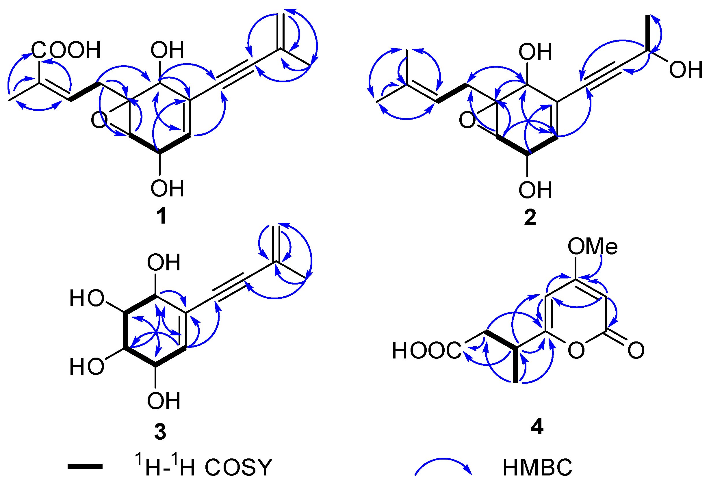
Aspergienyne O (1) was acquired as a yellowish oil. Its HR-ESI-MS peak at m/z 313.1048 ([M + Na]+, calcd for 313.1046) and the 13C NMR data (Table 1) suggested a molecular formula of C16H18O5 with eight degrees of unsaturation. The 1H NMR and HSQC data of 1 (Table 1) exhibited four olefinic groups [δH 6.72 (m), 5.77 (dd, J = 5.0, 1.9 Hz), 5.28 (m) and 5.25 (m)], a methylene [δH 3.03 (dd, J = 15.1, 8.8 Hz), and 2.54 (dd, J = 15.1, 6.9 Hz)], three oxymethines [δH 4.46 (dd, J = 5.0, 2.5 Hz), 4.19 (t, J = 1.9 Hz), and 3.34 (t, J = 2.5 Hz)], and two methyls [δH 1.90 (s) and 1.87 (s)]. The 13C NMR and HSQC data of 1 displayed sixteen carbons, such as a carbonyl carbon (δC 171.7), six sp2 carbons (δC 141.1, 134.3, 132.6, 128.4, 124.3, and 122.3), two sp carbons (δC 91.7 and 87.5), three sp3 methine carbons (δC 68.1, 66.1, and 60.3), one sp3 nonprotonated carbon (δC 62.5), one sp3 methylene group (δC 32.5), together with two methyl carbons (δC 23.5 and 12.9). The NMR data (Table 1) of 1 were highly similar to those of the previously reported biscognienyne D [7], except that the methyl group was replaced by the carboxyl group of 1. The HMBC signals from H-15/H-13 to C-16 (δC 171.7) supported this speculation (Figure 2). Comprehensive analysis of its 2D NMR signals not only confirmed the above hypothesis but also verified the two-dimensional structure shown in Figure 1.
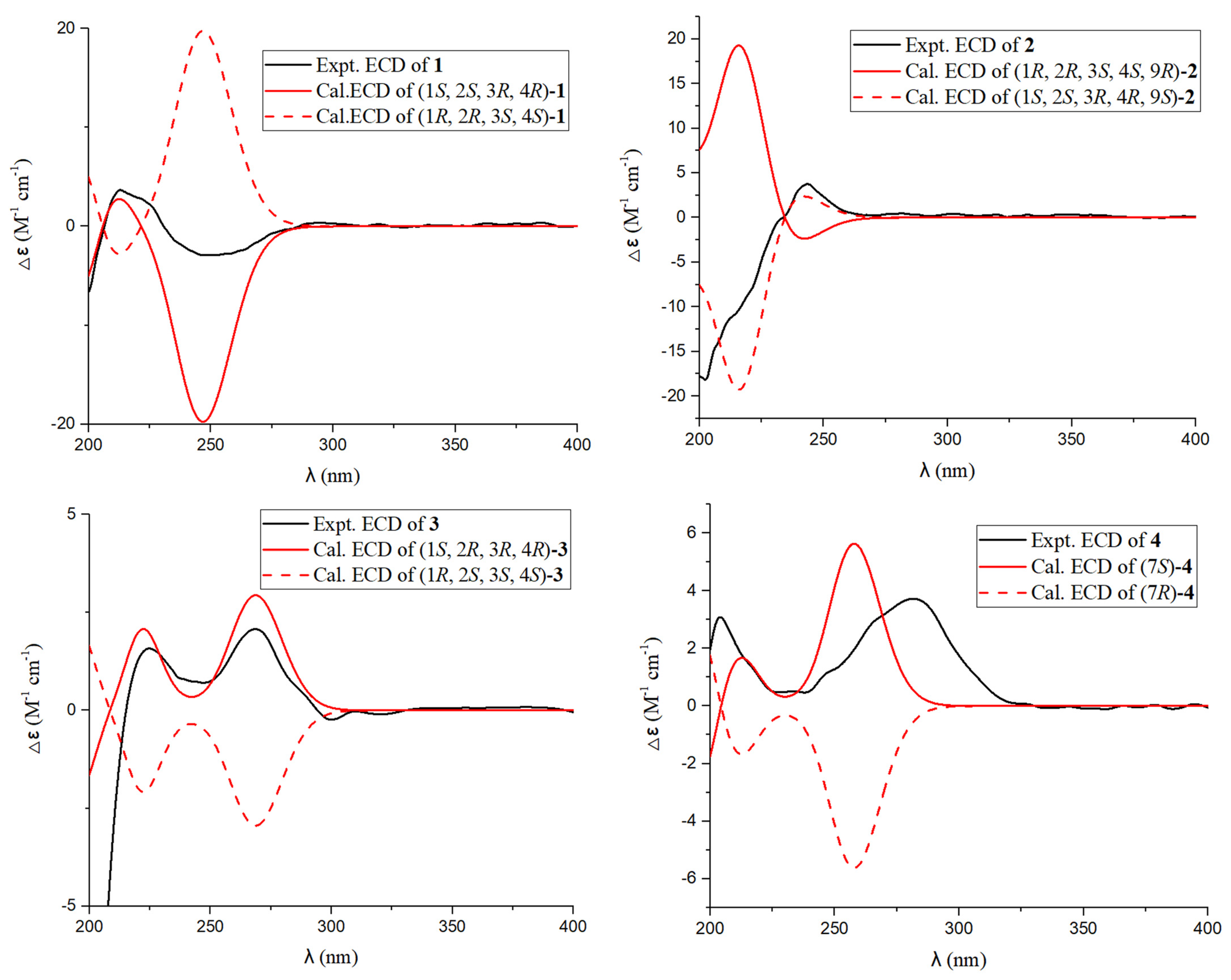
In the NOESY spectrum of 1 (Figure 3), there is a diagnostic association between the olefinic proton [δH 6.72 (m, H-13)] and the terminal methyl proton [δH 1.87 (s, H3-15)], and thus the geometry of the Δ13,14 double bond was defined as Z. Furthermore, the NOESY signals of H-1/H-12β suggested that H-1 and H-12β were β-oriented. Meanwhile, the small value of JH-3/H-4 (2.5 Hz) demonstrated a cis-orientation of these protons. The final configuration of 1 was elucidated as (1S,2S,3R,4R)-1 by means of comparing its experimental ECD spectrum to those calculated (Figure 4).
Aspergienyne P (2) was isolated as a yellowish oil. The analysis of the ion peak discovered at m/z 287.1255 ([M + Na]+, calcd for 287.1259) in the HR-ESI-MS report combined with its 13C NMR data gained the molecular formula C15H20O4. Comprehensive analysis of 1D NMR information (Table 1) of 2 proposed that 2 and 1 are very similar in structure, except for the replacement of 16-COOH and the Δ9,10 double bond in 1 by a methyl group and a hydroxyl group in 2, respectively, which was determined via the crucial HMBC signals from H-13/H-15 to C-16 (δC 18.1), H-16 (δH 1.69) to C-13/C-14/C-15, H-9 (δH 4.57) to C-7/C-8, H-10 to C-9 (δC 59.0), and the significant 1H-1H COSY signals (Figure 2) of H-9/H-10. The NOESY signals (Figure 3) of H-1/H-3 and H-1/H-4 suggested that H-1, H-3, and H-4 were on the same side. Consequently, (1S*, 2S*, 3R*, 4R*)-2 was tentatively established. The 13C NMR data of two potential isomers (2a and 2b) (Figure 5) were calculated according to the GIAO method to determine the complete relative configuration of 2. Due to the good correlation coefficient (R2 = 0.9978) (Figure 6) and the high results of the DP4+ probability analysis (100%) (Table S2) between the calculated and experimental 13C NMR chemical shifts, the (1S*,2S*,3R*,4R*,9S*)-2 was recommended. The absolute stereochemistry of 2 was determined as 1S, 2S, 3R, 4R, and 9S due to the coincidence of the calculated and experimental ECD spectral curves (Figure 4).
The C11H14O4 of aspergienyne Q (3) was determined on the basis of HR-ESI-MS analysis, which exhibited a [M +Na]+ ion at m/z 233.0785 (calcd for 233.0784). The NMR data (Table 2) of 3 was compared in detail with that of (1R,2R,3R,4S)-5-(3-Methylbut-3-en-1-yn-1-yl)-cyclohex-5-ene-1,2,3,4-tetraol [8], and combined with the HR-ESI-MS of 3, and the structure of compound 3 was determined. Moreover, the absolute structure of 3 was further confirmed by 2D NMR and ECD spectroscopy (Figure 2, Figure 3 and Figure 4). Thus, as shown in Figure 1, 3 is reported for the first time in this paper as a new natural product.
The molecular formula of 3-(4-methoxy-2-oxo-2H-pyran-6-yl)butanoic acid (4) was defined as C10H12O5 according to the HR-ESI-MS ion peak discovered at m/z 213.0765 ([M + H]+, calcd for 213.0763), implying five degrees of unsaturation. The 1H NMR spectrum displayed the presence of two olefins [δH 6.04 (d, J = 2.2 Hz) and 5.54 (d, J = 2.2 Hz)], a methylene [δH 2.58 (dd, J = 14.6, 7.4 Hz) and 2.40 (dd, J = 14.6, 7.5 Hz)], one methine proton [δH 3.10 (m)], a methyl proton [δH 1.26 (d, J = 7.0 Hz)], and one methoxy [δH 3.85 (s)]. Subsequently, the carbon resonances of one methine group (δC 36.4), one methylene (δC 40.7), one methyl (δC18.3), and one methoxy (δC 56.9) were observed in its 13C NMR spectrum, along with two carbonyl carbons (δC 176.0 and 167.4) and four olefin carbons (δC 173.0, 169.2, 100.7, and 88.4), as supported by its HSQC spectrum. The NMR data of 4 closely resembled those of pestalotiopyrone N [9], except for a Δ7,8 double bond in pestalotiopyrone N being replaced by one methine and one methylene present at C-7 and C-8 in 4, which was proved on the basis of the key HMBC signals from H-7 to C-5/C-6/C-9, H-8 to C-6/C-9 and H-10 to C-6/C-7, and the key 1H-1H COSY signals (Figure 2) of H-7/H-8 and H-7/H-10. Finally, the absolute configuration of (7S)-4 was elucidated via ECD calculations (Figure 4).
In addition to these four new isolates, five previously reported isolates were acquired, which were identified on the basis of comparing their spectral data with the reported data as pestalotiopyrone I (5) [10], ficipyrone A (6) [11], 4-(hydroxymethyl)-5,7-dimethoxy-6-methylisobenzofuran-1(3H)-one (7) [12], mycophenolic acid (8) [13], and terezine A (9) [14].

2.2. Results of Antiproliferative Activity

The MTT method was used to evaluate the anti-proliferative activity of all isolates on five human cancer cell lines. Among them, 9 displayed moderate anti-proliferation activity on HeLa cancer cells (IC50 =16.6 μM); moreover, 5 displayed anti-proliferation activity on the HeLa and A549 cancer cell lines (IC50 = 29.3 μM and IC50 = 45.4 μM, respectively). The other isolates displayed no notable anti-proliferation activity on five cancer cell lines (IC50 > 50 μM). The positive control was etoposide (IC50: 15.7 μM for HeLa cells, 8.2 μM for A549 cells).

3. Materials and Methods

3.1. General Experimental Procedures

The NMR data were measured by Bruker 400 MHz and 600 MHz instruments (Bruker, Bremen, Germany). The HR-ESI-MS reports and the Optical rotations were collected using an LC-MS spectrometer (Agilent 6545 Q-TOF) and a JASCO P-2000 polarimeter (Jasco, Tokyo, Japan), respectively. The other instruments and materials employed in the experiments were the same as those in our previous reports [15].

3.2. Fungal Material

According to the sequence and morphology of the internal transcriptional spacer (ITS) of the strain, the fungal strain collected from fresh fruits of the mangrove plant Kandelia candel in the Beihai was identified and designated as GXNU-Y85. Subsequently, we obtained its registration number OR999402, as we submitted the ITSrDNA of GXNU-Y85 to GenBank.

3.3. Fermentation, Extraction, and Isolation

This target strain was fermented for 28 days at 25 °C, where the medium consisted of 80 mL H2O (H2O with 0.5% sea salt) and 80 g of rice. Mycelium was collected and soaked in methanol (3 × 10 L) for 2 days to collect the crude extract (21.4 g), which was subsequently extracted 3 times using ethyl acetate. The extract (11.1 g) was isolated via a silica gel column, and six fractions (Fr. 1-Fr. 6) were obtained using a ratio of PE-EtOAc solvent (50:1 to 1:1, v/v). Fr. 5 (2.54 g) was further isolated through a Sephadex LH-20 column (100% methanol) to produce seven fractions (Fr. 5.1 to Fr. 5.7). Isolates 3 (5.6 mg), 8 (7.1 mg), and 5 (3.9 mg) were separated from Fr. 5.2 (0.84 g) based on semipreparative HPLC (50% MeOH–H2O; 7 mL/min; YMC-column, 4.6 mm I.D. × 250 mm, S-5 μm, 12 nm). Isolates 2 (4.6 mg), 7 (6.3 mg), 1 (7.6 mg), and 6 (4.8 mg) were acquired from Fr. 5.4 (0.92 g) on the basis of semipreparative HPLC (MeCN–H2O v/v, 40:60; 7 mL/min; YMC-column, 4.6 mm I.D. × 250 mm, S-5 μm, 12 nm). Fr. 5.7 (0.71 g) was further separated according to semipreparative HPLC (MeCN–H2O v/v, 40:60; 7 mL/min; YMC-column, 4.6 mm I.D. × 250 mm, S-5 μm, 12 nm) to yield 4 (5.2 mg) and 9 (6.1 mg).

3.3.1. Aspergienyne O (1)

Yellowish oil; [ α ] D 22 −13.75 (c 0.37, MeOH); UV (MeOH) λmax (log ε) 259.5 (3.25) nm; IR (KBr) νmax 3357, 2977, 2930, 2195, 1671, 1448, and 1039 cm−1; HR-ESI-MS m/z [M + Na]+ (313.1048); ECD (MeOH) λmax (Δε) 213.50 (+3.769), 247.74 (−2.965) nm; NMR data (in CD3OD) at Table 1.

3.3.2. Aspergienyne P (2)

Yellowish oil; [ α ] D 22 −41.26 (c 0.30, MeOH); UV (MeOH) λmax (log ε) 258.1 (3.15) nm; IR (KBr) νmax 3421, 1719, 1609, 1430, 1384, 1247, 830, and 787 cm−1; HR-ESI-MS m/z [M + Na]+ (287.1255); ECD (MeOH) λmax (Δε) 202.50 (−18.463), 243.09 (+4.011) nm; NMR data (in CD3OD) at Table 1.

3.3.3. Aspergienyne Q (3)

Yellowish oil; [ α ] D 22 +16.89 (c 0.69, MeOH); UV (MeOH) λmax (log ε) 259.2 (3.02) nm; IR (KBr) νmax 3428, 2955, 2870, 1656, 1459, 1382, 1370, 1038, 967, and 834 cm−1; HR-ESI-MS m/z [M + Na]+ (233.0785); ECD (MeOH) λmax (Δε) 224.76 (+1.570), 268.54 (+2.080) nm; NMR data (in CD3OD) at Table 2.

3.3.4. 3-(4-Methoxy-2-oxo-2H-pyran-6-yl)butanoic Acid (4)

Yellowish oil; [ α ] D 22 −15.03 (c 0.28, MeOH); UV (MeOH) λmax (log ε) 212.7 (3.08), 264.4 (3.31) nm; IR (KBr) νmax 3411, 2933, 1638, 1464, 1371, 1034, and 886 cm−1; HR-ESI-MS m/z [M + H]+ (213.0765); ECD (MeOH) λmax (Δε) 203.65 (+3.078), 281.71 (+3.744) nm; NMR data (in CD3OD) at Table 2.

3.4. ECD and NMR Calculations

The absolute stereochemistry of the new isolates was defined through the ECD and NMR calculations described in previous reports [16,17]. The Supporting Information provides a detailed description of the process.

3.5. Anti-Proliferative Activity Test

The anti-proliferative activity of 19 against five cancer cell lines (A549, MCF-7, Hela, 5-8F, and T24 cells) was evaluated by MTT assay as previously reported [18]. The positive control was etoposide.

4. Conclusions

In the present study, three new compounds (1, 2, and 4), one new natural compound (3), and five previously reported isolates (59) were acquired from the fungal strain Aspergillus sp. GXNU-Y85 by various chromatographic techniques. Extensive spectroscopic data (HRESIMS, NMR, and ECD) and quantum chemical calculations were used to determine the structures of these new compounds. Biological activity studies revealed that compounds 5 and 9 showed moderate anti-proliferative activity against HeLa and A549 cells (IC50 = 16.6–45.4 μM).

Supplementary Materials

The following supporting information can be downloaded at: https://www.mdpi.com/article/10.3390/md22060277/s1, NMR, IR, UR and HRESIMS spectra of 14. NMR spectra of 59.

Author Contributions

Z.L. and F.Q. conceived and designed the experiments. C.W. performed the experiments. F.W., P.T., Y.S., Q.L. and M.G. analyzed the data. C.W., F.Q. and Z.L. wrote the paper. All authors have read and agreed to the published version of the manuscript.

Funding

This study was supported by the Natural Science Foundation of Guangxi Province (2024GXNSFBA010005), and Basic Ability Improvement Project of Young and Middle-Aged Teachers in Guangxi Colleges (2024KY0597), the PhD Start-up Fund of Yulin Normal University (G2023ZK22).

Data Availability Statement

The authors declare that all data of this study are available within the article and its Supplementary Materials file or from the corresponding authors upon request.

Conflicts of Interest

The authors declare no conflicts of interest.

References

  1. Ancheeva, E.; Daletos, G.; Proksch, P. Bioactive secondary metabolites from endophytic fungi. Curr. Med. Chem. 2020, 27, 1836–1854. [Google Scholar] [CrossRef] [PubMed]
  2. Wang, Z.; Wang, L.; Pan, Y.; Zheng, X.; Liang, X.; Sheng, L.; Zhang, D.; Sun, Q.; Wang, Q. Research advances on endophytic fungi and their bioactive metabolites. Bioprocess Biosyst. Eng. 2023, 46, 165–170. [Google Scholar] [CrossRef] [PubMed]
  3. Kouipou Toghueo, R.M.; Boyom, F.F. Endophytic fungi from terminalia species: A comprehensive review. J. Fungi 2019, 5, 43. [Google Scholar] [CrossRef] [PubMed]
  4. Zheng, R.; Li, S.; Zhang, X.; Zhao, C. Biological activities of some new secondary metabolites isolated from endophytic fungi: A review study. Int. J. Mol. Sci. 2021, 22, 959. [Google Scholar] [CrossRef] [PubMed]
  5. Qin, F.; Luo, L.; Liu, Y.C.; Bo, X.L.; Wu, F.R.; Wang, F.F.; Tan, M.J.; Wei, Y.Q.; Dou, X.B.; Wang, C.Y.; et al. Diisoprenyl-cyclohexene-type meroterpenoids from a mangrove endophytic fungus Aspergillus sp. GXNU-Y65 and their anti-nonalcoholic steatohepatitis activity in AML12 cells. Phytochemistry 2024, 218, 113955. [Google Scholar] [PubMed]
  6. Qin, F.; Song, Z.S.; Luo, L.; Bo, X.L.; Wu, F.R.; Tan, M.J.; Wang, F.F.; Huang, X.S.; Wang, H.S. Diisoprenyl cyclohexene-type meroterpenoids with cytotoxic activity from a mangrove endophytic fungus Aspergillus sp. GXNU-Y85. Mar. Drugs 2024, 22, 58. [Google Scholar] [CrossRef]
  7. Akone, S.H.; Wang, H.; Mouelle, E.N.M.; Mándi, A.; Kurtán, T.; Koliye, P.R.; Hartmann, R.; Bhatia, S.; Yang, J.; Müller, W.E.; et al. Prenylated cyclohexene-type meroterpenoids and sulfur-containing xanthones produced by Pseudopestalotiopsis theae. Phytochemistry 2022, 197, 113124. [Google Scholar] [CrossRef]
  8. Buckler, J.N.; Meek, T.; Banwell, M.G.; Carr, P.D. Total synthesis of the cyclic carbonate-containing natural product aspergillusol B from D-(-)-tartaric acid. J. Nat. Prod. 2017, 80, 2088–2093. [Google Scholar] [CrossRef] [PubMed]
  9. Wang, J.J.; Peng, Q.Y.; Yao, X.G.; Liu, Y.H.; Zhou, X.F. New pestallic acids and diphenylketone derivatives from the marine alga-derived endophytic fungus Pestalotiopsis neglecta SCSIO41403. J. Antibiot. 2020, 73, 585588. [Google Scholar] [CrossRef] [PubMed]
  10. Rönsberg, D.; Debbab, A.; Mándi, A.; Wray, V.; Dai, H.F.; Kurtán, T.; Proksch, P.; Aly, A.H. Secondary metabolites from the endophytic fungus Pestalotiopsis virgatula isolated from the mangrove plant Sonneratia caseolaris. Tetrahedron Lett. 2013, 54, 3256–3259. [Google Scholar] [CrossRef]
  11. Liu, S.C.; Liu, X.Y.; Guo, L.D.; Che, Y.S.; Liu, L. 2H-pyran-2-one and 2H-furan-2-one derivatives from the plant endophytic fungus Pestalotiopsis fici. Chem. Biodivers. 2013, 10, 2007–2013. [Google Scholar] [CrossRef] [PubMed]
  12. Peng, J.Y.; Chen, P.W.; Li, C.W.; Liu, J.T.; Sun, Y.J.; Yang, M.Z.; Ding, B.; Huang, H.B.; Tao, Y.W. Phthalide metabolites produced by mangrove endophytic fungus Pestalotiopsis sp. SAS4. Magn. Reson. Chem. 2022, 60, 525–529. [Google Scholar] [CrossRef] [PubMed]
  13. Siebert, A.; Cholewinski, G.; Trzonkowski, P.; Rachon, J. Immunosuppressive properties of amino acid and peptide derivatives of mycophenolic acid. Eur. J. Med. Chem. 2020, 189, 112091. [Google Scholar] [CrossRef] [PubMed]
  14. Wang, Y.; Gloer, J.B.; Scott, J.A.; Malloch, D. Terezines A-D: New amino acid-derived bioactive metabolites from the coprophilous fungus Sporormiella teretispora. J. Nat. Prod. 1995, 58, 93–99. [Google Scholar] [CrossRef] [PubMed]
  15. Qin, F.; Wang, C.Y.; Kim, D.; Wang, H.S.; Zhu, Y.K.; Lee, S.K.; Yao, G.Y.; Liang, D. Nitidumpeptins A and B, cyclohexapeptides isolated from Zanthoxylum nitidum var. tomentosum: Structural elucidation, total synthesis, and antiproliferative activity in cancer cells. J. Org. Chem. 2021, 86, 1462–1470. [Google Scholar] [CrossRef] [PubMed]
  16. Qin, F.; Wang, C.Y.; Wang, C.G.; Chen, Y.; Li, J.J.; Li, M.S.; Zhu, Y.K.; Lee, S.K.; Wang, H.S. Undescribed isoquinolines from Zanthoxylum nitidum and their antiproliferative effects against human cancer cell lines. Phytochemistry 2023, 205, 113476. [Google Scholar] [CrossRef] [PubMed]
  17. Qin, F.; Wang, C.Y.; Hu, R.; Wang, C.G.; Wang, F.F.; Zhou, M.M.; Liang, D.; Liao, H.B.; Lee, S.K.; Wang, H.S. Anti-infammatory activity of isobutylamides from Zanthoxylum nitidum var. Tomentosum. Fitoterapia 2020, 142, 104486. [Google Scholar] [CrossRef] [PubMed]
  18. Huang, R.Z.; Jin, L.; Wang, C.G.; Xu, X.J.; Du, Y.; Liao, N.; Ji, M.; Liao, Z.X.; Wang, H.S. A pentacyclic triterpene derivative possessing polyhydroxyl ring A suppresses growth of HeLa cells by reactive oxygen species-dependent NF-κB pathway. Eur. J. Pharmacol. 2018, 838, 157–169. [Google Scholar] [CrossRef] [PubMed]
Figure 1. The chemical structures 19.
Marinedrugs 22 00277 g001
Figure 2. Key HMBC and 1H-1H COSY correlations of compounds 14.
Marinedrugs 22 00277 g002
Figure 3. Key NOESY correlations for 13.
Marinedrugs 22 00277 g003
Figure 4. Calculated and experimental ECD spectra of 14.
Marinedrugs 22 00277 g004
Figure 5. Conformations of low-energy conformers of structures 2a and 2b in MeOH.
Marinedrugs 22 00277 g005
Figure 6. Regression analyses of experimental versus calculated 13C NMR chemical shifts of model compounds 2a and 2b.
Marinedrugs 22 00277 g006
Table 1. 1H and 13C NMR Data for 1 and 2 in CD3OD.
No1 a2 b
δC, TypeδH (J in Hz)δC, TypeδH (J in Hz)
168.1, CH4.19, t (1.9)67.6, CH4.15, t (2.0)
262.5, C 63.2, C
360.3, CH3.34, t (2.5)60.3, CH3.3, s
466.1, CH4.46, dd (5.0, 2.5)66.1, CH4.41, dd (4.9, 2.0)
5134.3, CH5.77, dd (5.0, 1.9)134.2, CH5.74, dd (4.9, 2.3)
6124.3, C 124.1, C
787.5, C 82.3, C
891.7, C 92.7, C
9128.4, C 59.0, CH4.57, q (6.6)
10122.3, CH25.28, m, 5.25, m24.6, CH31.41, d (6.6)
1123.5, CH31.90, s
1232.5, CH23.03, dd (15.1, 8.8) 2.54, dd (15.1, 6.9)32.2, CH22.94, dd (14.6, 9.0) 2.14, dd (14.6, 6.2)
13136.2, CH6.72, m119.0, CH5.13, m
14132.6, C 136.7, C
1512.9, CH31.87, s26.0, CH31.73, s
16171.7, C 18.1, CH31.69, s
a 1H NMR at 600 MHz, 13C NMR at 150 MHz; b 1H NMR at 400 MHz, 13C NMR at 100 MHz.
Table 2. 1H (400 MHz) and 13C (100 MHz) NMR Data for 3 and 4 in CD3OD.
No3No4
δC, TypeδH (J in Hz)δC, TypeδH (J in Hz)
174.4, CH3.86, dd (7.2, 0.8)2167.4, C
273.4, CH3.71, dd (9.9, 7.2)388.4, CH5.54 d (2.2)
372.1, CH3.48, dd (9.9, 4.3)4173.9, C
467.6, CH4.23, dd (5.2, 4.3)5100.7, CH6.04 d (2.2)
5134.1, CH6.06, dd (5.2, 1.7)6169.2, C
6128.3, C 736.4, CH3.10 m
787.5, C 840.7, CH22.58 dd (14.6, 7.4),
2.40 dd (14.6, 7.5)
892.5, C 9176.0, C
9128.3, C 1018.3, CH31.26 d (7.0)
10122.5, CH25.30, m; 5.28, m4-OMe56.9, CH33.85 s
1123.5, CH31.91, m
Disclaimer/Publisher’s Note: The statements, opinions and data contained in all publications are solely those of the individual author(s) and contributor(s) and not of MDPI and/or the editor(s). MDPI and/or the editor(s) disclaim responsibility for any injury to people or property resulting from any ideas, methods, instructions or products referred to in the content.