
Exophilone, a Tetrahydrocarbazol-1-one Analogue with Anti-Pulmonary Fibrosis Activity from the Deep-Sea Fungus Exophiala oligosperma MCCC 3A01264


 ,
,
Abstract
1. Introduction
2. Results and Discussion
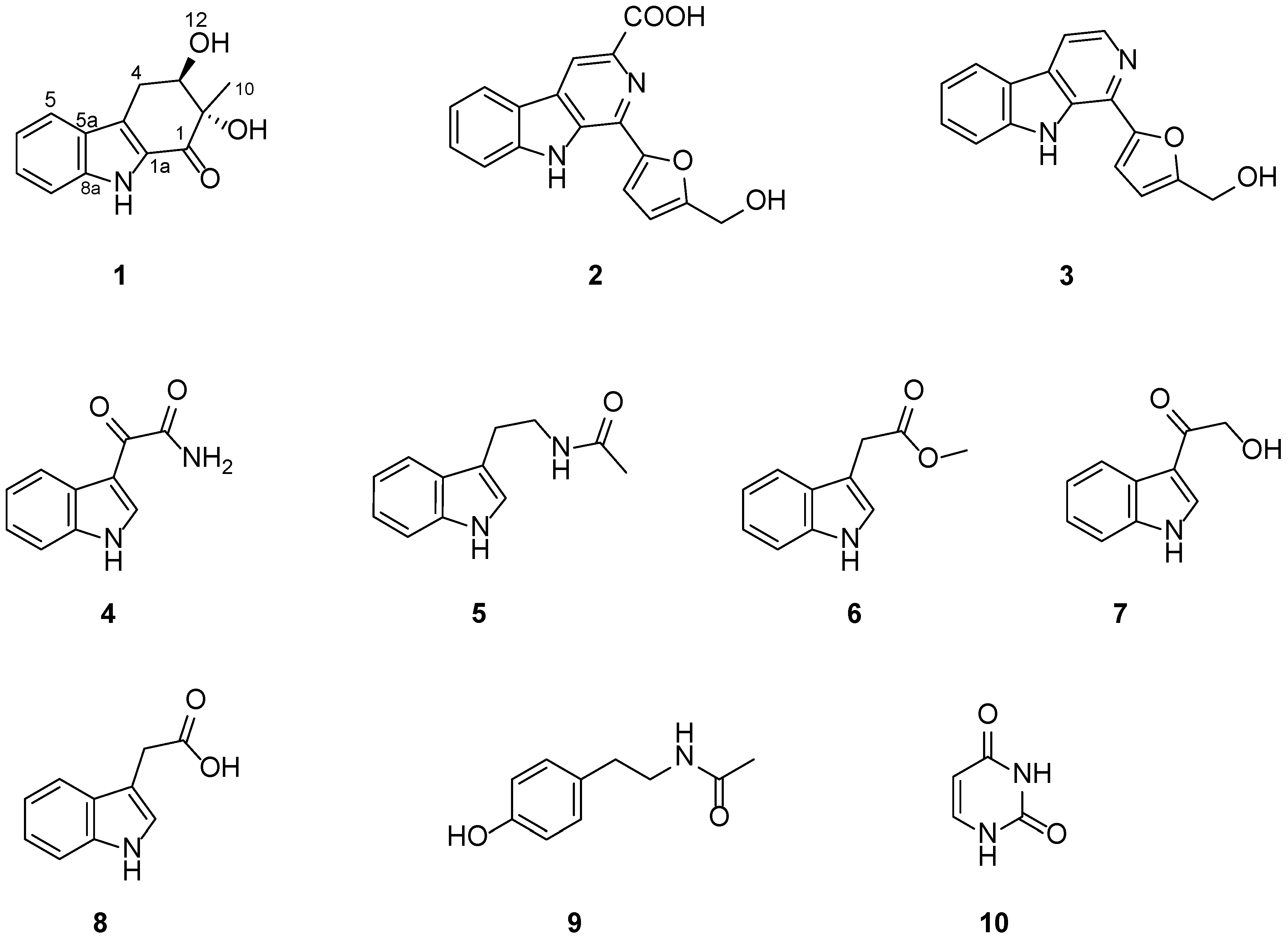
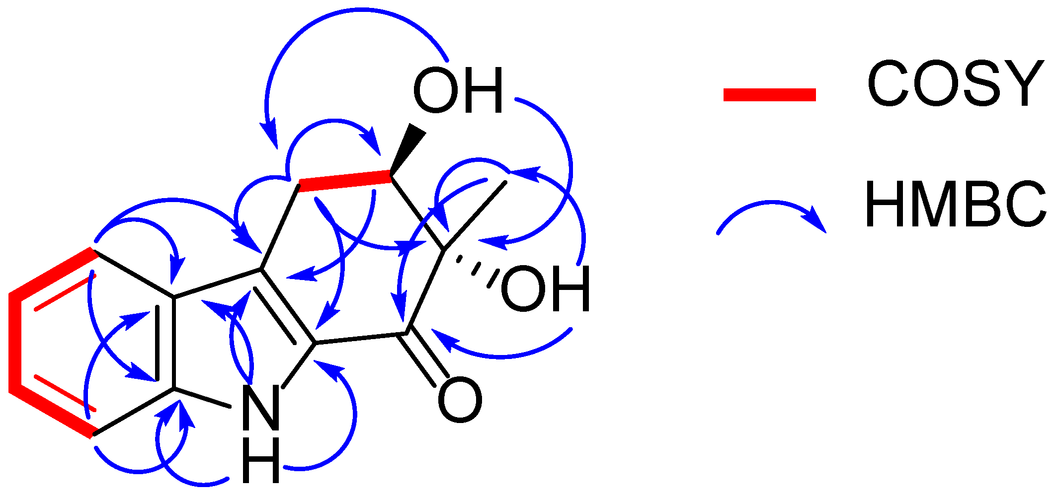
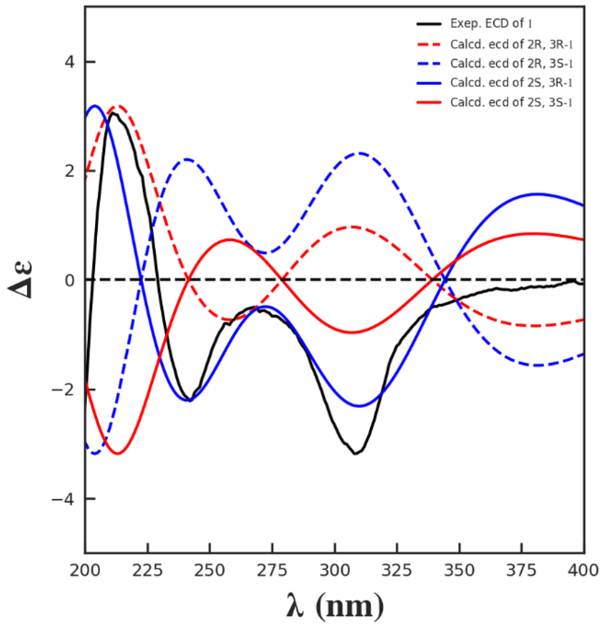
2.1. Structural Elucidation
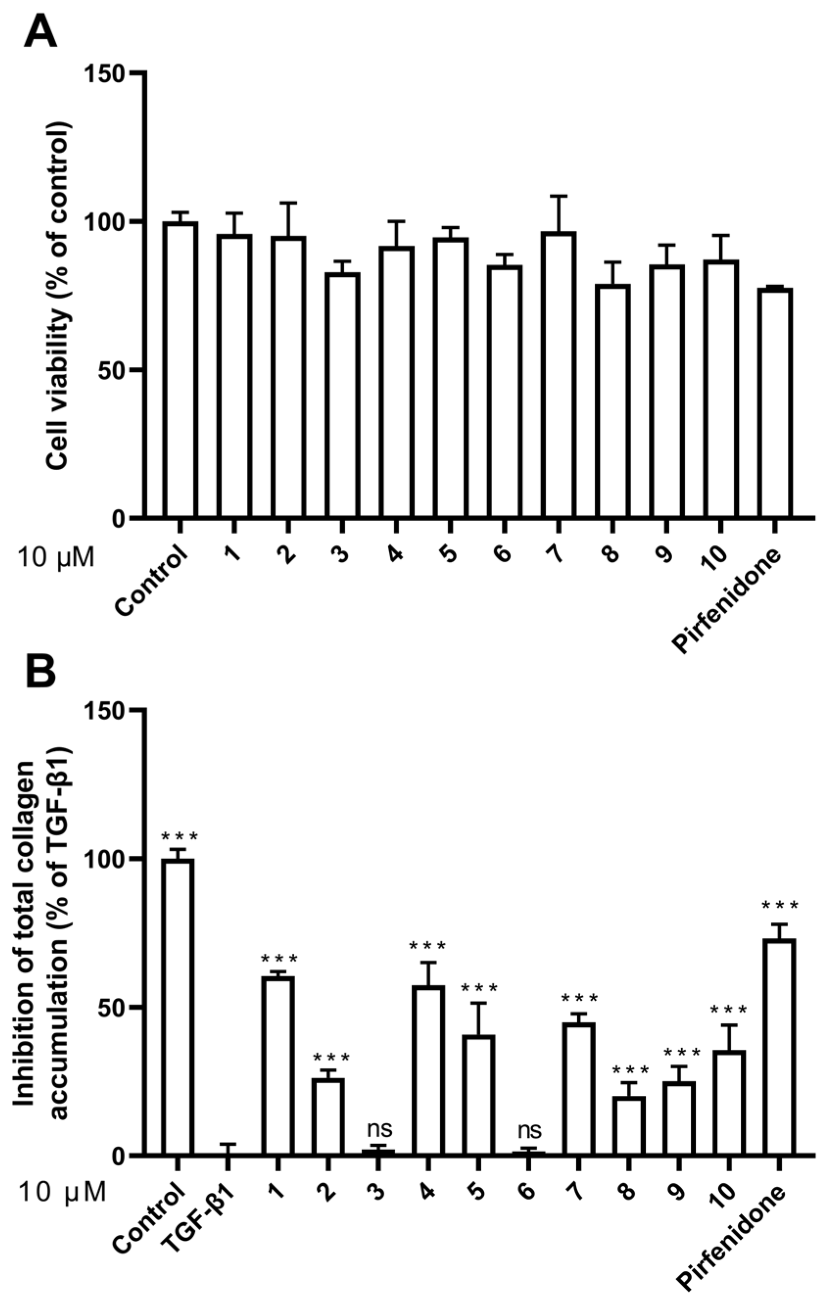
2.2. Effect of Compounds 1–10 on HFL1 Cell Viability
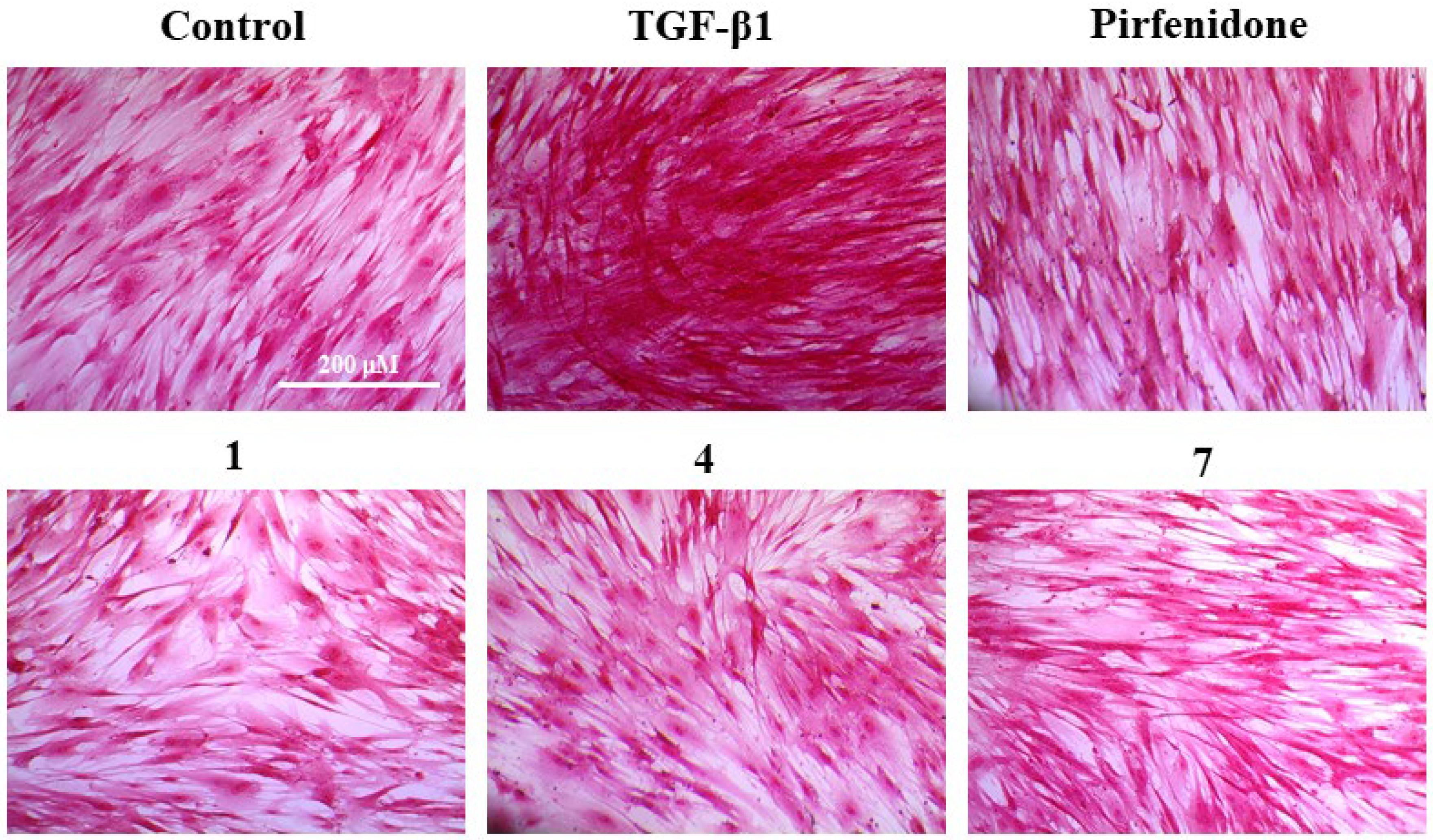
2.3. Effect of Compounds 1–10 on HFL1 Cell Collagen Accumulation
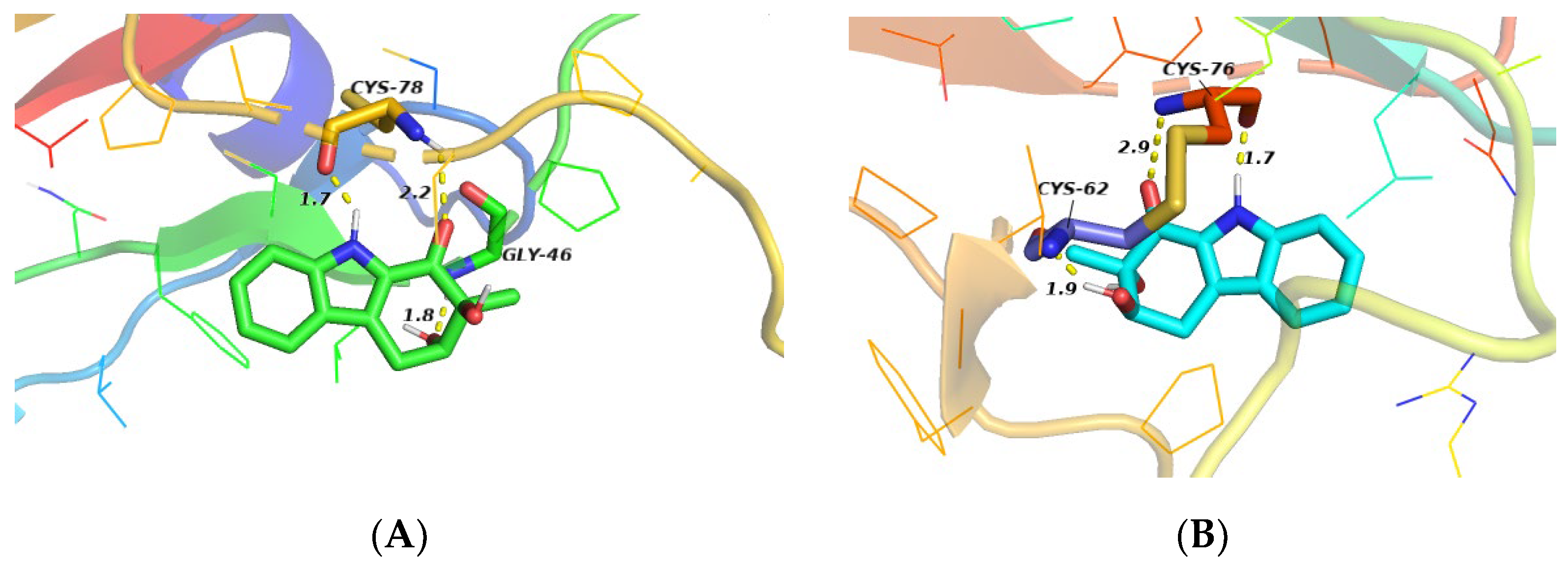
2.4. Molecular Docking Study
3. Discussion
4. Materials and Methods
4.1. General Procedures
4.2. Fungal Strain and Culture Method
4.3. Extraction and Isolation
4.4. Cell Culture and Cytotoxicity Assay
4.5. Collagen Accumulation Inhibition In Vitro
4.6. Molecular Docking
4.7. Statistical Analysis
5. Conclusions
Supplementary Materials
Author Contributions
Funding
Data Availability Statement
Conflicts of Interest
References
- Ul Arifeen, Z.M.; Ma, Y.-N.; Xue, Y.-R.; Liu, C.-H. Deep-sea fungi could be the new arsenal for bioactive molecules. Mar. Drugs 2019, 18, 9. [Google Scholar] [CrossRef]
- Ibrar, M.; Ullah, M.W.; Manan, S.; Farooq, U.; Rafiq, M.; Hasan, F. Fungi from the extremes of life: An untapped treasure for bioactive compounds. Appl. Microbiol. Biotechnol. 2020, 104, 2777–2801. [Google Scholar] [CrossRef] [PubMed]
- Rimawi, B.H.; Rimawi, R.H.; Mirdamadi, M.; Steed, L.L.; Marchell, R.; Sutton, D.A.; Thompson, E.H.; Wiederhold, N.P.; Lindner, J.R.; Boger, M.S. A case of Exophiala oligosperma successfully treated with voriconazole. Med. Mycol. Case Rep. 2013, 2, 144–147. [Google Scholar] [CrossRef] [PubMed]
- Hwang, B.-S.; Kim, H.-J.; Jeong, G.-S.; Oh, J.-S.; Rho, J.-R. Isolation and structure determination of two new carbazoles from Streptomyces ehimensis JB201. Bull. Korean Chem. Soc. 2010, 31, 3457–3459. [Google Scholar] [CrossRef]
- Karwehl, S.; Jansen, R.; Huch, V.; Stadler, M. Sorazolons, carbazole alkaloids from Sorangium cellulosum strain soce375. J. Nat. Prod. 2016, 79, 369–375. [Google Scholar] [CrossRef] [PubMed]
- Wu, X.; Huang, J.; Wang, J.; Xu, Y.; Yang, X.; Sun, M.; Shi, J. Multi-pharmaceutical activities of chinese herbal polysaccharides in the treatment of pulmonary fibrosis: Concept and future prospects. Front. Pharmacol. 2021, 12, 707491. [Google Scholar] [CrossRef] [PubMed]
- Ahmad Alhiyari, M.; Ata, F.; Islam Alghizzawi, M.; Bint, I.B.A.; Salih Abdulhadi, A.; Yousaf, Z. Post COVID-19 fibrosis, an emerging complicationof SARS-CoV-2 infection. IDCases 2021, 23, e01041. [Google Scholar] [CrossRef] [PubMed]
- Dimitroulis, I.A. Nintedanib: A novel therapeutic approach for idiopathic pulmonary fibrosis. Respir. Care 2014, 59, 1450–1455. [Google Scholar] [CrossRef]
- Richeldi, L.; du Bois, R.M.; Raghu, G.; Azuma, A.; Brown, K.K.; Costabel, U.; Cottin, V.; Flaherty, K.R.; Hansell, D.M.; Inoue, Y.; et al. Efficacy and safety of nintedanib in idiopathic pulmonary fibrosis. N. Engl. J. Med. 2014, 370, 2071–2082. [Google Scholar] [CrossRef]
- Roth, G.J.; Binder, R.; Colbatzky, F.; Dallinger, C.; Schlenker-Herceg, R.; Hilberg, F.; Wollin, S.-L.; Kaiser, R. Nintedanib: From discovery to the clinic. J. Med. Chem. 2015, 58, 1053–1063. [Google Scholar] [CrossRef]
- Ogura, T.; Azuma, A.; Inoue, Y.; Taniguchi, H.; Chida, K.; Bando, M.; Niimi, Y.; Kakutani, S.; Suga, M.; Sugiyama, Y.; et al. All-case post-marketing surveillance of 1371 patients treated with pirfenidone for idiopathic pulmonary fibrosis. Respir. Investig. 2015, 53, 232–241. [Google Scholar] [CrossRef] [PubMed]
- George, P.M.; Patterson, C.M.; Reed, A.K.; Thillai, M. Lung transplantation for idiopathic pulmonary fibrosis. Lancet Respir. Med. 2019, 7, 271–282. [Google Scholar] [CrossRef]
- Xue, L.; Deng, D.; Zheng, S.; Tang, M.; Yang, Z.; Pei, H.; Chen, Y.; Yang, T.; Liu, K.; Ye, H.; et al. Design, synthesis and discovery of 2(1H)-quinolone derivatives for the treatment of pulmonary fibrosis through inhibition of TGF-β/smad dependent and independent pathway. Eur. J. Med. Chem. 2020, 197, 112259. [Google Scholar] [CrossRef] [PubMed]
- Su, B.N.; Chang, L.C.; Park, E.J.; Cuendet, M.; Santarsiero, B.D.; Mesecar, A.D.; Mehta, R.G.; Fong, H.H.; Pezzuto, J.M.; Kinghorn, A.D. Bioactive constituents of the seeds of Brucea javanica. Planta Med. 2002, 68, 730–733. [Google Scholar] [CrossRef]
- Chen, P.-N.; Hao, M.-J.; Li, H.-J.; Xu, J.; Mahmud, T.; Lan, W.-J. Biotransformations of anthranilic acid and phthalimide to potent antihyperlipidemic alkaloids by the marine-derived fungus Scedosporium apiospermum F41–1. Bioorg. Chem. 2021, 116, 105375. [Google Scholar] [CrossRef]
- Bao, B.; Zhang, P.; Lee, Y.; Hong, J.; Lee, C.-O.; Jung, J.H. Monoindole alkaloids from a marine sponge Spongosorites sp. Mar. Drugs 2007, 5, 31–39. [Google Scholar] [CrossRef]
- Vaca, J.; Salazar, F.; Ortiz, A.; Sansinenea, E. Indole alkaloid derivatives as building blocks of natural products from Bacillus thuringiensis and Bacillus velezensis and their antibacterial and antifungal activity study. J. Antibiot. 2020, 73, 798–802. [Google Scholar] [CrossRef]
- Liu, Y.; Jung, J.H.; Zhang, S. Indole alkaloids from a sponge Sarcotragus species. Biochem. Syst. Ecol. 2006, 34, 453–456. [Google Scholar] [CrossRef]
- Tang, M.; Zhou, X.; Cai, J.; Chen, G. Chemical constituents from the fresh flower buds of Musa nana and their chemotaxonomic significance. Biochem. Syst. Ecol. 2021, 99, 104348. [Google Scholar] [CrossRef]
- Evidente, A.; Iacobellis, N.S.; Sisto, A. Isolation of indole-3-acetic acid methyl ester, a metabolite of indole-3-acetic acid from Pseudomonas amygdali. Experientia 1993, 49, 182–183. [Google Scholar] [CrossRef]
- Sobolevskaya, M.P.; Denisenko, V.A.; Moiseenko, A.S.; Shevchenko, L.S.; Menzorova, N.I.; Sibirtsev, Y.T.; Kim, N.Y.; Kuznetsova, T.A. Bioactive metabolites of the marine actinobacterium Streptomyces sp. KMM 7210. Russ. Chem. Bull. 2007, 56, 838–840. [Google Scholar] [CrossRef]
- Kan, S.; Chen, G.; Han, C.; Chen, Z.; Song, X.; Ren, M.; Jiang, H. Chemical constituents from the roots of Xanthium sibiricum. Nat. Prod. Res. 2011, 25, 1243–1249. [Google Scholar] [CrossRef] [PubMed]
- Deng, D.; Pei, H.; Lan, T.; Zhu, J.; Tang, M.; Xue, L.; Yang, Z.; Zheng, S.; Ye, H.; Chen, L. Synthesis and discovery of new compounds bearing coumarin scaffold for the treatment of pulmonary fibrosis. Eur. J. Med. Chem. 2020, 185, 111790. [Google Scholar] [CrossRef] [PubMed]
- Mnafgui, K.; Ghazouani, L.; Hajji, R.; Tlili, A.; Derbali, F.; da Silva, F.I.; Araújo, J.L.; de Oliveira Schinoff, B.; Bachega, J.F.R.; da Silva Santos, A.L.; et al. Oleuropein protects against cerebral ischemia injury in rats: Molecular docking, biochemical and histological findings. Neurochem. Res. 2021, 46, 2131–2142. [Google Scholar] [CrossRef] [PubMed]
- Intaraudom, C.; Rachtawee, P.; Suvannakad, R.; Pittayakhajonwut, P. Antimalarial and antituberculosis substances from Streptomyces sp. BCC26924. Tetrahedron 2011, 67, 7593–7597. [Google Scholar] [CrossRef]
- Zhao, F.; Liu, Z.-Q. Indole and its alkyl-substituted derivatives protect erythrocyte and DNA against radical-induced oxidation. J. Biochem. Mol. Toxicol. 2009, 23, 273–279. [Google Scholar] [CrossRef]
- Dudala, S.S.; Venkateswarulu, T.C.; Kancharla, S.C.; Kodali, V.P.; Babu, D.J. A review on importance of bioactive compounds of medicinal plants in treating idiopathic pulmonary fibrosis (special emphasis on isoquinoline alkaloids). Future J. Pharm. Sci. 2021, 7, 156. [Google Scholar] [CrossRef]


| Position | δC, Type | δH, Mult. (J in Hz) |
|---|---|---|
| 1 | 192.4, CO | |
| 2 | 77.3, C | |
| 3 | 74.2, CH | 4.03, m |
| 4 | 27.5, CH2 | 2.76, m 3.28, m |
| 4a | 123.6, C | |
| 5a | 125.3, C | |
| 5 | 121.2, CH | 7.66, d (7.8) |
| 6 | 119.7, CH | 7.08, ddd (7.8, 7.2, 1.2) |
| 7 | 126.2, CH | 7.30, ddd (7.8, 7.2, 1.2) |
| 8 | 112.8, CH | 7.38, d (7.8) |
| 8a | 138.9, C | |
| 9 | NH | 11.59, brs |
| 1a | 129.5, C | |
| 10 | 18.6, CH3 | 1.24, s |
| 11 | OH | 5.25, brs |
| 12 | OH | 5.16, d (3.6) |
| Compound | Inhibition (%) | Survival Rate (%) |
|---|---|---|
| 1 | 60.44 ± 1.54 | 98.14 ± 6.20 |
| 2 | 26.28 ± 2.53 | 98.28 ± 11.15 |
| 3 | 2.19 ± 1.34 | 81.69 ± 3.36 |
| 4 | 57.37 ± 7.65 | 91.56 ± 10.17 |
| 5 | 40.88 ± 10.52 | 96.11 ± 1.80 |
| 6 | 1.46 ± 1.16 | 87.01 ± 1.13 |
| 7 | 44.96 ± 2.82 | 99.25 ± 12.96 |
| 8 | 20.15 ± 4.49 | 78.49 ± 8.92 |
| 9 | 25.11 ± 4.97 | 84.93 ± 7.86 |
| 10 | 35.62 ± 8.39 | 86.96 ± 9.81 |
| pirfenidone | 73.14 ± 4.72 | 77.57 ± 0.52 |
Publisher’s Note: MDPI stays neutral with regard to jurisdictional claims in published maps and institutional affiliations. |
© 2022 by the authors. Licensee MDPI, Basel, Switzerland. This article is an open access article distributed under the terms and conditions of the Creative Commons Attribution (CC BY) license (https://creativecommons.org/licenses/by/4.0/).
Share and Cite
Hong, M.-J.; Hao, M.-J.; Zhang, G.-Y.; Li, H.-J.; Shao, Z.-Z.; Liu, X.-P.; Ma, W.-Z.; Xu, J.; Mahmud, T.; Lan, W.-J. Exophilone, a Tetrahydrocarbazol-1-one Analogue with Anti-Pulmonary Fibrosis Activity from the Deep-Sea Fungus Exophiala oligosperma MCCC 3A01264. Mar. Drugs 2022, 20, 448. https://doi.org/10.3390/md20070448
Hong M-J, Hao M-J, Zhang G-Y, Li H-J, Shao Z-Z, Liu X-P, Ma W-Z, Xu J, Mahmud T, Lan W-J. Exophilone, a Tetrahydrocarbazol-1-one Analogue with Anti-Pulmonary Fibrosis Activity from the Deep-Sea Fungus Exophiala oligosperma MCCC 3A01264. Marine Drugs. 2022; 20(7):448. https://doi.org/10.3390/md20070448
Chicago/Turabian StyleHong, Ming-Jun, Meng-Jiao Hao, Guang-Yu Zhang, Hou-Jin Li, Zong-Ze Shao, Xiu-Pian Liu, Wen-Zhe Ma, Jun Xu, Taifo Mahmud, and Wen-Jian Lan. 2022. "Exophilone, a Tetrahydrocarbazol-1-one Analogue with Anti-Pulmonary Fibrosis Activity from the Deep-Sea Fungus Exophiala oligosperma MCCC 3A01264" Marine Drugs 20, no. 7: 448. https://doi.org/10.3390/md20070448
APA StyleHong, M.-J., Hao, M.-J., Zhang, G.-Y., Li, H.-J., Shao, Z.-Z., Liu, X.-P., Ma, W.-Z., Xu, J., Mahmud, T., & Lan, W.-J. (2022). Exophilone, a Tetrahydrocarbazol-1-one Analogue with Anti-Pulmonary Fibrosis Activity from the Deep-Sea Fungus Exophiala oligosperma MCCC 3A01264. Marine Drugs, 20(7), 448. https://doi.org/10.3390/md20070448