Review

Botryllus schlosseri as a Unique Colonial Chordate Model for the Study and Modulation of Innate Immune Activity

1
Regenerative Medicine and Stem Cell Research Center, The Shraga Segal Department of Microbiology, Immunology, and Genetics, Faculty of Health Sciences, Ben Gurion University of the Negev, Beer Sheva 8410501, Israel
2
Laboratory of Connective Tissue, Centro Nacional de Investigación y Atención de Quemados, Instituto Nacional de Rehabilitación “Luis Guillermo Ibarra Ibarra”, Calzada Mexico-Xochimilco No. 289, Col. Arenal de Guadalupe, Tlalpan, Mexico City 14389, Mexico
3
Institute for Stem Cell Biology and Regenerative Medicine, Stanford University School of Medicine, Hopkins Marine Station, Stanford University, Chan Zuckerberg Biohub, Pacific Grove, CA 93950, USA
*
Author to whom correspondence should be addressed.
O.G. and E.A.M.-T. contributed equally to this work.
Mar. Drugs 2021, 19(8), 454; https://doi.org/10.3390/md19080454
Submission received: 30 June 2021 / Revised: 4 August 2021 / Accepted: 5 August 2021 / Published: 9 August 2021

Abstract

:
Understanding the mechanisms that sustain immunological nonreactivity is essential for maintaining tissue in syngeneic and allogeneic settings, such as transplantation and pregnancy tolerance. While most transplantation rejections occur due to the adaptive immune response, the proinflammatory response of innate immunity is necessary for the activation of adaptive immunity. Botryllus schlosseri, a colonial tunicate, which is the nearest invertebrate group to the vertebrates, is devoid of T- and B-cell-based adaptive immunity. It has unique characteristics that make it a valuable model system for studying innate immunity mechanisms: (i) a natural allogeneic transplantation phenomenon that results in either fusion or rejection; (ii) whole animal regeneration and noninflammatory resorption on a weekly basis; (iii) allogeneic resorption which is comparable to human chronic rejection. Recent studies in B. schlosseri have led to the recognition of a molecular and cellular framework underlying the innate immunity loss of tolerance to allogeneic tissues. Additionally, B. schlosseri was developed as a model for studying hematopoietic stem cell (HSC) transplantation, and it provides further insights into the similarities between the HSC niches of human and B. schlosseri. In this review, we discuss why studying the molecular and cellular pathways that direct successful innate immune tolerance in B. schlosseri can provide novel insights into and potential modulations of these immune processes in humans.

1. Introduction

The study of the immunological mechanisms that allow animals to regenerate and to recognize allogeneic tissues is an important trait in the field of transplantation. In vertebrates, T cells are the principal effector cells of the adaptive immune system for the nonself recognition. Interestingly, innate cells are activated by inflammatory signals released by damaged or stressed cells resulting from the allogeneic transplantation; this innate immunity is necessary for T-cell activation and the concomitant rejection response [1,2]. For example, following HSC transplantation, the hyperactivation of natural killer (NK) cells results in NK cell-mediated production of proinflammatory signals that induces and sustains the T-cell-mediated graft-versus-host disease (GVHD) response [3,4]. Innate immunity also plays an important role in the tolerance of the embryo tissues during pregnancy. In this case, innate responses are highly associated with pregnancy disorders such as recurrent pregnancy loss, preterm birth, and preeclampsia [5,6,7,8,9,10,11].
To study the cellular and molecular mechanisms guiding innate immune systems for tolerance or rejection, we review the idea of studying an emerging model of a colonial chordate, Botryllus schlosseri. B. schlosseri has been studied for decades and has very interesting natural phenomena which are based on innate immune responses, from natural transplantation or rejection to synchronized program cell removal and whole-body regeneration. This organism is a tunicate; it is part of the closest sister group of to vertebrates, and it lacks lymphocyte-based adaptive immunity [12,13,14]. This enables the study of the cellular and molecular mechanisms that direct tolerance rejection and regeneration in a system that lacks T- and B-cell-mediated immunity. In the recent decade, new tools and advancements have been added to the model of B. schlosseri, from the genome project [15] and gene sets of different immunological phenomena to isolation of immune cells and HSC transplantation [16,17]. B. schlosseri’s genome (~600 Mbp) was fully sequenced and annotated in 2013 [15], finding that many protein-encoding genes share significant homology with at least 75% of human genes [15]. In this review, we explain the different natural innate immunity-based phenomena in B. schlosseri and how they relate to mammalian processes. Additionally, we review the tools and gene sets available to study on the cellular and molecular levels those immunological phenomena in vivo and in vitro using this unique model organism.

Botryllus schlosseri

B. schlosseri is an invasive marine colonial tunicate from the phylum Chordata, which can now be found all over the world [18,19]. Tunicates were named after the gelatinous “tunic” structure that covers their body and were proposed by Charles Darwin as a fundamental clue in the evolution of vertebrates [20]; indeed, molecular phylogenetics studies found them to be the vertebrates’ closest living invertebrate relatives [21,22].
B. schlosseri has the ability to reproduce either sexually or asexually [23,24]. Following fertilization, a series of classic embryonic developmental stages over a 6 day period results in a tadpole larva conceived in the sexual pathway [23]. This larval stage is actually a major clue reflecting the close relationship to vertebrates, as it features characteristics such as a tail, notochord, neural tube, and striated musculature (Figure 1A) [15,22,23,25]. The swimming larva settles on a substrate within a few hours after hatching and metamorphoses into a sessile oozoid with an invertebrate-like body plan (Figure 1B), which initiates a cyclical blastogenic process that results in the formation of a colony of genetically identical zooids and buds [23,26]. The budding cycle takes 7 days and begins with secondary buds turning into primary buds which, at the end of the cycle, replace the previous generation of zooids, whose cells die through programmed cell death and are cleared through programed cell removal (Figure 1C) [17,23,27,28,29]. This cyclical budding process is mediated by stem cells, which sustain the organism throughout its life, and which are responsible for organogenesis in the asexual reproduction cycle and gametogenesis in the sexual reproduction pathway [17,30,31,32,33,34,35].
B. schlosseri has different levels of naturally occurring immune responses. When two genetically compatible colonies that share at least one allele in their Botryllus histocompatibility factor (BHF) gene touch, they fuse, share circulation, and form a chimera (Figure 1D, top) [36,37,38]. However, incompatible colonies, which do not share any allele in the BHF gene, undergo a rejection response, creating points of rejection between the colonies with necrotic tissue (Figure 1D, bottom) [31,33,34,36,37,38]. Interestingly, in fused animals on a semi-compatible level, in some cases, one chimeric partner is eliminated by an inflammatory process of allogeneic resorption which is comparable to human chronic rejection (Figure 1E) [16,39].
Another fascinating phenomenon that takes place when two compatible colonies fuse circulation is stem-cell competition, whereby stem cells from one colony infiltrate the reproductive organs of the other colony, overtaking gametogenesis and giving rise to progeny of its own genotype; thus, the stem cells represent biological units of natural selection. Stem-cell competition is an example of a new field that evolved from studying B. schlosseri chimeras. Cell competition in B. schlosseri was demonstrated in an experiment that used genetic markers to track the genotype of somatic and germline tissues within a two-colony chimera, revealing the expression of only one genotype in the germline tissues of both colonies, as well as one to few genotypes in somatic cells [31,33,34]. Follow-up studies further demonstrated that this clone takeover is mediated by stem cells [31]. In order to translate the study of stem-cell competition to mice, tetra-chimera mice were developed by injecting three distinct colored embryonic stem cells into blastocytes, each labeled with a different fluorescent reporter [40]. As the embryos developed, many adjacent seminiferous tubules yielded several fluorescent signals; however, only two to three different colors in each testicle remained after maturation [41]. The interpretation suggests that, while all clones entered the genital ridges bilaterally, only one of them remained in the adult mice [42]. In the blood-forming system, aging and disease processes have been attributed to stem-cell competition. During aging, there is an increase in the proportion of stem cells producing myeloid vs. lymphoid blood populations, which may be attributed to stem-cell competition, as observed in old versus young mice and humans [43,44,45,46,47]. Clonal expansion and stem-cell competition were clearly shown in the stepwise progression of aberrant preleukemic clones toward leukemias [48,49,50,51,52,53], as well as in the clonal expansion of smooth muscle cells in atherosclerosis in mice and humans [54,55]. These are examples of how studies in B. schlosseri have led to discoveries in mammalian development, aging, cancer, and atherosclerosis [42].

2. Natural Transplantation Phenomena (Fusion/Rejection Mechanisms)

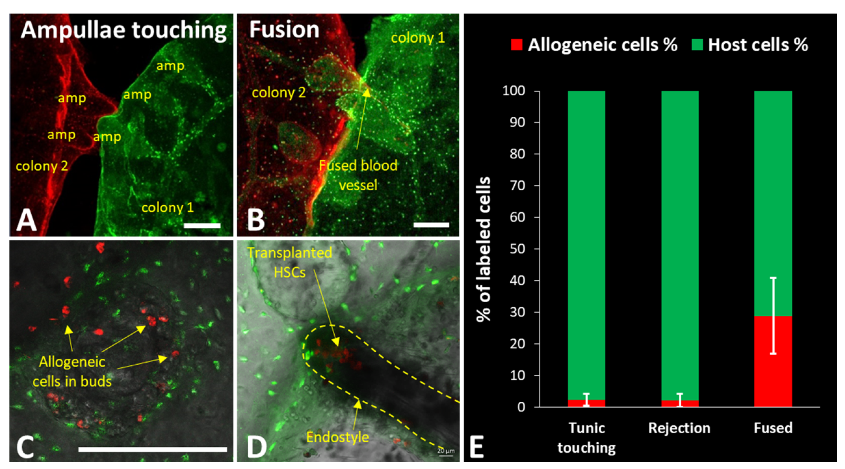
When two colonies of B. schlosseri touch with the ampullae (end point of their blood vessels), they experience a process of self/nonself recognition [36,38,56]. Colonies recognize each other as self and fuse their vasculatures to form a natural parabiont (Figure 1D top) if they share at least a single allele of the polymorphic histocompatibility gene, Botryllus histocompatibility factor (BHF) [38]. Upon the establishment of a common vasculature system, the immune cells and the stem cells can freely flow from one partner of the chimera to the other, resembling parabionts in mammalian experimental systems (Figure 2A–C). On the somatic level, chimerism can be stable for a long period of time, when both genotypes are present and visible. Since stem cells are also free to move within the chimera, a hierarchical competition of stem cells occurs in the chimeric colony where “winner” cells will be heritable dominant, giving rise to gonads of a single germline origin while maintaining somatic chimeras [33,34]. Heritable germline winner and loser strains reflect genetically determined stem-cell phenotypes [31,32,35]. Interestingly, in some cases of fused colonies, one of the partners will be resorbed. This usually happens during the blastogenic cycle, while there is a programmed cell removal of the old zooids; however, an inflammatory process also prevents the developmental process of the new zooids from buds [16,39]. This process is termed allogeneic resorption (Figure 1E).
Genetically noncompatible colonies undergo an immune rejection response where inflammatory and cytotoxic cells are involved, creating necrotic areas between the touching ampullae (Figure 1D bottom) [17,36,38]. The basis for the cytotoxicity rejection response is represented by cytotoxic morula cells (MCs), which seem to work according to the “missing self-hypothesis”, resembling human natural killer (NK) cells. Without the inhibitory recognition of compatible BHF, MCs kill the target cells, resulting in a necrotic lesion at the points of rejection (Figure 1D bottom) [17]. This allorecognition is ascribed as the invertebrate counterpart of transplantation immunity [16].

3. Zooid Resorption and Regeneration as a Model for Programed Cell Removal

A B. schlosseri colony evolves from a single tadpole-like larva that developed through classical chordate embryogenesis from a fertilized egg. Blastogenesis begins when the larva metamorphoses into a filtering oozoid that carries a bud, which develops into an individual adult zooid. Zooids grow buds that develop and replace them every week, forming colonies (Figure 1A–C). Each colony is composed of filtering adults (zooids), primary buds, and secondary buds. The blastogenetic cycle is defined by changes across these three generations which occur every week at 20 °C [24].
Zooids live for 1 week and get resorbed when the new generation replaces them. This “takeover” event is mediated by the coordination of different molecular and cellular processes, in which the circulating blood cells are involved. Before the takeover takes place, a diffuse programmed cell death is triggered, from the anterior to the posterior of the zooid [57], by a mitochondria-dependent apoptotic pathway. This has been evidenced by an increase in chromatin condensation [27], the activation of caspases (caspase-3 and -9) [58], the overexpression of mammalian apoptotic molecules (i.e., Bax, Fas, and FasL), and the downregulation of the antiapoptotic Bcl-2 [59]. Phosphatidylserine and oxidized lipids are expressed on the plasma membrane surface of affected cells as phagocyte recognition signals [27]. In the next steps of the takeover, circulating phagocytic cells (positive to CD36 antibodies) infiltrate senescent tissues, engulf apoptotic cells, and then also die by apoptosis (Figure 1C) [27,60].
Zooid resorption generates a substantial amount of biological material, which is recycled by developing buds to maintain blastogenic development [61]. The resorption of zooids and the allogeneic resorption (detailed in part E) that takes place in B. schlosseri chimeras share similarities in the immune response, as both involve the constant crosstalk among apoptosis, self/nonself recognition, and phagocytosis [62]. As this crosstalk occurs under natural conditions, B. schlosseri represents a valuable model to study, at different levels (genes, metabolites, signaling molecules, and cellular functions), the mechanisms that guide programmed cell removal, which enables the regeneration of new zooids.

4. Botryllus schlosseri as a Model to Study the Innate Immunity

B. schlosseri has an efficient immune system that not only fights and prevents infections, but also orchestrates histocompatibility, incompatibility, rejection, and zooid resorption processes. Interestingly, when its genome was annotated and compared with invertebrate and vertebrate genomes, it was found that numerous genes associated with immune system and hematopoiesis, including ZBTB1, MEFV, DSG3, NQ01, NQO2, and BHLHE40, which are involved in leukocyte development, as well as an additional set of genes that could attributed to precursors of human hematopoietic lineages, could be detected in B. schlosseri but not in other invertebrate species or solitary tunicate species [15].
Immune responses in B. schlosseri are mediated by homeostatic cell turnover and the licensing of innate cytotoxic cells, which collaborate with activated phagocytes. These immune effector cells are circulating blood cells that currently exhibit phagocytic and cytotoxic cell activity. Among phagocytic cells, B. schlosseri has a myeloid lineage that shares a large set of genes with mammalian myeloid lineages [17]; it also has amoebocytes and large phagocytes which are morphologically more related to hemocytes of arthropods and echinoderms [63]. These phagocytes engulf microorganisms and damaged self-cells, contain phagosomes with hydrolytic enzymes, lipids, and lipofuscins [64], and can be subdivided into static (in the circulatory system epithelia) and mobile (circulating through the colony) populations [65].
MCs of B. schlosseri have been characterized as cytotoxic cells; they are the most abundant circulatory cell type and have large cytoplasmic granules containing an inactive form of phenoloxidase (PO) [64]. Gene expression analysis has shown that they express a tunicate-specific gene repertoire and a set of genes (15%) sharing homology with vertebrate lymphocytes [17,63].
At the molecular level, BsTLR1 is expressed in both phagocytes and MCs of B. schlosseri as a member of the TLR receptor family, which is actively involved in self/nonself recognition [66]. Blood circulating cells also express a gene of a type II transmembrane protein that is related to CD94 and NKR-p1 receptors of human NK cells and T lymphocytes. This protein has been found to be upregulated during the allorecognition process [67]. Rhamnose-binding lectin (BsRBL) has been identified as an effector molecule that activates phagocytes, thus inducing the release of cytokine-like molecules that are recognized by the anti-IL1α and anti-TNFɣ antibodies [68]. Furthermore, activated phagocytes signal through Ras-like small GTPases, MAPKs, and NF-κB networks to trigger the recognition response of foreign cells [69].
The work in B. schlosseri has guided us to hypothesize that innate immune mechanisms during tissue maintenance, allorecognition, and regeneration are conserved and highly important for the initiation of the adaptive immune response in mammals. Therefore, this model allows the study of the orchestrating cellular and molecular processes around these immune responses, focusing on the innate immune responses. This information can then be translated to human immunity, with a particular impact on the improvement of therapeutic strategies for stem cells, tissue, and organ transplantation.
Moreover, the immune defenses of tunicates have made them a potential source of various natural drug resources with great potential for pharmacological applications. For example, the hemagglutinating activity of lectins in ascidian hemolymph has an important immune role [70]; marine lectins have been investigated as potential antimicrobial and antiviral agents, as well as compounds with immunomodulatory and cytotoxic effects on tumor cells [71]. Five homologous transcripts of rhamnose-binging lectins (RBLs) have been identified to enhance phagocytosis in B. schlosseri [70]. Furthermore, components of the complement alternative pathway (C3 and Bf orthologs) [72,73] and components of the lectin pathway (mannose-binding lectin, ficolin, and mannose-associated serine protease 1) are transcribed by MCs and associated with nonself recognition, opsonization, and clearance of microbes and apoptotic cells [74].
The previously mentioned PO enzyme, which is degranulated and released by B. schlosseri MCs, is a bioactive molecule with cytotoxic activity against microbial infections (i.e., yeast cells and bacterial spores), as well as nonself cells (i.e., incompatible blood) [70,75]. MCs are also the main source of the soluble cytokine-like proinflammatory molecules IL-1-α and TNF-α, which are suggested to be released in the presence of incompatible cells and microbes [70]. Further studies are needed to explore the drug potential of these B. schlosseri molecules and their possible pharmacological applications.
Several other compounds with antifungal, antidiabetic, antioxidant, and antitumor potential have been identified in tunicates. More in-depth information about this topic was reviewed in [76,77].

5. Botryllus as a Model for HSC Transplantation

B. schlosseri’s attributes have shown many similarities to vertebrates and mammals, whether in its blood circulation, stem-cell biology, or immune characteristics [31,33,56]. Similarly, to vertebrates, B. schlosseri stem cells reside in unique niches, where their status is assumed to be regulated on a spectrum where one end represents quiescence and the other represents differentiation/expansion. In 2005, Laird et al. showed stem-cell-based transplantation [31]; in 2008, Voskoboynik et al. successfully identified the endostyle niche and isolated somatic stem cells of B. schlosseri from it (Figure 2D) [35]. This advancement led to further studies focused on characterizing the niche and the stem cells.
HSCs, which are at the top of the hierarchy when it comes to blood/immune cells, maintain the organism’s blood and immune systems throughout its lifetime. They have been thoroughly studied in several organisms, the most important of which are humans and mice [78,79,80,81]. In 2018, Rosental et al. [17] successfully mapped whole transcriptomes of cells and tissues in the hematopoietic system of B. schlosseri, such as HSCs and their niche (endostyle), progenitor cells, and immune effector cells. They sorted 23 separate populations, mapped their transcriptomes, and identified a cluster of cells in which 235 genes were differentially upregulated, showing a significant gene activity homology to human and mammalian HSCs. Through transplantation assays, they showed those that enriched HSC differentiation to other Botryllus blood cells, as well as those home to the endostyle-niche (Figure 2D). For the characterization of the endostyle as an HSC niche, they compared transcriptome data from 10 endostyles to 34 whole colonies, finding 327 genes that were significantly elevated and shared with the upregulated genes in mouse and human hematopoietic bone marrow. This suggests a common origin for the endostyle niche in B. schlosseri, as well as the vertebrate hematopoietic bone marrow, beyond the homology HSCs and myeloid lineage-derived immune cells [17,63].
This recent work, taken together with previous research, makes B. schlosseri a complete model for HSC transplantation, considering their ability to isolate the HSCs, their interaction with the immune effector cells, and their localization to the HSC niche (Figure 2D). This includes the ability to analyze the level of transplantation-induced chimerism by flow cytometry (Figure 2E).

6. Allogeneic Resorption as a Model for Chronic Rejection

In many cases, after the fusion between two colonies of B. schlosseri, one of the semi-compatible partners will get reabsorbed within several weeks through an inflammatory process that prevents the regeneration of the new zooids (Figure 1E) [39]. The driving mechanisms of this process were elucidated by Corey et al., who identified cytotoxic MC as a key immune effector cell type in the process of allogeneic resorption [16]. The presence of MC resulted in gene expression changes in the “losing” partner that trigger cell-death programs and developmental processes defects. When allogeneic MCs were adoptively transplanted to colonies, they caused an inflammatory response preventing the development of next-generation zooids (in comparison to mock injections or non-MC donors), demonstrating that MCs are the drivers of this process. The isolation and RNA-sequencing of buds and zooids from resorbing versus non-resorbing parts of the chimeras provided a list of differentially expressed genes, revealing an upregulation in the expression of regulatory genes of host defense and proinflammatory markers.
Genes related to the complement system, such as MASP1, MASP2, and C3, were identified to have expression changes during allogeneic resorption, along with TNF-associated proteins (TRAF3 and TRAF4), coagulation components (KLKB1, KLK3, F2, F8), cell death (CASP2/7/9), and lysosomal proteinases (CTSV, CTSF). These data confirm that the process of allogeneic resorption comprises different crosstalk events which can be further studied in B. schlosseri.
The interleukin family member IL-17 resulted to be a key upregulated gene (60-fold increase). IL-17 is secreted by innate immune cells and is involved in the clearance of fungi and extracellular bacteria [82]. IL-17 acts as a key regulatory cytokine, and its upregulation results in tissue damage due to excessive inflammation, chronic inflammation [83], autoimmunity [84], and chronic GVHD in higher vertebrates [85]. In comparison, in B. schlosseri, the use of recombinant B. schlosseri IL-17 protein led to a significantly upregulated cellular cytotoxicity in a dose-dependent manner [16]. This result, taken together with the gene sets showing the upregulation of classical inflammatory responses, shows the parallels to human chronic rejection.

7. Prospect of a General Allogeneic Model

Currently, allogeneic HSC transplantation is used to treat several diseases in humans; however, this clinical process is highly complex and requires prophylactic treatment to prevent immune rejection [86]. Some of the more prominent options for prophylaxis include calcineurin inhibitors, rapamycin, mycophenolate mofetil with (or without) anti-thymocyte globulin, or, in the case of acute GVHD, use of systemic corticosteroids like methylprednisolone [87]. Despite recent advancements and the use of prophylaxis, acute GVHD is diagnosed in close to half of the allogeneic HSC transplantation procedures, and it is associated with poor prognosis [88]. Chronic rejection is only exacerbated by resistance to corticosteroid treatment; the inability to control chronic rejection leads some patients to require a re-transplantation which increases clinical risks [88]. In higher vertebrates, T cells have a major role in chronic rejection, GVHD, and pregnancy disorders [89,90]. Elucidating the immune-related mechanisms behind the activation of these effector cells in an allogeneic setting will give us a better understanding of how to circumvent their cytolytic activation and positively modulate the process of chronic rejection. NK cells and T cells in humans share the characteristic of identifying allogeneic self/nonself and are activated by either the identification of nonself or the lack of self.
In B. schlosseri allogeneic rejection occurs in a similar fashion to vertebrates, despite its more innate immune-based system. As previously discussed, BHF in B. schlosseri shares some attributes with human MHC [91], in that its recognition as “self” leads to a major inhibitory mechanism of cytotoxicity in allorecognition. BHF’s inhibitory effects on cytotoxicity [17], combined with observational evidence of fusion of colonies that share at least one BHF allele, suggest that the mechanism of cellular toxicity during allorecognition in this tunicate stems from the ‘missing self’ and can be compared to recognition by NK cells in advanced vertebrates [63]. Taken together, these findings demonstrate similarities in the innate immune responses between B. schlosseri and humans on the cellular and molecular levels. While the driving force in each organism is different, points of interaction such as immune pathways and key recognition molecules could be studied in an easy access model and then translated into mammalian complex models.

8. Conclusions

The advancements in research and scientific tools in the B. schlosseri model have promoted B. schlosseri as an interesting model to study innate immune system responses, more specifically in transplantations. One of the more prominent research tools, the Botryllus genome project [15], enabled analysis on the genetic and molecular levels, thus identifying the BHF [38] on the fusion histocompatibility locus [92]. Moreover, the genome project confirmed the location of the tunicates as the closest invertebrate group to vertebrates, whereby many immune genes are shared with mammals and their hematopoietic system [15].
Furthermore, as mentioned above, B. schlosseri has natural occurring phenomena which resemble many basic immunological processes, such as (I) rejection as acute rejection in transplantation, (II) fusion as natural parabionts that share stem cells, (III) stem-cell competition and chimerism, (IV) natural weekly cycle of zooid resorption and new bud development, working through classical programmed cell removal and regeneration mechanisms comparable to vertebrates, and (V) allogeneic resorption as a chronic rejection process.
Moreover, there were several gene expression sets obtained during the research of the above-described processes, which enabled the search of candidate genes and pathways that affect those immune-associated processes, for example, gene sets of allogeneic resorption [16], fusion and histocompatibility-associated genes [38], 23 different cellular populations and endostyle [17], and zooid regeneration and developmental processes [23].
Taken together, the tools and advancements in B. schlosseri, along with the ability of in vivo and ex vivo cellular immune profiles, with cellular and molecular manipulations (from morpholinos to recombinant proteins), represent the foundation for future discoveries on immune activation mechanisms in a simple model, which is relevant for human immune research.

Author Contributions

Conceptualization, B.R.; writing—original draft preparation, O.G., E.A.M.-T., T.L., S.T. and B.R.; writing—review and editing, O.G., E.A.M.-T., T.L., S.T., T.R., A.V., B.R. and O.G.-Y.; supervision, B.R.; project administration, O.G.-Y.; funding acquisition, A.V. and B.R. All authors have read and agreed to the published version of the manuscript.

Funding

The work of B.R. is supported by the Israel Science Foundation (ISF), grant number 1416/19, and HFSP Research Grant, RGY0085/2019. B.R. has received funding from the European Research Council (ERC) under the European Union’s Horizon 2020 research and innovation program under grant agreement No. 948476. A.V. is supported by the NIH R21AG062948 and the Chan Zuckerberg investigator program.

Institutional Review Board Statement

Not applicable.

Informed Consent Statement

Not applicable.

Conflicts of Interest

The authors declare no conflict of interest.

References

  1. Oberbarnscheidt, M.H.; Lakkis, F.G. Innate allorecognition. Immunol. Rev. 2014, 258, 145–149. [Google Scholar] [CrossRef] [Green Version]
  2. Zecher, D.; Li, Q.; Williams, A.L.; Walters, J.T.; Baddoura, F.K.; Chalasani, G.; Rothstein, D.M.; Shlomchik, W.D.; Demetris, A.J.; Lakkis, F.G. Innate immunity alone is not sufficient for chronic rejection but predisposes healed allografts to T cell-mediated pathology. Transpl. Immunol. 2012, 26, 113–118. [Google Scholar] [CrossRef] [PubMed] [Green Version]
  3. Ghadially, H.; Ohana, M.; Elboim, M.; Gazit, R.; Gur, C.; Nagler, A.; Mandelboim, O. NK cell receptor NKp46 regulates graft-versus-host disease. Cell Rep. 2014, 7, 1809–1814. [Google Scholar] [CrossRef] [Green Version]
  4. Simonetta, F.; Alvarez, M.; Negrin, R.S. Natural Killer Cells in Graft-versus-Host-Disease after Allogeneic Hematopoietic Cell Transplantation. Front. Immunol. 2017, 8, 465. [Google Scholar] [CrossRef]
  5. Geldenhuys, J.; Rossouw, T.M.; Lombaard, H.A.; Ehlers, M.M.; Kock, M.M. Disruption in the Regulation of Immune Responses in the Placental Subtype of Preeclampsia. Front. Immunol. 2018, 9, 1659. [Google Scholar] [CrossRef]
  6. Gomez-Lopez, N.; Stlouis, D.; Lehr, M.A.; Sanchez-Rodriguez, E.N.; Arenas-Hernandez, M. Immune cells in term and preterm labor. Cell. Mol. Immunol. 2014, 11, 571–581. [Google Scholar] [CrossRef] [Green Version]
  7. Kwak-Kim, J.; Bao, S.; Lee, S.K.; Kim, J.W.; Gilman-Sachs, A. Immunological Modes of Pregnancy Loss: Inflammation, Immune Effectors, and Stress. Am. J. Reprod. Immunol. 2014, 72, 129–140. [Google Scholar] [CrossRef] [PubMed]
  8. Moffett, A.; Chazara, O.; Colucci, F. Maternal allo-recognition of the fetus. Fertil. Steril. 2017, 107, 1269–1272. [Google Scholar] [CrossRef] [Green Version]
  9. Rosental, B.; Brusilovsky, M.; Hadad, U.; Oz, D.; Appel, M.Y.; Afergan, F.; Yossef, R.; Rosenberg, L.A.; Aharoni, A.; Cerwenka, A.; et al. Proliferating Cell Nuclear Antigen Is a Novel Inhibitory Ligand for the Natural Cytotoxicity Receptor NKp44. J. Immunol. 2011, 187, 5693–5702. [Google Scholar] [CrossRef] [PubMed]
  10. Shemesh, A.; Kugel, A.; Steiner, N.; Yezersky, M.; Tirosh, D.; Edri, A.; Teltsh, O.; Rosental, B.; Sheiner, E.; Rubin, E.; et al. NKp44 and NKp30 splice variant profiles in decidua and tumor tissues: A comparative viewpoint. Oncotarget 2016, 7, 70912–70923. [Google Scholar] [CrossRef] [PubMed]
  11. Shemesh, A.; Tirosh, D.; Sheiner, E.; Tirosh, N.B.; Brusilovsky, M.; Segev, R.; Rosental, B.; Porgador, A. First Trimester Pregnancy Loss and the Expression of Alternatively Spliced NKp30 Isoforms in Maternal Blood and Placental Tissue. Front. Immunol. 2015, 6, 189. [Google Scholar] [CrossRef] [Green Version]
  12. Azumi, K.; De Santis, R.; De Tomaso, A.; Rigoutsos, I.; Yoshizaki, F.; Pinto, M.R.; Marino, R.; Shida, K.; Ikeda, M.; Ikeda, M.; et al. Genomic analysis of immunity in a Urochordate and the emergence of the vertebrate immune system: “waiting for Godot”. Immunogenetics 2003, 55, 570–581. [Google Scholar] [CrossRef]
  13. Flajnik, M.F. A cold-blooded view of adaptive immunity. Nat. Rev. Immunol. 2018, 18, 438–453. [Google Scholar] [CrossRef]
  14. Hirano, M.; Das, S.; Guo, P.; Cooper, M.D. Chapter 4—The Evolution of Adaptive Immunity in Vertebrates. In Advances in Immunology; Frederick, W., Ed.; Academic Press: Cambridge, MA, USA, 2011; Volume 109, pp. 125–157. [Google Scholar] [CrossRef]
  15. Voskoboynik, A.; Neff, N.F.; Sahoo, D.; Newman, A.M.; Pushkarev, D.; Koh, W.; Passarelli, B.; Fan, H.C.; Mantalas, G.L.; Palmeri, K.J.; et al. The genome sequence of the colonial chordate, Botryllus schlosseri. eLife 2013, 2, e00569. [Google Scholar] [CrossRef]
  16. Corey, D.M.; Rosental, B.; Kowarsky, M.; Sinha, R.; Ishizuka, K.J.; Palmeri, K.J.; Quake, S.R.; Voskoboynik, A.; Weissman, I.L. Developmental cell death programs license cytotoxic cells to eliminate histocompatible partners. Proc. Natl. Acad. Sci. USA 2016, 113, 6520–6525. [Google Scholar] [CrossRef] [Green Version]
  17. Rosental, B.; Kowarsky, M.; Seita, J.; Corey, D.M.; Ishizuka, K.J.; Palmeri, K.J.; Chen, S.-Y.; Sinha, R.; Okamoto, J.; Mantalas, G.; et al. Complex mammalian-like haematopoietic system found in a colonial chordate. Nature 2018, 564, 425. [Google Scholar] [CrossRef]
  18. Ben-Shlomo, R.; Reem, E.; Douek, J.; Rinkevich, B. Population genetics of the invasive ascidian Botryllus schlosseri from South American coasts. Mar. Ecol. Prog. Ser. 2010, 412, 85. [Google Scholar] [CrossRef]
  19. Stoner, D.S.; Ben-Shlomo, R.; Rinkevich, B.; Weissman, I.L. Genetic variability of Botryllus schlosseri invasions to the east and west coasts of the USA. Mar. Ecol. Prog. Ser. 2002, 243, 93. [Google Scholar] [CrossRef]
  20. Darwin, C.; Kebler, L. On the Origin of Sspecies by Means of Natural Selection, or, The Preservation of Favoured Races in the Struggle for Life; J. Murray: London, UK, 1859; Volume 1, p. 502. [Google Scholar]
  21. Delsuc, F.; Brinkmann, H.; Chourrout, D.; Philippe, H. Tunicates and not cephalochordates are the closest living relatives of vertebrates. Nature 2006, 439, 965. [Google Scholar] [CrossRef] [PubMed]
  22. Manni, L.; Lane, N.J.; Joly, J.S.; Gasparini, F.; Tiozzo, S.; Caicci, F.; Zaniolo, G.; Burighel, P. Neurogenic and non-neurogenic placodes in ascidians. J. Exp. Zoology. Part B Mol. Dev. Evol. 2004, 302, 483. [Google Scholar] [CrossRef]
  23. Kowarsky, M.; Anselmi, C.; Hotta, K.; Burighel, P.; Zaniolo, G.; Caicci, F.; Rosental, B.; Neff, N.F.; Ishizuka, K.J.; Palmeri, K.J.; et al. Sexual and asexual development: Two distinct programs producing the same tunicate. Cell Rep. 2021, 34, 108681. [Google Scholar] [CrossRef]
  24. Manni, L.; Gasparini, F.; Hotta, K.; Ishizuka, K.J.; Ricci, L.; Tiozzo, S.; Voskoboynik, A.; Dauga, D. Ontology for the Asexual Development and Anatomy of the Colonial Chordate Botryllus schlosseri. PLoS ONE 2014, 9, e96434. [Google Scholar] [CrossRef] [Green Version]
  25. Manni, L.; Burighel, P. Common and divergent pathways in alternative developmental processes of ascidians. BioEssays 2006, 28, 902–912. [Google Scholar] [CrossRef]
  26. Milkman, R. Genetic and developmental studies on Botryllus schlosseri. Biol. Bull. 1967, 132, 229–243. [Google Scholar] [CrossRef] [PubMed]
  27. Cima, F.; Basso, G.; Ballarin, L. Apoptosis and phosphatidylserine-mediated recognition during the take-over phase of the colonial life-cycle in the ascidian Botryllus schlosseri. Cell Tissue Res. 2003, 312, 369–376. [Google Scholar] [CrossRef] [PubMed]
  28. Lauzon, R.J.; Ishizuka, K.J.; Weissman, I.L. A cyclical, developmentally-regulated death phenomenon in a colonial urochordate. Dev. Dyn. 1992, 194, 71–83. [Google Scholar] [CrossRef] [PubMed]
  29. Lauzon, R.J.; Patton, C.W.; Weissman, I.L. A morphological and immunohistochemical study of programmed cell death in Botryllus schlosseri (Tunicata, Ascidiacea). Cell Tissue Res. 1993, 272, 115–127. [Google Scholar] [CrossRef]
  30. Laird, D.J.; De Tomaso, A.W.; Cooper, M.D.; Weissman, I.L. 50 million years of chordate evolution: Seeking the origins of adaptive immunity. Proc. Natl. Acad. Sci. USA 2000, 97, 6924–6926. [Google Scholar] [CrossRef] [PubMed] [Green Version]
  31. Laird, D.J.; De Tomaso, A.W.; Weissman, I.L. Stem Cells are Units of Natural Selection in a Colonial Ascidian. Cell 2005, 123, 1351–1360. [Google Scholar] [CrossRef] [Green Version]
  32. Rinkevich, Y.; Voskoboynik, A.; Rosner, A.; Rabinowitz, C.; Paz, G.; Oren, M.; Douek, J.; Alfassi, G.; Moiseeva, E.; Ishizuka, K.J.; et al. Repeated, Long-Term Cycling of Putative Stem Cells between Niches in a Basal Chordate. Dev. Cell 2013, 24, 76–88. [Google Scholar] [CrossRef] [Green Version]
  33. Stoner, D.S.; Rinkevich, B.; Weissman, I.L. Heritable germ and somatic cell lineage competitions in chimeric colonial protochordates. Proc. Natl. Acad. Sci. USA 1999, 96, 9148–9153. [Google Scholar] [CrossRef] [PubMed] [Green Version]
  34. Stoner, D.S.; Weissman, I.L. Somatic and germ cell parasitism in a colonial ascidian: Possible role for a highly polymorphic allorecognition system. Proc. Natl. Acad. Sci. USA 1996, 93, 15254–15259. [Google Scholar] [CrossRef] [PubMed] [Green Version]
  35. Voskoboynik, A.; Soen, Y.; Rinkevich, Y.; Rosner, A.; Ueno, H.; Reshef, R.; Ishizuka, K.J.; Palmeri, K.J.; Moiseeva, E.; Rinkevich, B.; et al. Identification of the Endostyle as a Stem Cell Niche in a Colonial Chordate. Cell Stem Cell 2008, 3, 456–464. [Google Scholar] [CrossRef] [PubMed] [Green Version]
  36. Sabbadin, A. Le basi genetiche della capacita di fusione fra colonie in Botryllus schlosseri (Ascidiacea). Rend Accad Naz Lincei Ser VIII 1962, 32, 1031–1035. [Google Scholar]
  37. Scofield, V. Allorecognition and microbial infection: Roles in the evolution of sex and immunity. In The Origin and Evolution of Sex; Halvorson, H., Monroy, A., Eds.; Alan R. Liss: New York, NY, USA, 1985; Volume 7, p. 213. [Google Scholar]
  38. Voskoboynik, A.; Newman, A.M.; Corey, D.M.; Sahoo, D.; Pushkarev, D.; Neff, N.F.; Passarelli, B.; Koh, W.; Ishizuka, K.J.; Palmeri, K.J.; et al. Identification of a Colonial Chordate Histocompatibility Gene. Science 2013, 341, 384. [Google Scholar] [CrossRef] [Green Version]
  39. Rinkevich, B.; Tartakover, S.; Gershon, H. Contribution of morula cells to allogeneic responses in the colonial urochordate Botryllus schlosseri. Mar. Biol. 1998, 131, 227–236. [Google Scholar] [CrossRef]
  40. Ueno, H.; Weissman, I.L. Clonal Analysis of Mouse Development Reveals a Polyclonal Origin for Yolk Sac Blood Islands. Dev. Cell 2006, 11, 519. [Google Scholar] [CrossRef] [Green Version]
  41. Ueno, H.; Turnbull, B.B.; Weissman, I.L. Two-step oligoclonal development of male germ cells. Proc. Natl. Acad. Sci. USA 2009, 106, 175–180. [Google Scholar] [CrossRef] [Green Version]
  42. Weissman, I.L. Stem cells are units of natural selection for tissue formation, for germline development, and in cancer development. Proc. Natl. Acad. Sci. USA 2015, 112, 8922–8928. [Google Scholar] [CrossRef] [Green Version]
  43. Beerman, I.; Bhattacharya, D.; Zandi, S.; Sigvardsson, M.; Weissman, I.L.; Bryder, D.; Rossi, D.J. Functionally distinct hematopoietic stem cells modulate hematopoietic lineage potential during aging by a mechanism of clonal expansion. Proc. Natl. Acad. Sci. USA 2010, 107, 5465–5470. [Google Scholar] [CrossRef] [Green Version]
  44. Pang, W.W.; Price, E.A.; Sahoo, D.; Beerman, I.; Maloney, W.J.; Rossi, D.J.; Schrier, S.L.; Weissman, I.L. Human bone marrow hematopoietic stem cells are increased in frequency and myeloid-biased with age. Proc. Natl. Acad. Sci. USA 2011, 108, 20012–20017. [Google Scholar] [CrossRef] [Green Version]
  45. Rossi, D.J.; Bryder, D.; Seita, J.; Nussenzweig, A.; Hoeijmakers, J.; Weissman, I.L. Deficiencies in DNA damage repair limit the function of haematopoietic stem cells with age. Nat. Cell Biol. 2007, 447, 725–729. [Google Scholar] [CrossRef]
  46. Rossi, D.J.; Bryder, D.; Zahn, J.M.; Ahlenius, H.; Sonu, R.; Wagers, A.J.; Weissman, I.L. Cell intrinsic alterations underlie hematopoietic stem cell aging. Proc. Natl. Acad. Sci. USA 2005, 102, 9194–9199. [Google Scholar] [CrossRef] [Green Version]
  47. Rossi, D.J.; Jamieson, C.H.; Weissman, I.L. Stems Cells and the Pathways to Aging and Cancer. Cell 2008, 132, 681–696. [Google Scholar] [CrossRef] [Green Version]
  48. Corces, M.; Hong, W.-J.; Weissman, I.L.; Medeiros, B.C.; Majeti, R. Preleukemic mutations in human acute myeloid leukemia affect epigenetic regulators and persist in remission. Proc. Natl. Acad. Sci. USA 2014, 111, 2548–2553. [Google Scholar] [CrossRef] [PubMed] [Green Version]
  49. Jaiswal, S.; Jamieson, C.H.; Pang, W.W.; Park, C.Y.; Chao, M.P.; Majeti, R.; Traver, D.; Van Rooijen, N.; Weissman, I.L. CD47 Is Upregulated on Circulating Hematopoietic Stem Cells and Leukemia Cells to Avoid Phagocytosis. Cell 2009, 138, 271–285. [Google Scholar] [CrossRef] [PubMed] [Green Version]
  50. Jamieson, C.H.; Ailles, L.E.; Dylla, S.J.; Muijtjens, M.; Jones, C.; Zehnder, J.L.; Gotlib, J.; Li, K.; Manz, M.G.; Keating, A.; et al. Granulocyte-macrophage progenitors as candidate leukemic stem cells in blast-crisis CML. N. Engl. J. Med. 2004, 351, 657–667. [Google Scholar] [CrossRef] [PubMed] [Green Version]
  51. Jan, M.; Snyder, T.M.; Corces-Zimmerman, M.R.; Vyas, P.; Weissman, I.L.; Quake, S.R.; Majeti, R. Clonal Evolution of Preleukemic Hematopoietic Stem Cells Precedes Human Acute Myeloid Leukemia. Sci. Transl. Med. 2012, 4, 149ra118. [Google Scholar] [CrossRef] [Green Version]
  52. Miyamoto, T.; Weissman, I.L.; Akashi, K. AML1/ETO-expressing nonleukemic stem cells in acute myelogenous leukemia with 8;21 chromosomal translocation. Proc. Natl. Acad. Sci. USA 2000, 97, 7521–7526. [Google Scholar] [CrossRef] [Green Version]
  53. Sykes, S.M.; Kokkaliaris, K.D.; Milsom, M.D.; Levine, R.L.; Majeti, R. Clonal evolution of preleukemic hematopoietic stem cells in acute myeloid leukemia. Exp. Hematol. 2015, 43, 989–992. [Google Scholar] [CrossRef] [PubMed] [Green Version]
  54. Kojima, Y.; Volkmer, J.-P.; McKenna, K.; Civelek, M.; Lusis, A.J.; Miller, C.L.; Direnzo, D.; Nanda, V.; Ye, J.; Connolly, A.J.; et al. CD47-blocking antibodies restore phagocytosis and prevent atherosclerosis. Nature 2016, 536, 86. [Google Scholar] [CrossRef] [Green Version]
  55. Wang, B.; Zhang, F.; Wang, S.; Yang, R.; Chen, C.; Zhao, W. Imaging endogenous HClO in atherosclerosis using a novel fast-response fluorescence probe. Chem. Commun. 2020, 56, 2598. [Google Scholar] [CrossRef] [PubMed]
  56. Scofield, V.L.; Schlumpberger, J.M.; West, L.A.; Weissman, I.L. Protochordate allorecognition is controlled by a MHC-like gene system. Nature 1982, 295, 499. [Google Scholar] [CrossRef] [PubMed]
  57. Burighel, P.; Schiavinato, A. Degenerative regression of the digestive tract in the colonial ascidian Botryllus schlosseri (Pallas). Cell Tissue Res. 1984, 235, 309–318. [Google Scholar] [CrossRef] [PubMed]
  58. Ballarin, L.; Burighel, P.; Cima, F. A Tale of Death and Life: Natural Apoptosis in the Colonial Ascidian Botryllus schlosseri (Urochordata, Ascidiacea). Curr. Pharm. Des. 2008, 14, 138. [Google Scholar] [CrossRef] [PubMed]
  59. Ballarin, L.; Schiavon, F.; Manni, L. Natural Apoptosis during the Blastogenetic Cycle of the Colonial Ascidian Botryllus schlosseri: A Morphological Analysis. Zoöl. Sci. 2010, 27, 96. [Google Scholar] [CrossRef]
  60. Voskoboynik, A.; Rinkevich, B.; Weiss, A.; Moiseeva, E.; Reznick, A.Z. Macrophage involvement for successful degeneration of apoptotic organs in the colonial urochordate Botryllus schlosseri. J. Exp. Biol. 2004, 207, 2409. [Google Scholar] [CrossRef] [Green Version]
  61. Lauzon, R.J.; Ishizuka, K.J.; Weissman, I.L. Cyclical Generation and Degeneration of Organs in a Colonial Urochordate Involves Crosstalk between Old and New: A Model for Development and Regeneration. Dev. Biol. 2002, 249, 333–348. [Google Scholar] [CrossRef] [Green Version]
  62. Rinkevich, B.; Weissman, I.L. Botryllus schlosseri (tunicata) whole colony irradiation: Do senescent zooid resorption and immunological resorption involve similar recognition events? J. Exp. Zoöl. 1990, 253, 189. [Google Scholar] [CrossRef]
  63. Rosental, B.; Raveh, T.; Voskoboynik, A.; Weissman, I.L. Evolutionary perspective on the hematopoietic system through a colonial chordate: Allogeneic immunity and hematopoiesis. Curr. Opin. Immunol. 2020, 62, 91. [Google Scholar] [CrossRef]
  64. Franchi, N.; Ballarin, L. Immunity in Protochordates: The Tunicate Perspective. Front. Immunol. 2017, 8, 674. [Google Scholar] [CrossRef] [PubMed]
  65. Lauzon, R.J.; Brown, C.; Kerr, L.; Tiozzo, S. Phagocyte dynamics in a highly regenerative urochordate: Insights into development and host defense. Dev. Biol. 2013, 374, 357–373. [Google Scholar] [CrossRef] [PubMed] [Green Version]
  66. Peronato, A.; Franchi, N.; Loriano, B. BsTLR1: A new member of the TLR family of recognition proteins from the colonial ascidian Botryllus schlosseri. Fish Shellfish. Immunol. 2020, 106, 967. [Google Scholar] [CrossRef] [PubMed]
  67. Khalturin, K.; Becker, M.; Rinkevich, B.; Bosch, T.C. Urochordates and the origin of natural killer cells: Identification of a CD94/NKR-P1-related receptor in blood cells of Botryllus. Proc. Natl. Acad. Sci. USA 2003, 100, 622–627. [Google Scholar] [CrossRef] [PubMed] [Green Version]
  68. Franchi, N.; Schiavon, F.; Carletto, M.; Gasparini, F.; Bertoloni, G.; Tosatto, S.C.; Ballarin, L. Immune roles of a rhamnose-binding lectin in the colonial ascidian Botryllus schlosseri. Immunobiolgy 2011, 216, 725–736. [Google Scholar] [CrossRef]
  69. Franchi, N.; Schiavon, F.; Betti, M.; Canesi, L.; Ballarin, L. Insight on signal transduction pathways involved in phagocytosis in the colonial ascidian Botryllus schlosseri. J. Invertebr. Pathol. 2013, 112, 260–266. [Google Scholar] [CrossRef]
  70. Ballarin, L. Immunobiology of compound ascidians, with particular reference to Botryllus schlosseri: State of art. Invertebr. Surviv. J. 2008, 5, 54. [Google Scholar]
  71. Cheung, R.C.F.; Wong, J.H.; Pan, W.; Chan, Y.S.; Yin, C.M.; Dan, X.L.; Ng, T.B. Marine lectins and their medicinal applications. Appl. Microbiol. Biotechnol. 2015, 99, 3755. [Google Scholar] [CrossRef] [PubMed]
  72. Franchi, N.; Ballarin, L. Preliminary characterization of complement in a colonial tunicate: C3, Bf and inhibition of C3 opsonic activity by compstatin. Dev. Comp. Immunol. 2014, 46, 430. [Google Scholar] [CrossRef]
  73. Peronato, A.; Drago, L.; Rothbacher, U.; Macor, P.; Ballarin, L.; Franchi, N. Complement system and phagocytosis in a colonial protochordate. Dev. Comp. Immunol. 2020, 103, 103530. [Google Scholar] [CrossRef]
  74. Nicola, F.; Loriano, B. Morula cells as key hemocytes of the lectin pathway of complement activation in the colonial tunicate Botryllus schlosseri. Fish Shellfish. Immunol. 2017, 63, 157. [Google Scholar] [CrossRef]
  75. Ballarin, L.; Menin, A.; Franchi, N.; Bertoloni, G.; Cima, F. Morula cells and non-self recognition in the compound ascidian Botryllus schlosseri. Invertebr. Surviv. J. 2005, 2, 1. [Google Scholar]
  76. Palanisamy, S.K.; Rajendran, N.M.; Marino, A. Natural Products Diversity of Marine Ascidians (Tunicates; Ascidiacea) and Successful Drugs in Clinical Development. Nat. Prod. Bioprosp. 2017, 7, 1–111. [Google Scholar] [CrossRef] [Green Version]
  77. Ramesh, C.; Tulasi, B.R.; Raju, M.; Thakur, N.; Dufosse, L. Marine Natural Products from Tunicates and Their Associated Microbes. Mar. Drugs 2021, 19, 308. [Google Scholar] [CrossRef]
  78. Copelan, E.A. Hematopoietic Stem-Cell Transplantation. N. Engl. J. Med. 2006, 354, 1813. [Google Scholar] [CrossRef] [PubMed]
  79. Doulatov, S.; Notta, F.; Laurenti, E.; Dick, J.E. Hematopoiesis: A Human Perspective. Cell Stem Cell 2012, 10, 120. [Google Scholar] [CrossRef] [PubMed] [Green Version]
  80. Osawa, M.; Hanada, K.-I.; Hamada, H.; Nakauchi, H. Long-Term Lymphohematopoietic Reconstitution by a Single CD34-Low/Negative Hematopoietic Stem Cell. Science 1996, 273, 242–245. [Google Scholar] [CrossRef] [PubMed] [Green Version]
  81. Spangrude, G.J.; Heimfeld, S.; Weissman, I.L. Purification and characterization of mouse hematopoietic stem cells. Science 1988, 241, 58–62. [Google Scholar] [CrossRef]
  82. Weaver, C.T.; Hatton, R.D.; Mangan, P.R.; Harrington, L.E. IL-17 Family Cytokines and the Expanding Diversity of Effector T Cell Lineages. Annu. Rev. Immunol. 2007, 25, 821. [Google Scholar] [CrossRef] [PubMed] [Green Version]
  83. Miossec, P.; Kolls, J.K. Targeting IL-17 and TH17 cells in chronic inflammation. Nat. Rev. Drug Discov. 2012, 11, 763. [Google Scholar] [CrossRef] [PubMed]
  84. Zhu, S.; Qian, Y. IL-17/IL-17 receptor system in autoimmune disease: Mechanisms and therapeutic potential. Clin. Sci. 2012, 122, 487–511. [Google Scholar] [CrossRef] [Green Version]
  85. van der Waart, A.B.; van der Velden, W.J.; Blijlevens, N.M.; Dolstra, H. Targeting the IL17 Pathway for the Prevention of Graft-Versus-Host Disease. Biol. Blood Marrow Transpl. 2014, 20, 752. [Google Scholar] [CrossRef] [Green Version]
  86. Carnevale-Schianca, F.; Leisenring, W.; Martin, P.J.; Furlong, T.; Schoch, G.; Anasetti, C.; Appelbaum, F.R.; Carpenter, P.A.; Deeg, H.J.; Kiem, H.-P.; et al. Longitudinal Assessment of Morbidity and Acute Graft-versus-Host Disease after Allogeneic Hematopoietic Cell Transplantation: Retrospective Analysis of a Multicenter Phase III Study. Biol. Blood Marrow Transpl. 2009, 15, 749. [Google Scholar] [CrossRef] [Green Version]
  87. Holt, C.D. Overview of Immunosuppressive Therapy in Solid Organ Transplantation. Anesthesiol. Clin. 2017, 35, 365. [Google Scholar] [CrossRef]
  88. Ustun, C.; Jillella, A.; Shah, R.; Sterling, K.; DeRemer, D.; Savage, N.; Awan, F.; Gossage, J.R.; Dillard, T.; Martin, P.J. Second allo-SCT from a different donor can improve severe steroid-resistant gut GVHD. Bone Marrow Transpl. 2010, 45, 1658. [Google Scholar] [CrossRef] [PubMed]
  89. Lindahl, K.F.; Wilson, D.B. Histocompatibility antigen-activated cytotoxic T lymphocytes. II. Estimates of the frequency and specificity of precursors. J. Exp. Med. 1977, 145, 508. [Google Scholar] [CrossRef] [PubMed] [Green Version]
  90. Macdonald, W.A.; Chen, Z.; Gras, S.; Archbold, J.; Tynan, F.E.; Clements, C.S.; Bharadwaj, M.; Kjer-Nielsen, L.; Saunders, P.M.; Wilce, M.C.; et al. T Cell Allorecognition via Molecular Mimicry. Immunity 2009, 31, 897. [Google Scholar] [CrossRef] [Green Version]
  91. Taketa, D.A.; De Tomaso, A.W. Botryllus schlosseri allorecognition: Tackling the enigma. Dev. Comp. Immunol. 2015, 48, 254. [Google Scholar] [CrossRef] [Green Version]
  92. De Tomaso, A.W.; Nyholm, S.V.; Palmeri, K.J.; Ishizuka, K.J.; Ludington, W.B.; Mitchel, K.; Weissman, I.L. Isolation and characterization of a protochordate histocompatibility locus. Nature 2005, 438, 454. [Google Scholar] [CrossRef] [PubMed]
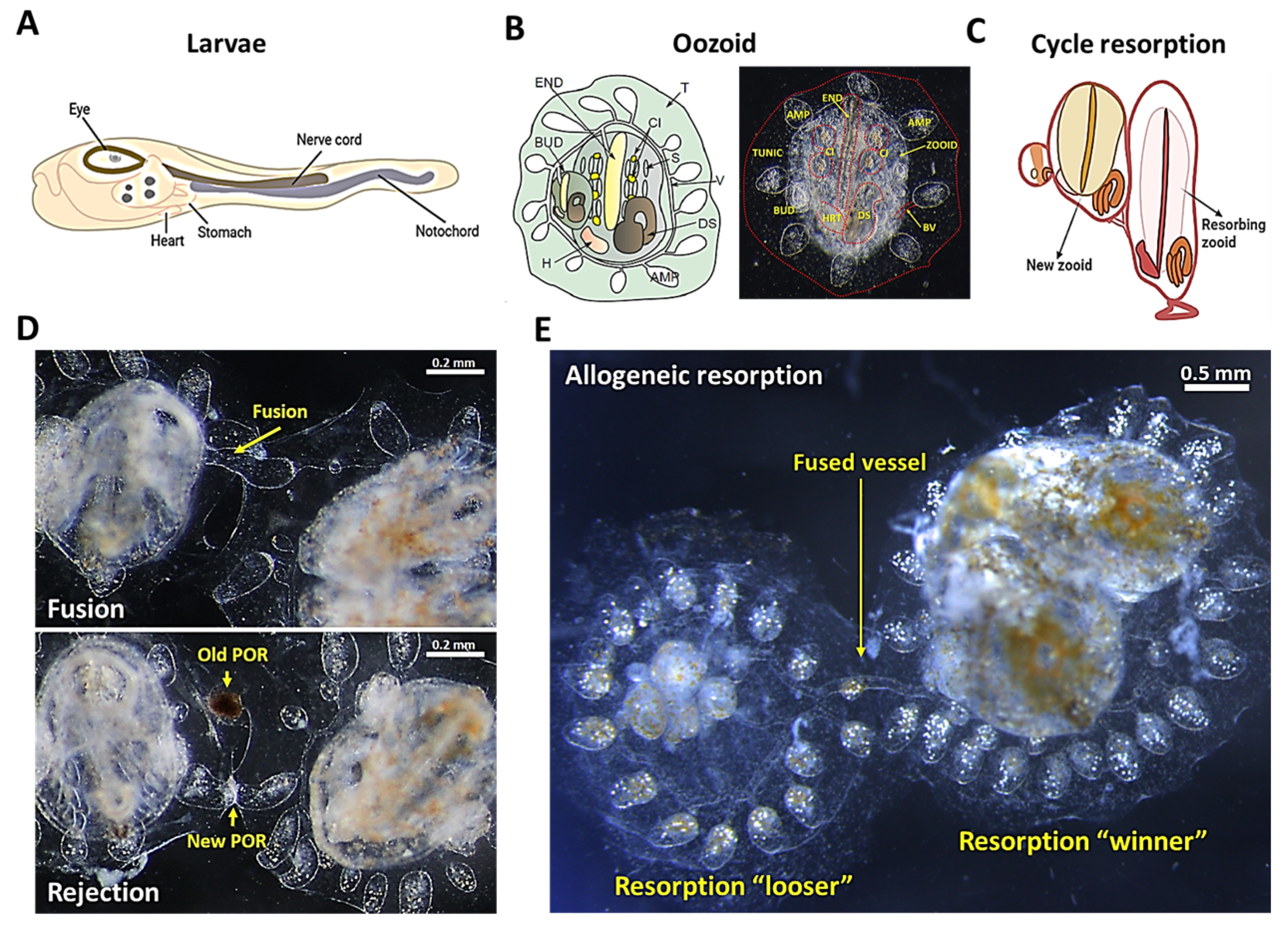
Figure 1. The anatomy of B. schlosseri and its different levels of naturally occurring immune responses. (A) Diagram of the tunicate larvae tadpole phase showing the nerve cord and the notochord. (B) Diagram and live imaging of the ventral view of a zooid (Z) and primary bud (BUD), embedded within a tunic (TUN) and connected with vasculature (V), which terminates in ampullae (AMP). The zooid has a branchial sac conformed by the endostyle (END), stigmata (S), cell islands (CI), digestive system (DS), and heart (H). (C) Diagram showing the “takeover” phase in the weekly cycle of zooid regeneration mediated by noninflammatory programmed cell removal of the resorbing old zooid. (D) Live imaging of two B. schlosseri colonies undergoing fusion (arrows show fused vasculature (top)) and rejection (arrows show points of rejection (POR) (bottom)). (E) Live imaging from the allogeneic resorption process, where one colony is the “loser” (which is resorbed), while the other is the “winner”, demonstrating normal developmental stages. (A,C) were created using BioRender; (B,D) were reproduced with permission from [17], Springer Nature Limited, Berlin, Germany, 2018; (E) was reproduced with permission from [16], National Academy of Sciences, 2016.
Marinedrugs 19 00454 g001
Figure 2. Transplantation in vivo using exogenous labeling in B. schlosseri. (A) Two colonies differentially labeled with CFSE (green) and CellTracker Deep Red, prior to fusion. (B) Fused colonies through the blood vessel and exchanged allogeneic cells can be seen in the confocal image of the live colonies. The transparent body of B. schlosseri is used to follow transplantation in vivo. (C) Transplanted allogeneic cells in a developing secondary bud, followed in a live colony. (D) Transplanted HSCs (exogenous lipophilic dye DiD, red) in the endostyle stem-cell niche in live animals. Bars = 200 µm. (E) Measurement by flow cytometry of transplanted allogeneic cell abundance 3 weeks after transplantation; average and SD of three pairs of touching animals without response, rejecting animals, and fused colonies. This shows the ability of measurement of transplantation success or modulation. (A,B,D) were reproduced with permission from [17], Springer Nature Limited, 2018; (C) was reproduced with permission from [16], National Academy of Sciences, 2016.
Marinedrugs 19 00454 g002
Publisher’s Note: MDPI stays neutral with regard to jurisdictional claims in published maps and institutional affiliations.