Article

Highly Oxygenated Constituents from a Marine Alga-Derived Fungus Aspergillus giganteus NTU967

1
Faculty of Pharmacy, School of Pharmaceutical Sciences, National Yang-Ming University, Taipei 11221, Taiwan
2
Department of Medical Research, China Medical University Hospital, China Medical University, Taichung 404, Taiwan
3
Department of Medicine, Mackay Medical College, New Taipei City 25245, Taiwan
4
Graduate Institute of Natural Products, College of Pharmacy, Kaohsiung Medical University, Kaohsiung 80708, Taiwan
5
Biodiversity Research Center, Academia Sinica, Taipei 11529, Taiwan
6
Institute of Marine Biology and Center of Excellence for the Oceans, National Taiwan Ocean University, Keelung 20224, Taiwan
7
Department of Chinese Pharmaceutical Sciences and Chinese Medicine Resources, China Medical University, Taichung 40447, Taiwan
8
Department of Biotechnology, Asia University, Taichung 41354, Taiwan
9
Chinese Medical Research Center, China Medical University, Taichung 40447, Taiwan
10
Institute of Fisheries Science, National Taiwan University, Taipei 10617, Taiwan
*
Author to whom correspondence should be addressed.
These authors contributed equally to this work.
Mar. Drugs 2020, 18(6), 303; https://doi.org/10.3390/md18060303
Submission received: 9 May 2020 / Revised: 26 May 2020 / Accepted: 4 June 2020 / Published: 6 June 2020

Abstract

:
Agar-based disc diffusion antimicrobial assay has shown that the ethyl acetate extract of the fermented broth of Aspergillus giganteus NTU967 isolated from Ulva lactuca exhibited significant antimicrobial activity in our preliminary screening of bioactive fungal strains. Therefore, column chromatography of the active principles from liquid- and solid–state fermented products of the fungal strain was carried out, and which had led to isolation of eleven compounds. Their structures were determined by spectral analysis to be seven new highly oxygenated polyketides, namely aspergilsmins A–G (17), along with previously reported patulin, deoxytryptoquivaline, tryptoquivaline and quinadoline B. Among these, aspergilsmin C (3) and patulin displayed promising anticancer activities against human hepatocellular carcinoma SK-Hep-1 cells and prostate cancer PC-3 cells with IC50 values between 2.7–7.3 μM. Furthermore, aspergilsmin C (3) and patulin exhibited significant anti-angiogenic functions by impeding cell growth and tube formation of human endothelial progenitor cells without any cytotoxicity.

1. Introduction

The so called marine-derived fungi have been isolated from a wide array of marine organisms such as mangroves, algae, sponges and corals, whose habitats distribute from deep sea to intertidal zone. Among these, algae-derived fungi have been reported to be the largest source of secondary metabolites with diversified bioactivities [1,2], and that can be exploited potentially as lead compounds for new drug development. It was reported that by employing one of the post-genomic strategies, one strain many compounds (OSMAC), on the cultivation of the fungal strains could enhance the quantity and diversity of fungal secondary metabolites [3,4]. The OSMAC approach usually involved the manipulation of culturing parameters, such as media formulation, temperature, agitation, luminosity, aeration, etc. [5,6,7]. In addition. easy to scale-up and quality control of the fungal metabolites would be another major advantage that made fungi to be one of the best options for natural product research and new lead discovery.
Taiwan is an island located at tropical and subtropical region with highly diversified marine algal species [8], indicating an abundant resource of the fungal endophytes. However, the chemical investigation on the local algae-derived fungal strains are still rare so far. Thus, an efficient agar-based disc diffusion assay was applied for the preliminary biological screening against Escherichia coli, Staphylococcus aureus, Candida albicans and Cryptococcus neoformans [9], the ethyl acetate extracts of fermented broths of Aspergillus giganteus NTU967 derived from the green alga Ulva lactuca were found to exhibit significant inhibition zone against S. aureus and C. neoformans. Therefore, chemical investigation on both the fermented products of Aspergillus giganteus NTU967 was performed, and this has resulted in the isolation and identification of seven previously unreported highly oxygenated polyketides 17 (Figure 1) together with four known compounds. This study describes the isolation and characterization of the new compounds together with their bioactivities (see Supplementary Materials).

2. Results and Discussion

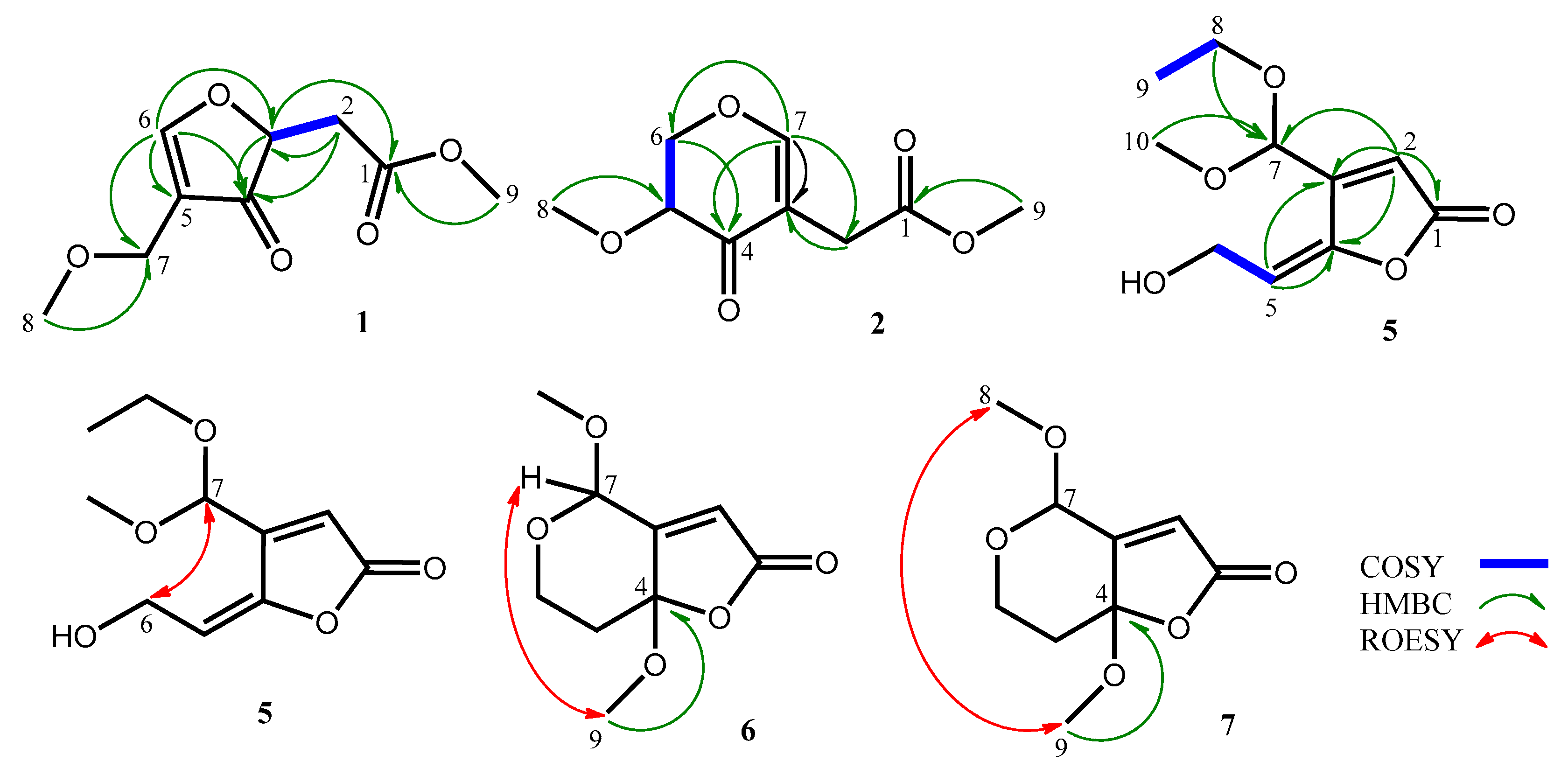
2.1. Isolation and Characterization of Secondary Metabolites

In this study, the green alga Ulva lactuca-derived fungal strain Aspergillus giganteus NTU967 was cultured in both solid- and liquid–state culturing conditions in order to enrich the diversity of the fungal secondary metabolites and eleven chemical entities including seven new compounds 17 and four previously reported compounds, patulin, deoxytryptoquivaline, tryptoquivaline and quinadoline B, were obtained from the fermented products. Of the known compounds isolated, patulin, a highly oxygenated C7 mycotoxin, with a hemiacetal functionality is apt to racemize naturally to form a pair of enantiomers and was first characterized in 1943 under the name of tercinin as a potential antimicrobial agent [10]. Recently, patulin in combination with oxaliplatin were found to exhibit synergism against human colorectal cancer [11]. Deoxytryptoquivaline, tryptoquivaline and quinadoline—three quinazolone-containing alkaloids—were identified by comparison of spectroscopic data with literatures [12,13]. In addition to be isolated from Aspergillus spp., a series of tryptoquivaline analogs have ever been obtained from a marine sea fan-derived fungus Neosartorya siamensis [14].
Compound 1, obtained as colorless oil, was determined to have a molecular formula of C9H12O5, as evidenced by its 13C NMR spectrum (Table 1) and a pseudomolecular ion [M + Na]+ at m/z 223.0574 (calcd. 223.0582 for C9H12O5Na) in the positive mode of HRESIMS analysis. The IR absorptions at 1745 and 1672 revealed the presence of an ester carbonyl and a conjugated ketone functionality, respectively. The 1H NMR (CD3OD, 400 MHz) of compound 1 revealed two oxygen-bearing three-proton signals at δH 3.35 (H3-8) and 3.71 (H3-9), one methylene signal at δH 2.61 (d, J = 6.2 Hz, H2-2), one oxygen-bearing methylene signal at δH 4.55 (H2-7), one oxygen-bearing methine signal at δH 4.55 (H-3) and an olefinic methine signal at δH 7.66 (s, H-6) (Table 2). The 13C NMR (CD3OD, 125 MHz) coupled with phase-sensitive HSQC spectrum of compound 1 showed nine signals including two methoxyl carbons at δC 57.7 (C-8) and 52.7 (C-9), one methylene carbon at δC 36.7 (C-2), one oxymethylene carbon at δC 75.9 (C-7), one oxygenated methine carbon at δC 69.7 (C-3), one oxygenated olefinic carbon at δC 166.1 (C-6), one nonprotonated olefinic carbon at δC 116.2 (C-5), one ester carbonyl carbon at δC 172.9 (C-1) and one conjugated ketone carbon at δC 191.8 (C-4) (Table 1). On account of the molecular formula C9H12O5, the double bond equivalent (DBE) of compound 1 was four including a double bond and two carbonyl groups. Thus, there should be one additional ring in compound 1. Further two dimensional NMR analysis including one cross-peak of δH 2.61 (H2-2)/δH 4.55 (H-3) in the COSY spectrum of compound 1 in combination with key correlations of δH 2.61 (H2-2)/δC 69.7 (C-3) and 191.8 (C-4); δH 4.55 (H-3)/δC 172.9 (C-1) and 191.8 (C-4); δH 7.66 (H-6)/δC 69.7 (C-3), 191.8 (C-4), 116.2 (C-5) and 75.9 (C-7); δH 4.55 (H2-7)/δC 191.8 (C-4); δH 3.35 (H3-8)/δC 75.9 (C-7); δH 3.71 (H3-9)/δC 172.9 (C-1) in the HMBC spectrum of compound 1 (Figure 2), corroborated the gross structure of 1. The optical rotation value of compound 1 ([α] 27D −0.36) was close to zero, revealing that compound 1 could be a racemate.
The molecular formula of 2, C9H12O5, was deduced through analysis of its 13C NMR and HRESIMS data. Its IR spectrum revealed the presence of an ester carbonyl (1737 cm−1) and a conjugated ketone group (1673 cm−1). The 1H NMR spectrum of compound 2 coupled with phase-sensitive HSQC spectrum showed two methoxyl signals at δH 3.45 (s, H3-8) and 3.67 (s, H3-9), one methylene signal at δH 3.14 (s, H2-2), one oxygenated methylene signal at δH 4.49 (d, J = 4.7 Hz, H2-6), one carbinoyl methine signal at δH 3.70 (t, J = 4.7 Hz, H-5), and one olefinic methine signal at δH 7.50 (s, H-7) (Table 2). Nine carbon resonances including two methoxyl at δC 58.7 (C-8) and 52.5 (C-9), one oxygenated methylene at δC 72.3 (C-6), one methylene at δC 31.1 (C-2), one olefinic methine at δC 164.0 (C-7), one carbinoyl methine at δC 77.3 (C-5), one nonprotonated olefinic carbon at δC 111.7 (C-3), one ester carbonyl carbon at δC 173.6 (C-1), and one ketone carbon at δC 191.3 were observed in the 13C NMR of compound 2 (Table 1), which were supported by phase-sensitive HSQC spectrum. On account of the molecular formula of 2, C9H12O5, compound 2 would contain a ring in addition to a double bond, a ketone and an ester carbonyl to fit the unsaturation number. Further comprehensive analysis of two dimensional NMR spectra of compound 2 (Figure 2), one cross-peak of δH 3.70 (H-5)/δH 4.49 (H2-6) in the COSY spectrum together with key cross-peaks of δH 3.14 (H2-2)/δC 111.7 (C-3); δH 4.49 (H2-6)/δC 191.3 (C-4); δH 7.50 (H-7)/δC 31.3 (C-2), 111.7 (C-3), 191.3 (C-4) and 72.3 (C-6); δH 3.45 (H3-8)/δC 77.3 (C-5); δH 3.67 (H3-9)/δC 173.6 (C-1) in the HMBC spectrum established the structure of 2.
The physical and NMR data of compound 3 and compound 4 were almost compatible with those of patulin except that an additional methyl and an additional ethyl were observed in the 1H and 13C NMR spectra of compound 3 and compound 4 (Table 1 and Table 2), respectively. The pseudomolecular ion [M + H]+ at m/z 169.0493 and 183.0655 in the HRESIMS of compounds 3 and 4, 14 and 28 Da more than that of patulin, confirmed that compound 3 and compound 4 were the methyl and ethyl derivatives of patulin, respectively. In the HMBC spectra of compounds 3 and 4, distinctive cross-peaks of δH 3.54 (H3-8)/δC 95.9 (C-7) and δH 3.71 and 3.91 (H2-8)/δC 94.8 (C-7) indicated the methyl and ethyl groups were attached to C-7 of compounds 3 and 4, respectively. The structures of compound 3 and compound 4 were thus elucidated to be as shown.
Compound 5, obtained as colorless oil, was determined to have a molecular formula of C10H14O5, as evidenced by its HRESIMS analysis and 13C NMR spectrum (Table 1). It contained a hydroxy, a γ-lactone carbonyl and a double bond due to the IR absorption bands at 3435, 1768 and 1643 cm−1, respectively. Interpretations of the 1H NMR data along with the HSQC spectrum of compound 5 showed two methyl signals at δH 3.36 (s, H3-10) and 1.24 (t, J = 7.1 Hz, H3-10), two oxygenated methylene signals at δH 4.40 (d, J = 6.9 Hz, H2-6) and 3.63 (m, H2-8), two olefinic methine signals at δH 6.31 (s, H-2) and 5.85 (t, J = 6.9 Hz, H-5) and one dioxygenated methine signal at δH 5.52 (s, H-7) (Table 2). The 13C NMR data of compound 5 coupled with its phase-sensitive HSQC assignments showed one methoxyl carbon at δC 53.2 (C-10), one methyl carbon at δC 15.4 (C-9), two oxygenated methylene carbons at δC 62.8 (C-8) and 57.3 (C-6), two olefinic methine carbons at δC 119.7 (C-2) and 115.3 (C-5), one dioxygenated methine carbon at δC 97.6 (C-7) and three nonprotonated carbons at δC 169.6 (C-1), 156.5 (C-3) and 148.2 (C-4) (Table 1). Correlations of δH 5.85 (H-5)/δH 4.40 (H2-6) and δH 3.63 (H2-8)/δH 1.24 (H3-9) in the COSY spectrum of compound 5 accompanied with key correlations including δH 6.31 (H-2)/δC 169.6 (C-1), 156.5 (C-3), 148.2 (C-4) and 97.6 (C-7); δH 5.58 (H-5)/δC 156.5 (C-3) and 148.2 (C-4); δH 3.63 (H2-8)/δC 97.6 (C-7); δH 3.36 (H-10)/δC 97.6 (C-7) in the HMBC spectrum of compound 5 (Figure 2), established the gross structure of 5. The configuration of Δ4 in compound 5 was determined to be E form based on a key correlation of δH 4.40 (H2-6)/δH 5.52 (H-7) in the ROESY of compound 5 (Figure 2). Since compound 5 had an acetal carbon at its C-7 and an optical rotation value close to zero ([α] 27D +0.02), compound 5 was deduced to be an acetal racemate.
The 1H NMR data of compounds 6 and 7 were almost identical with that of compound 3 except that an olefinic proton at δH 6.02 (H-5) in compound 3 was substituted by a methylene group at δH 1.91 and 2.38 (H2-5) in compound 6 and δH 1.99 and 2.34 (H2-5) in compound 7 and an additional methoxyl functionality at δH 3.23 (H3-9) and δH 3.19 (H3-9) was observed in compound 6 and compound 7 (Table 2), respectively. These changes also reflected in their 13C NMR data (Table 1), in which two olefinic signals at δC 150.9 (C-4) and 109.3 (C-5) in compound 3 were replaced by a ketal carbon signal (δC-4 107.6 in 6; δC-4 107.2 in 7) along with a methylene carbon resonance (δC-5 40.3 in 6; δC-5 41.9 in 7). The additional methoxyl groups in compounds 6 and 7 were deduced to be attached at C-4 due to key cross-peaks of H3-9/C-4 in the HMBC spectra of both compound 6 and compound 7 (Figure 2). In the ROESY spectra of compounds 6 and 7, a key correlation of δH 3.23 (H3-9)/δH 5.11 (H-7) confirmed that two methoxyls in compound 6 were oriented in different phases, while a key cross-peak of δH 3.46 (H3-8)/δH 3.19 (H3-9) indicated that two methoxyls in compound 7 were located on the same side (Figure 2). The structures of compounds 6 and 7 were thus established as shown in Figure 1.

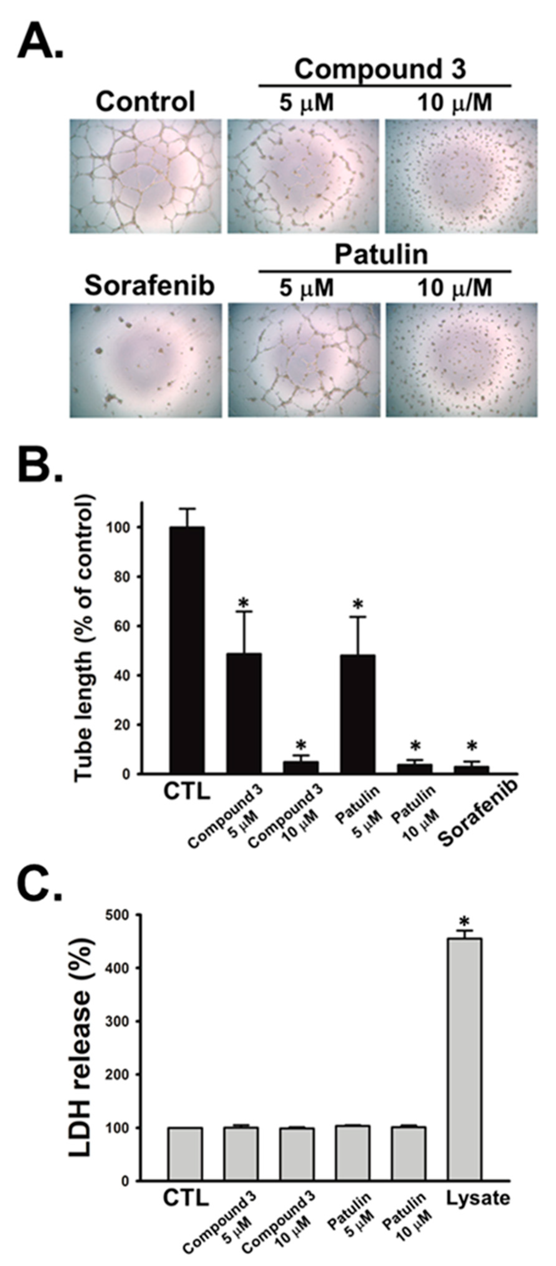
2.2. Anticancer and Anti-Angiogenic Assays of Secondary Metabolites

All eleven pure isolates were subjected to biological assays. Among these, compound 3 exerted promising anticancer activities against human hepatocellular carcinoma SK-Hep-1 cells and prostate cancer PC-3 cells with IC50 values of 2.7 ± 0.2 and 7.3 ± 0.3 μM (Table 3). Paclitaxel, a well-known anticancer agent, was used as the positive control. Additionally, we evaluated the anti-angiogenic activities of all the pure isolates against human endothelial progenitor cells (EPCs). As shown in Table 3, compound 3 and patulin exhibited most potent anti-angiogenic activities by suppressing EPCs growth with IC50 values of 4.6 ± 0.3 and 4.7 ± 0.2 μM, respectively. Since capillary-like tubules are the essential characteristic of angiogenesis, we next performed tube formation assay to validate the anti-angiogenic effects of compound 3 and patulin in EPCs with sorafenib as a positive control. The results showed that compound 3 and patulin concentration-dependently inhibited capillary tube formation of EPCs (Figure 3A,B). Furthermore, it was found that compound 3 and patulin did not induce the release of lactate dehydrogenase (LDH) in EPCs (Figure 3C), suggesting that these two compounds display anti-angiogenesis property without the cytotoxic fashion. Compound 4 bearing an ethoxyl group at its C-7 instead of a hydroxy and a methoxyl at C-7 of patulin and 3, respectively, reduced its bioactivity significantly. It was thus speculated that the size of the functional group attached at C-7 and an olefinic functionality at C-4 of patulin could play crucial roles in the anticancer and anti-angiogenic activities. These findings provide evidences that both compound 3 and patulin may serve as the potential natural products to block tumor angiogenesis for cancer treatment.

3. Materials and Methods

3.1. General Experimental Procedures

Optical rotations and UV were measured on a JASCO P-2000 polarimeter (Tokyo, Japan) and Thermo UV-Visible Heλios α Spectrophotometer (Bellefonte, CA, USA), respectively. 1H and 13C NMR were acquired on Bruker AVIII HD 400 and Bruker AVIII-500 spectrometer (Ettlingen, Germany). Low and high resolution mass spectra were obtained using an API4000 triple quadrupole mass spectrometer (Applied Biosystems, Foster City, CA, USA) and Q Exactive Plus Hybrid Quadrupole-Orbitrap Mass Spectrometer (Thermo Fisher Scientific, Bremen, Germany), respectively. IR spectra were recorded on a JASCO FT/IR 4100 spectrometer (Tokyo, Japan). Sephadex LH-20 (GE Healthcare, Uppsala, Sweden) and Diaion HP-20 (Mitsubishi Chemical, Tokyo, Japan) was used for open column chromatography. An HPLC pump L-7100 (Hitachi, Japan) equipped with a refractive index detector (Bischoff, Leonberg, Germany) was used for compound purification. All the organic solvents were purchased from Merck (Darmstadt, Germany).

3.2. Fungal Strain and Culture

Aspergillus giganteus NTU967 was isolated from the marine green alga Ulva lactuca collected from the northeast coast of Taiwan and was identified by sequencing of the internal transcribed spacer regions of the rDNA (ITS) and β-tubulin gene. A BLAST search of the ITS sequence (GenBank accession no. MH250052) was not conclusive and led to the best matches as Aspergillus clavatoanicus, A. clavatus, A. giganteus and A. longivesica (query coverage 94–100%, identity 98–99%) while a BLAST search of the β-tubulin gene resulted the best matches as A. giganteus (query coverage 97–100%, identity 98.48–99.63%). For liquid culture, the mycelium of Aspergillus giganteus NTU967 was inoculated into 5 L serum bottles, each containing 2 g Peptone (Becton, Dickinson and Company, Sparks, MD, USA), 1 g yeast extract (Becton, Dickinson and Company, Sparks, MD, USA), 10 g Dextrose (Becton, Dickinson and Company, Sparks, MD, USA) and 2.5 L deionized water. The fermentation was conducted with aeration at 25–30 °C for 16 days. For solid culture, the mycelium of Aspergillus giganteus NTU967 was inoculated into 500 mL flasks, each containing 50 g brown rice (Santacruz, Taiwan), 2% yeast extract (Becton, Dickinson and Company, Sparks, MD, USA), 1% sodium tartrate and 1% KH2HPO4 in 20 mL deionized water. The solid culture was conducted at 25–30 °C for 30 days.

3.3. Extraction and Isolation of Secondary Metabolites

For liquid culture, the filtered fermented broth (15.0 L) of Aspergillus giganteus NTU967 was partitioned three times with 30 L EtOAc, then concentrated in vacuum to dryness (8.0 g). Subsequently, the crude extract was redissolved in 20 mL MeOH, then applied onto a Sephadex LH-20 column (2.5 cm i.d. × 68 cm) eluted with MeOH at a flow rate of 2.5 mL/min. Each fraction (20 mL) collected was checked for its compositions by TLC using CH2Cl2-MeOH (10:1, v/v) for development, and dipping in vanillin–H2SO4 was used in the detection of compounds with similar skeletons. All the fractions were combined into four samples I–VI. Subsequently, sample III with antimicrobial activity was precoated with 15.0 g Diaion HP-20 gel, then applied onto a Diaion HP-20 column (4.5 cm i.d. × 30 cm) eluted with mixtures of H2O/MeOH in a stepwise gradient mode with a flow rate of 2.0 mL/min to obtain four subsamples I–IV. Subsample III eluted by 75% MeOH was rechromatographed on a semipreparative reversed-phase column (Phenomenex Luna 5 μ PFP, 10 × 250 mm) with 35% MeOHaq as eluent, 2 mL/min, to afford 1 (30.4 mg, tR = 13.5 min), 2 (15.2 mg, tR = 16.9 min) and 3 (27.4 mg, tR = 27.5 min). Subsample III was further purified on a semipreparative reversed-phase column (Thermo Hypersil 5 μ C18, 10 × 250 mm) with 25% MeCNaq as eluent, 2 mL/min, to give 4 (22.8 mg, tR = 35.1 min), 5 (18.4 mg, tR = 23.4 min), 6 (16.6 mg, tR = 26.1 min) and 7 (35.6 mg, tR = 27.6 min).
For solid–state culture, the fermented products were lyophilized, ground into powder (750 g) and extracted three times with equal volumes of methanol. Extracts were first partitioned with n-hexane and the methanol layers suspended in deionized H2O, then partitioned with ethyl acetate and concentrated to obtain dried ethyl acetate extract (7.0 g). For compound separation, the ethyl acetate extract was subjected to Sephadex LH-20 column chromatography (2.5 i.d. × 68.0 cm), using methanol as the eluent at a flow rate of 2.5 mL/min to give 30 fractions (20.0 mL/fr.). All the fractions were combined into 6 samples as I–VI based on the results of TLC analysis and antimicrobial assay. Sample III with antimicrobial activity was precoated with 20.0 g Diaion HP-20 gel, then applied onto a Diaion HP-20 column (4.5 cm i.d. × 30 cm) eluted with mixtures of H2O/MeOH in a stepwise gradient mode with a flow rate of 2.0 mL/min to get four subsamples I–IV. Subsample II eluted by 50% MeOH was rechromatographed on a semipreparative reversed-phase column (BIOSIL Pro-ODS-U 5 μ, 10 × 250 mm) with 15% MeOHaq as eluent, 2 mL/min, to obtain patulin (16.7 mg, tR = 20.1 min). Subsample IV eluted by 100% MeOH was further purified on a semipreparative reversed-phase column (Phenomenex Luna 5 µ PFP, 10 × 250 mm) with 75% MeOHaq as eluent, 2 mL/min, to afford quinadoline B (27.7 mg, tR = 11.8 min), deoxytryptoquivaline (21.0 mg, tR = 23.0 min) and tryptoquivaline (16.4 mg, tR = 36.2 min).
Aspergilsmin A (1): Colorless oil; [α] 27D −0.36 (c = 0.05, MeOH); IR (ZnSe) νmax: 2951, 1745, 1672, 1611, 1456, 1438, 1399, 1333, 1308, 1283, 1254, 1196, 1171, 1105, 1055, 1033 and 1009 cm−1; UV λmax (MeOH) (log ε) 261 (3.9) nm; 1H and 13C NMR spectroscopic data: see Table 1 and Table 2; HRESIMS [M + Na]+ at m/z 223.0574 (calcd. 223.0582 for C9H12O5Na).
Aspergilsmin B (2): Colorless oil; [α] 27D +1.22 (c = 0.05, MeOH); IR (ZnSe) νmax: 2947, 1737, 1673, 1619, 1443, 1406, 1344, 1291, 1257, 1157, 1097, 1056, 1043, 1024, 1011 and 854 cm−1; UV λmax (MeOH) (log ε) 268 (3.9) nm; 1H and 13C NMR spectroscopic data: see Table 1 and Table 2; HRESIMS [M + Na]+ at m/z 223.0573 (calcd. 223.0582 for C9H12O5Na).
Aspergilsmin C (3): Colorless oil; [α] 27D −3.52 (c = 0.05, MeOH); IR (ZnSe) νmax: 2955, 1780, 1536, 1443, 1406, 1344, 1210, 1065 and 868 cm−1; UV λmax (MeOH) (log ε) 274 (4.0) nm; 1H and 13C NMR spectroscopic data: see Table 1 and Table 2; HRESIMS [M + H]+ at m/z 169.0493 (calcd. 169.0501 for C8H9O4).
Aspergilsmin D (4): Colorless oil; [α] 27D −2.14 (c = 0.05, MeOH); IR (ZnSe) νmax: 2945, 1780, 1635, 1404, 1092 and 1019 cm−1; UV λmax (MeOH) (log ε) 275 (4.0) nm; 1H and 13C NMR spectroscopic data: see Table 1 and Table 2; HRESIMS [M + H]+ at m/z 183.0655 (calcd. 183.0657 for C9H11O4).
Aspergilsmin E (5): Colorless oil; [α] 27D +0.02 (c = 0.05, MeOH); IR (ZnSe) νmax: 3435, 1768, 1643, 1053 and 1008 cm−1; UV λmax (MeOH) (log ε) 223 (3.7) and 270 (4.0) nm; 1H and 13C NMR spectroscopic data: see Table 1 and Table 2; HRESIMS [M + H]+ at m/z 215.0915 (calcd. 215.0947 for C10H15O5).
Aspergilsmin F (6): Colorless oil; [α] 27D −0.53 (c = 0.05, MeOH); IR (ZnSe) νmax: 2951, 1768, 1643, 1456, 1396, 1207, 1163, 1082, 1022 and 972 cm−1; UV λmax (MeOH) (log ε) 257 (4.1) nm; 1H and 13C NMR spectroscopic data: see Table 1 and Table 2; HRESIMS [M + Na]+ at m/z 223.0579 (calcd. 223.0582 for C9H12O5Na).
Aspergilsmin G (7): Colorless oil; [α] 27D −1.86 (c = 0.05, MeOH); IR (ZnSe) νmax: 2971, 2842, 1766, 1456, 1435, 1358, 1329, 1266, 1205, 1196, 1171, 1129, 1041, 1034 and 915 cm−1; UV λmax (MeOH) (log ε) 271 (4.0) nm; 1H and 13C NMR spectroscopic data: see Table 1 and Table 2; HRESIMS [M + Na]+ at m/z 223.0574 (calcd. 223.0582 for C9H12O5Na).

3.4. Cell Culture

The human hepatocellular carcinoma cell line SK-Hep-1 and hormone refractory prostate cancer cell line PC-3 were purchased from the American Type Cell Culture Collection (Manassas, VA, USA) and maintained in DMEM medium containing 10% fetal bovine serum (FBS), penicillin (100 units/mL) and streptomycin (100 μg/mL). Cells were maintained in humidified air containing 5% CO2 at 37 °C. All cell culture reagents were purchased from Gibco-BRL life technologies (Grand Island, NY, USA). The isolation and maintenance of human CD34-positive endothelial progenitor cells (EPCs) were conducted using the standard method as previously described [15].

3.5. Biologic Assay for Anticancer Activity

SK-Hep-1 and PC-3 cancer cells were seeded onto 96-well plates in a density of 5 × 10³ cells per well. Overnight, cells were treated with the tested compounds for 48 h. Then, anticancer activity was determined by the SRB assay according to previously described procedures [16].

3.6. Biologic Assay for Anti-Angiogenic Activity

For EPCs’ cell growth assay, EPCs were cultured in 96-well plates at a density of 5 × 10³ cells in each well. Overnight, the culture medium was replaced with MV2 complete medium containing 2% FBS in the presence of the tested compounds for 48 h. The reaction was terminated after 48 h of incubation with 50% TCA. After the TCA fixation, every well was incubated for 15 min incubation with 0.4% sulforhodamine B in 1% acetic acid. The plates were then washed before the dye was dissolved by 10-mM Tris buffer. Absorbance density values were read by an enzyme-linked immunosorbent assay (ELISA) reader (515 nm).
For EPCs’ tube formation assay, EPCs were seeded with the density of 1.25 × 10⁴ cells per well in Matrigel-coated 96-well plates and incubated in an MV2 complete medium containing 2% FBS and the tested compounds for 24 h. EPCs differentiation and capillary-like tube formation was taken with the inverted phase contrast microscope. The long axis of each tube was measured with MacBiophotonics Image J software in 3 randomly chosen fields per well.
For EPCs’ cytotoxicity assay, EPCs (5 × 10³ cells/well) were seeded onto 96-well plates and incubated with MV2 complete medium containing 2% FBS and the tested compounds for 24 h. Then, the quantification of LDH release in the medium was done with a cytotoxicity assay kit.

4. Conclusions

In this report, we have identified seven new polyketides 17 along with four known compounds from a marine algicolous fungal strain Aspergillus giganteus NTU967. Of the compounds identified, compound 3 and its known analog patulin exhibited promising anticancer as well as significant anti-angiogenic activities when compared with the clinically used drugs.

Supplementary Materials

The following are available online at https://www.mdpi.com/1660-3397/18/6/303/s1, Figures S1–S38: All the NMR spectra of compounds 17. Figure S39: The structures of known compounds isolated in this study. Table S1: Cytotoxicities of aspergilsmins A-G (17), patulin, deoxytryptoquivaline, tryptoquivaline and quinadoline B against PC-3 and SK-Hep-1 cells.

Author Contributions

J.-J.C. drafted the manuscript initially. S.-W.W. performed the biological assays. Y.-R.C. analyzed and interpreted the data. K.-L.P. identified the fungal strain. Y.-H.K. revised the manuscript and gave suggestions. T.-Y.S. isolated and purified all the secondary metabolites. T.-H.L. designed and conceived the whole study. All authors checked and approved the final manuscript.

Funding

This work was supported by a grant from the Ministry of Science and Technology (MOST107-2320-B-002-017-MY3) of Taiwan to T.-H.L.

Acknowledgments

We thank S.-L.H. and S.-Y.S. in the Instrumentation Center of the College of Science, National Taiwan University for the NMR and MS data acquisition, respectively.

Conflicts of Interest

The authors declare no conflict of interest.

References

  1. Rateb, M.E.; Ebel, R. Secondary metabolites of fungi from marine habitats. Nat. Prod. Rep. 2011, 28, 290–344. [Google Scholar] [CrossRef] [PubMed]
  2. Xu, L.; Meng, W.; Cao, C.; Wang, J.; Shan, W.; Wang, Q. Antibacterial and antifungal compounds from marine fungi. Mar. Drugs 2015, 13, 3479–3513. [Google Scholar] [CrossRef] [PubMed]
  3. Li, W.; Ding, L.; Wang, N.; Xu, J.; Zhang, W.; Zhang, B.; He, B.; Wu, B.; Jin, H. Isolation and characterization of two new metabolites from the sponge-derived fungus Aspergillus sp. LS34 by OSMAC approach. Mar. Drugs 2019, 17, 283. [Google Scholar] [CrossRef] [PubMed] [Green Version]
  4. Pan, R.; Bai, X.; Chen, J.; Zhang, H.; Wang, H. Exploring structural diversity of microbe secondary metabolites using OSMAC strategy: A literature review. Front. Microbiol. 2019, 10, 294. [Google Scholar] [CrossRef] [PubMed] [Green Version]
  5. Hoang, T.P.T.; Roullier, C.; Boumard, M.C.; Robiou du Pont, T.; Nazih, H.; Gallard, J.F.; Pouchus, Y.F.; Beniddir, M.A.; Grovel, O. Metabolomics-driven discovery of meroterpenoids from a mussel-derived penicillium ubiquetum. J. Nat. Prod. 2018, 81, 2501–2511. [Google Scholar] [CrossRef] [PubMed]
  6. Selegato, D.M.; Freire, R.T.; Pilon, A.C.; Biasetto, C.R.; de Oliveira, H.C.; de Abreu, L.M.; Araujo, A.R.; da Silva Bolzani, V.; Castro-Gamboa, I. Improvement of bioactive metabolite production in microbial culture – A systems approach by OSMAC and deconvolution-based 1H NMR quantification. Magn. Reson. Chem. 2019, 57, 458–471. [Google Scholar] [CrossRef] [PubMed]
  7. Wang, W.J.; Li, D.Y.; Li, Y.C.; Hua, H.M.; Ma, E.L.; Li, Z.L. Caryophyllene sesquiterpenes from the marine-derived fungus Ascotricha sp. ZJ-M-5 by the one strain-many compounds strategy. J. Nat. Prod. 2014, 77, 1367–1371. [Google Scholar] [CrossRef] [PubMed]
  8. Huang, S.F. Seaweeds of Northeastern Taiwan; National Taiwan Museum: Taipei, Taiwan, 2000; pp. 6–7. [Google Scholar]
  9. Lee, M.S.; Yang, Y.L.; Wu, C.Y.; Chen, Y.L.; Lee, C.K.; Tzean, S.S.; Lee, T.H. Efficient identification of fungal antimicrobial principles by tandem MS and NMR database. J. Food Drug Anal. 2019, 27, 860–868. [Google Scholar] [CrossRef] [PubMed]
  10. Raistrick, H. Patulin in the common cold collaborative research on a derivative of Penicillin patulum Bainier. Lancet 1943, 242, 625. [Google Scholar] [CrossRef]
  11. Alam, N.; Yu, J.Q.; Beale, P.; Hug, F. Dose and sequence dependent synergism from the combination of oxaliplatin with emetine and patulin against colorectal cancer. Anticancer Agents Med. Chem. 2019, 20, 264–273. [Google Scholar] [CrossRef] [PubMed]
  12. Buechi, G.; Luk, K.C.; Kobbe, B.; Townsend, J.M. Four new mycotoxins of Aspergillus clavatus related to tryptoquivaline. J. Org. Chem. 1977, 42, 244–246. [Google Scholar] [CrossRef] [PubMed]
  13. Koyama, N.; Inoue, Y.; Sekine, M.; Hayakawa, Y.; Homma, H.; Omura, S.; Tomoda, H. Relative and absolute stereochemistry of quinadoline B, an inhibitor of lipid droplet synthesis in macrophages. Org. Lett. 2008, 10, 5273–5276. [Google Scholar] [CrossRef] [PubMed]
  14. Prata-Sena, M.; Ramos, A.A.; Buttachon, S.; Castro-carvalho, B.; Marques, P.; Dethoup, T.; Kijjoa, A.; Rocha, E. Cytotoxic activity of secondary metabolites from marine-derived fungus Neosartorya siamensis in human cancer cells. Phytother. Res. 2016, 30, 1862–1871. [Google Scholar] [CrossRef] [PubMed]
  15. Lee, M.S.; Wang, G.J.; Pang, K.L.; Lee, C.K.; Kuo, Y.H.; Lin, R.K.; Lee, T.H. Angiogenesis inhibitors and anti-inflammatory agents from Phoma sp. NTOU4195. J. Nat. Prod. 2016, 79, 2983–2990. [Google Scholar] [CrossRef] [PubMed]
  16. Thakur, R.S.; Ahirwar, B. A steroidal derivative from Trigonella foenumgraecum L. that induces apoptosis in vitro and in vivo. J. Food Drug Anal. 2019, 27, 231–239. [Google Scholar] [CrossRef] [PubMed] [Green Version]
Figure 1. Chemical structures of compounds 17 and patulin.
Marinedrugs 18 00303 g001
Figure 2. COSY and key HMBC and ROESY correlations of compounds 1, 2, 5, 6 and 7.
Marinedrugs 18 00303 g002
Figure 3. Effects of compound 3 and patulin on tube formation and cytotoxicity of human EPCs. (A) EPCs were treated with compound 3, patulin and sorafenib (10 μM) for 24 h. Tubular morphogenesis was recorded by the inverted phase contrast microscope. Representative images of EPCs’ tube formation were shown; (B) tube formation was quantified by measuring the length of tubes using ImageJ software; (C) Cells were treated with the indicated compounds for 24 h, then the cytotoxicity was determined using LDH assay Data represent the mean ± S.E.M. of 4 independent experiments. * p < 0.05 compared with the control group.
Marinedrugs 18 00303 g003
Table 1. 13C NMR spectroscopic data for compounds 17 (δ in ppm, mult.).
No.1 a,b2 a,b3 a,b4 a,b5 a,b6 a,b7 a,b
1172.9 s173.6 s170.6 s170.6 s169.6 s170.6 s170.6 s
236.7 t31.1 t111.9 d111.1 d119.7 d117.1 d119.9 d
369.7 d111.7 s147.5 s147.6 s156.5 s163.9 s160.9 s
4191.8 s191.3 s150.9 s151.3 s148.2 s107.6 s107.2 s
5116.2 s77.3 d109.3 d109.4 d115.3 d40.3 t41.9 t
6166.1 d72.3 t60.0 t60.0 t57.3 t62.3 t58.3 t
775.9 t164.0 d95.9 d94.8 d97.6 d98.8 d97.4 d
857.7 q58.7 q56.5 q65.6 t62.8 t57.3 q55.5 q
952.7 q52.5 q 15.4 q15.4 q51.2 q52.2 q
10 53.2 q
a Measured in CD3OD (125 MHz); b Multiplicties were obtained from phase-sensitive HSQC experiments.
Table 2. 1H NMR spectroscopic data for compounds 17 (δ in ppm, mult., J in Hz).
No.1 a2 b3 b4 b5 b6 b7 b
1
22.61 d (6.2)3.14 s6.60 s6.05 s6.31 s6.12 s6.23 s
34.55 c
4
5 3.70 t (4.7)6.02 m6.02 m5.85 t (6.9)1.91 m1.99 m
2.38 dt (13.5, 2.1)2.34 d (13.2)
67.66 s4.49 d (4.7)4.37 dd (17.5, 4.4)4.36 dd (17.3, 4.4)4.40 d (6.9)3.76 td (12.2, 2.1)3.71 m
4.56 dd (17.5, 2.5)4.56 dd (17.3, 2.6) 4.01 m4.10 td (12.0, 2.0)
74.55 c7.50 s5.68 s5.79 s5.52 s5.11 s5.52 s
83.35 s3.45 s3.54 s3.71 m3.63 m3.60 s3.46 s
3.91 m
93.71 s3.67 s 1.26 t (7.1)1.24 t (7.1)3.23 s3.19 s
10 3.36 s
a Measured in CD3OD (400 MHz); b Measured in CD3OD (500 MHz); c Signals were overlapped and were picked up from HSQC spectrum.
Table 3. Anticancer and anti-angiogenic activities of compound 3 and patulin.
CompoundsAnticancer (IC50, μM)Anti-Angiogenesis (IC50, μM)
SK-Hep-1 aPC-3 bEPC c
Compound 32.7 ± 0.27.3 ± 0.34.6 ± 0.3
Patulin2.9 ± 0.12.7 ± 0.14.7 ± 0.2
Paclitaxel d0.011 ± 0.0020.013 ± 0.002
Sorafenib d4.8 ± 0.3
a Hepatocellular carcinoma cells; b Prostate cancer cells; c Endothelial progenitor cells; d Positive control.

