Article

The Biosynthesis of Rare Homo-Amino Acid Containing Variants of Microcystin by a Benthic Cyanobacterium

1
Department of Microbiology, University of Helsinki, Viikinkaari 9, FI-00014 Helsinki, Finland
2
Institute of Biotechnology, University of Helsinki, Viikinkaari 5D, FI-00014 Helsinki, Finland
3
Department of Biological and Environmental Science, University of Jyväskylä, FI-40014 Jyväskylä, Finland
*
Author to whom correspondence should be addressed.
Mar. Drugs 2019, 17(5), 271; https://doi.org/10.3390/md17050271
Submission received: 1 April 2019 / Revised: 2 May 2019 / Accepted: 5 May 2019 / Published: 7 May 2019
(This article belongs to the Special Issue Marine Bacterial Toxins)

Abstract

:
Microcystins are a family of chemically diverse hepatotoxins produced by distantly related cyanobacteria and are potent inhibitors of eukaryotic protein phosphatases 1 and 2A. Here we provide evidence for the biosynthesis of rare variants of microcystin that contain a selection of homo-amino acids by the benthic cyanobacterium Phormidium sp. LP904c. This strain produces at least 16 microcystin chemical variants many of which contain homophenylalanine or homotyrosine. We retrieved the complete 54.2 kb microcystin (mcy) gene cluster from a draft genome assembly. Analysis of the substrate specificity of McyB1 and McyC adenylation domain binding pockets revealed divergent substrate specificity sequences, which could explain the activation of homo-amino acids which were present in 31% of the microcystins detected and included variants such as MC-LHty, MC-HphHty, MC-LHph and MC-HphHph. The mcy gene cluster did not encode enzymes for the synthesis of homo-amino acids but may instead activate homo-amino acids produced during the synthesis of anabaenopeptins. We observed the loss of microcystin during cultivation of a closely related strain, Phormidium sp. DVL1003c. This study increases the knowledge of benthic cyanobacterial strains that produce microcystin variants and broadens the structural diversity of known microcystins.

1. Introduction

Microcystins are potent hepatotoxins, which inhibit eukaryotic protein phosphatases type 1 and 2A [1]. Microcystins are frequently detected during blooms of cyanobacteria in freshwater environments and are linked to poisoning of humans and animals [2,3]. Microcystins have an unusual chemical structure with more than 240 reported chemical variants [4]. The microcystin chemical structure can be summarized as cyclo-D-Ala1-X2-D-MeAsp3-Z4-Adda5-D-Glu6-Mdha7 where X and Z are variable L-amino acids. The bulk of microcystin chemical variation can be attributed to the amino acids found at positions two (X) and four (Z).
Microcystins are synthesized by large hybrid multifunctional enzymes known as nonribosomal peptide synthetases (NRPS) and polyketide synthases (PKS) [5,6,7,8]. The chemical variability of microcystins is mainly attributed to a substrate promiscuity of microcystin biosynthetic enzymes, which allows the incorporation of a diverse range of proteinogenic/non-proteinogenic amino acids, non-proteinogenic organic acids and different degrees of methylation [9]. This is due to differences in substrate specificity of the NRPS enzymes, the deletion of enzymatic domains or loss/gain of tailoring enzymes [1,8].
Homo-amino acids have an extra methylene (CH2) group in the carbon side chain and are frequently found in cyanobacterial natural products [9,10,11,12,13,14,15,16]. Cyanobacteria have been found to produce microcystins that contain homo-amino acids in positions X and Z, such as homotyrosine (Hty), homophenylalanine (Hph), homoisoleucine (Hil), and homoarginine (Har) [4,10,11,12,13]. However, the biosynthetic origins of such microcystins are unclear.
Phormidium is a genus of cyanobacteria belonging to Oscillatoriales order that includes unbranched filamentous strains. Phormidium is not well defined taxonomically and further studies are needed to improve its separation with other genera of Oscillatoriales [17], which complicates the interpretation of reports about Phormidium in the scientific literature. Cyanobacteria belonging to Phormidium genus have been isolated from mangrove [18], saline-alkaline lakes [19], thermal springs [20], high altitude lakes [21] and mats/biofilms in Antarctic lakes [22] among others. Phormidium spp. isolated from Egypt, USA, Brazil and Spain have been reported to produce microcystins [13,23,24,25]. Putative Phormidium spp. producers of microcystin were reported to be associated to the poisoning of a dog in New Zealand [26]. However, analysis based on 16S rRNA sequences indicates a close similarity of these strains with Planktothrix [26]. Microcystin-LR was detected from Phormidium strains isolated from benthic environments in USA [24] and here we further analyze selected strains from that study. We describe the microcystin biosynthetic gene cluster from Phormidium sp. LP904c and show that this strain produces rare homo-amino acid containing microcystins in addition to microcystin-LR. We also report the loss of the microcystin synthesis by Phormidium sp. DVL1003c after 11 years of cultivation in our laboratory.

2. Results

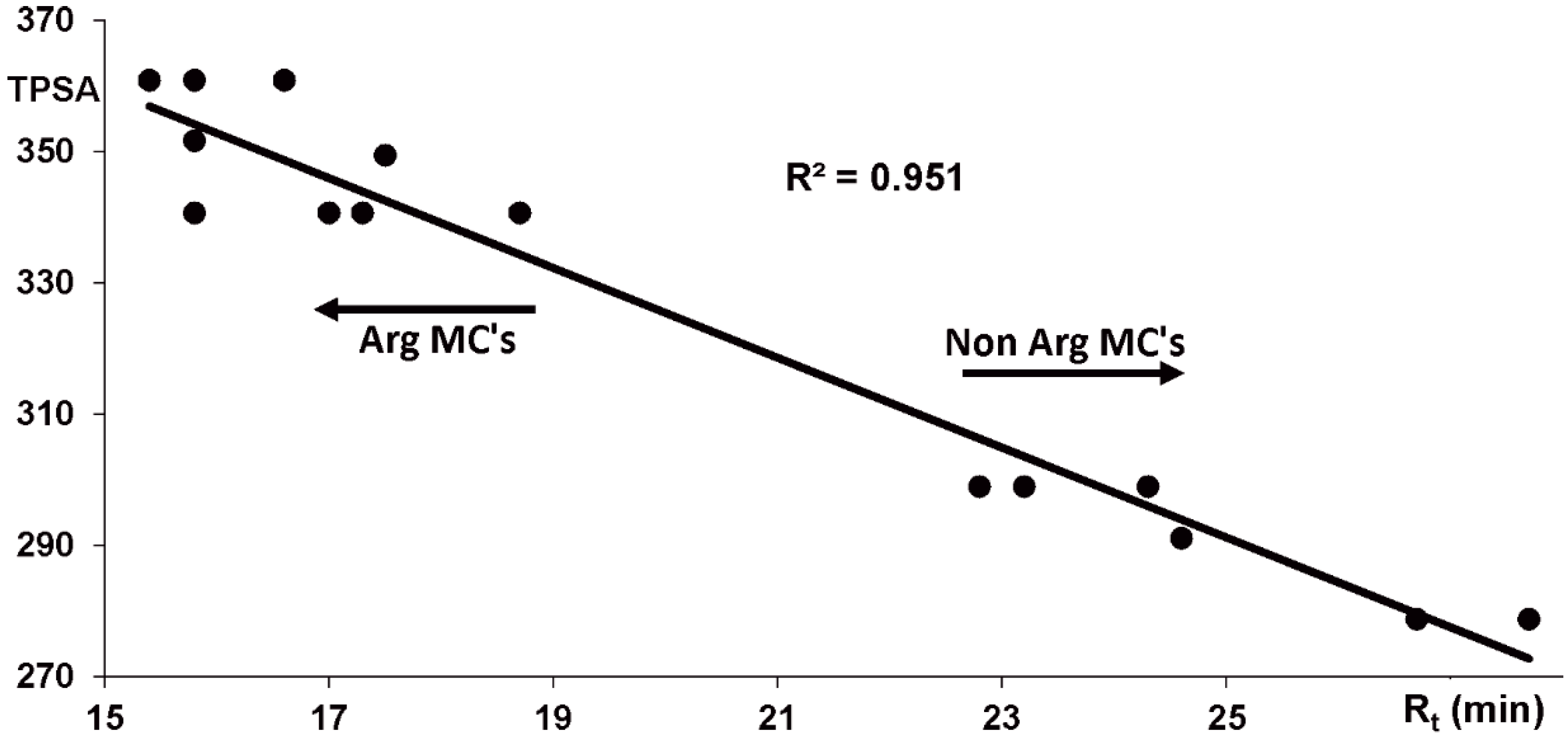
2.1. Phormidium Strains Producing Unusual Microcystins (MCs)

Phormidium sp. LP904c is a benthic cyanobacterium isolated from the Lake Perris in Riverside County, California (USA) that is reported to produce microcystin-LR. This cyanobacterium was found to produce diverse microcystins varying in the positions X and Z (Figure 1A). Further variations were related to the presence or absence of methylation in the aspartic acid (Aa3) or Adda (Aa5) and for the presence of a methylated serine or dehydroalanine instead of the N-methyldehydroalanine (Aa7) (Figure 2).
MC-LR was the main variant as previously reported [24] and accounted for 55% of the total microcystin produced by this strain. However, the strain produces a range of other microcystins including MC-LHty, MC-HphHty, MC-LHph and MC-HphHph (Figure 2, Table 1). A number of amino acids were found in position X and Z, such as Leu, Arg, Phe, Trp, Tyr, Met and unusual homo-amino acids such as Hph and Hty (Figure 2, Table 1).
Phormidium sp. DVL1003c (Figure 1B) was isolated from a freshwater reservoir in Riverside County, California. Strains DVL1003c and LP904c were shown to synthesize similar microcystin variants in 2005 (Table 1, Supplementary Figures S1–S3). Structural characterizations of microcystins produced by Phormidium sp. LP904c were done using high-performance liquid chromatography/electrospray ionization ion-trap mass spectrometry (HPLC-ESI-ITMS) and ultra-high performance liquid chromatography-quadrupole time-of-flight mass spectrometry (UPLC-QTOF). MCs identified were divided to two groups, ten arginine (Arg) containing MCs (Numbers 1–10) and six non Arg MCs (Numbers 11–16) (Table 1). All Arg MCs eluted first, followed by the more hydrophobic non Arg MCs (Supplementary Figures S2 and S3). These two MC groups fragment differently as protonated molecules resulting in distinct product ion spectra (Supplementary Figure S4). Both spectral groups were highly similar to spectra recorded in similar ion trap conditions [27]. In Arg containing MC structures fragmentation prefers the formation of ion 4a–6c (relative intensities (RI) from 51 to 100%, Arg4-Adda5-Glu6, Supplementary Figures S4 and S5) which have an m/z 599 when methyl group 2 is present. When the methyl group is absent, e.g., [DMAdda5]MC, an m/z 585 is detected. Serine in MC structure generates a base peak of [M + H − H2O]+ which was seen in the spectra of [MeSer7]MC-LR and which lowered the relative intensity of 4a-6c ion (Supplementary Figure S4). Ions 4a-6c – CO (RI 26–94%) and 4a–6c – NH3 (RI 6–28%) were also present (Supplementary Figure S4). The presence of ion 4a–7c (RI 4–20%, Arg4-Adda5-Glu6-Aa7, Supplementary Figure S5) shows the identity of the Aa7. Other diagnostic intense ions were 4a-2c/7a-5c (RI 8–38%) which showed the identity of amino acids 3 and 6, and 7a–4c (RI 45–94%, Aa7-Ala1-Aa2-MeAsp3-Arg4) together with other ions showed the identity of amino acids 4 and 5. Ion 5y-5x-7c was present in all the protonated MC product ion spectra except in the [DMAdda5]MC-LR spectra because cleavage at position 5y is not so favorable when there is hydroxyl group instead of the methoxy group vicinal to the 5y bond [28]. In the presence of methyl group 3 the ion mass is m/z 375 or 393 when Aa7 is MeSer and when it is absent the mass is m/z 361, such as in the [Dha7]MC-LR. Ion 5y-5x-7c identifies amino acids 5, 6 and 7. Identification of amino acids 1 and 2 using MS is challenging because the intensities of the diagnostic ions are near the noise level but for example ion 4a–1c (m/z 753) was clearly present in the spectrum of MC-MR showing the presence of methionine in this microcystin (Supplementary Figures S4 and S5).
The base peak is [M + H − C9H10O]+ in the product ion spectra of protonated non Arg microcystins [29]. In the Phormidium sp. LP904c microcystins spectra, the most intense peak was [M + H − NH3]+ followed by [M + H − C9H10O]+ (same as 5y), [M + H − (NH3 + C9H10O)]+ (same as 5y-5x-4c) and [M + H − H2O]+ (Supplementary Table S1). The ion 5y-5x-7c was present in all protonated MC product ion spectra identifying amino acids 5, 6 and 7 (Supplementary Figure S4). Medium intensity ions 5y-5x-1c (m/z 446) and 5y-5x-2c, which are present in all non Arg MCs, identify the amino acids 1 and 2. Ions 5y-5x-3c and 5y-5x-4c identify amino acids 3 and 4. The low intensity of ion 5y-5x-3c in some MCs weakened the certainty of amino acids 2 and 4. Product ion spectra of sodiated non-Arg microcystins are presented in Supplementary Figure S6 and product ion assignments are presented in Supplementary Table S2. These data support the assignments of the protonated product ions but do not clearly enhance the identity of amino acids 2 and 4. Additional proof for the MC structures gave the accurate masses measured from the protonated molecules error being lower than ±2.4 ppm (Table 1). The immonium and other diagnostic ions of amino acids 2, 4 measured with UPLC-QTOF were also in agreement with the presented MC structures (Supplementary Table S3).
In addition to the mass spectral behavior, the chromatographic retention behavior was consistent with the structures of microcystins produced by Phormidium spp. DVL1003c and LP904c (Supplementary Figures S2 and S3). Polar surface areas obtained with the topological method from the microcystin 3D molecular structures correlated well (R2 = 0.951) with the measured retention times obtained from reversed phase chromatography (Figure 3 and Supplementary Figure S7). Large macrocyclic structures with many polar groups did not give the best polar surface areas with a topological method. Despite this, Topological Polar Surface Area (TPSA) values of microcystins, which are macrocyclic and polar groups containing molecules correlated well with the retention times. These results show that there is no discrepancy between the derived microcystin structures and their surface polarities.
Microcystin contents of Phormidium spp. LM603a (5–11b), LM603d (12–29d) (Figure 1C), and LS703b (1–2, Figure 1D) strains were very similar with the Phormidium spp. DVL1003c and LP904c (Supplementary Figure S8). However, recent chemical analysis indicates that microcystin is no longer produced by Phormidium sp. DVL1003c and we decided to choose strain LP904c for further analysis.

2.2. Microcystin and Homophenylalanine Biosynthetic Genes from Phormidium

A draft genome sequence from Phormidium sp. LP904c (7.7 Mb, 108 scaffolds, 129,707 N50) was obtained to identify the microcystin biosynthetic gene cluster (mcy) The complete 54.2 kb mcy gene cluster was identified through tBLASTn searches (Figure 4). The microcystin biosynthetic gene cluster from Phormidium sp. LP904c encodes ten genes organized in a bidirectional operon (Figure 4). There are three NRPS genes (mcyA, mcyB and mcyC), one PKS gene (mcyD), two hybrid NRPS-PKS genes (mcyE and mcyG), an aspartate racemase (mcyF), an ABC transporter (mcyH), a dehydrogenase (mcyI) and a methyltransferase (mcyJ) (Figure 4 and Supplementary Table S4). Most of the protein sequences from the enzymes of the microcystin biosynthetic pathway in Phormidium sp. LP904c are similar to sequences from Planktothrix spp. (Supplementary Table S4). The organization of mcy genes is identical to the Planktothrix agardhii CYA126 except for the positions of mcyI and mcyJ (Figure 4). Many of the microcystin variants produced by Phormidium sp. LP904c contain homo-amino acids (Figure 2 and Table 1). However, the mcy biosynthetic gene cluster from Phormidium sp. LP904c lacked obvious enzymes for the biosynthesis of homo-amino acids (Figure 4).

2.3. McyB1 and McyC from Phormidium sp. LP904c

The adenylation domains of McyB1 and McyC are responsible for the selection and activation of the amino acids in the positions X and Z of the microcystin, respectively. Sequence information based on microcystin biosynthetic genes from Phormidium sp. DVL1003c was obtained using PCR products based on specific primers prior to loss of microcystin biosynthesis by this strain [10]. These DNA sequences were translated to amino acid sequences and the predicted adenylation domain binding pockets of McyB1 and McyC from different cyanobacteria were obtained (Table 2 and Table 3). Both McyB1 and McyC adenylation domains binding pockets from Phormidium sp. LP904c and Phormidium sp. DVL1003c differ at the positions 236, 239 and 278 (in addition to position 331 in the McyB1 and exception in position 278 in McyC of DVL1003c) when compared to the same adenylation domains from other strains previously described in the literature (Table 2 and Table 3).
Phylogenetic analysis based on the conserved sequences of 16S rRNA genes indicates the relationship of the studied strain to the other cyanobacteria sequences available in the National Center for Biotechnology Information (NCBI) GenBank database (Figure 5). Phormidium spp. DVL1003c and LP904c are grouped with other Phormidium, Oscillatoria and Lyngbya strains in the 16S rRNA gene phylogenetic tree (Figure 5). Other strains producing microcystin belonging to the order Nostocales, Oscillatoriales, Stigonematales and Chroococcales were included in the phylogenetic tree of the 16S rRNA gene and a broad distribution of these strains based on their relativeness to similar strains can be seen (Figure 5).
The concatenated phylogenetic tree based on microcystin biosynthetic enzymes McyD and McyE indicates the relationship of the Phormidium sp. LP904c sequences and other cyanobacteria producers of microcystins (Supplementary Figure S9). The studied strains are grouped with close related Phormidium and Planktothrix strains. However, recombination can be seen analyzing the phylogenetic history based on condensation and adenylation domains from the McyB1 and McyC (Supplementary Figure S10). Condensation domains sequences are divided into two big groups according to each enzyme: McyB1 and McyC (Supplementary Figure S10A). In contrast, the adenylation domain phylogenetic tree shows the recombination events occurred between the gene sequences (Supplementary Figure S10B). Adenylation domains from Phormidium spp. LP904c and DVL1003c McyB1 are grouped with domains from McyB1 and McyC from Dolichospermum and Hapalosiphon (Supplementary Figure S10B), while the adenylation domain from McyC in Phormidium sp. LP904c and DVL1003c is grouped with sequences from the McyB1 domains from Planktothrix spp.

3. Discussion

3.1. Benthic Cyanobacteria Producing Unusual Microcystins

Cyanobacteria are known for the production of toxins implicated in the toxicosis of animals and humans [2]. The hepatotoxin microcystin producing cyanobacteria have been detected mainly from planktonic strains present in freshwater environments distributed worldwide [2]. However, microcystins have been reported from benthic Fischerella and Phormidium strains [13,23,24,25,26,34,35]. Phormidium sp. LP904c was previously described to produce a high amount (441 ± 109 µgL−1) of microcystin and the main variant detected was microcystin-LR [24]. The present study shows the benthic Phormidium sp. LP904c producing altogether 16 microcystins. Different isolates from diverse genera of cyanobacteria have been related to the hepatotoxin production, such as Anabaena/Dolichospermum spp., Nostoc sp., Plectonema sp. and Phormidium sp. from Egypt [23], Phormidium sp. from USA, New Zealand and Brazil [13,24,26] and Fischerella sp. from Brazil and Australia [34,35,36]. Microcystin has been detected in benthic environmental samples from diverse places worldwide, including Switzerland [37], Spain [38,39], Australia [40], Antarctica [41] and the Arctic [42]. These wide distributions of toxic samples and the rise of the number of cyanobacterial isolates from diverse places in the world highlights the need to monitor these benthic communities for toxin production.

3.2. Microcystins Containing Homo-Amino Acids

The results show Phormidium sp. LP904c producing, in addition to the main variant microcystin-LR (55%), a variation of amino acids in position X and Z (Figure 2, Table 1). Leucine, arginine, phenylalanine, tryptophan, tyrosine, methionine and rare homo-amino acids Hph and Hty were detected (Figure 2, Table 1). Microcystins varying in different parts of its chemical structure were also detected, such as D-Asp3, L-MeSer7, Dha7 and DMAdda5 (Figure 2, Table 1). Microcystins are synthesized by nonribosomal peptide synthetases and polyketide synthases [5,9]. These nonribosomal pathways are known for the ability to incorporate proteinogenic and non-proteinogenic amino acids to the growing peptide chain. Phormidium sp. LP904c has been shown in this study to produce diverse variants of microcystins containing Hty and Hph at positions X and Z (Figure 2 and Table 1). Altogether, 31% of the microcystin variants produced contained homo-amino acids. The homo-amino acids are 14 mass units bigger than the proteinogenic amino acid due the presence of a methylene (CH2) group in the carbon side chain. Phormidium sp. CENA270 isolated from Brazil produces a range of [D-Leu1] microcystin variants, including Har at the position Z [13]. Hty has been found in diverse cyanobacterial compounds, such as aeruginosins, cyanopeptolins and microginins [9,43]. Hph was detected in pahayokolides [44] and microcystin [45]. Some microcystins, spumigins, lyngbyazothrins, tychonamides, schizotrin A, portoamides and anabaenopeptins contain Hph and/or Hty in their chemical structure [9,15,46,47,48]. HphA, HphB and HphCD were shown to be involved in the synthesis of Hph from L-Phe in Nostoc punctiforme PCC 73102 [14]. Interestingly, Escherichia coli containing the cyanobacterial hph genes could also convert L-Tyr to Hty [14]. Phormidium sp. LP904c has the hphABCD genes present in another part of the genome as part of the anabaenopeptin gene cluster [16]. The mcy biosynthetic gene cluster of Phormidium sp. LP904c did not encode enzymes for the biosynthesis of homo-amino acids and it is plausible that the McyB1 and McyC adenylation domains activate the homo-amino acids produced as part of the biosynthesis of anabaenopeptins.

3.3. Microcystin Gene Cluster from Phormidium

The microcystin gene cluster has been described from Microcystis, Planktothrix, Dolichospermum, Nostoc and Fischerella [5,6,7,8,49]. In this study, the microcystin biosynthetic gene cluster from Phormidium sp. LP904c is presented. The Phormidium microcystin biosynthetic enzymes have high similarity to Planktothrix amino acid sequences and it is as well mostly unidirectional, with exception of mcyI (dehydrogenase) and mcyJ (methyltransferase) (Figure 4 and Supplementary Table S4). However, the thioestherase McyT present in Planktothrix agardhii CYA 126 [6] is absent in the Phormidium microcystin gene cluster. The new mcy gene cluster sequence further expands the known genetic diversity of the microcystin family and will allow methods to detect the toxin based on gene sequences to be further refined.
The adenylation domains from the NRPSs McyB1 and McyC are responsible for the selection and activation of the amino acid to be incorporated in the positions X and Z of microcystin, respectively [5,6,7,8]. In the present study, the gene regions of mcyB1 and mcyC from Phormidium sp. LP904c were analyzed to address their variation compared with previously described adenylation domains binding pockets. The adenylation domains have eight to ten amino acids responsible for the substrate specificity in the NRPS [31,50,51]. The binding pocket in the McyB1 and McyC adenylation domains from Phormidium sp. LP904c are unusual compared to other adenylation domains from other strains producing microcystins (Table 2 and Table 3). The McyC binding pocket is predicted to activate leucine, even though no microcystins with a leucine in the position Z were detected. The adenylation domains of McyA1, McyA2, McyB1 and McyC were found to be affected by recombination events which might have resulted in the high number of chemical variants in the positions 7, 1, 2 and 4 of the microcystin, respectively [10,11,13,52,53,54,55,56,57]. The adenylation domain sequences of McyB1 and McyC from Phormidium sp. LP904c might have gone through recombination events because their sequences group with sequences from different enzymes (Supplementary Figure S10). Adenylation domains from nostopeptolide [58] and cyanopeptolin [59] biosynthetic pathways have been shown to activate more than one amino acid. The synthesis of high numbers of microcystin variants could be attributed to the multispecificity of the adenylation domains, specificity-regulation and gatekeeping function of the condensation domains, tolerance of the condensation domains for recombination and point mutations after recombination events that change the specificity of the adenylation-condensation domains [60]. Further analysis based on biochemical assays of adenylation domains could improve the knowledge of amino acids incorporated by these systems.

3.4. Lack of Microcystin from Phormidium sp. DVL1003c

Phormidium sp. DVL1003c was first analyzed by our group for the synthesis of microcystin in 2005 and documented on the microcystin variants and mcy synthetase genes. In addition, Izaguirre and collaborators (2007) reported the synthesis of microcystin-LR by this strain [24]. Surprisingly, the culture maintained in the University of Helsinki Culture Collection is no longer able to produce microcystins. Other Phormidium strains which have been cultivated under identical conditions still produce almost the same microcystins variants as DVL1003c was able to produce (Supplementary Figure S8). Thus, this study presents one example of a cyanobacterial strain which loses the ability to synthesize a natural product under laboratorial conditions.

3.5. Diversity of Benthic Phormidium Strains

Phormidium spp. isolated from Egypt, USA, Brazil and Spain have been reported to produce microcystins [23,24,25]. In the phylogenetic tree constructed with 16S rRNA gene sequences, microcystin-producing strains of Phormidium are grouped together with other Oscillatoriales such as Oscillatoria and Lyngbya (Figure 5). The low taxonomic agreement among these strains opens the possibilities for discovery of new benthic cyanobacteria yet to be assigned to some genera and that can be a potential microcystin producer.

4. Materials and Methods

4.1. Cultivation of Phormidium Strains

The Phormidium strains were isolated from Riverside County, California, USA: DVL1003c was isolated from a freshwater reservoir, strains LM603a (5–11b) and LM603d (12–29d) were isolated from Lake Mathews, strain LP904c (3–7b) from Lake Perris and strain LS703b (1–2) from Lake Skinner [24]. The cultures were grown in 50 mL of Z8 medium at 22 °C under constant light.

4.2. Chemical Analysis

Freeze dried cyanobacterial biomass was extracted with methanol as previously described [13]. Methanol extracts were analyzed with low resolution HPLC-ESI-ITMS (Agilent 1100 Series LC/MSD Ion Trap XCT Plus, Agilent Technologies, Palo Alto, CA, USA) to obtain the chromatographic data and product ion spectra of protonated microcystins. A 10 μL sample was injected into a Zorbax C8 column (4.6 × 150 mm, 5 μm, Agilent technologies, Palo Alto, CA, USA) which was eluted for 7.5 min isocratically with 5% isopropanol (+0.05% trifluoracetic acid (TFA)) (solvent B) in 0.05% TFA then to 16% of B in 2.5 min, to 46% of B in 40 min, to 100% of B in 10 min and finally isocratically for 5 min at 40 °C with a flow rate of 0.6 mL min−1 so that the total length of the elution program was 65 min. Mass spectral data was accumulated in Ultra Scan positive electrospray ionization mode (26,000 m/z s−1) at scan range of m/z 100–1200 and by averaging three spectra.
High resolution UPLC-QTOF (Acquity I-Class UPLC-Synapt G2-Si HDMS, Waters Corp., Milford, MA, USA) analyses of microcystins were performed from the methanol extract of Phormidium sp. LP904c (3–7b). One μL sample was injected to Cortecst UPLC® C18+ column (2.1 × 50 mm, 1.6 μm, Waters) which was eluted at 40 °C with a flow rate of 0.3 mL min−1 from 20% acetonitrile (+0.1% HCOOH) (solvent B) in 0.1% HCOOH to 95% of B in 5 min, was kept there for 2 min, then back to 20% of B in 0.5 min and finally kept there for 2.5 min before next run. QTOF was calibrated with sodium formate giving a calibrated mass range of m/z 91.036–1178.651. Leucine enkephalin was used at 10 s intervals as a lock mass reference compound. Mass spectral data was accumulated in positive electrospray ionization Resolution Mode at scan range of m/z 50–1200.
The chemical characterization of certain microcystin variants was analyzed based on HPLC-ESI-ITMS and UPLC-QTOF results. Microcystin structures derived from mass spectral data was matched with chromatographic retention behavior. Polar surface areas of microcystins were calculated with a topological method [30], which gave TPSA (topological polar surface area) values for the microcystins.

4.3. DNA Extraction, PCR and Genome Sequencing and Assembly

The genomic DNA extraction from strain Phormidium sp. DVL1003c was obtained as described in [10]. Fragments of the 16S rRNA (JQ771628.1) and mcyB1CDE genes were obtained by PCR and sequenced as described in [10]. Alternatively, the genomic DNA extraction from strain Phormidium sp. LP904c was obtained as described in [15,61]. An isolated DNA sample from Phormidium sp. LP904c was checked using a NanoDrop 1000 spectrophotometer (Thermo Scientific) to measure the concentration and an Agilent TapeStation (Agilent Technologies) to assess the quality. High-molecular DNA was subjected to library (Illumina TruSeq® PCR Free 350bp, Illumina, San Diego, CA, USA) construction and sequenced by Illumina HiSeq2500 platform (Illumina, San Diego, CA, USA) with a paired ends 100 cycles run. The genome data (1 Gb) of Phormidium sp. LP904c was firstly checked by Spades (version 3.7.1) [62] for read correction and removal of erroneous ones, and then assembled using Newbler (version 3.0) [63]. Non-cyanobacterial sequences from contaminants were identified by Kraken 1.0 [64] and removed using a custom script. The accession number of complete sequences of the microcystin gene cluster from Phormidium sp. LP904c is MK870090 and the mcyB1CDE genes from Phormidium sp. DVL1003c are MK924153-MK924156.

4.4. Bioinformatics Analysis

These sequences obtained from mcyB1C were translated using BioEdit [65]. The predictions of the binding pockets of McyC and McyB1 adenylation domains was performed using the bioinformatics tool PKS/NRPS analysis [66], alignment of the sequences using BioEdit [47] and NRPSpredictor2 [32,33]. The phylogenetic tree based on 16S rRNA gene sequences was constructed in Molecular Evolutionary Genetic Analysis (MEGA) 5 [67] using the maximum likelihood method (K2+G+I model) and 1000 bootstrap replicates. A concatenated phylogenetic tree constructed using McyD and McyE sequences was obtained using the neighbor-joining method (Poisson model + G) and 1000 bootstrap replications. The same parameters were used to construct the phylogenetic tree using adenylation and condensation domains from McyB1 and McyC.

5. Conclusions

Benthic cyanobacteria belonging to Phormidium genus isolated from freshwater environments were shown to produce microcystin containing homo-amino acids. This finding increases the knowledge of microcystin variants produced by Phormidium. Furthermore, we describe the microcystin biosynthetic genes from Phormidium sp. LP904c, which resembles the ones found in Planktothrix strains. Interestingly, Phormidium sp. DVL1003c ceased the synthesis of microcystins during the laboratorial maintenance in our laboratory. Further work is necessary to unveil why two strains kept under the same cultivation conditions had different fates.

Supplementary Materials

The following are available online at https://www.mdpi.com/1660-3397/17/5/271/s1: Figure S1: Relative amounts (%) of microcystin variants in Phormidium sp. LP904c (yellow) and DVL1003c (green).; Figure S2: Ultraviolet (UV), total ion current (TICC) and extracted ion (EIC) chromatograms obtained with HPLC-ITMS of protonated microcystins found from Phormidium sp. LP904c.; Figure S3: Ultraviolet (UV), total ion current (TICC) and extracted ion (EIC) chromatograms obtained with HPLC-ITMS of protonated microcystins found from Phormidium sp. DVL1003c.; Figure S4: Product ion spectra of protonated microcystins from Phormidium sp. LP904c obtained with HPLC-ITMS.; Figure S5: Coding for the product ions generated from the protonated and sodiated microcystins.; Figure S6: Product ion spectra of sodiated non Arg microcystins from Phormidium sp. LP904c obtained with HPLC-ITMS. Mass/charge values of protonated microcystins and their retention times (min) are marked to the spectra.; Figure S7: Effect of polar surface areas (PSA) to the retention times (Rt, min) of Phormidium sp. DVL1003c microcystins calculated with a topological polar surface area (TPSA) method (Ertl et al., 2000).; Figure S8: Microcystin variants produced by studied Phormidium strains and relative amount produced.; Figure S9: Concatenated phylogenetic tree of the McyD and McyE amino acid sequences constructed using neighbor-joining with 1000 bootstrap replications.; Figure S10: Phylogenetic tree constructed using condensation (A) and adenylation (B) domains from McyB1 (blue) and McyC (pink) amino acid sequences.; Table S1: Assignments, ion masses (m/z) and intensities (%) of the protonated non Arg microcystins of the most important product ions.; Table S2: Assignments, ion masses (m/z) and intensities (%) of the sodiated non Arg microcystins of the most important product ions.; Table S3: Microcystin variants, retention times (Rt), relative amounts (RA) and small diagnostic ions (from protonated MCs) with corresponding amino acids from Phormidium sp. LP904c by HPLC-ITMS and UPLC-QTOF. RAs were calculated from the sum of the peak areas of the extracted ion chromatograms of different ion species (H, Na, K and 2H) of microcystins.; Table S4: Sequence similarity of the microcystin gene cluster from Phormidium sp. LP904c obtained by BLASTp.

Author Contributions

D.P.F. and K.S. designed the study. T.K.S., D.P.F. and J.J. wrote the article. D.P.F., T.K.S., J.J., M.W., A.H., S.S. performed the experimental work. T.K.S., D.P.F., H.W. and D.O.A. performed bioinformatics analysis. D.P.F. and K.S. contributed for the reagents and equipment.

Funding

The work is supported by grants from the Academy of Finland grant (1273798) to Kaarina Sivonen and (259505) to David P. Fewer, Jane and Aatos Erkko Foundation to Kaarina Sivonen, São Paulo Research Foundation to Danillo O. Alvarenga (2018/01563-2), and NordForsk NCoE program “NordAqua” (Project Number 82845). to Kaarina Sivonen. Tania Keiko Shishido is supported by Academy of Finland (308266).

Acknowledgments

We would like to thank George Izaguirre for providing the strains for this study and Lyudmila Saari for maintaining the strains.

Conflicts of Interest

The authors declare no conflict of interest.

References

  1. Dittmann, E.; Fewer, D.P.; Neilan, B.A. Cyanobacterial toxins: Biosynthetic routes and evolutionary roots. FEMS Microbiol. Rev. 2013, 37, 23–43. [Google Scholar] [CrossRef] [PubMed]
  2. Sivonen, K. Cyanobacterial toxins. In Encyclopedia of Microbiology; Moselio, S., Ed.; Elsevier: Oxford, UK, 2009; pp. 290–307. [Google Scholar]
  3. Merel, S.; Walker, D.; Chicana, R.; Snyder, S.; Baurès, E.; Thomas, O. State of knowledge and concerns on cyanobacterial blooms and cyanotoxins. Environ. Int. 2013, 59, 303–327. [Google Scholar] [CrossRef] [PubMed]
  4. Meriluoto, J.; Spoof, L.; Codd, G. Handbook of Cyanobacterial Monitoring and Cyanotoxin Analysis; John Wiley & Sons, Ltd.: Chichester, UK, 2017; ISBN 978-1-119-06868-6. [Google Scholar]
  5. Tillett, D.; Dittmann, E.; Erhard, M.; Von Döhren, H.; Börner, T.; Neilan, B.A. Structural organization of microcystin biosynthesis in Microcystis aeruginosa PCC7806: An integrated peptide polyketide synthetase system. Chem. Biol. 2000, 7, 753–764. [Google Scholar] [CrossRef]
  6. Christiansen, G.; Fastner, J.; Erhard, M.; Börner, T.; Dittmann, E. Microcystin biosynthesis in Planktothrix: Genes, Evolution, and Manipulation. J. Bacteriol. 2003, 185, 564–572. [Google Scholar] [CrossRef] [PubMed]
  7. Rouhiainen, L.; Vakkilainen, T.; Siemer, B.L.; Buikema, W.; Haselkorn, R.; Sivonen, K. Genes coding for hepatotoxic heptapeptides (microcystins) in the cyanobacterium Anabaena strain 90. Appl. Environ. Microbiol. 2004, 70, 686–692. [Google Scholar] [CrossRef]
  8. Fewer, D.P.; Wahlsten, M.; Österholm, J.; Jokela, J.; Rouhiainen, L.; Kaasalainen, U.; Rikkinen, J.; Sivonen, K. The genetic basis for O-acetylation of the microcystin toxin in cyanobacteria. Chem. Biol. 2013, 20, 861–869. [Google Scholar] [CrossRef]
  9. Welker, M.; von Döhren, H. Cyanobacterial peptides-nature’s own combinatorial biosynthesis. FEMS Microbiol. Rev. 2006, 30, 530–563. [Google Scholar] [CrossRef] [PubMed]
  10. Fewer, D.P.; Rouhiainen, L.; Jokela, J.; Wahlsten, M.; Laakso, K.; Wang, H.; Sivonen, K. Recurrent adenylation domain replacement in the microcystin synthetase gene cluster. BMC Evol. Biol. 2007, 7, 183. [Google Scholar] [CrossRef] [PubMed]
  11. Tooming-Klunderud, A.; Fewer, D.P.; Rohrlack, T.; Jokela, J.; Rouhiainen, L.; Sivonen, K.; Kristensen, T.; Jakobsen, K.S. Evidence for positive selection acting on microcystin synthetase adenylation domains in three cyanobacterial genera. BMC Evol. Biol. 2008, 8, 256. [Google Scholar] [CrossRef] [PubMed]
  12. Alexova, R.; Haynes, P.A.; Ferrari, B.C.; Neilan, B.A. Comparative protein expression in different strains of the bloom-forming cyanobacterium Microcystis aeruginosa. Mol. Cell. Proteom. 2011, 10, M110.003749. [Google Scholar] [CrossRef]
  13. Shishido, T.K.; Kaasalainen, U.; Fewer, D.P.; Rouhiainen, L.; Jokela, J.; Wahlsten, M.; Fiore, M.F.; Yunes, J.S.; Rikkinen, J.; Sivonen, K. Convergent evolution of [D-Leucine(1)] microcystin-LR in taxonomically disparate cyanobacteria. BMC Evol. Biol. 2013, 13, 86. [Google Scholar] [CrossRef] [PubMed]
  14. Koketsu, K.; Mitsuhashi, S.; Tabata, K. Identification of homophenylalanine biosynthetic genes from the cyanobacterium Nostoc punctiforme PCC73102 and application to its microbial production by Escherichia coli. Appl. Environ. Microbiol. 2013, 79, 2201–2208. [Google Scholar] [CrossRef]
  15. Rouhiainen, L.; Jokela, J.; Fewer, D.P.; Urmann, M.; Sivonen, K. Two alternative starter modules for the non-ribosomal biosynthesis of specific anabaenopeptin variants in Anabaena (Cyanobacteria). Chem. Biol. 2010, 17, 265–273. [Google Scholar] [CrossRef] [PubMed]
  16. Shishido, T.K.; Jokela, J.; Fewer, D.P.; Wahlsten, M.; Fiore, M.F.; Sivonen, K. Simultaneous production of anabaenopeptins and namalides by the cyanobacterium Nostoc sp. CENA543. ACS Chem. Biol. 2017, 12, 2746–2755. [Google Scholar] [CrossRef]
  17. Komárek, J.; Anagnostidis, K. Cyanoprokaryota 2. Teil: Oscillatoriales. In Süßwasserflora von Mitteleuropa; Büdel, B., Gärtner, G., Krienitz, L., Schagerl, M., Eds.; Elsevier/Spektrum Akademischer Verlag: Heidelberg, Germany, 2005; pp. 390–391. [Google Scholar]
  18. Silva, C.S.; Genuário, D.B.; Vaz, M.G.; Fiore, M.F. Phylogeny of culturable cyanobacteria from Brazilian mangroves. Syst. Appl. Microbiol. 2014, 37, 100–112. [Google Scholar] [CrossRef]
  19. Andreote, A.P.; Vaz, M.G.; Genuário, D.B.; Barbiero, L.; Rezende-Filho, A.T.; Fiore, M.F. Nonheterocytous cyanobacteria from Brazilian saline-alkaline lakes. J. Phycol. 2014, 50, 675–684. [Google Scholar] [CrossRef]
  20. Bravakos, P.; Kotoulas, G.; Skaraki, K.; Pantazidou, A.; Economou-Amilli, A. A polyphasic taxonomic approach in isolated strains of Cyanobacteria from thermal springs of Greece. Mol. Phylogenet. Evol. 2016, 98, 147–160. [Google Scholar] [CrossRef] [PubMed]
  21. Singh, Y.; Khattar, J.; Singh, D.P.; Rahi, P.; Gulati, A. Limnology and cyanobacterial diversity of high altitude lakes of Lahaul-Spiti in Himachal Pradesh, India. J. Biosci. 2014, 39, 643–657. [Google Scholar] [CrossRef]
  22. Hawes, I.; Sumner, D.Y.; Andersen, D.T.; Mackey, T.J. Legacies of recent environmental change in the benthic communities of Lake Joyce, a perennially ice-covered Antarctic lake. Geobiology 2011, 9, 394–410. [Google Scholar] [CrossRef]
  23. Mohamed, Z.A.; El-Sharouny, H.M.; Ali, W.S.M. Microcystin production in benthic mats of cyanobacteria in the Nile River and irrigation canals, Egypt. Toxicon 2006, 47, 584–590. [Google Scholar] [CrossRef] [PubMed]
  24. Izaguirre, G.; Jungblut, A.D.; Neilan, B.A. Benthic cyanobacteria (Oscillatoriaceae) that produce microcystin-LR, isolated from four reservoirs in southern California. Water Res. 2007, 41, 492–498. [Google Scholar] [CrossRef]
  25. Cantoral Uriza, E.A.; Asencio, A.D.; Aboal, M. Are we underestimating benthic cyanotoxins? Extensive sampling results from Spain. Toxins 2017, 9, 385. [Google Scholar] [CrossRef]
  26. Wood, S.A.; Heath, M.W.; Holland, P.T.; Munday, R.; McGregor, G.B.; Ryan, K.G. Identification of a benthic microcystin-producing filamentous cyanobacterium (Oscillatoriales) associated with a dog poisoning in New Zealand. Toxicon 2010, 55, 897–903. [Google Scholar] [CrossRef] [PubMed]
  27. Mayumi, T.; Kato, H.; Imanishi, S.; Kawasaki, Y.; Hasegawa, M.; Harada, K. Structural characterization of microcystins by LC/MS/MS under Ion Trap conditions. J. Antibiot. 2006, 59, 710–719. [Google Scholar] [CrossRef] [PubMed]
  28. Yuan, M.; Namikoshi, M.; Otsuki, A.; Sivonen, K. Effect of amino acid side-chain on fragmentation of cyclic peptide ions: Differences of electrospray ionization collision-induced decomposition mass spectra of toxic heptapeptide microcystins containing ADMAdda instead of Adda. Eur. Mass. Spectrom 1998, 4, 287–298. [Google Scholar] [CrossRef]
  29. Diehnelt, C.W.; Dugan, N.R.; Peterman, S.M.; Budde, W.L. Identification of microcystin toxins from a strain of Microcystis aeruginosa by Liquid Chromatography Introduction into a hybrid linear Ion Trap-Fourier Transform Ion Cyclotron Resonance Mass Spectrometer. Anal. Chem. 2006, 78, 501–512. [Google Scholar] [CrossRef]
  30. Ertl, P.; Rohde, B.; Selzer, P. Fast calculation of molecular polar surface area as a sum of fragment-based contributions and its application to the prediction of drug transport properties. J. Med. Chem. 2000, 43, 3714–3717. [Google Scholar] [CrossRef]
  31. Stachelhaus, T.; Mootz, H.D.; Marahiel, M.A. The specificity-conferring code of adenylation domains in nonribosomal peptide synthetases. Chem. Biol. 1999, 6, 493–505. [Google Scholar] [CrossRef]
  32. Rausch, C.; Weber, T.; Kohlbacher, O.; Wohlleben, W.; Hudson, D.H. Specificity prediction of adenylation domains in nonribosomal peptide synthetases (NRPS) using transductive support vector machines (TSVMs). Nucleic Acids Res. 2005, 33, 5799–5808. [Google Scholar] [CrossRef]
  33. Röttig, M.; Medema, M.H.; Bin, K.; Weber, T.; Rausch, C.; Kohbacher, O. NRPSpredictor2—A web server for predicting NRPS adenylation domain specificity. Nucleic Acids Res. 2011, 39, W362–W367. [Google Scholar] [CrossRef]
  34. Fiore, M.F.; Genuário, D.B.; da Silva, C.S.; Shishido, T.K.; Moraes, L.A.; Cantúsio Neto, R.; Silva-Stenico, M.E. Microcystin production by a freshwater spring cyanobacterium of the genus Fischerella. Toxicon 2009, 53, 754–761. [Google Scholar] [CrossRef]
  35. Cirés, S.; Alvarez-Roa, C.; Wood, S.A.; Puddick, J.; Loza, V.; Heimann, K. First report of microcystin-producing Fischerella sp. (Stigonematales, Cyanobacteria) in tropical Australia. Toxicon 2014, 88, 62–66. [Google Scholar] [CrossRef]
  36. Gaget, V.; Humpage, A.R.; Huang, Q.; Monis, P.; Brookes, J.D. Benthic cyanobacteria: A source of cylindrospermopsin and microcystin in Australian drinking water reservoirs. Water Res. 2017, 124, 454–464. [Google Scholar] [CrossRef] [PubMed]
  37. Mez, K.; Beattie, K.; Codd, G.; Hanselmann, K.; Hauser, B.; Naegeli, H.; Preisig, H. Identification of a microcystin in benthic cyanobacteria linked to cattle deaths on alpine pastures in Switzerland. Eur. J. Phycol. 1997, 32, 111–117. [Google Scholar] [CrossRef]
  38. Aboal, M.; Puig, M.A. Intracellular and dissolved microcystin in reservoirs of the river Segura basin, Murcia, SE Spain. Toxicon 2005, 45, 509–518. [Google Scholar] [CrossRef] [PubMed]
  39. Hurtado, I.; Aboal, M.; Zafra, E.; Campillo, D. Significance of microcystin production by benthic communities in water treatment systems of arid zones. Water Res. 2008, 42, 1245–1253. [Google Scholar] [CrossRef] [PubMed]
  40. Dasey, M.; Ryan, N.; Wilson, J.; McGregor, G.; Fabbro, L.; Neilan, B.A.; Burns, B.P.; Kankaanpää, H.; Morrison, L.F.; Codd, G.A.; et al. Investigations into the taxonomy, toxicity and ecology of benthic cyanobacterial accumulations in Myall Lake, Australia. Mar. Freshw. Res. 2005, 56, 45–55. [Google Scholar] [CrossRef]
  41. Hitzfeld, B.C.; Lampert, C.S.; Spaeth, N.; Mountfort, D.; Kaspar, H.; Dietrich, D.R. Toxin production in cyanobacterial mats from ponds on the McMurdo Ice Shelf, Antarctica. Toxicon 2000, 38, 1731–1748. [Google Scholar] [CrossRef]
  42. Kleinteich, J.; Wood, S.A.; Puddick, J.; Schleheck, D.; Küpper, F.C.; Dietrich, D. Potent toxins in Arctic environments—Presence of saxitoxins and an unusual microcystin variant in Arctic freshwater ecosystems. Chem. Biol. Interact. 2013, 206, 423–431. [Google Scholar] [CrossRef]
  43. Mehner, C.; Müller, D.; Krick, A.; Kehraus, S.; Löser, R.; Gütschow, M.; Maier, A.; Fiebig, H.-H.; Brun, R.; König, G.M. A novel beta-amino acid in cytotoxic peptides from the cyanobacterium Tychonema sp. Eur. J. Org. Chem. 2008, 2008, 1732–1739. [Google Scholar] [CrossRef]
  44. An, T.; Kumar, T.K.; Wang, M.; Liu, L.; Lay, J.O., Jr.; Liyanage, R.; Berry, J.; Gantar, M.; Marks, V.; Gawley, R.E.; et al. Structures of pahayokolides A and B, cyclic peptides from a Lyngbya sp. J. Nat. Prod. 2007, 70, 730–735. [Google Scholar] [CrossRef] [PubMed]
  45. Namikoshi, M.; Sivonen, K.; Evans, W.R.; Carmichael, W.W.; Rouhiainen, L.; Luukkainen, R.; Rinehart, K.L. Structures of three new homotyrosine-containing microcystins and a new homophenylalanine variant from Anabaena sp. strain 66. Chem. Res. Toxicol. 1992, 5, 661–666. [Google Scholar] [CrossRef]
  46. Fewer, D.P.; Jokela, J.; Rouhiainen, L.; Wahlsten, M.; Koskenniemi, K.; Stal, L.J.; Sivonen, K. The non-ribosomal assembly and frequent occurrence of the protease inhibitors spumigins in the bloom-forming cyanobacterium Nodularia spumigena. Mol. Microbiol. 2009, 73, 924–937. [Google Scholar] [CrossRef] [PubMed]
  47. Zainuddin, E.N.; Jansen, R.; Nimtz, M.; Wray, V.; Preisitsch, M.; Lalk, M.; Mundt, S. Lyngbyazothrins A–D, antimicrobial cyclic undecapeptides from the cultured cyanobacterium Lyngbya sp. J. Nat. Prod. 2009, 72, 1373–1378. [Google Scholar] [CrossRef] [PubMed]
  48. Leão, P.N.; Pereira, A.R.; Liu, W.T.; Ng, J.; Pevzner, P.A.; Dorrestein, P.C.; König, G.M.; Vasconcelos, V.M.; Gerwick, W.H. Synergistic allelochemicals from a freshwater cyanobacterium. Proc. Natl. Acad. Sci. USA 2010, 107, 11183–11188. [Google Scholar] [CrossRef] [PubMed]
  49. Heck, K.; Alvarenga, D.O.; Shishido, T.K.; Varani, A.M.; Dörr, F.A.; Pinto, E.; Rouhiainen, L.; Jokela, J.; Sivonen, K.; Fiore, M.F. Biosynthesis of microcystin hepatotoxins in the cyanobacterial genus Fischerella. Toxicon 2018, 141, 43–50. [Google Scholar] [CrossRef] [PubMed]
  50. Conti, E.; Stachelhaus, T.; Marahiel, M.A.; Brick, P. Structural basis for the activation of phenylalanine in the non-ribosomal biosynthesis of gramicidin S. EMBO J. 1997, 16, 4174–4183. [Google Scholar] [CrossRef]
  51. Challis, G.L.; Ravel, J.; Townsend, C.A. Predictive, structure-based model of amino acid recognition by nonribosomal peptide synthetase adenylation domains. Chem. Biol. 2000, 7, 211–224. [Google Scholar] [CrossRef]
  52. Mikalsen, B.; Boison, G.; Skulberg, O.M.; Fastner, J.; Davies, W.; Gabrielsen, T.M.; Rudi, K.; Jacobsen, K.S. Natural variation in the microcystin synthetase operon mcyABC and impact on microcystin production in Microcystis strains. J. Bacteriol. 2003, 185, 2774–2785. [Google Scholar] [CrossRef]
  53. Tanabe, Y.; Kaya, K.T.; Watanabe, M.M. Evidence for recombination in the microcystin synthetase (mcy) genes of toxic cyanobacteria Microcystis spp. J. Mol. Evol. 2004, 58, 633–641. [Google Scholar] [CrossRef] [PubMed]
  54. Kurmayer, R.; Christiansen, G.; Gumpenberger, M.; Fastner, J. Genetic identification of microcystin ecotypes in toxic cyanobacteria of the genus Planktothrix. Microbiology 2005, 151, 1525–1533. [Google Scholar] [CrossRef] [PubMed]
  55. Kurmayer, R.; Gumpenberger, M. Diversity of microcystin genotypes among populations of the filamentous cyanobacteria Planktothrix rubescens and Planktothrix agardhii. Mol. Ecol. 2006, 15, 3849–3861. [Google Scholar] [CrossRef]
  56. Tooming-Klunderud, A.; Mikalsen, B.; Kristensen, T.; Jakobsen, K.S. The mosaic structure of the mcyABC operon in Microcystis. Microbiology 2008b, 154, 1886–1899. [Google Scholar] [CrossRef] [PubMed]
  57. Christiansen, G.; Yoshida, W.Y.; Blom, J.F.; Portmann, C.; Gademann, K.; Hemscheidt, T.; Kurmayer, R. Isolation and structure determination of two microcystins and sequence comparison of the McyABC adenylation domains in Planktothrix species. J. Nat. Prod. 2008, 71, 1881–1886. [Google Scholar] [CrossRef]
  58. Hoffmann, D.; Hevel, J.M.; Moore, R.E.; Moore, B.S. Sequence analysis and biochemical characterization of the nostopeptolide A biosynthetic gene cluster from Nostoc sp. GSV224. Gene 2003, 311, 171–180. [Google Scholar] [CrossRef]
  59. Rounge, T.B.; Rohrlack, T.; Kristensen, T.; Jakobsen, K.S. Recombination and selectional forces in cyanopeptolin NRPS operons from highly similar, but geographically remote Planktothrix strains. BMC Microbiol. 2008, 8, 141. [Google Scholar] [CrossRef]
  60. Meyer, S.; Kehr, J.C.; Mainz, A.; Dehm, D.; Petras, D.; Süssmuth, R.D.; Dittmann, E. Biochemical dissection of the natural diversification of microcystin provides lessons for synthetic biology of NRPS. Cell. Chem. Biol. 2016, 23, 462–471. [Google Scholar] [CrossRef]
  61. Wang, H.; Sivonen, K.; Rouhiainen, L.; Fewer, D.P.; Lyra, C.; Rantala-Ylinen, A.; Vestola, J.; Jokela, J.; Rantasärkkä, K.; Li, Z.; et al. Genome-derived insights into the biology of the hepatotoxic bloom-forming cyanobacterium Anabaena sp. strain 90. BMC Genom. 2012, 13, 613. [Google Scholar] [CrossRef]
  62. Nurk, S.; Bankevich, A.; Antipov, D.; Gurevich, A.A.; Korobeynikov, A.; Lapidus, A.; Prjibelski, A.D.; Pyshkin, A.; Sirotkin, A.; Sirotkin, Y.; et al. Assembling single-cell genomes and mini-metagenomes from chimeric MDA products. J. Comput. Biol. 2013, 20, 714–737. [Google Scholar] [CrossRef]
  63. Margulies, M.; Egholm, M.; Altman, W.E.; Attiya, S.; Bader, J.S.; Bemben, L.A.; Berka, J.; Braverman, M.S.; Chen, Y.J.; Chen, Z.; et al. Genome sequencing in microfabricated high-density picolitre reactors. Nature 2005, 437, 376–380. [Google Scholar] [CrossRef]
  64. Wood, D.E.; Salzberg, S.L. Kraken: Ultrafast metagenomic sequence classification using exact alignments. Genome Biol. 2014, 15, R46. [Google Scholar] [CrossRef] [PubMed]
  65. Hall, T.A. BioEdit: A user-friendly biological sequence alignment editor and analysis program for Windows 95/98/NT. Nucl. Acids Symp. Ser. 1999, 41, 95–98. [Google Scholar]
  66. Bachmann, B.O.; Ravel, J. In silico prediction of microbial secondary metabolic pathways from DNA sequence data. Methods Enzymol. 2009, 458, 181–217. [Google Scholar] [PubMed]
  67. Tamura, K.; Peterson, D.; Peterson, N.; Stecher, G.; Nei, M.; Kumar, S. MEGA5: Molecular evolutionary genetics analysis using maximum likelihood, evolutionary distance, and maximum parsimony methods. Mol. Biol. Evol. 2011, 28, 2731–2739. [Google Scholar] [CrossRef] [PubMed]
Figure 1. Photomicrography of the studied Phormidium strains. (A) LP904c. (B) DVL1003c. (C) LM603d. (D) LS703b.
Marinedrugs 17 00271 g001
Figure 2. Structural variation in the 16 microcystins detected in the Phormidium sp. LP904c. The column charts indicate the relative amounts of amino acids (%) detected in the X and Z positions of microcystins. Hty = homotyrosine, Hph = homophenylalanine.
Marinedrugs 17 00271 g002
Figure 3. Effect of polar surface areas (PSA) to the retention times (Rt, min) of Phormidium sp. LP904c microcystins calculated with a topological polar surface area (TPSA) method [30]. High correlation (R² = 0.951) shows that the proposed microcystin structures fit well to the measured retention times.
Marinedrugs 17 00271 g003
Figure 4. The 54.2 kb microcystin (mcy) gene cluster from Phormidium sp. LP904c. The respective sequences that will be transcribed to NRPS (nonribosomal peptide synthetase), PKS (polyketides synthase) and tailoring enzymes are indicated in the gene cluster.
Marinedrugs 17 00271 g004
Figure 5. A phylogenetic tree of the 16S rRNA gene sequences constructed using maximum likelihood with 1000 bootstrap. Strains detected producing microcystin are highlighted in bold and the Phormidium spp. DVL1003c and LP904c studied here are highlighted in green. Planktonic Anabaena have recently been taxonomically assigned to the genus Dolichospermum.
Marinedrugs 17 00271 g005
Table 1. Protonated ion masses of microcystin (MC) variants from Phormidium sp. LP904c and their relative amounts (%) identified from either Phormidium sp. LP904c or DVL1003c by HPLC-ESI-ITMS. Peak areas of the extracted ion chromatograms of the protonated microcystins were used in relative amount calculations. Hty = homotyrosine, Hph = homophenylalanine, [M + H]+ (m/z) = mass per charge value of protonated microcystins, Rt (min) = retention time in minutes, RA = relative amount, Aa = amino acid. Additional information presented in Figure S1.
No.MicrocystinAa in PositionRt[M + H]+ (m/z)ErrorRA (%)
XZ(min)CalculatedMeasured(ppm)LP904cDVL1003c
1MC-YRYR15.41045.53531045.5329–2.35<1<1
2MC-HtyRHtyR15.81059.55101059.5510–0.0144
3MC-MRMR15.81013.51251013.51482.2611
4[DMAdda5]MC-LRLR15.8981.5404981.5404–0.04<1<1
5[L-MeSer7]MC-LRLR16.61013.56661013.5649–1.73<1<1
6[D-Asp3]MC-LRLR17981.5404981.5404–0.04<1<1
7MC-LRLR17995.5560995.55660.515549
8MC-FRFR17.31029.54041029.5401–0.3397
9[Dha7]MC-LRLR17.5981.5404981.5404–0.04<1<1
10MC-HphRHphR18.71043.55601043.55610.011714
11MC-LYLY22.81002.51831002.5178–0.5112
12MC-LHtyLHty23.21016.53391016.53470.73512
13MC-HphHtyHphHty24.31064.53391064.5337–0.2438
14MC-LWLW24.61025.53421025.53430.00<1<1
15MC-LHphLHph26.71000.53901000.5369–2.15<11
16MC-HphHphHphHph27.71048.53901048.54031.20<1<1
Table 2. Specificity codes inferred from the protein sequence of first adenylation domain of McyB1 according to [31] or NRPSpredictor2 [32,33]. Conserved amino acids are highlighted in light blue and variable amino acids are highlighted in dark green. Homo-amino acids are highlighted in red.
OrganismStrainMcyB1P *%Aa #
235236239278299301322330331517
Phormidium sp.LP904cDICVFGLVHKSer-Thr60Leu, Hph, Phe, Hty, Tyr, Arg
Phormidium sp.DVL1003cDICVFGLVHKSer-Thr60Leu, Hph, Phe, Hty, Tyr, Arg
Planktothrix agardhiiCYA 126/8DALFFGLVDKLeu100Arg, Leu
Planktothrix agardhii213DALFFGLVDKLeu100Arg, Leu
Planktothrix agardhiiNIVA-CYA56/3DALFFGLVDKLeu100Leu, Arg, Tyr
Planktothrix prolificaNIVA-CYA 98DALFFGLVDKLeu100Leu, Arg
Planktothrix rubescensNIVA-CYA 407DALLFGLVDKLeu90Leu, Arg, Hty
Dolichospermum sp. 90DVWFFGLVDKSer80Leu, Arg, Hil
Dolichospermum flos-aquae18B6DVWSFGLVDKSer80Arg, X
Dolichospermum lemmermannii66 ADVWSFGLVYKSer70Hty, X, Hph, Leu, Tyr, Phe
Nostoc sp. 152DALFFGLIYKLeu80Leu, Hil, X, Val
Nostoc sp.IO-102-IDIKNFGAIVKGln50Leu, X, Phe, Hil, Tyr
Fischerella sp.PCC9339DVLIFGLIYKPro70Leu
Hapalosiphon hibernicusBZ-3-1DVWFFGLVDKSer80Leu, Arg
Microcystis aeruginosaPCC 7806DAWFLGNVVKLeu100Val
Microcystis aeruginosaFCY-28DGWTIGAVEKArg90Leu
Microcystis aeruginosaFCY-26DGWTIGAVEKArg90Ni
Microcystis aeruginosaUV027DVWTIGAVEKArg100Arg
Microcystis aeruginosaK139DAWFLGNVVKLeu100Leu
Microcystis aeruginosaDIANCHI905DAWFLGNVVKLeu100Ni
Microcystis aeruginosaPCC 9807DAWFLGNVVKLeu100Ni
Microcystis aeruginosaPCC 7941DAWFLGNVVKLeu100Ni
Microcystis aeruginosaPCC 9443DGWTIGAVEKArg90Ni
Microcystis aeruginosaNIES-843DGWTIGAVEKArg90Arg, Leu, Tyr
Microcystis aeruginosaSPC777DGWTIGAVEKArg90Arg
Microcystis viridisNIES 102DGWTIGAVEKArg90Hil, Leu, Arg, Tyr, Trp, Phe, Hty, X
P * prediction by NRPSpredictor2; Aa # amino acid detected by LC-MS (Supplementary Table S5); X. MC contains an unknown amino acid or the overall amino acid content is not known; Ni. No information. Planktonic Anabaena have recently been taxonomically assigned to the genus Dolichospermum.
Table 3. Specificity codes inferred from the protein sequence of the adenylation domain of McyC according to [31] or NRPS predictor2 [32,33]. Conserved amino acids are highlighted in light blue and variable amino acids are highlighted in dark green. Homo-amino acids are highlighted in red.
OrganismStrainMcyCP *%Aa #
235236239278299301322330331517
Phormidium sp.LP904cDALFFGLVDKLeu100Arg, Hty, Hph, Trp
Phormidium sp.DVL1003cDALCFGLVDKLeu100Arg, Hty, Hph, Trp
Planktothrix agardhiiCYA 126/8DPWGFGLVDKGln70Arg
Planktothrix agardhii213DPWCFGLVDKGln70Arg
Planktothrix agardhiiNIVA-CYA 56/3DPWGFGLVDKGln70Arg
Planktothrix prolificaNIVA-CYA 98DPWGFGLVDKGln70Arg
Planktothrix rubescensNIVA-CYA 407DPWGFGLVDKGln70Arg
Dolichospermum sp. 90DVWCFGLVDKSer80Arg
Dolichospermum flos-aquae18B6DVWSFGLVDKSer80Arg
Dolichospermum lemmermannii66 ADVWSFGLVDKSer80Arg
Nostoc sp. 152DVWNFGFIDKGln70Arg, Har
Nostoc sp.IO-102-IDVWNFGFVDKGlu70Arg
Fischerella sp.PCC9339DVWFFGLVD-Ser70Arg
Hapalosiphon hibernicusBZ-3-1DVWFFGLVDKSer80Ala, Leu, Val
Microcystis aeruginosaPCC 7806DVWTIGAVDKArg100Arg
Microcystis aeruginosaFCY-28DVWTIGAVDKArg100Ni
Microcystis aeruginosaFCY-26DVWTIGAVDKArg100Ni
Microcystis aeruginosaUV027DVWTIGAVDKArg100Arg
Microcystis aeruginosaK139DVWTIGAVEKArg100Arg
Microcystis aeruginosaDIANCHI905DVWTIGAVDKArg100Ni
Microcystis aeruginosaPCC 9807DVWTIGIVDKArg90Ni
Microcystis aeruginosaPCC 7941DVWTIGAVDKArg100Ni
Microcystis aeruginosaPCC 9443DVWTIGIVDKArg90Ni
Microcystis aeruginosaNIES-843DVWTIGAVDKArg100Arg
Microcystis aeruginosaSPC777DVWTIGAVDKArg100Arg
Microcystis viridisNIES 102DVWTIGAVDKArg100Arg
P * prediction by NRPSpredictor2; Aa # amino acid detected by LC-MS (Supplementary Table S5); X. MC contains an unknown amino acid or the overall amino acid content is not known; Ni. No information. Planktonic Anabaena have recently been taxonomically assigned to the genus Dolichospermum.

