‘Messy’ Processing of χ-conotoxin MrIA Generates Homologues with Reduced hNET Potency
Abstract
1. Introduction
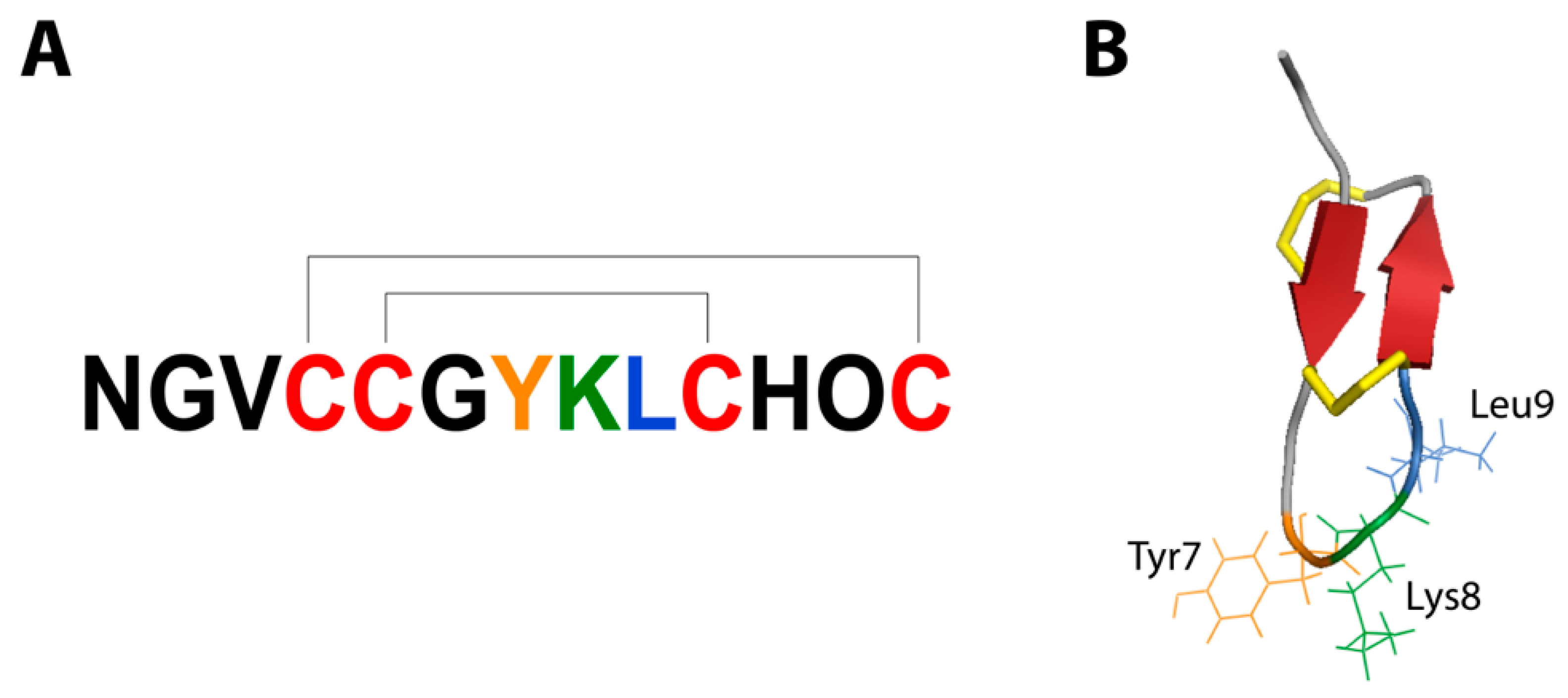
2. Results and Discussion
2.1. Peptide Synthesis
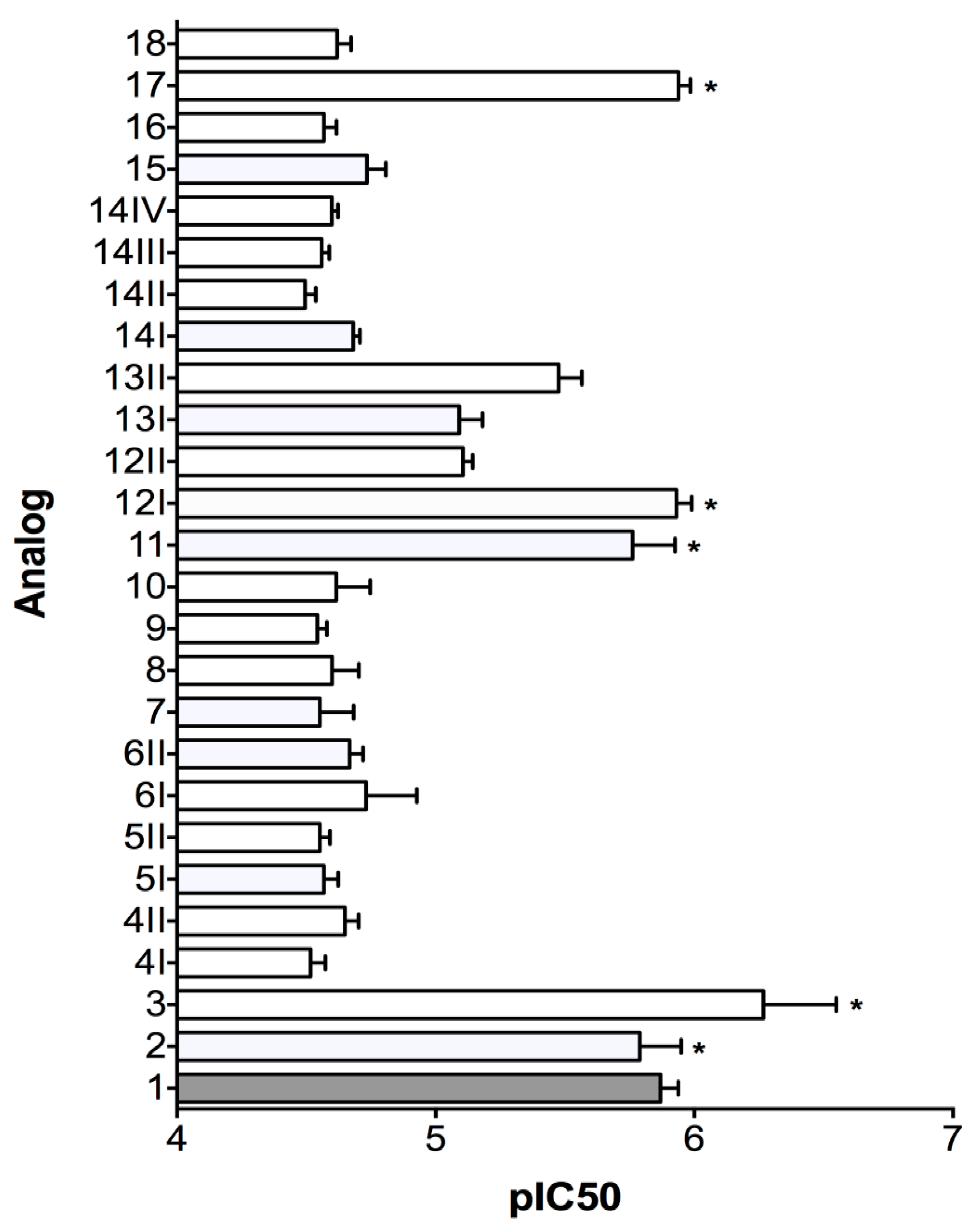
2.2. Norepinephrine Uptake Inhibition Assay
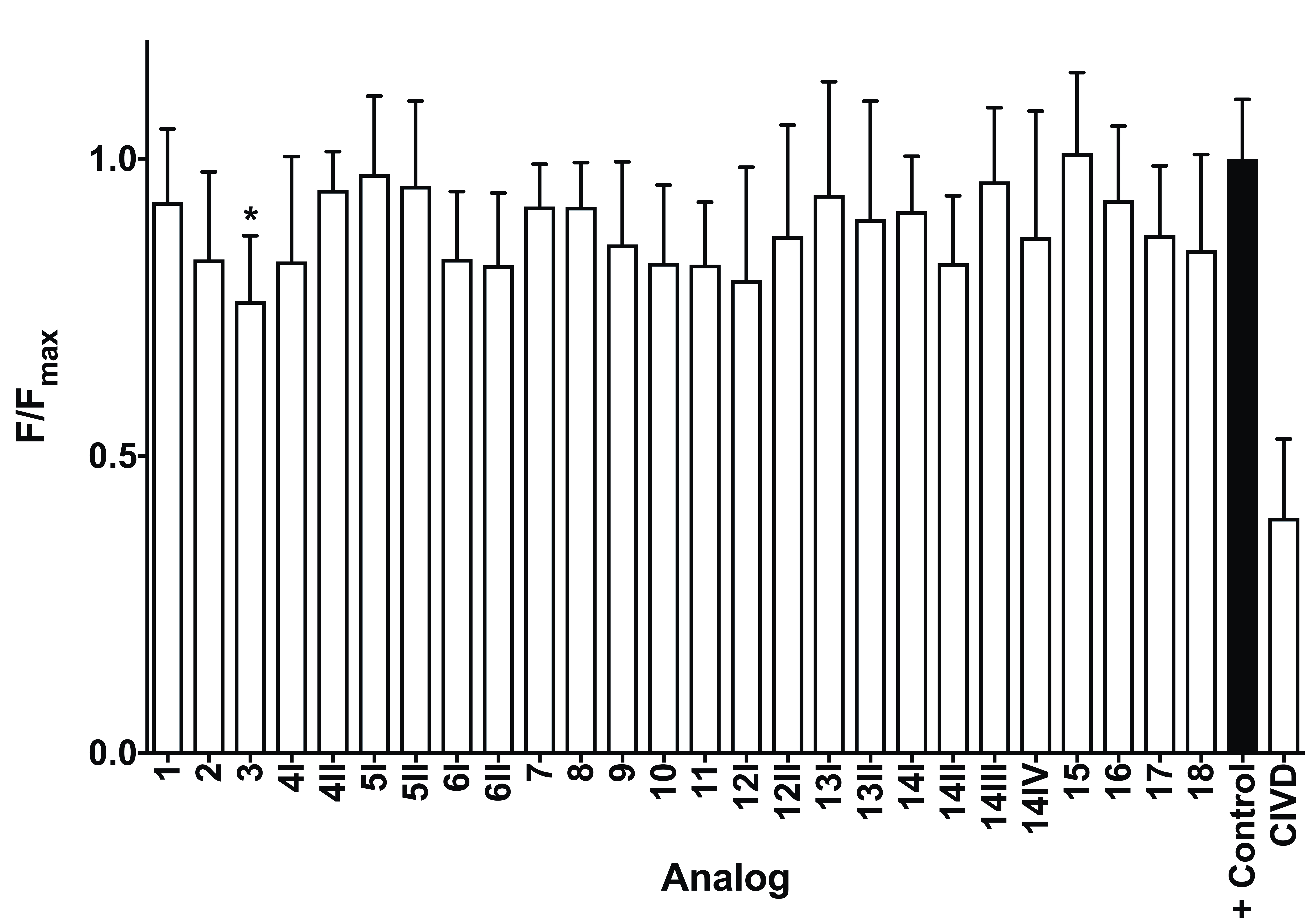
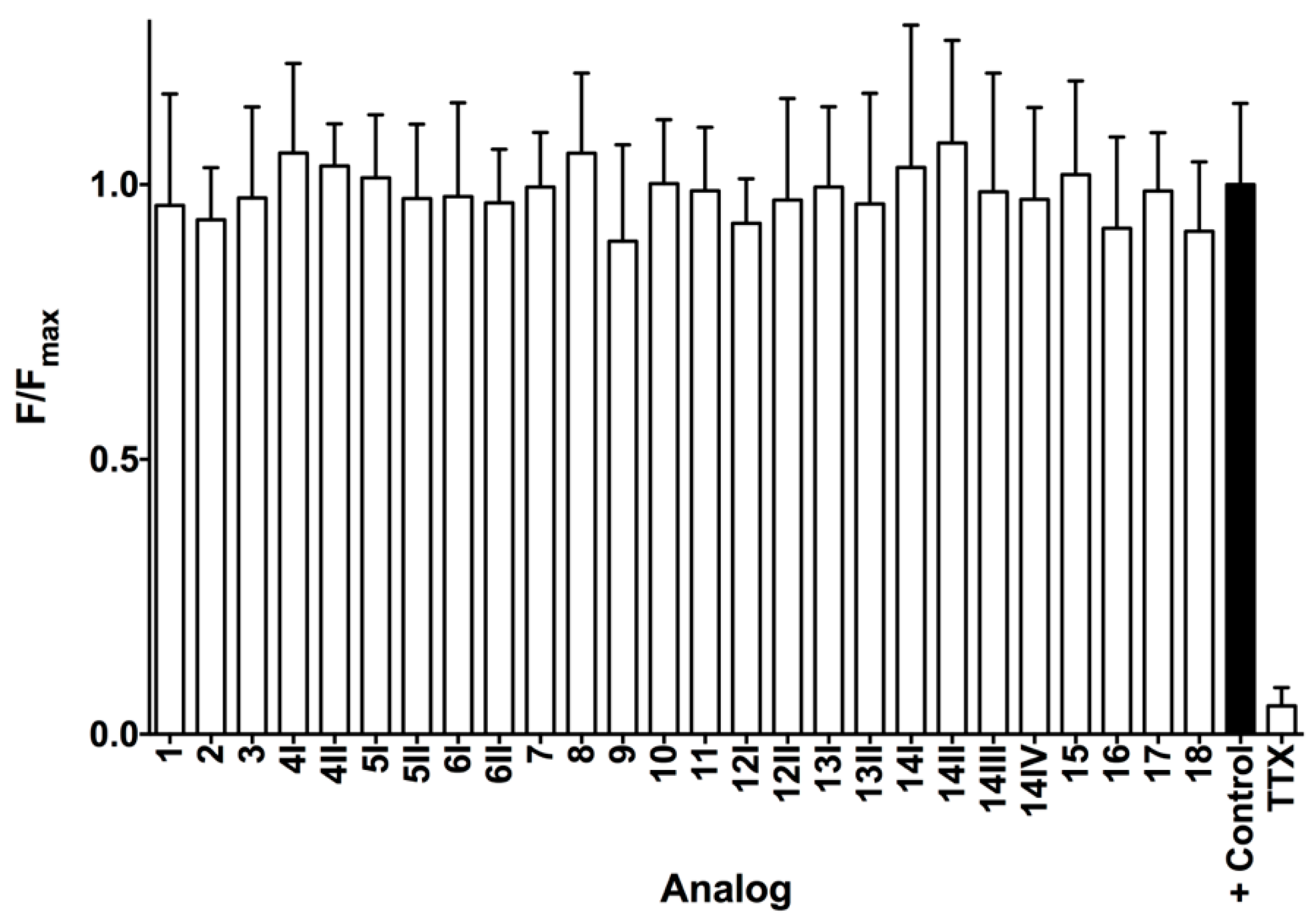
2.3. Calcium Influx Assays
3. Materials and Methods
3.1. Materials and Apparatus
3.2. Peptide Synthesis
3.3. NE Uptake Inhibition Assay
3.4. Calcium Influx Assays
4. Conclusions
Author Contributions
Funding
Conflicts of Interest
References
- Akondi, K.B.; Muttenthaler, M.; Dutertre, S.; Kaas, Q.; Craik, D.J.; Lewis, R.J.; Alewood, P.F. Discovery, synthesis, and structure–activity relationships of conotoxins. Chem. Rev. 2014, 114, 5815–5847. [Google Scholar] [CrossRef] [PubMed]
- Lewis, R.J.; Garcia, M.L. Therapeutic potential of venom peptides. Nat. Rev. Drug Discov. 2003, 2, 790–802. [Google Scholar] [CrossRef] [PubMed]
- Lewis, R.J. Conotoxins as selective inhibitors of neuronal ion channels, receptors and transporters. IUBMB Life 2004, 56, 89–93. [Google Scholar] [CrossRef] [PubMed]
- Dutertre, S.; Jin, A.-H.; Kaas, Q.; Jones, A.; Alewood, P.F.; Lewis, R.J. Deep venomics reveals the mechanism for expanded peptide diversity in cone snail venom. Mol. Cell. Proteom. 2013, 12, 312–329. [Google Scholar] [CrossRef] [PubMed]
- Lavergne, V.; Dutertre, S.; Jin, A.-H.; Lewis, R.J.; Taft, R.J.; Alewood, P.F. Systematic interrogation of the Conus marmoreus venom duct transcriptome with ConoSorter reveals 158 novel conotoxins and 13 new gene superfamilies. BMC Genom. 2013, 14, 708. [Google Scholar] [CrossRef] [PubMed]
- Prashanth, J.R.; Brust, A.; Jin, A.-H.; Alewood, P.F.; Dutertre, S.; Lewis, R.J. Cone snail venomics: From novel biology to novel therapeutics. Future Med. Chem. 2014, 6, 1659–1675. [Google Scholar] [CrossRef] [PubMed]
- Sharpe, I.A.; Gehrmann, J.; Loughnan, M.L.; Thomas, L.; Adams, D.A.; Atkins, A.; Palant, E.; Craik, D.J.; Adams, D.J.; Alewood, P.F. Two new classes of conopeptides inhibit the α1-adrenoceptor and noradrenaline transporter. Nat. Neurosci. 2001, 4, 902–907. [Google Scholar] [CrossRef] [PubMed]
- Lewis, R.J. Discovery and development of the χ-conopeptide class of analgesic peptides. Toxicon 2012, 59, 524–528. [Google Scholar] [CrossRef] [PubMed]
- Brust, A.; Palant, E.; Croker, D.E.; Colless, B.; Drinkwater, R.; Patterson, B.; Schroeder, C.I.; Wilson, D.; Nielsen, C.K.; Smith, M.T. χ-Conopeptide pharmacophore development: Toward a novel class of norepinephrine transporter inhibitor (Xen2174) for pain. J. Med. Chem. 2009, 52, 6991–7002. [Google Scholar] [CrossRef] [PubMed]
- Vetter, I.; Mozar, C.A.; Durek, T.; Wingerd, J.S.; Alewood, P.F.; Christie, M.J.; Lewis, R.J. Characterisation of Nav types endogenously expressed in human SH-SY5Y neuroblastoma cells. Biochem. Pharmacol. 2012, 83, 1562–1571. [Google Scholar] [CrossRef] [PubMed]
- Yin, K.; Baillie, G.J.; Vetter, I. Neuronal cell lines as model dorsal root ganglion neurons: A transcriptomic comparison. Mol. Pain 2016, 12. [Google Scholar] [CrossRef] [PubMed]
- Fields, G.B.; Noble, R.L. Solid phase peptide synthesis utilizing 9-fluorenylmethoxycarbonyl amino acids. Int. J. Pept. Protein Res. 1990, 35, 161–214. [Google Scholar] [CrossRef]
- Simpson, L.S.; Zhu, J.Z.; Widlanski, T.S.; Stone, M.J. Regulation of chemokine recognition by site-specific tyrosine sulfation of receptor peptides. Chem. Biol. 2009, 16, 153–161. [Google Scholar] [CrossRef] [PubMed]
- Bryan-Lluka, L.J.; Bonisch, H.; Lewis, R.J. χ-Conopeptide MrIA partially overlaps desipramine and cocaine binding sites on the human norepinephrine transporter. J. Biol. Chem. 2003, 278, 40324–40329. [Google Scholar] [CrossRef] [PubMed]
- Sharpe, I.A.; Palant, E.; Schroeder, C.I.; Kaye, D.M.; Adams, D.J.; Alewood, P.F.; Lewis, R.J. Inhibition of the norepinephrine transporter by the venom peptide χ-MrIA. Site of action, Na+ dependence, and structure-activity relationship. J. Biol. Chem. 2003, 278, 40317–40323. [Google Scholar] [CrossRef] [PubMed]
- Sousa, S.R.; Vetter, I.; Ragnarsson, L.; Lewis, R.J. Expression and pharmacology of endogenous Cav channels in SH-SY5Y human neuroblastoma cells. PLoS ONE 2013, 8, e59293. [Google Scholar] [CrossRef] [PubMed]


| Analog | Sequence | Relative Abundance | Expected MW (Da) | Observed MW (Da) |
|---|---|---|---|---|
| 1 | NGVCCGYKLCHPC * | 100.00 | 1391.7 | 1392.0 |
| 2 | NGVCCGYKLCHPC | 49.77 | 1392.7 | 1392.8 |
| 3 | ZGVCCGYKLCHPC | 0.73 | 1389.7 | 1389.8 |
| 4I | ZGVCCGYKLCHP | 0.05 | 1288.5 | 2577.6 |
| 4II | 2577.6 | |||
| 5I | ZGVCCGYKLC | 0.34 | 1054.3 | 2106.0 |
| 5II | 2107.0 | |||
| 6I | NGVCCGYKLC | 0.12 | 1057.3 | 2115.8 |
| 6II | 2113.8 | |||
| 7 | ZGVCCGYKL | 0.03 | 951.1 | 951.6 |
| 8 | ZGVCCGYKL * | 0.01 | 950.1 | 950.4 |
| 9 | NGVCCGYK | 0.02 | 841.0 | 841.6 |
| 10 | ILRGILRNGVCC * | 0.02 | 1313.7 | 1314.0 |
| 11 | GILRNGVCCGYKLCHPC | 0.33 | 1831.2 | 1834.0 |
| 12-I | LRNGVCCGYKLCHOC | 0.14 | 1678.0 | 1678.0 |
| 12-II | 1677.8 | |||
| 13I | VCCGYKLCHOC | 0.88 | 1237.5 | 1237.4 |
| 13II | 1237.4 | |||
| 14I | CGYKLCHOC | 0.21 | 1037.2 | 2073.6 |
| 14II | 2072.8 | |||
| 14III | 2073.0 | |||
| 14IV | 2071.8 | |||
| 15 | GYKLCHOC | 0.04 | 934.1 | 934.6 |
| 16 | YKLCHOC | 0.02 | 877.1 | 877.6 |
| 17 | GVCCGYKLCHPC | 0.01 | 1278.6 | 1278.6 |
| 18 | GVCCGY(SO3−)KLCHOC | 0.11 | 1377.5 | 1376.6 |
© 2019 by the authors. Licensee MDPI, Basel, Switzerland. This article is an open access article distributed under the terms and conditions of the Creative Commons Attribution (CC BY) license (http://creativecommons.org/licenses/by/4.0/).
Share and Cite
Ziegman, R.; Brust, A.; Jha, P.; Cardoso, F.C.; Lewis, R.J.; Alewood, P.F. ‘Messy’ Processing of χ-conotoxin MrIA Generates Homologues with Reduced hNET Potency. Mar. Drugs 2019, 17, 165. https://doi.org/10.3390/md17030165
Ziegman R, Brust A, Jha P, Cardoso FC, Lewis RJ, Alewood PF. ‘Messy’ Processing of χ-conotoxin MrIA Generates Homologues with Reduced hNET Potency. Marine Drugs. 2019; 17(3):165. https://doi.org/10.3390/md17030165
Chicago/Turabian StyleZiegman, Rebekah, Andreas Brust, Prerna Jha, Fernanda C. Cardoso, Richard J. Lewis, and Paul F. Alewood. 2019. "‘Messy’ Processing of χ-conotoxin MrIA Generates Homologues with Reduced hNET Potency" Marine Drugs 17, no. 3: 165. https://doi.org/10.3390/md17030165
APA StyleZiegman, R., Brust, A., Jha, P., Cardoso, F. C., Lewis, R. J., & Alewood, P. F. (2019). ‘Messy’ Processing of χ-conotoxin MrIA Generates Homologues with Reduced hNET Potency. Marine Drugs, 17(3), 165. https://doi.org/10.3390/md17030165


