1. Introduction
Jasplakinolide (
1) (also known as jaspamide) is a cyclodepsipeptide that was first reported in 1986 by Ireland [
1] and Crews [
2] from the marine sponge
Jaspis (syn.:
Doryplores)
splendens (order Astrophorida, family Ancorinidae). Biosynthetically, jasplakinolide features a polyketide–peptide hybrid derived from the PKS-NRPS pathway [
3]. Jasplakinolide revealed potent pharmacological activities such as antifungal [
4], anthelmintic [
1], insecticidal [
2] and most profoundly its highly potent cytotoxic activity with IC
50 values in the nanomolar range [
5]. Since then, an in-depth investigation has been directed toward the identification of its mode of action as a cytotoxic agent, which revealed that
1 exerts its cytotoxic activity by disrupting the actin cytoskeleton as a potent stabilizer of filamentous actin (F-actin) [
6]. Subsequently, many research interests have been invested in marine sponges producing cyclodepsipeptides with mixed skeletons structurally related to jasplakinolide, even if they are taxonomically apart from
Jaspis sponges such as
Geodia and
Cymbastela (geodiamolides) [
7,
8,
9,
10,
11,
12] or
Suberites sponges (seragamides) [
13]. In addition to the cyclodepsipeptides obtained from
Jaspis sponges, some acyclic congeners have been reported that have been considered as plausible hydrolytic products according to the determined biosynthetic gene clusters of chondramide C [
14]. These feature three main biosynthetic phases starting with PKS portion formation and ending with the addition of a tyrosine moiety prior to the ring closure creating a macrocycle. To end up with an opened congener, this may be a biosynthetic pathway devoid of the cyclization step or unexpectedly activating a second enzyme cluster to reopen the 19-membered ring at either the ester bond between C-1 and C-15 or the amide bond between C-3 and tyrosine-
NH [
15].
In a continuation of our ongoing research targeting new and bioactive cytotoxic jasplakinolide derivatives, we previously explored the extract of three specimens of the Indonesian marine sponge
Jaspis splendens, affording jasplakinolides Q and R, which were new congeners, together with the parent compound jasplakinolide (
1) [
16]. The remaining semi-pure fractions of the extract were combined and re-fractionated by vacuum liquid chromatography (VLC) using the C18 stationary phase with a gradient elution using water and methanol as a mobile phase. The resulting VLC fractions were then re-evaluated by a de-replication strategy to identify unreported molecular weights of jasplakinolides, which may indicate the existence of new jasplakinolide congeners.
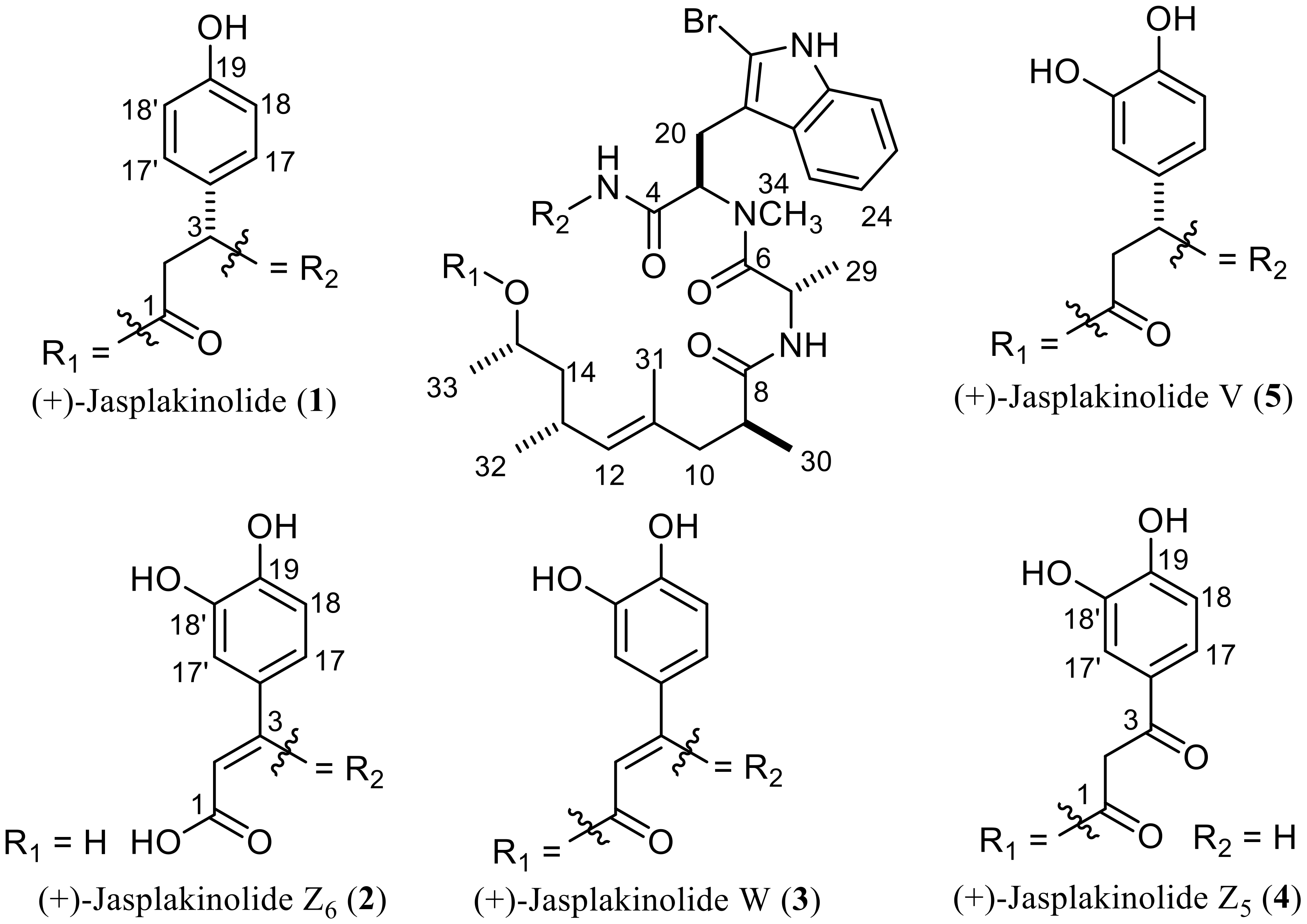
In this study we report the isolation, identification and bioactivity assessment of a new acyclic natural jasplakinolide (
2) and a structural revision for a previously reported derivative (
4) (
Figure 1), in addition to two known cyclic derivatives (
1 and
5).
2. Results and Discussion
Compound
2 was obtained as an amorphous solid. The HRESIMS spectrum of
2 revealed pseudomolecular ion peaks at
m/z 758.2749 [M + NH
4]
+ (calcd for C
36H
49BrN
5O
8;
m/z 758.2759) and
m/z 763.2311 [M + Na]
+ (calcd for C
36H
45BrN
4NaO
8;
m/z 763.2313), indicating the molecular formula C
36H
45BrN
4O
8, which differs from that of
1 by two oxygen atoms [
17]. The UV spectrum of
2 revealed an absorption maximum (λ
max) at 326 nm in addition to those at 220 and 280 nm, suggesting the presence of an acyclic jasplakinolide derivative [
17].
The
1H and
13C NMR data of
2 (
Table 1) were similar to those of
1 except for the presence of downfield-shifted aromatic protons of an ABX system, ascribed for a catechol moiety, which replaced those of the AA’BB’ system of
1 (
Figure 2). A further difference between the
1H NMR spectra of both compounds was the presence of an olefinic singlet at δ
H 5.13 in the spectrum of
2, which was found to be connected to a sp
2 methine carbon at δ
C 99.7, as observed in the HMQC spectrum.
Further spectral information of the structure of 2 was provided by 1H NMR measurement in DMSO-d6, which showed four downfield proton resonances at δH 11.62 (s), 10.95 (s), 9.41 (br s) and 9.07 (br s). These were assigned to the NH signal of the pyrrole ring of tryptophan, a terminal carboxylic acid group of a β-tyrosine unit and the two phenolic hydroxyl groups of the catechol moiety, respectively.
Based on previous findings,
2 was primarily suggested to be an acyclic jasplakinolide derivative of (+)-jasplakinolide W (
3) [
17].
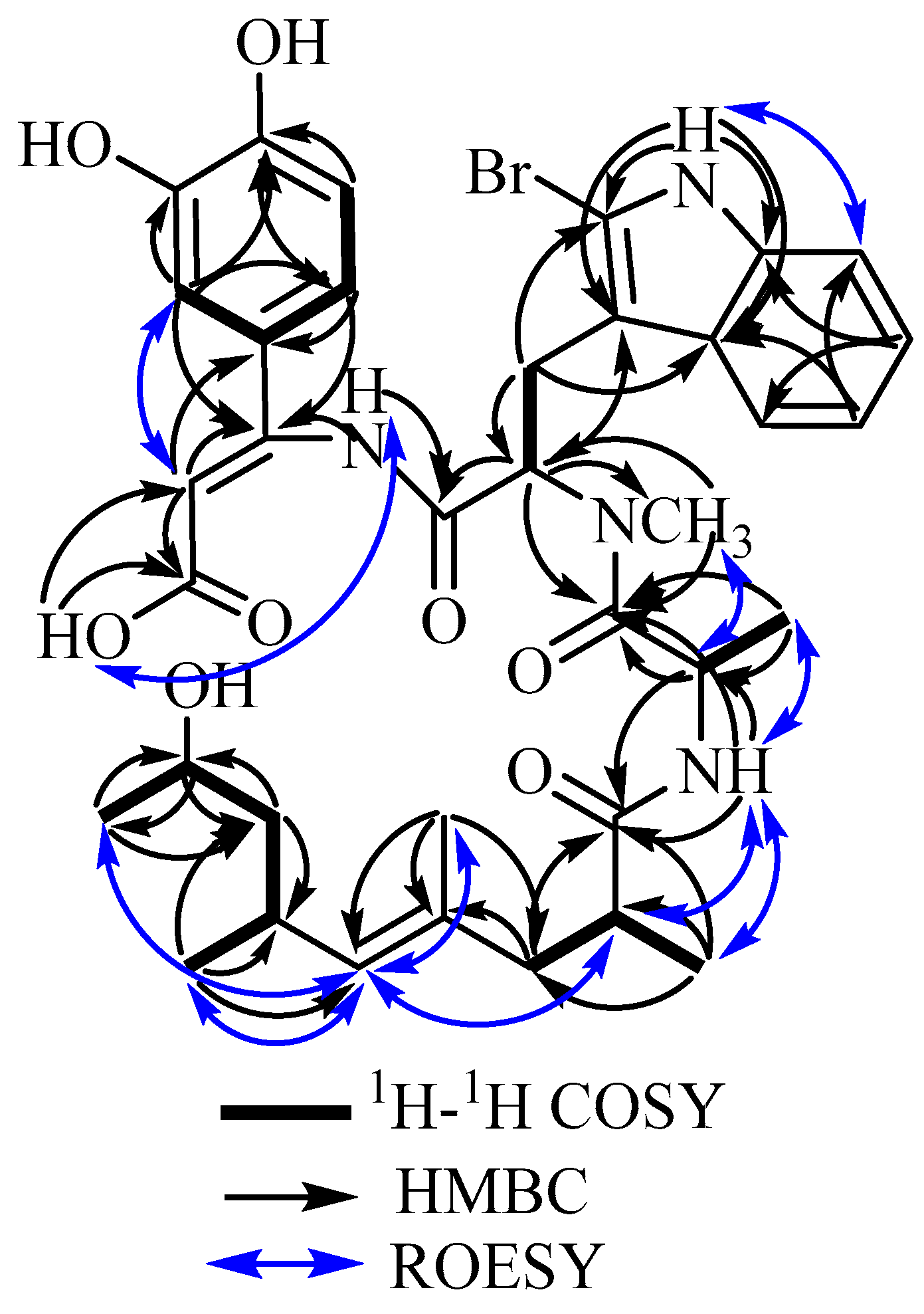
Further evidence for the structure of
2 was provided via 2D NMR measurements including
1H–
1H COSY, HMBC, HMQC and ROESY experiments (see
supplementary materials, Figures S5–S8). Key correlations exhibited in the COSY spectrum confirmed the ABX system of the catechol moiety. The HMBC spectrum showed correlations from the olefinic singlet proton at δ
H 5.13 to three quaternary carbons at δ
C 168.3, 153.4 and 125.8, which were assigned to C-1, C-3 and C-16, respectively, confirming the presence of a double bond between C-2 and C-3. Further HMBC correlations were found from a broad singlet proton at δ
H 6.80 (3–N
H–4) to two quaternary carbons C-3 and C-4 (δ
C 168.3), which indicate the connection between the tryptophan and the
β-dihydroxyphenyl alanine moiety. Based on the structural evidence from NMR spectra and the molecular formula,
2 was assigned as an acyclic jasplakinolide derivative that features a
β-dihydroxyphenyl alanine with a double bond between C-2 and C-3 instead of a
β-tyrosine unit as found in
1. The stereochemistry of the double bond between C-2 and C-3 was assigned as
cis according to the priority and based on the strong ROESY correlations (
Figure 3, as well as
Figure S8a in the supplementary materials) from H-2 to H-17′ and H-17, whereas no correlation was found between H-2 and the amide proton, which connects the
β-dioxyphenyl alanine group to the brominated tryptophan moiety.
Based on the close similarity of the ROESY spectrum obtained for
2 to those of
1 and
3, together with the optical rotation (+47.6 (0.6, MeOH)), it was unambiguously confirmed that
2 is an acyclic derivative of
3, lacking the depside bond between C-1 and C-15. Following the previously reported series of acyclic jasplakinolides [
17],
2 was trivially named (+)-jasplakinolide Z
6.
Compound
4 was obtained as a white amorphous solid and interestingly its HRESIMS spectrum showed pseudomolecular ion peaks at
m/z 758.2749 [M + NH
4]
+ (calcd for C
36H
49BrN
5O
8;
m/z 758.2759) and
m/z 763.2306 [M + Na]
+ (calcd for C
36H
45BrN
4NaO
8;
m/z 763.2302), determining its molecular formula as C
36H
45BrN
4O
8, identical to that of
2. The UV spectrum of
4 revealed an additional absorption maximum (λ
max) at 310.7 nm to the usual maximal absorption wavelengths at 220 and 280 nm found for other derivatives, indicating the presence of an acyclic jasplakinolide congener. Based on the determined molecular formula and UV spectrum of
4 and by comparison to the previously reported jasplakinolides, it was suggested to be the reported derivative (+)-jasplakinolide Z
5 [
17]. 1D and 2D NMR data of
4 revealed a similar pattern of benzenoid moiety in
β-tyrosine residue with an additional hydroxyl group (catechol ring) with three proton resonances being part of an ABX spin system similar to that found in
2 (
Table 1). In addition, the
1H NMR data revealed two doublets at δ
H 3.87 and δ
H 3.73 (d,
J = 16.5 Hz), which were directly correlated through the HMQC spectrum to a methylene carbon (δ
C 45.8). In addition, the gHMBC spectrum of
4 featured clear correlations from a doublet at δ
H 3.87 to two carbons at δ
C 168.1 and 192.3 with the latter having key correlations to aromatic protons at δ
H 7.39 (d,
J = 2.0 Hz, H-17’), δ
H 7.37 (dd,
J = 8.3, 2.0 Hz, H-17) together with
4JH-C (
ω) correlation to the proton signal at δ
H 6.82 (d,
J = 8.3 Hz, H-18), indicating the existence of a ketocarbonyl functionality at C-3, which indicates the cleavage of the 19-membered macrocyclic ring of jasplakinolide at the C3–N bond. By further comparing the 1D and 2D NMR spectral data with those from the literature,
4 was found to be very similar to (+)-jasplakinolide Z
5.
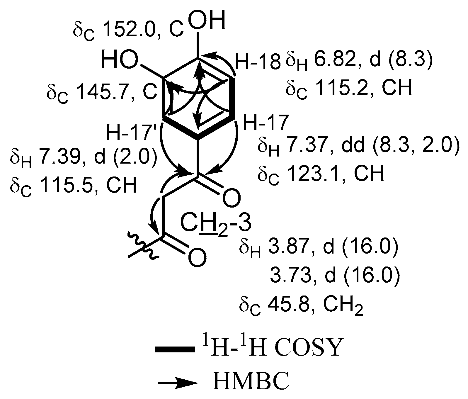
However, by a careful inspection of the 2D NMR spectra of
4 and by comparing them to the reported spectral data of (+)-jasplakinolide Z
5, several discrepancies were clearly observed between the previously reported proton and carbon NMR assignment of the protons and carbons of the aromatic ring of tyrosine (catechol moiety) in the literature [
17] and those obtained in this work. The major differences were noticed for the proton and carbon NMR assignments at C-17’, C-18 and C-19 (
Figure 4). Further confirmation of the assignment presented in this work was provided by 2D NMR data including
1H–
1H COSY and gHMBC correlations, which revealed clear
3JH-C correlations from H-17 and H-17’ to an oxygenated aromatic carbon at δ
C 152.0 (C-19), in addition to the correlations from H-18 and H-17’ to another oxygen-bearing aromatic carbon at δ
C 145.7 (C-18’). All this evidence supported the final assignment of the revised NMR data of (+)-jasplakinolide Z
5 [
17] as depicted in
Table 1 and
Figure 4.
Based on the plausible common biosynthetic pathway of all isolated compounds and the parent compound jasplakinolide (1), they are expected to have the same absolute configuration.
All the isolated compounds were assessed for their antiproliferative activity against mouse lymphoma (L5178Y) cells using the microculture tetrazolium (MTT assay) and compared to kahalalide F as a positive control (IC
50 = 4.3 µM). Results revealed that all the isolated compounds possess potent cytotoxic activity with IC
50 values in the low micromolar to nanomolar range. Among the tested compounds,
2 was the least potent (IC
50 = 3.2 µM) derivative compared to
4,
5 and
1, which showed IC
50 values in the nanomolar range (<100 nM). This indicates that the 19-membered ring of jasplaklinolide is not a major structural requirement for its activity. The main factor that might influence the antiproliferative activity of the acyclic jasplakinolide congeners is its lipophilic character, as noticed when comparing
2 with
4, where the former has a free carboxylic acid functionality imparting higher polarity to the molecule compared to
4. Further proof was provided by a comparison of the antiproliferative activity of three acyclic jasplakinolides Z
1–Z
3 against a panel of human cancer cells, where the latter two derivatives are the methyl and ethyl esters of the former one, respectively [
17]. Results also revealed that the hydrolysis of the depside bond, yielding a free carboxylic acid group significantly diminished its activity, which was almost turned back to be equivalent to the parent compound by converting the carboxylic acid group into an ester functionality. This can be attributed to the increased lipophilicity of the ester derivatives that may facilitate their cellular uptake and hence recuperate the activity of the compound.
Adding to the main structure-activity relationship (SAR) characteristics influencing the antiproliferative activity of jasplakinolide [
17], the 19-membered macrocyclic ring is not necessary for activity, since it can be opened to yield acyclic congeners with equivalent activities, provided that they keep the lipophilic character sufficient to facilitate cellular uptake.
3. Experimental Section
3.1. General Experimental Procedures
Vacuum liquid chromatography (VLC) was carried out using C18 reversed-phase and a gradient elution was applied, going from water to methanol with a 10% gradient interval. A Perkin-Elmer-241 MC polarimeter (Perkin-Elmer, Akron, Ohio, USA) was used for measuring the optical rotation. LRESIMS was recorded on an LCMS HP1100 Agilent Finnigan LCQ Deca XP Thermoquest machine (Thermo Electron Corporation, San Jose, California, USA) and HRESIMS were determined on a FTHRMS-Orbitrap (Thermo Fischer Scientific, Bremen, Germany) mass spectrometer. For analytical HPLC analysis, samples were injected into a Dionex Ultimate 3000 LC system equipped with a photodiode array detector (UVD340S) (Dionex, Munich, Germany) with detection channels at 235, 254, 280 and 340 nm. Ready-made separation columns (125 L × 4 mm ID) were prefilled with Eurosphere-10C18 (Knauer, Berlin, Germany) and used a linear gradient from 90% H2O (pH 2.0) to 100% MeOH over 40 min with a flow rate of 1 mL/min. TLC analysis was carried out using aluminum sheets precoated with silica gel 60 F254 (Merck, Darmstadt, Germany).
Preparative HPLC separations were performed on a LaChrom-Merck Hitachi HPLC system (Tokyo, Japan), pump L-7100(Hitachi, Tokyo, Japan), UV detector L-7400 (Hitachi, Tokyo, Japan) using a column (Knauer, 300 L × 8 mm ID, prefilled with 100 C18 Eurosphere, flow rate 5 mL/min, UV detection at 280 nm), and the solvent system consisted of a linear gradient of MeOH and nanopure H2O running 40%–90% MeOH over 30 min.
1D (1H and 13C NMR) and 2D NMR (COSY, gHMBC, gHMQC and ROESY) spectra were recorded on Bruker AVANCE DMX 600 or 300 spectrometers (Bruker, Fällanden, Switzerland) using chloroform-d, DMSO-d6 or methanol-d4 as solvents (Sigma-Aldrich, Taufkirchen, Germany).
3.2. Biological Material
In August 2008, sponge specimens were collected on three neighboring islands of East Kalimantan (Indonesia), namely Samama, Panjang and Shoal Islands, at 10-meter depths. Numbers of voucher specimens are RMNH Por. 4234, 4266 and 4299, respectively. They were taxonomically identified as Jaspis splendens (order Astrophorida, family Ancorinidae) by Dr. Nicole de Voogd at the National Museum of Natural History, Leiden, Netherlands, where the voucher specimens are kept. HPLC and LCMS analyses of the three samples revealed that they were identical with regard to their peptide derivatives. Hence, the materials were combined in order to obtain a sufficient amount of compounds for subsequent structure elucidation.
3.3. Extraction and Isolation
The animal was freeze-dried and the material (500 g) was extracted with methanol (3 × 2 L) and filtered. The extracts were then combined, evaporated under reduced pressure to dryness and processed as follows. The methanolic extract (80 g) was dissolved in water and partitioned with n-hexane, ethyl acetate and then n-butanol. The bioactive ethyl acetate soluble fraction (2 g) was loaded on C18 reversed-stationary phase and subjected to VLC using a linear gradient from water to MeOH to afford 11 fractions (Fr. I-XI). Fr. VIII, eluted with methanol:water (7:3) (670 mg), revealed several peaks with UV absorption spectra similar to that of jasplakinolide. An aliquot of 300 mg of Fr. VIII was further purified by preparative HPLC, equipped with a C18 ready-made column using a gradient elution of MeOH and nanopure H2O, 40%–90% MeOH over 30 min, to give 1 (140.0 mg), 2 (46.8 mg), 4 (33.7 mg) and 5 (21.0 mg).
(+)-Jasplakinolide Z6 (
2): amorphous yellow solid;
+47.6° (
c 0.60, MeOH); UV (MeOH) λ
max 220, 280, 326.1 nm;
1H-NMR and
13-C NMR see
Table 1; HRESIMS
m/z 758.2749 [M + NH
4]
+ (calcd for C
36H
49BrN
5O
8;
m/z 758.2759) and
m/z 763.2311 [M + Na]
+ (calcd for C
36H
45BrN
4NaO
8;
m/z 763.2313).
(+)-Jasplakinolide Z5 (
4): white amorphous solid;
+58.0° (
c 0.40, MeOH); UV (MeOH) λ
max 220, 280, 310.7 nm;
1H-NMR and
13-C NMR see
Table 1; HRESIMS
m/z 758.2749 [M + NH
4]
+ (calcd for C
36H
49BrN
5O
8;
m/z 758.2759) and
m/z 763.2306 [M + Na]
+ (calcd for C
36H
45BrN
4NaO
8;
m/z 763.2302).
3.4. Cell Proliferation Assay
Cytotoxicity was tested against L5178Y mouse lymphoma cells using the microculture tetrazolium (MTT) assay as described earlier [
18,
19]. All experiments were carried out in triplicate and repeated three times. As controls, media with 0.1% EGMME/DMSO was included in the experiments.


