Article

Preparation of Antioxidant Peptide by Microwave- Assisted Hydrolysis of Collagen and Its Protective Effect Against H2O2-Induced Damage of RAW264.7 Cells

1
Zhejiang Provincial Engineering Technology Research Center of Marine Biomedical Products; School of Food and Pharmacy, Zhejiang Ocean University, Zhoushan 316022, China
2
Hangzhou Institute for Food and Drug Control, Hangzhou 310052, China
*
Author to whom correspondence should be addressed.
Mar. Drugs 2019, 17(11), 642; https://doi.org/10.3390/md17110642
Submission received: 16 October 2019 / Revised: 7 November 2019 / Accepted: 12 November 2019 / Published: 14 November 2019
(This article belongs to the Special Issue Active Compounds from Sea Cucumber)

Abstract

:
Antioxidant peptides have elicited interest for the versatility of their use in the food and pharmaceutical industry. In the current study, antioxidant peptides were prepared by microwave-assisted alkaline protease hydrolysis of collagen from sea cucumber (Acaudina molpadioides). The results showed that microwave irradiation significantly improved the degree of hydrolysis of collagen and the hydroxyl radical (OH⋅) scavenging activity of hydrolysate. The content and OH⋅ scavenging activity of collagen peptides with molecular weight ≤ 1 kDa (CPS) in the hydrolysate obtained at 250 W increased significantly compared with the non-microwave-assisted control. CPS could scavenge OH⋅ and 2,2-diphenyl-1-picrylhydrazyl (DPPH) radical in a dose-dependent manner. The scavenging activity of OH⋅ and DPPH radical was 93.1% and 41.2%, respectively, at CPS concentration of 1 mg/mL. CPS could significantly promote RAW264.7 cell proliferation and reduce the Reactive Oxygen Species (ROS) level of H2O2-induced damage in RAW264.7 cells in a dose-dependent manner. Furthermore, all CPS-treated groups exhibited an increase in superoxide dismutase (SOD) and glutathione peroxidase (GSH-Px) and a decrease in malondialdehyde (MDA) level compared with the control. These results showed that CPS could effectively protect RAW264.7 cells from H2O2-induced damage, implying the potential utilization of CPS as a natural antioxidant for food and pharmaceutical applications.

1. Introduction

Bioactive peptides are short peptides with some bioactive functions, such as antioxidant, antimicrobial, antitumor, or antihypertensive activity. Bioactive peptides have been widely used in health-food, nutraceuticals, and pharmaceutical preparations due to their significant biological functions and efficient absorption [1]. As one of several promising bioactive peptides, antioxidant peptides have become a topic of great interest because of the association between many human diseases and oxidative stress [2]. When humans are subjected to oxidative stress, large amounts of reactive oxygen species (ROS) are produced. Abundant quantities of ROS (superoxide, hydrogen peroxide, and hydroxyl) tend to react with biological molecules, such as protein and DNA, often leading to liver disease, heart disease, and cancer because of oxidative damage of cells [3,4]. ROS-induced oxidative cell damage is usually accompanied by an increase in lipid peroxides [5]. It was known that malondialdehyde (MDA) is the major secondary metabolite of lipid peroxidation induced by ROS. The content of MDA is a marker of lipid peroxidation and the degree of cell damage [6]. The superoxidase dismutase (SOD) and glutathione peroxidase (GSH-Px) are the major antioxidant enzymes, which play a key role in protecting cells from oxidative damage [7]. Antioxidants can protect the body from damage by removing excess ROS, reducing the MDA, and improving the activities of SOD and GSH-Px. Therefore, as a natural antioxidant, antioxidant peptides have great potential of utilization in pharmaceutical industries.
At present, antioxidant peptides are mainly obtained by separation and purification directly from the plant or animal tissues, chemical synthesis, microbial fermentation, or enzymatic hydrolysis of protein. However, the separation and purification of antioxidant peptides directly from the plant or animal tissues, and fermentation products are expensive. Chemical synthesis of peptides also requires sophisticated and expensive equipment and reagents, and the chemical reagents involved may be harmful to humans and/or cause environmental pollution. Therefore, enzymatic digestion of protein has been the most common method for preparation of antioxidant peptides due to the simplicity, safety, low-cost, and sustainability of the approach [8]. Collagen is an important structural protein rich in hydroxyproline, which has been employed in food and medical applications for decades with excellent biocompatibility and safety [9,10]. For example, the collagens from echinoderms (sea urchin, starfish, and sea cucumber) can be used to develop collagen barrier-membranes for guided tissue regeneration due to the good mechanical performances [11]. However, the large molecular weight of the compound creates barriers in absorption and utilization in the body. Therefore, collagen peptides, smaller proteins hydrolyzed from collagen, have received increasing attention because of their physicochemical properties and biological activity [12,13,14]. However, one of the limitations of the collagen peptides production process is the length of time required in conventional hydrolysis methods due to its dense fiber structure [8]. This limitation can potentially be overcome by using microwave assisted hydrolysis. Studies have shown that microwaves cause different biological and chemical effects depending, among other factors, upon strength, frequencies and length of exposure [15]. Microwave radiation penetrates into the interior of the protein molecule, exposing the hidden enzymatic sites and enabling a more thorough hydrolysis [16]. Microwave-assisted enzymatic hydrolysis of protein can, therefore, significantly reduce the overall hydrolysis time and increase the yield of peptide by intensifying molecular movements and collisions [8]. Previous studies had also demonstrated microwave-assisted enzymatic hydrolysis to be one of the most effective methods to prepare peptides [8,17,18].
Sea cucumbers, of which more than 1400 species exist around the world, are important marine resources of protein containing high amounts of collagen in their body wall [19]. Several researchers have reported the development and characteristics of various collagens from sea cucumbers [20,21,22,23]. The common sea cucumber, Acaudina molpadioides, is widely distributed in East China Sea, especially in the coastal areas of Ningbo and Zhoushan. Traditionally, due to the lack of key technologies for industrialization, Acaudina molpadioides resources have not been utilized to their potential and continue to sell for a low price. The preparation of collagen peptides using Acaudina molpadioides has great potential due to the good biological function of collagen peptides and inexpensive raw materials of Acaudina molpadioides [24]. Studies have shown that collagen peptides of sea cucumber have a demonstrated ability to scavenge free radicals and other peroxides [25]. In previous studies, we had prepared an antioxidant peptide from collagen of Acaudina molpadioides (ASC-Am) by microwave-assisted neutral protease hydrolysis [26]. Different proteases hydrolyze different sites of the protein to produce peptides of different amino acid sequences. Therefore, in this study, we focus on production of antioxidant peptides from ASC-Am by microwave-assisted alkaline protease hydrolysis. Furthermore, the protective effect of antioxidant peptides against peroxide (H2O2)- induced damage of RAW264.7 cells was investigated by measuring cell proliferation, ROS, MDA, SOD, and GSH-Px levels.

2. Results and Discussion

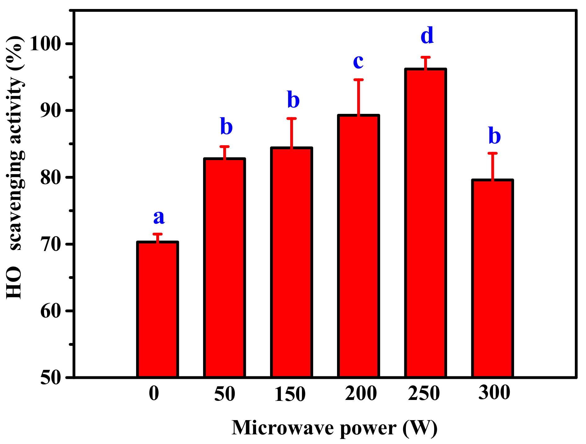
2.1. Effect of Microwave Assisted on Alkaline Protease Hydrolysis of ASC-Am

The effect of microwave power on OH· scavenging activity of hydrolysate by alkaline protease digestion of ASC-Am was investigated (Figure 1). The results demonstrated that OH· scavenging activities were significantly improved at all tested microwave powers (50–300 W) as compared to non-microwave assisted hydrolysis. The enhanced OH· scavenging activity when the microwave power increased from 50 to 250 W suggested that the higher microwave power enabled the higher probability of contact between ASC-Am and alkaline protease, resulting in more small peptides with antioxidant activity. However, excessive molecular collisions cause the denaturation of alkaline protease, which may make the OH· scavenging activity begin to decrease when the microwave power continues to increase to 300 W. The maximum OH· scavenging activity was observed at 250 W of microwave power, which increased from 70.3% in non-microwave assisted to 96.2% in this treatment. Therefore, 250 W was selected as the optimal power for microwave-assisted alkaline protease digestion of ASC-Am.
In addition, we investigated the content and molecular weight distribution of the peptides in hydrolyzate under microwave assisted digestion with 250 W. As shown in Table 1, compared with 0 W, the total content of CPL at 250 W decreased, but the content of CPM and CPS increased by 48% and 30.2%, respectively. Furthermore, the proportions of CPM and CPS in hydrolyzate obtained at 250 W were both higher than those at 0 W. These results suggested that microwave assisted hydrolysis can significantly promote the breakdown of ASC-Am into smaller molecular weight peptides. The results of this study were similar to those of Uluko et al. (2015), which showed that the hydrolyzed product under microwave irradiation increased by 184.9% compared to the control [27].
The molecular weight of peptides has a significant effect on the antioxidant activity [28]. It was evident that the peptides with smaller molecular weight revealed a stronger antioxidant activity (Figure 2), which was similar to the results of previous reports [29]. In addition, compared with those under 0 W of microwave power, the OH· scavenging activities of CPL, CPM, and CPS under 250 W power were significantly improved. The reasons for this result may be: (1) microwave radiation probably exposed more cleavage sites of collagen for alkaline protease hydrolysis, resulting in an improvement in antioxidant activity of the peptides due to altered amino acid sequences of peptides; (2) the microwaves probably altered the amino acid sequence of the alkaline protease, resulting in a change in the cleavage site of alkaline protease.

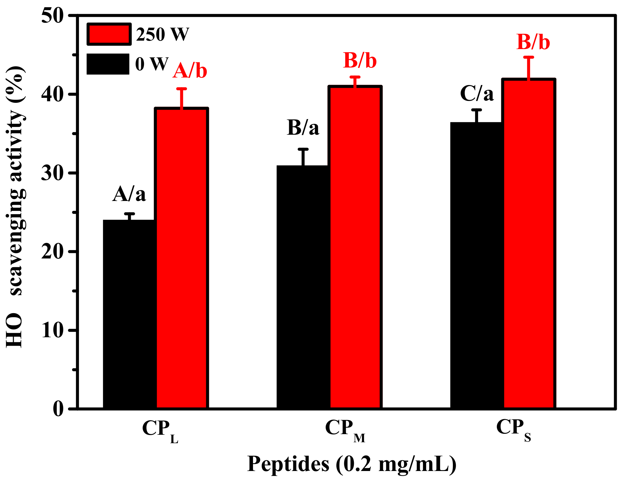
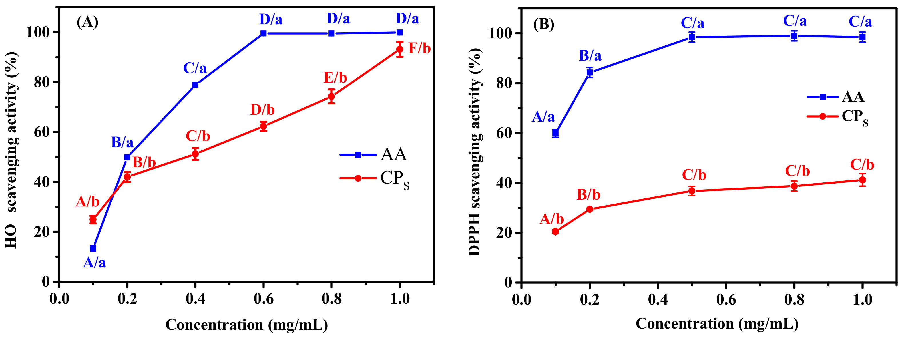
2.2. Antioxidant Activity of CPS

In order to better investigate its antioxidant properties, the DPPH and OH· scavenging assays at different concentration of CPS were studied and compared with the positive controls containing ascorbic acid (AA). The CPS The OH· scavenging activity of CPS increased from 24.9% at 0.1 mg/mL to 93.1% at 1.0 mg/mL (Figure 3A), indicating it scavenged OH· in a concentration-dependent manner. The EC50 value of CPS was 0.4 mg/mL for OH·, which was lower than that of the peptide BSH-III (Mw ≤ 1 kDa) from protein hydrolysate obtained from bluefin leatherjacket skin (IC50 of 0.746 mg/mL) [30]. It is known that OH· can have a destructive effect on many biological macromolecules, such as proteins and nucleic acids; therefore, the high OH· scavenging activity of CPS suggested the potential utilization of CPS as a natural antioxidant for reducing or eliminating damage induced by OH· in food and pharmaceutical applications.
DPPH radicals are often used in antioxidant experiments because of their high stability [31]. The mechanism of DPPH assay is based on the reduction of DPPH solution in the presence of a hydrogen donor, leading to the formation of the non-radical form DPPH-H [32]. The scavenging of DPPH radicals by CPS was well correlated with the concentration of CPS (Figure 3B). The DPPH scavenging activity of CPS was found to be 20.5% at a concentration of 0.1 mg/mL, which increased to 41.2% at 1.0 mg/mL, still much lower than that of AA. However, the DPPH scavenging activity of CPS was significantly higher than that of the protein hydrolysate fraction (Mw < 1 kDa) from bluefin leatherjacket skin and skate cartilage [30,33].

2.3. Antioxidant Activity Evaluated by H2O2-Induced Injury Cell Model

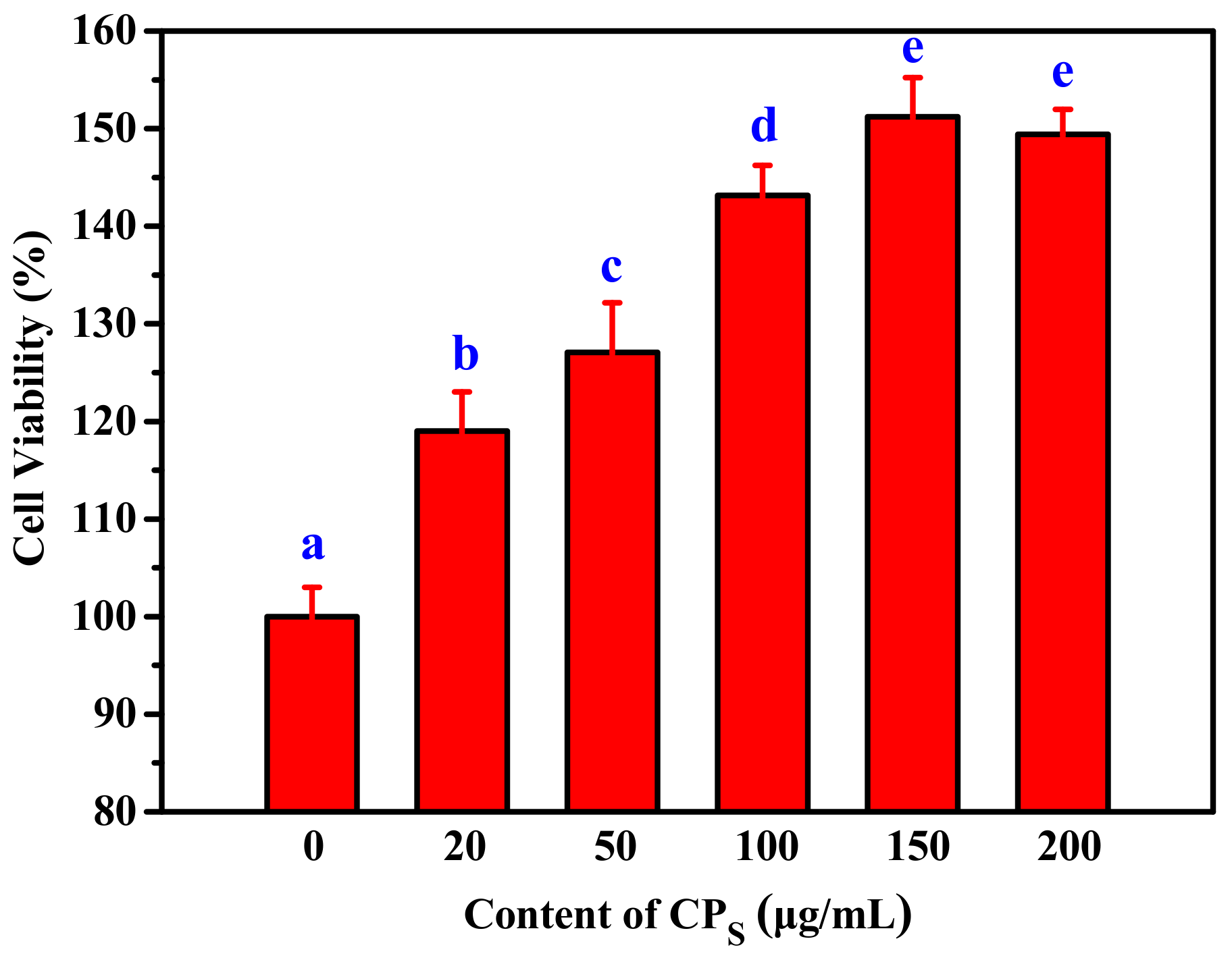
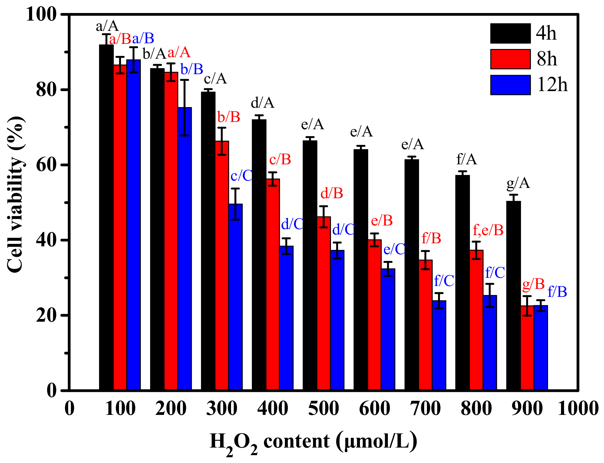
2.3.1. The Effect of H2O2 and CPs on the Proliferation of RAW264.7 Cells

To investigate the effect of H2O2 on viability of RAW264.7 cells, they were treated with different concentrations of H2O2. RAW264.7 cells revealed a significant decrease in viability with increasing concentration of H2O2 and treatment time (Figure 4). When the concentration of H2O2 was greater than 300 μM and the treatment time was longer than 8 h, the cell viability was significantly inhibited. Cell viability exposed to 500 µmol/L H2O2 for 8 h was 46.2% of the control value. In the subsequent experiments, the RAW264.7 cells were treated with 500 µmol/L H2O2 for 8 h to study the effect of CPS on H2O2-induced injury.
The effect of CPS-treated on the proliferation of RAW264.7 cells was shown in Figure 5. The viability of RAW264.7 cells in CPS treatment group was significantly improved compared to the control group when the concentration of CPS increased from 20 µg/mL to 150 µg/mL (Figure 5). This demonstrated that CPS could effectively promote the proliferation of RAW264.7 cells in a dose-dependent manner. However, a significant increase in cell viability was not observed when cells were treated with 200 µg/mL. Therefore, 100 µg/mL, 150 µg/mL and 200 µg/mL were selected as the low-, middle- and high-dose groups for subsequent experiments, respectively.

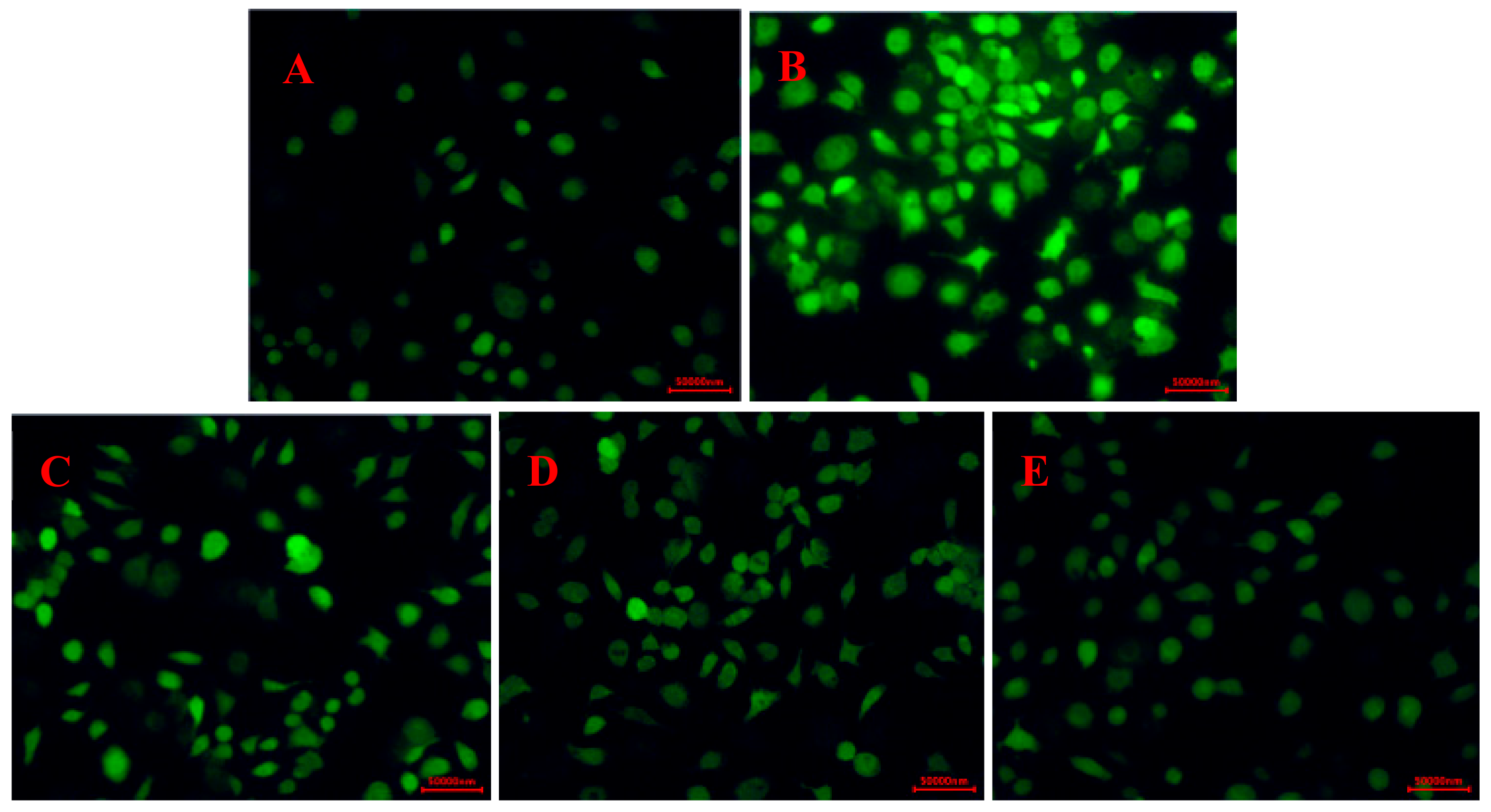
2.3.2. Effects of CPS on the ROS Levels in RAW264.7 Cells

In this section, the effect of CPS on the ROS levels in RAW264.7 cells was investigated. The fluorescence intensity in RAW264.7 following H2O2 treatment was significantly larger than that of the control group (Figure 6). However, the addition of CPS effectively reduced the ROS level relative to the model group, and the ROS level gradually decreased with the increase in CPS concentration. The results suggested that the protective effects of CPS against H2O2-induced injury of RAW264.7 cells may have resulted from inhibition of intracellular ROS production.

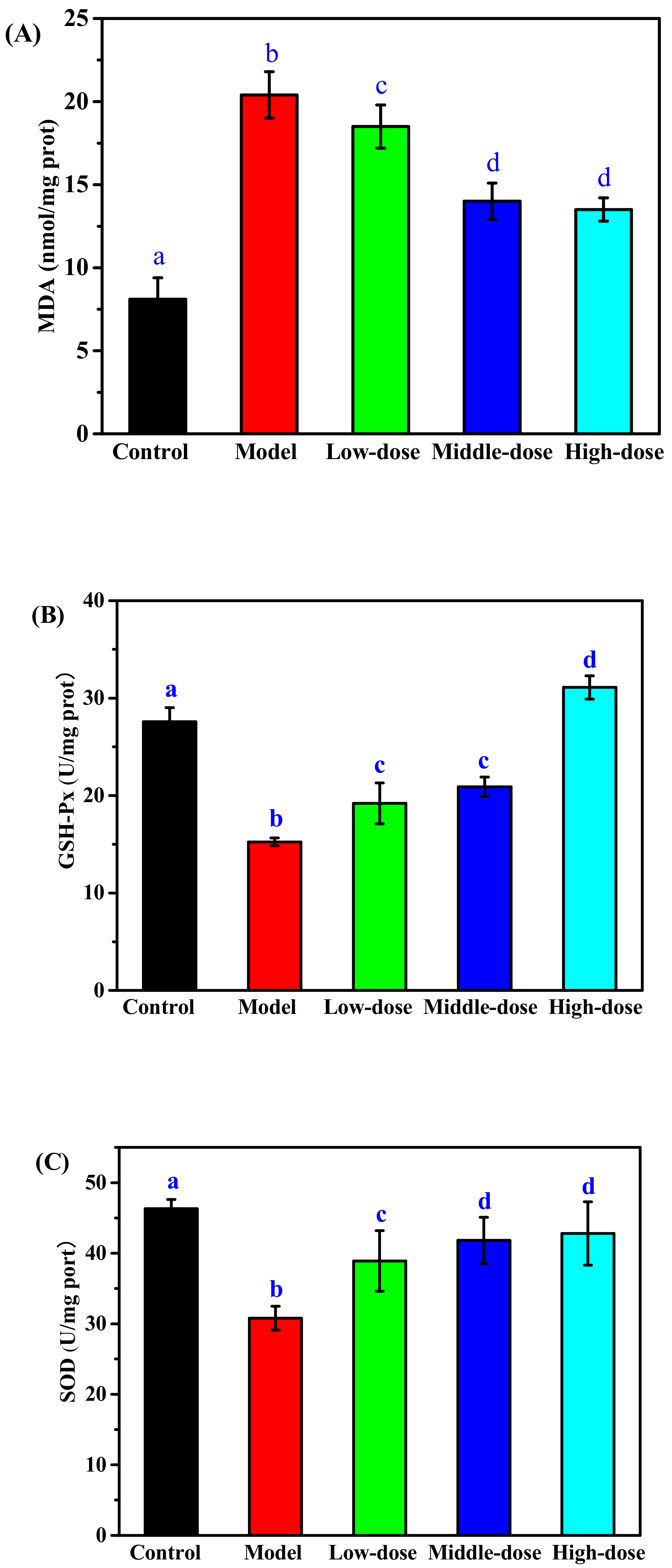
2.3.3. Effect of CPS on the Level of MDA, SOD, and GSH-Px in Cells

To evaluate the antioxidant activity of CPS, we investigated the effect of CPS-pretreated on the level of MDA, GSH-Px, and SOD respectively in H2O2-induced oxidative damage of RAW264.7 cells. The MDA level in the cells significantly increased following H2O2 treatment as compared to the control group (Figure 7A), indicating that the RAW264.7 cells were damaged by H2O2. A significantly lower MDA level compared to the control group was observed in the CPS-treated groups, especially in the middle- and high-dose group, indicating that CPS decreased the oxidative damage level of cells caused by H2O2. Effects of CPS on GSH-Px and SOD activity of RAW264.7 cells were shown in Figure 7B,C. The results showed that H2O2 caused a significant decrease in GSH-Px and SOD activity in RAW264.7 cells. Fortunately, the levels of GSH-Px and SOD were both markedly promoted in CPS-treated groups as compared to the control group. The group with the higher dose exhibited the highest level of GSH-Px, but the results between the low-dose and middle-dose groups were not statistically significant. The highest levels of SOD were observed in the high-dose and middle-dose groups, but the difference between them was not statistically significant. Qiu et al. had reported that collagen peptides could up-regulate the levels of SOD and GSH-Px, and down-regulate the contents of MDA, playing a protective role in antioxidant effects on Drosophila [34]. The collagen peptides from cod skin protected liver tissue against oxidative injure by increasing the activity of SOD and decreasing MDA [35]. Therefore, these results in this study demonstrated that collagen peptides from sea cucumber Acaudina molpadioides could protect RAW264.7 cells against H2O2-induced injury by inhibition of lipid peroxides and enhancement of antioxidant enzyme activity.

3. Materials and Methods

3.1. Chemicals and Reagents

Sea cucumber Acaudina molpadioides was provided from Zhoushan Jingzhou Aquatic Food Co., Ltd., in Zhejiang Province of China. Fetal calf serum was provided by GIBCO (Invitrogen Corporation, Carlsbad, California, USA). The 2,2-dipehnyl-1-picryldydrazyl (DPPH), 3-(4,5- Dimethylthiazol-2-yl)-2,5-diphenyl tetrazolium bromide (MTT) and dimethyl sulfoxide (DMSO) were purchased from Sigma-Aldrich (Shanghai, China). Assay kits for the MDA, SOD, and GSH-Px were purchased from the Nanjing Jiancheng Bioengineering Institute (Nanjing, China). ROS detection kit was purchased from Beyotime Company (Jiangsu, China). All other reagents were analytical grade.

3.2. Preparation of Collagen Peptides

ASC-Am was prepared from the body wall of Acaudina molpadioides according to the method reported previously [26]. Two grams of ASC-Am were added into 200 mL buffer solution (pH 10.0) and pretreated with irradiation (0, 50, 150, 200, 250, and 300 W) for 30 minutes each in a microwave (XH-300A; Beijing Xianghu, China). Alkaline protease (≥200 U/mg, Shanghai Ryon Biological Technology Co., Ltd., Shanghai, China) was added to the sample for hydrolysis at 45 °C. After 1 h, the alkaline protease was inactivated in boiling water for 10 min. After centrifuging the hydrolysates at 10,000 rpm for 10 min, the supernatants were freeze-dried, and the scavenging activity of hydroxyl radical (OH⋅) was measured at the concentration of 0.2 mg/mL.
The hydrolysates obtained under the optimal microwave power were separated by ultrafiltration membranes with size exclusion of 5 kDa and 1 kDa. Three fractions (CPS, collagen peptides with Mw ≤ 1 kDa; CPM, collagen peptides with 1 kDa < Mw ≤ 5 kDa; and CPL, collagen peptides with Mw > 5 kDa) were collected and freeze-dried separately to measure the peptide content and scavenging activity of OH⋅ radical at the concentration of 0.2 mg/mL.

3.3. Antioxidant Activity Measurements

3.3.1. Scavenging Activity of Hydroxyl Radical

The scavenging activity of OH⋅ of peptide was determined using the Fenton method [36]. Two mL peptide samples were mixed with 2 mL of phosphate buffer (0.2 M, pH 7.4), 1 mL of 1,10-phenanthroline (0.75 mM), 1 mL of FeSO4 (0.75 mM), and 1 mL of 0.3% H2O2 (v/v). The mixture was heated at 37 °C for 30 min, and the absorbance (A1) measured at 510 nm. The scavenging activity of OH⋅ was calculated using the following formula, where A0 is the absorbance of the blank with 2 mL of water instead of the peptide solution and A2 the absorbance of mixture with 1 mL of deionized water instead of the FeSO4 solution.
OH· scavenging activity (%) = [1−(A1 − A2)/A0] × 100%

3.3.2. Scavenging Activity of DPPH Radical

The scavenging activity of DPPH radical was tested according to previously reported methods [37]. The samples of peptides were dissolved in water with different concentrations 0.1, 0.2, 0.5, 0.8, and 1.0 mg/mL. Two mL of peptide samples were mixed with 2 mL of DPPH solution (0.2 mM) and 1 mL of ethanol. The mixture was incubated for 30 min at room temperature and then centrifuged at 5000 rpm for 5 min. The absorbance of the sample (As) was measured at 517 nm using an ultraviolet-visible spectrophotometer. The scavenging activity of DPPH was calculated as shown in the formula:
DPPH scavenging activity (%) = [1−(As − Ab)/Ac] × 100%
where Ac is the absorbance of the control group with 2 mL of water instead of the peptide sample; Ab is the absorbance of blank with 2 mL of ethanol in place of the DPPH solution.

3.4. The Effect of H2O2 and Peptides on the Proliferation of RAW264.7 Cells

RAW264.7 cells (purchased from Chinese Academy of Sciences) were grown in DMEM medium supplemented with 10% fetal bovine serum and incubated at 37 °C in a humidified atmosphere with 5% CO2. The peptides were dissolved in DMEM medium and diluted to different concentrations (20–200 µg/mL). Cell viability was determined using MTT assay. RAW264.7 cells were seeded in 96-well plates at 1 × 10⁵ cells/mL and incubated at 37 °C with 5% CO2 for 24 h. The cells were treated with different concentrations of H2O2 (100, 200, 300, 400, 500, 600, 700, 800, and 900 μ mol/L) or peptides. A volume of 20 µL of MTT solution (2 mg/mL) was added to each well and incubated for 4 h. The supernatant was removed and 100 µL of Dimethyl sulfoxide (DMSO) was added into each well to dissolve the formazan crystals. The plates were shaken for 5 min to dissolve the crystals completely, and absorbance was measured at 490 nm. The cell viability with H2O2 or peptide treatment was evaluated as follows:
Cell viability (%) = AS/AN × 100%
where AS and AN were the absorbance of treatment and control wells, respectively.

3.5. Reactive Oxygen Species (ROS) Assay

Production of ROS was monitored using a ROS Assay Kit (Beyotime Biotechnology Co., Ltd., Shanghai, China). RAW 264.7 cells were cultured in 96-well plates with a density of 1 × 10⁵ cells/mL for 24 h. Cells were treated with various concentrations of peptides for 24 h, and then exposed to H2O2 (500 μ mol/L) for 8 h. The experiments were implemented in the control group (not treated with peptides and H2O2), the model group (treated with 500 µM H2O2), the low-dose group (treated with 100 µg/mL peptides + 500 µM H2O2), the middle-dose group (treated with 150 µg/mL peptides + 500 µM H2O2), and the high-dose group (treated with 200 µg/mL peptides + 500 µM H2O2). RAW264.7 were incubated for 20 min in a 37 °C cell incubator with 5 µL 2′,7′- dichlorodihydrofluorescein diacetate (DCFH-DA). Cells were washed three times with serum-free cell culture medium and then observed under a fluorescence microscope.

3.6. Assays for Antioxidant Enzyme Activity

RAW264.7 cells (1 × 10⁵ cells/mL) were seeded in 96-well plates, and exposed to 500 µmol/L H2O2 for 8 h with or without various concentrations of peptide pre-treatment for 24 h. RAW264.7 cells were washed with Phosphate-buffered saline (PBS), and then lysed with cell lysate. The supernatants were collected following centrifugation at 2000 × g for 5 min at 4 °C. The activity of SOD, GSH-Px, and MDA content was determined by using corresponding diagnostic kits according to the manufacturer’s instructions (Nanjing Jiancheng Bioengineering Institute, Nanjing, China).

3.7. Statistical Analysis

Each experiment was carried out in triplicate. Data were presented as means and standard deviations. Results were analyzed using Microsoft Excel 2010 (Redmond, WA, USA), and significant differences (p < 0.05) between data were identified by Duncan’s multiple range test in the software SPSS (SPSS Inc., Chicago, IL, USA).

4. Conclusions

Microwave-assisted hydrolysis of collagen is a promising method for preparation of antioxidant peptides. Microwave radiation can significantly increase the content and antioxidant activity of small molecular weight peptides. The hydrolyzate fragment CPS (Mw ≤ 1 kDa) obtained by microwave-assisted alkaline protease hydrolysis of collagen from sea cucumber Acaudina molpadioides exhibited the good scavenging activities of DPPH and OH•. CPS also exhibited a significant protective effect on H2O2-injured RAW264.7 cells by promoting cell proliferation, reducing the levels of ROS and MDA, and enhancing antioxidant enzyme (SOD and GSH-Px) activity. Therefore, it is recommended, as a result of the current study, to explore CPS as a potential natural antioxidant and utilize microwave-assisted hydrolysis as a method of obtaining high levels of antioxidant peptides from naturally occurring high-molecular weight protein molecules.

Author Contributions

H.-X.J. designed the experiments and wrote the paper; Y.L. performed the experiments; J.L. analyzed the data; S.-J.L. and Z.-S.Y. provided experimental materials and equipment.

Funding

The research was funded by the Fundamental Research Funds for the Provincial Universities of Zhejiang Province (2019JZ00005) and Natural Science Foundation of Zhejiang province (LQ20D060004).

Conflicts of Interest

The authors declare no conflict of interest. The funders had no role in the design of the study; in the collection, analyses, or interpretation of data; in the writing of the manuscript, or in the decision to publish the results.

References

  1. Liu, R.; Zheng, W.; Li, J.; Wang, L.; Wu, H.; Wang, X.; Shi, L. Rapid identification of bioactive peptides with antioxidant activity from the enzymatic hydrolysate of Mactra veneriformis by UHPLC-Q-TOF mass spectrometry. Food Chem. 2015, 167, 484–489. [Google Scholar] [CrossRef] [PubMed]
  2. Reyes Jara, A.M.; Liggieri, C.S.; Bruno, M.A. Preparation of soy protein hydrolysates with antioxidant activity by using peptidases from latex of Maclura pomifera fruits. Food Chem. 2018, 264, 326–333. [Google Scholar] [CrossRef] [PubMed]
  3. Dinstel, R.R.; Cascio, J.; Koukel, S. The antioxidant level of Alaska’s wild berries: High, higher and highest. Int. J. Circumpolar Health 2013, 72. [Google Scholar] [CrossRef]
  4. Lin, X.; Bai, D.; Wei, Z.; Zhang, Y.; Huang, Y.; Deng, H.; Huang, X. Curcumin attenuates oxidative stress in RAW264.7 cells by increasing the activity of antioxidant enzymes and activating the Nrf2-Keap1 pathway. PLoS ONE 2019, 14, e0216711. [Google Scholar] [CrossRef] [PubMed]
  5. Li, S.; Song, Z.; Liu, T.; Liang, J.; Yuan, J.; Xu, Z.; Sun, Z.; Lai, X.; Xiong, Q.; Zhang, D. Polysaccharide from Ostrea rivularis attenuates reproductive oxidative stress damage via activating Keap1-Nrf2/ARE pathway. Carbohydr. Polym. 2018, 186, 321–331. [Google Scholar] [CrossRef] [PubMed]
  6. Xiao, X.; Liua, J.; Zhu, X.; Hua, Y.; Wang, C.; Zhang, Y. Protective effects of protopine on hydrogen peroxide-induced oxidative injury of PC12 cells via Ca antagonism and antioxidant mechanisms. Eur. J. Pharmacol. 2008, 591, 21–27. [Google Scholar] [CrossRef]
  7. Duranti, G.; Ceci, R.; Sgrò, P.; Sabatini, S.; Luigi, L.D. Influence of the PDE5 inhibitor tadalafil on redox status and antioxidant defense system in C2C12 skeletal muscle cells. Cell Stress Chaperones 2017, 22, 389–396. [Google Scholar] [CrossRef]
  8. Huang, Y.; Ruan, G.; Qin, Z.; Li, H.; Zheng, Y. Antioxidant activity measurement and potential antioxidant peptides exploration from hydrolysates of novel continuous microwave-assisted enzymolysis of the Scomberomorus niphonius protein. Food Chem. 2017, 223, 89–95. [Google Scholar] [CrossRef]
  9. Zhong, M.; Chen, T.; Hu, C.; Ren, C. Isolation and characterization of collagen from the body wall of sea cucumber Stichopus monotuberculatus. J. Food Sci. 2015, 80, C671–C679. [Google Scholar] [CrossRef]
  10. Zhang, J.; Liu, S.; Zhang, Y.; Lu, Y.; Wang, M.; Wang, G.; Liu, X. Purification and antioxidant ability of peptide from egg in sea cucumber Apostichopus japonicus. Int. J. Food Prop. 2016, 20, 306–317. [Google Scholar] [CrossRef]
  11. Ferrario, C.; Leggio, L.; Leone, R.; Di Benedetto, C.; Guidetti, L.; Cocce, V.; Ascagni, M.; Bonasoro, F.; La Porta, C.A.M.; Candia Carnevali, M.D.; et al. Marine-derived collagen biomaterials from echinoderm connective tissues. Mar. Environ. Res. 2017, 128, 46–57. [Google Scholar] [CrossRef] [PubMed]
  12. Wu, J.; Fujioka, M.; Sugimoto, K.; Mu, G.; Ishimi, Y. Assessment of effectiveness of oral administration of collagen peptide on bone metabolism in growing and mature rats. J. Bone Miner. Metab. 2004, 22, 547–553. [Google Scholar] [CrossRef] [PubMed]
  13. Ohara, H.; Iida, H.; Ito, K.; Takeuchi, Y.; Nomura, Y. Effects of Pro-Hyp, a collagen hydrolysate-derived peptide, on hyaluronic acid synthesis using in vitro cultured synovium cells and oral ingestion of collagen hydrolysates in a guinea pig model of osteoarthritis. Biosci. Biotechnol. Biochem. 2010, 74, 2096–2099. [Google Scholar] [CrossRef] [PubMed]
  14. Midori, T.; Yoh-Ichi, K.; Yoshihiro, N.J.B.B. Biochemistry, effects of collagen peptide ingestion on UV-B-induced skin damage. Biosci. Biotechnol. Biochem. 2009, 73, 930–932. [Google Scholar]
  15. Izquierdo, F.J.; Peñas, E.; Baeza, M.L.; Gomez, R. Effects of combined microwave and enzymatic treatments on the hydrolysis and immunoreactivity of dairy whey proteins. Int. Dairy J. 2008, 18, 918–922. [Google Scholar] [CrossRef]
  16. Xu, Z.; Fang, Y.; Chen, Y.; Yang, W.; Ma, N.; Pei, F.; Kimatu, B.M.; Hu, Q.; Qiu, W. Protective effects of Se-containing protein hydrolysates from Se-enriched rice against Pb(2+)-induced cytotoxicity in PC12 and RAW264.7 cells. Food Chem. 2016, 202, 396–403. [Google Scholar] [CrossRef]
  17. Lin, Y.J.; Le, G.W.; Wang, J.Y.; Li, Y.X.; Shi, Y.H.; Sun, J. Antioxidative peptides derived from enzyme hydrolysis of bone collagen after microwave assisted acid pre-treatment and nitrogen protection. Int. J. Mol. Sci. 2010, 11, 4297–4308. [Google Scholar] [CrossRef]
  18. Zhang, H.; Yu, L.; Yang, Q.; Sun, J.; Bi, J.; Liu, S.; Zhang, C.; Tang, L. Optimization of a microwave-coupled enzymatic digestion process to prepare peanut peptides. Molecules 2012, 17, 5661–5674. [Google Scholar] [CrossRef]
  19. Abedin, M.Z.; Karim, A.A.; Ahmed, F.; Latiff, A.A.; Gan, C.Y.; Che, G.F.; Islam Sarker, M.Z. Agriculture, isolation and characterization of pepsin-solubilized collagen from the integument of sea cucumber (Stichopus vastus). J. Sci. Food Agric. 2013, 93, 1083–1088. [Google Scholar] [CrossRef]
  20. Lin, S.J.; Xue, Y.P.; San, E.L.; Keong, T.C.; Chen, L.F.; Zheng, Y.G. Extraction and characterization of pepsin soluble collagen from the body wall of sea cucumber Acaudina leucoprocta. J. Aquat. Food Prod. Technol. 2017, 26, 502–515. [Google Scholar] [CrossRef]
  21. Liu, Z.; Oliveira, A.C.; Su, Y.C. Purification and characterization of pepsin-solubilized collagen from skin and connective tissue of giant red sea cucumber (Parastichopus californicus). J. Agric. Food Chem. 2010, 58, 1270–1274. [Google Scholar] [CrossRef] [PubMed]
  22. Liu, Z.Q.; Tuo, F.Y.; Song, L.; Liu, Y.X.; Dong, X.P.; Li, D.M.; Zhou, D.Y.; Shahidi, F. Action of trypsin on structural changes of collagen fibres from sea cucumber (Stichopus japonicus). Food Chem. 2018, 256, 113–118. [Google Scholar] [CrossRef] [PubMed]
  23. Zhu, B.W.; Dong, X.P.; Zhou, D.Y.; Gao, Y.; Yang, J.F.; Li, D.M.; Zhao, X.K.; Ren, T.T.; Ye, W.X.; Tan, H.; et al. Physicochemical properties and radical scavenging capacities of pepsin-solubilized collagen from sea cucumber Stichopus japonicus. Food Hydrocoll. 2012, 28, 182–188. [Google Scholar] [CrossRef]
  24. Zhuang, Y.; Sun, L.; Zhao, X.; Wang, J.; Hou, H.; Li, B. Antioxidant and melanogenesis-inhibitory activities of collagen peptide from jellyfish (Rhopilema esculentum). J. Sci. Food Agric. 2009, 89, 1722–1727. [Google Scholar] [CrossRef]
  25. Zhou, X. Technology, antioxidant peptides isolated from sea cucumber Stichopus Japonicus. Eur. Food Res. Technol. 2012, 234, 441–447. [Google Scholar] [CrossRef]
  26. Jin, H.X.; Xu, H.P.; Li, Y.; Zhang, Q.W.; Xie, H. Preparation and evaluation of peptides with potential antioxidant activity by microwave assisted enzymatic hydrolysis of collagen from sea cucumber Acaudina molpadioides obtained from Zhejiang Province in China. Mar. Drugs 2019, 17, 169. [Google Scholar] [CrossRef]
  27. Uluko, H.; Zhang, S.; Liu, L.; Tsakama, M.; Lu, J.; Lv, J. Effects of thermal, microwave, and ultrasound pretreatments on antioxidative capacity of enzymatic milk protein concentrate hydrolysates. J. Funct. Foods 2015, 18, 1138–1146. [Google Scholar] [CrossRef]
  28. Chi, C.F.; Hu, F.Y.; Wang, B.; Li, Z.R.; Luo, H.Y. Influence of amino acid compositions and peptide profiles on antioxidant capacities of two protein hydrolysates from skipjack tuna (Katsuwonus pelamis) dark muscle. Mar. Drugs 2015, 13, 2580–2601. [Google Scholar] [CrossRef]
  29. Kong, X.; Zhou, H.; Hua, Y. Preparation and antioxidant activity of wheat gluten hydrolysates (WGHs) using ultrafiltration membranes. J. Sci. Food Agric. 2008, 88, 920–926. [Google Scholar] [CrossRef]
  30. Chi, C.-F.; Wang, B.; Hu, F.-Y.; Wang, Y.-M.; Zhang, B.; Deng, S.-G.; Wu, C.-W. Purification and identification of three novel antioxidant peptides from protein hydrolysate of bluefin leatherjacket (Navodon septentrionalis) skin. Food Res. Int. 2015, 73, 124–129. [Google Scholar] [CrossRef]
  31. Ma, Y.L.; Zhu, D.Y.; Thakur, K.; Wang, C.H.; Wang, H.; Ren, Y.F.; Zhang, J.G.; Wei, Z.J. Antioxidant and antibacterial evaluation of polysaccharides sequentially extracted from onion ( Allium cepa L.). Int. J. Biol. Macromol. 2018, 111, 92–101. [Google Scholar] [CrossRef] [PubMed]
  32. Bao, Y.; Zhao, M.; Shi, J.; Ning, Y.; Jiang, Y.J.F.C. Effect of ultrasonic treatment on the recovery and DPPH radical scavenging activity of polysaccharides from longan fruit pericarp. Food Chem. 2008, 106, 685–690. [Google Scholar]
  33. Pan, X.; Zhao, Y.Q.; Hu, F.Y.; Wang, B. Preparation and identification of antioxidant peptides from protein hydrolysate of skate ( Raja porosa ) cartilage. J. Funct. Foods 2016, 25, 220–230. [Google Scholar] [CrossRef]
  34. Qiu, W.; Chen, X.; Tian, Y.; Wu, D.; Du, M.; Wang, S. Protection against oxidative stress and anti-aging effect in Drosophila of royal jelly-collagen peptide. Food Chem. Toxicol. 2019, 110881. [Google Scholar] [CrossRef] [PubMed]
  35. Han, Y.; Xie, J.; Gao, H.; Xia, Y.; Chen, X.; Wang, C. Hepatoprotective effect of collagen peptides from cod skin against liver oxidative damage in vitro and in vivo. Cell Biochem. Biophys. 2015, 71, 1089–1095. [Google Scholar] [CrossRef] [PubMed]
  36. Lu, C.; Li, H.; Li, C.; Chen, B.; Shen, Y. Chemical composition and radical scavenging activity of Amygdalus pedunculata Pall leaves’ essential oil. Food Chem. Toxicol. 2018, 119, 368–374. [Google Scholar] [CrossRef]
  37. Aissi, O.; Boussaid, M.; Messaoud, C. Essential oil composition in natural populations of Pistacia lentiscus L. from Tunisia: Effect of ecological factors and incidence on antioxidant and antiacetylcholinesterase activities. Ind. Crop. Prod. 2016, 91, 56–65. [Google Scholar] [CrossRef]
Figure 1. Effect of microwave power on OH· scavenging activity of collagen hydrolysate. The concentration of hydrolyzed product was 2 mg/mL. Values with different letters are significantly different (p < 0.05).
Marinedrugs 17 00642 g001
Figure 2. The OH· scavenging activity of peptides with different Mw (CPL, Mw > 5 kDa; CPM, 1 kDa < Mw ≤ 5 kDa; CPS, Mw ≤ 1 kDa) from hydrolysate of ASC-Am. (a–b) Values with different letters indicated significant differences in the same samples at different microwave powers (P < 0.05). (A–C) Values with different letters indicated significant differences in different samples at the same microwave powers (p < 0.05).
Marinedrugs 17 00642 g002
Figure 3. The scavenging activity of OH· (A) and DPPH (B) of CPS obtained by alkaline protease hydrolysis of ASC-Am at 250 W of microwave power. Ascorbic acid (AA) was used as a positive control. (a–b) Values with different letters indicated significant differences in different samples at the same concentrations (p < 0.05). (A–F) Values with different letters indicated significant differences in the same samples at the different concentrations (p < 0.05).
Marinedrugs 17 00642 g003
Figure 4. The effect of H2O2 on the viability of RAW264.7 cells. (a–g). Values with different letters indicated significant differences in the same time at different concentrations of H2O2 (p < 0.05). (A–C) Values with different letters indicated significant differences in the same concentrations of H2O2 at different times (p < 0.05).
Marinedrugs 17 00642 g004
Figure 5. Effect of CPS on viability of RAW264.7 cells. Values with different letters are significantly different (p < 0.05).
Marinedrugs 17 00642 g005
Figure 6. Effect of CPS on ROS levels in RAW264.7 cells induced by H2O2. (A) Control group; (B) model group (500 µM H2O2); (C) low-dose group (100 µg/mL CPS + 500 µM H2O2); (D) middle-dose group (150 µg/mL CPS + 500 µM H2O2); (E) high-dose group (200 µg/mL CPS + 500 µM H2O2).
Marinedrugs 17 00642 g006
Figure 7. Effect of CPS on levels of MDA (A), GSH-Px (B), and SOD (C) in RAW264.7 cells induced by H2O2. Model group (500 µM H2O2), low-dose group (100 µg/mL CPS + 500 µM H2O2), middle-dose group (150 µg/mL CPS + 500 µM H2O2), high-dose group (200 µg/mL CPS + 500 µM H2O2). Values with different letters are significantly different (p < 0.05).
Marinedrugs 17 00642 g007
Table 1. Effect of microwave-assisted hydrolysis of ASC-Am on peptide content and molecular weight distribution.
HydrolysatesContent (%)Mw Distribution (%)
0 W250 W0 W250 W
CPL (Mw > 5 kDa)10090.3 ± 4.321.7 ± 2.115.7 ± 1.5
CPM (1 kDa < Mw ≤ 5 kDa)100148.0 ± 8.520.3 ± 1.224.0 ± 1.8
CPS (Mw ≤ 1 kDa)100130.2 ± 6.458.0 ± 3.160.3 ± 2.9

