Article

Glycosaminoglycans from a Sea Snake (Lapemis curtus): Extraction, Structural Characterization and Antioxidant Activity

1
Key Laboratory of Marine Drugs, Ministry of Education, Shandong Provincial Key laboratory of Glycoscience and Glycoengineering, School of Medicine and Pharmacy, Ocean University of China, Qingdao 266003, China
2
Laboratory for Marine Drugs and Bioproducts of Qingdao National Laboratory for Marine Science and Technology, Qingdao 266237, China
3
Marine Biomedical Research Institute of Qingdao, Qingdao 266071, China
4
Ocean College of Hainan University, Haikou 570228, China
*
Author to whom correspondence should be addressed.
Mar. Drugs 2018, 16(5), 170; https://doi.org/10.3390/md16050170
Submission received: 10 April 2018 / Revised: 7 May 2018 / Accepted: 10 May 2018 / Published: 18 May 2018
(This article belongs to the Collection Marine Polysaccharides)

Abstract

:
Sea snakes have wide application prospects in medicine, health food and other fields. Several novel polysaccharides were successfully obtained from the skin and the meat of a sea snake (Lapemis curtus). The structures of polysaccharides LSP3 and LMP3, which were extracted and purified from Lapemis curtus, were determined to be new and highly heterogenic glycosaminoglycans (GAGs) by means of FT-IR, ESI-MS/MS and NMR. LSP3 is a hybrid dermatan sulfate (DS) and composed of 48% 4-sulfated disaccharides (Di4S), 42% 6-sulfated disaccharides (Di6S) and 5% disulfated disaccharides (Di2,6S), while LMP3 is a hybrid chondroitin sulfate (CS) and composed of 70% Di4S, 20% Di6S, and 8% Di2,6S. More importantly, LSP3 and LMP3 showed a strong scavenging ability of 1,1-diphenyl-2-picrylhydrazyl (DPPH) radicals, iron (Fe2+) chelating activity and total antioxidant capacity in vitro, especially LSP3, with high contents of uronic acid and sulfate, which possessed a higher scavenging ability of DPPH radicals than other fractions. These data suggested that the sea snake polysaccharides could be promising candidates for natural antioxidant ingredients.

Graphical Abstract

1. Introduction

Glycosaminoglycans (GAGs) are polyanionic polysaccharides composed of amino sugar and uronic acid, which are widely distributed in animals. According to the difference of disaccharide composition and glycosidic bond, GAGs are generally divided into four groups, namely, chondroitin sulfate and dermatan sulfate (CS/DS), keratan sulfate (KS), heparin and heparan sulfate (HP/HS), and hyaluronic acid (HA) [1]. CS and DS have been confirmed to be closely related to inflammation, immune response, cardiovascular disease, tumorigenesis, infection, wound repair and fibrosis, and especially iduronic acid (IdoA), which influences multiple cellular properties [2].
Antioxidant activity is a focus of intensive scientific investigations because of the ever-increasing demand of food and pharmaceutical industries to develop natural antioxidant compounds [3]. The process of oxidative stress plays a major role in the development of chronic and degenerative illness, such as cancer, autoimmune disorders, aging, cataract, rheumatoid arthritis, as well as cardiovascular and neurodegenerative diseases [4]. The human body can counteract oxidative stress by producing antioxidants to protect cells from oxidation. Among these antioxidants, CS and DS have increasingly attracted interest of many research groups [5].
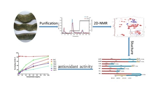
Sea snakes are the largest group of marine reptiles that inhabit the tropical and subtropical waters of the Indian and Pacific Oceans [6]. Sea snakes have wide application prospects in medicine, health food and other fields. The oil in the viscera of the Erabu sea snake is used as a functional food in Japan [7]. The current research on sea snakes mainly focuses on the snake venom, which is mainly considered to be used in the treatment of infectious, hematological, inflammatory, cardiovascular, and malignant diseases [8]. To the best of our knowledge, there has been no systematic study of sea snake polysaccharides. In this study, several polysaccharides were extracted and purified from the skin and meat of Lapemis curtus, and their physicochemical properties, structures, and antioxidant activities were studied (Figure 1). The aim of this study is to provide a basis for further research and development of sea snake polysaccharides.

2. Results and Discussion

2.1. Chemical Composition

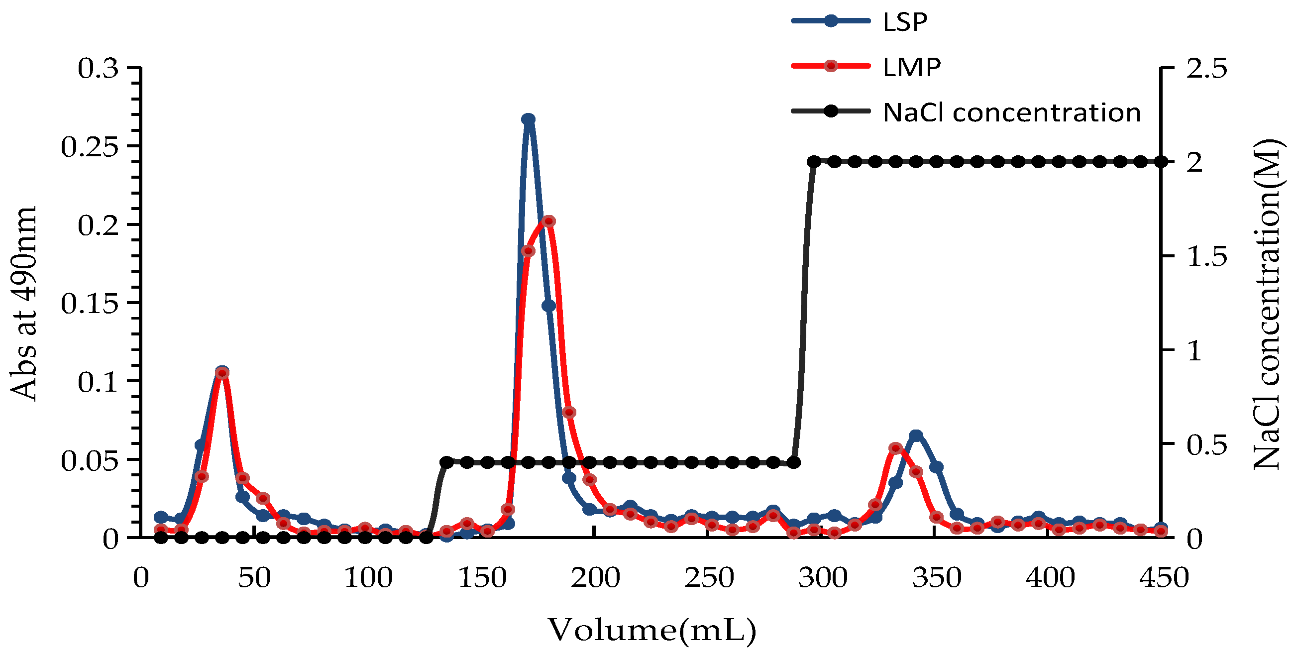
Two crude polysaccharides (LSP and LMP) were obtained by enzymolysis extraction from the skin and the meat of Lapemis curtus, respectively. LSP and LMP were further fractionated on a Q Sepharose Fast Flow column (Figure 2), and three sub-fractions of LSP (LSP1, LSP2, and LSP3) and three sub-fractions of LMP (LMP1, LMP2, and LMP3) were obtained. Physicochemical properties of these polysaccharide sub-fractions were analyzed (as shown in Table 1). Non-sulfated polymers were eluted by pure water and a low concentration of NaCl solution (0.4 mol/L), and sulfated polymers were eluted by a high concentration of NaCl solution (2.0 mol/L), both from LSP and LMP. In addition, the fractions eluted by a high concentration of NaCl solution possessed a higher content of uronic acid and a lower content of protein than that of fractions eluted by pure water and a low concentration of NaCl solution. LSP3 displayed a higher content of uronic acid and degree of sulfation than that of LMP3.
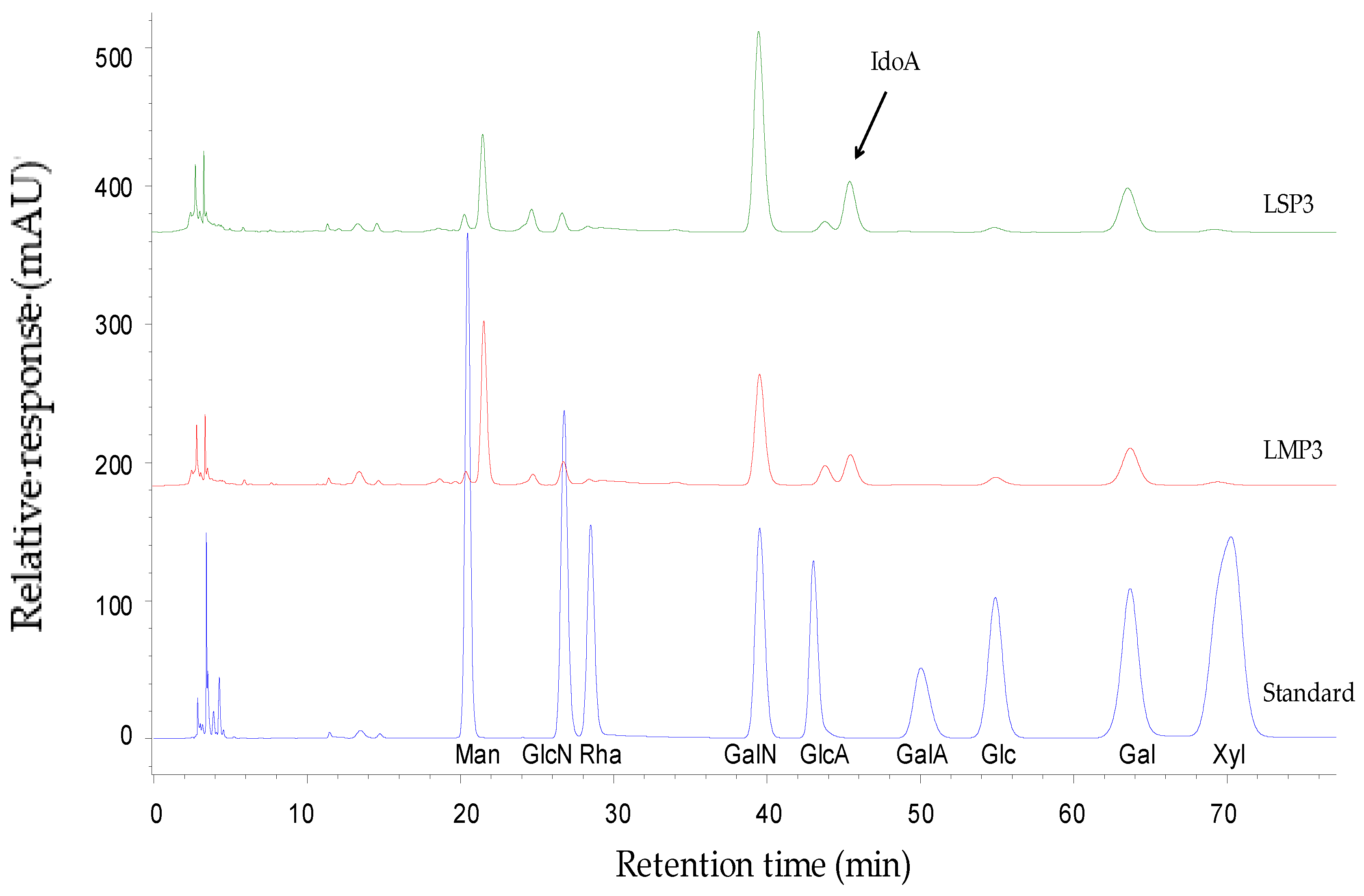
Results of monosaccharide composition analysis showed that LSP1 and LMP1, LSP2 and LMP2 were mainly composed of mannose (Man), N-acetylglucosamine (GlcNAc), glucose (Glc) and galactose (Gal), with different molar ratios (Table 1). LSP3 and LMP3 were mainly composed of iduronic acid (IdoA), glucuronic acid (GlcA), N-acetylgalactosamine (GalNAc) and Gal (Figure 3). However, LSP3 contained a high content of IdoA, while LMP3 contained a high content of GlcA. Results of disaccharide compositions analysis showed that LSP3 and LMP3 contained mainly ΔDi6S and ΔDi4S at a molar ratio of 1:1.1 and 1:3.5, respectively (Table 1), that is to say, LSP3 possessed a higher content of ΔDi6S and a lower content of ΔDi4S than that of LMP3. In addition, disulfated disaccharides (ΔDi2,6S) was detected in LSP3 and LMP3, and the contents of ΔDi2,6S were 4.9% and 8.0%, respectively.

2.2. FT-IR Spectroscopy of LSP3 and LMP3

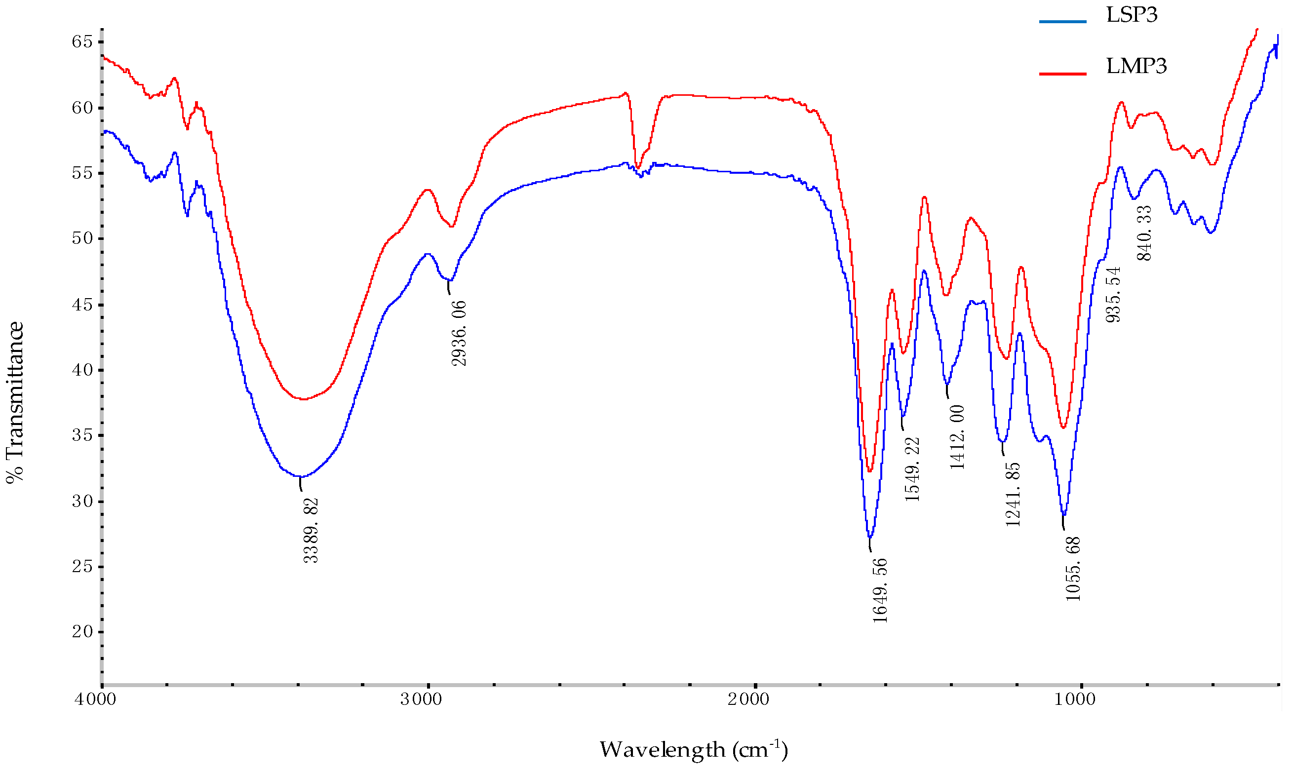
The FT-IR spectra of LSP3 and LMP3 were shown in Figure 4. The broad and intense absorption at 3389 cm−1 was attributed to the O–H stretching vibration. The characteristic C–H stretching vibration of sugar ring was at 2936 cm−1. The signal at 1649 cm−1 was attributed to the H–O–H vibration. The peak at 1412 cm−1 was assigned to the O–H in-plane bending vibration. The signal at 1055 cm−1 was related to the C–O–C skeletal vibration. The signals at 1241 cm−1 and 840 cm−1 were attributed to the stretching vibrations of S=O and C–O–S [9,10], respectively. The peak at 1549 cm−1 was assigned to the N–H variable angle vibration. Therefore, it was concluded that LSP3 and LMP3 contain sulfate groups and carboxyl groups, which are in accordance with the structural features of GAGs.

2.3. ESI-MS Analysis of LSP3 and LMP3

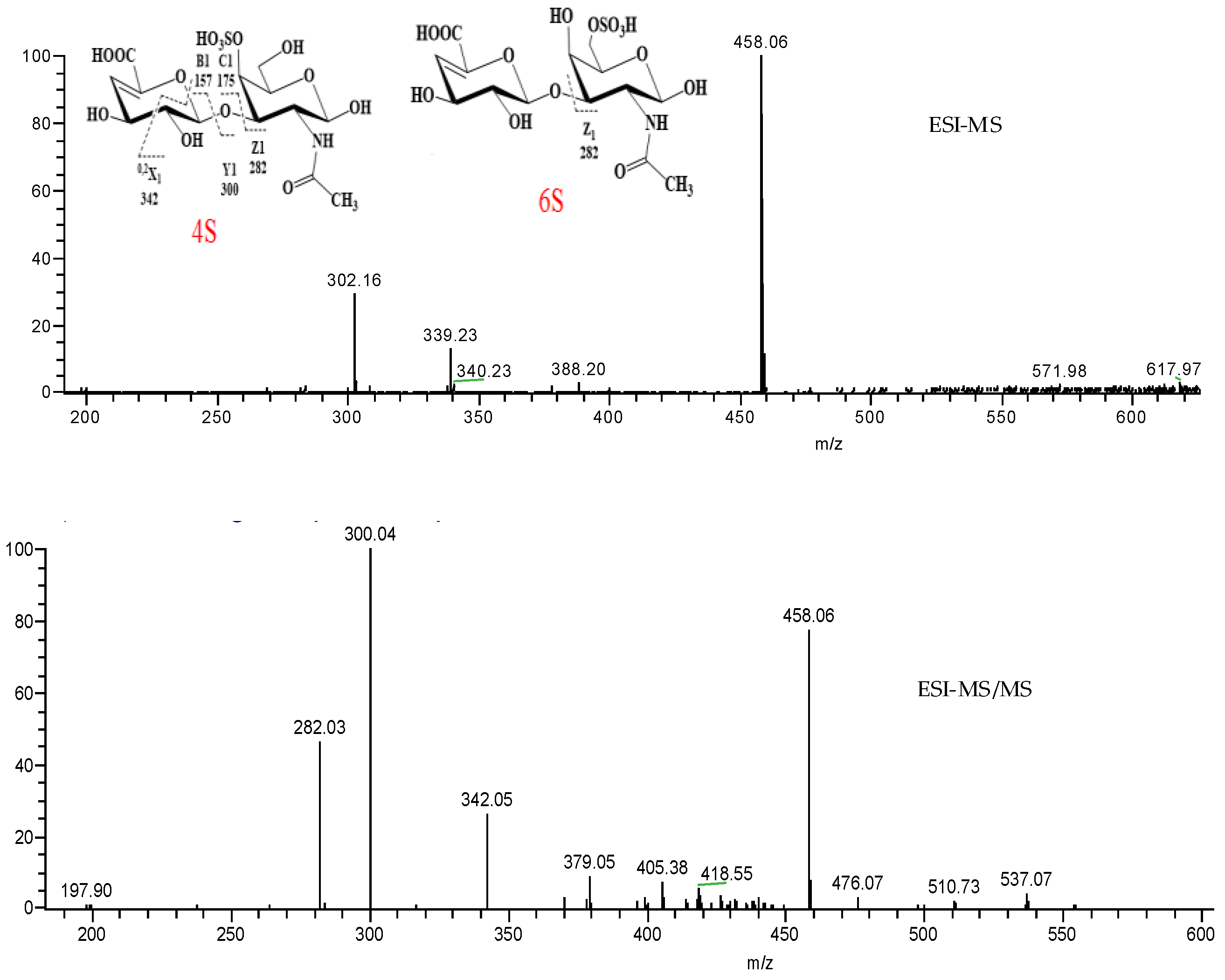
ESI-MS is a soft ionization technique commonly coupled with liquid chromatography for identification of oligosaccharides. As shown in Figure 5, the main deprotonated ion [M − H] of LSP3 was produced at m/z 458.06 in negative ESI-MS mode, which is in accordance with the structure of ΔHexA-GalNAc, with one sulfate group digested by chondroitinase ABC afterwards. In order to identify the substitution and the linkage of this fraction, the singly charged molecule ion [M − H] m/z 458.06 was further selected as the precursor ion for ESI-MS/MS analysis. The ion m/z 282.03, produced from ΔDi6S cleavage, was assigned as Z1 from the reducing terminal. The ions m/z 342.05, m/z 300.04 and m/z 282.03, produced from ΔDi4S cleavage, were assigned as 0,2X1, Y1 and Z1 from the reducing terminal [11], respectively. The results showed that LSP3 contains mainly ΔDi6S and ΔDi4S, which are in close agreement with the analysis of disaccharide compositions. The ESI-MS spectrum of LMP3 is essentially identical to that of LSP3.

2.4. NMR Spectroscopy Analysis of LSP3 and LMP3

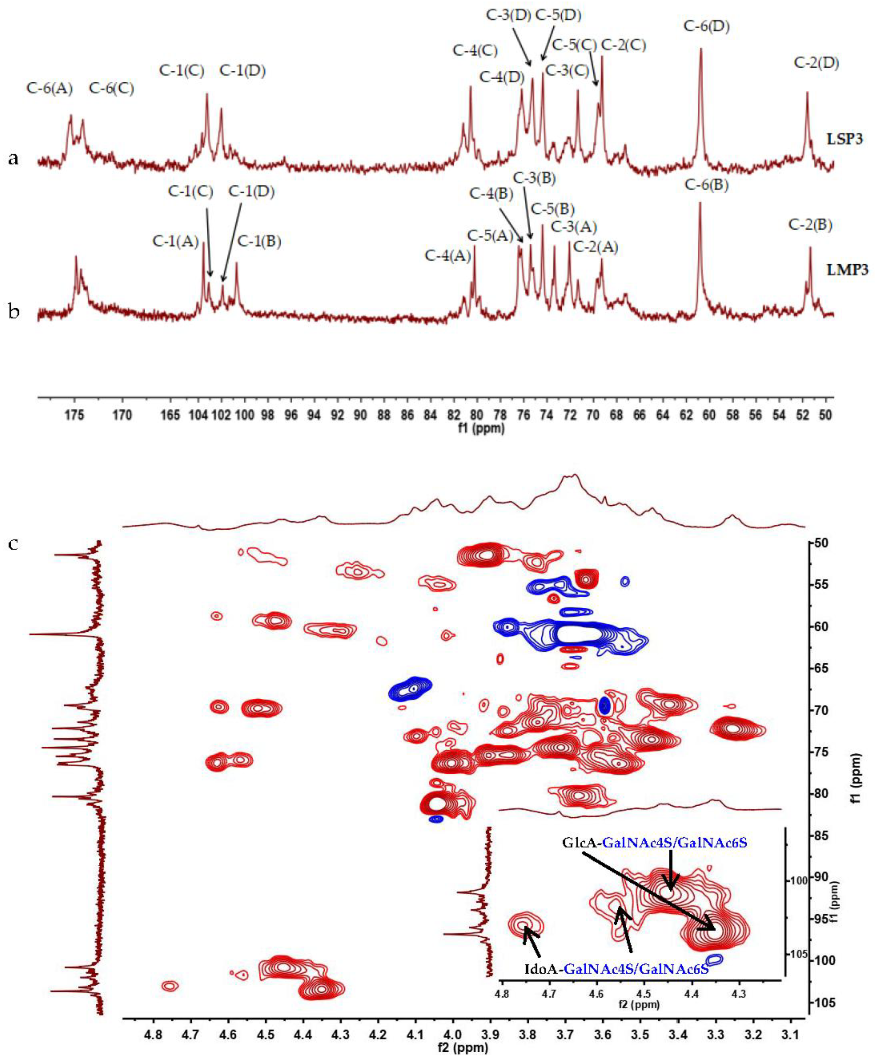
The structures of LSP3 and LMP3 were further elucidated by means of 1H-NMR and 13C-NMR (Figure 6) spectroscopy. Take LMP3, for example: four major anomeric carbon signals at δ 103.35 ppm (A), 101.08 ppm (B), 103.13 ppm (C) and 101.68 ppm (D) were observed in the 13C-NMR spectrum (Figure 6b), and four protons at δ 4.35 ppm (A), 4.44 ppm (B), 4.77 ppm (C) and 4.56 ppm (D) were observed accordingly in the 1H-NMR spectrum. The presence of CS and DS units in the structures of LMP3 were confirmed by two series of signals related to uronic acid (A, C) and GalNAc (B, D) residues, as the previous report indicated [1]. A series of 2D-NMR (NOESY, TOCSY, COSY, HSQC, HMBC) experiments allowed almost complete assignment of GlcA-GalNAc4S, GlcA-GalNAc6S, IdoA-GalNAc4S and IdoA-GalNAc6S disaccharide fragments of LSP3 and LMP3 (as listed in Table 2) [1,12,13]. The signals of B(H1)-B(C1) (4.44 ppm/101.08 ppm) and D(H1)-D(C1) (4.56 ppm/101.68 ppm) in the HSQC spectrum of LMP3 (Figure 6c) were attributed to GlcA-GalNAc4S and IdoA-GalNAc4S disaccharides of LMP3, due to the different chemical environment for GalNAc sugar ring. By calculating the signal intensities of C-1(A)/C-1(C), it was found that LSP3 and LMP3 contained GlcA and IdoA, with a molar ratio of 2.9:7.1 and 7.0:3.0, respectively. The structures of LSP3 and LMP3 were determined as CS/DS chains. LSP3 is a DS-rich GAG and LMP3 is a CS-rich GAG.
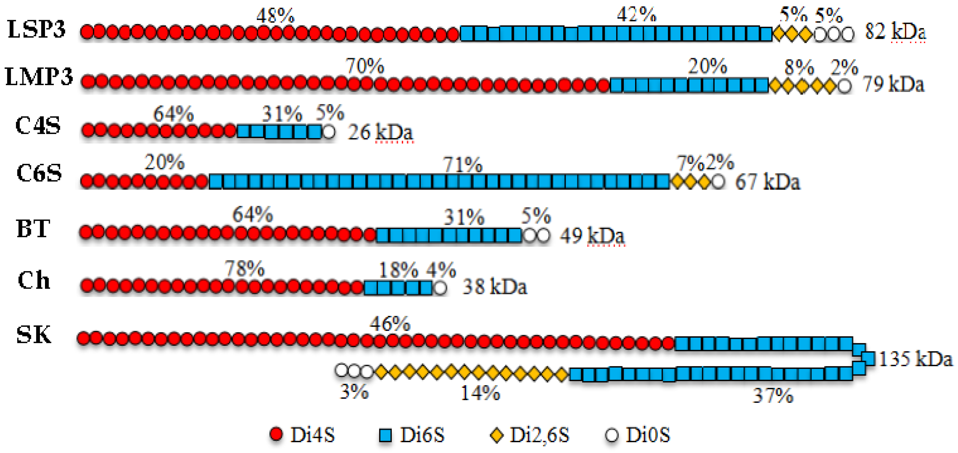
GAGs in marine animals are different to those of terrestrial organisms, mainly in terms of molecular weight and sulfation pattern. CS extracted from squid possesses antiviral and anti-metastatic activities. DS from sea squirts and hybrids CS/DS from sharks can promote the outgrowth of neurite, and are useful for nerve regeneration [14]. CS and DS are complex molecules with potential impacts on many biological systems, and it is important to consider the sulfation pattern and the size of the molecules to better understand the structure/function relationships of CS/DS [15]. The structural schematic representations of LSP3 and LMP3 were shown in Figure 7, based on the analysis of disaccharide compositions, ESI-MS/MS and NMR. The structures of LSP3 and LMP3 are different in sulfation patterns and molecule sizes compared with the structures of CS extracted from the cartilage of different animal species [15]. LSP3 and LMP3 possessed higher molecular weight than the C4S, bovine trachea (BT) and chicken sternum (Ch) of terrestrial organisms. Especially, LSP3 and LMP3 contain significant amounts of disulfated disaccharides (Di2,6S), which is only found in ocean animals, like sharks and skates. Therefore, LSP3 and LMP3 are new structure types of GAGs, which might display different functions.

2.5. Antioxidant Activity of Fractions

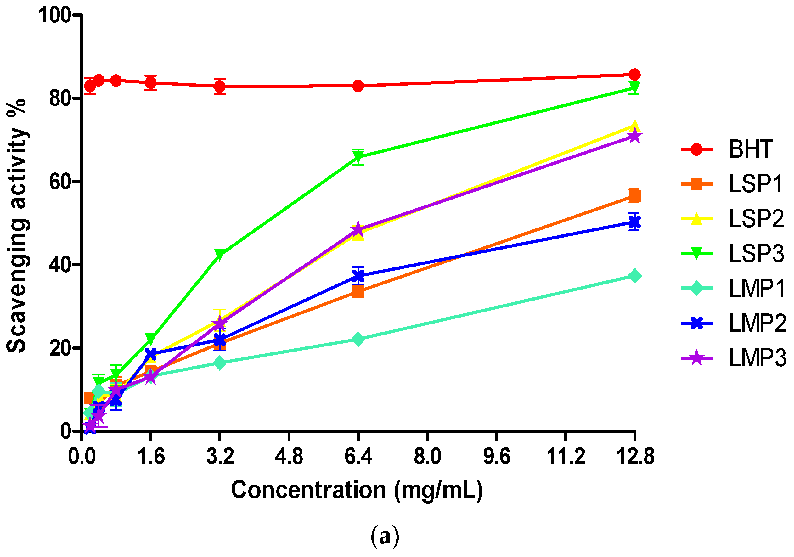
DPPH is a useful reagent to evaluate the free radical scavenging ability of hydrogen-donating antioxidants, which can transfer hydrogen atoms or electrons to DPPH radicals. Iron is the most abundant transition metal in biological systems and plays critical roles in redox systems. ABTS [2,2′-azino-bis(3-ethylbenzothiazoline-6-sulfonic acid)] radical cation scavenging assay is an excellent tool for determining the antioxidant activity of hydrogen-donating antioxidants and chain-breaking antioxidants. The results showed that the DPPH radicals’ scavenging ability, ferrous chelating power and total antioxidant capacity of each Lapemis curtus polysaccharides was concentration-dependent (Figure 8), and all the fractions exhibited significant antioxidant activity, especially for LSP3.
The fractions LSP1, LSP2, LSP3 from the skin of Lapemis curtus exhibited stronger scavenging activity than that of fractions LMP1, LMP2, LMP3 from the meat of Lapemis curtus, which eluted at the same concentration of NaCl on a Q-Sepharose Fast Flow column. Especially, the scavenging ability of LSP3 on DPPH radicals was up to 65.8% at 6.4 mg/mL (Figure 8a), which was similar to that of Butylated hydroxytoluene (BHT). The fractions LMP1, LMP2 from the meat of Lapemis curtus exhibited higher chelating activities than that of fractions LSP1, LSP2 from the skin of Lapemis curtus. LMP2 exhibited the most effective chelating activity fraction, followed by LSP2 and LSP3. In addition, the chelating activity of LMP2 was up to 93.7% at 3.2 mg/mL (Figure 8b), which was similar to that of Ethylene diamine tetraacetic acid (EDTA). The highest total antioxidant capacities were recorded in LSP2 and LSP3 (Figure 8c), with 57.8% and 49.2% of total antioxidant capacities at 6.4 mg/mL, respectively.
The results showed that DPPH and Fe2+ were more sensitive to Lapemis curtus polysaccharides than ABTS. LSP3 and LMP3 with high DS/CS content possessed a strong scavenging ability of DPPH radicals, iron (Fe2+) chelating activity and total antioxidant capacity. In addition, LSP3 possessed stronger antioxidant activities than that of LMP3, which may be related to its high content of uronic acid and degree of sulfation. LSP2 possessed a strong scavenging ability of DPPH radicals, iron (Fe2+) chelating activity and total antioxidant capacity, which may be related to its high contents of GlcNAc [16] and proteins. The polysaccharides from Lapemis curtus showed great potential for future human health applications.

3. Materials and Methods

3.1. Materials and Reagents

Lapemis curtus was provided by the Ocean College of Hainan University (Haikou, Hainan Province, China). Chondroitinase ABC was provided by the Marine Biomedical Research Institute of Qingdao (Qingdao, Shandong Province, China). Neutral protease of Bacillus subtilis was purchased from Novozymes (Copenhagen, Denmark), papain was purchased from AppliChem (Darmstadt, Germany), and trypsin was purchased from Amresco (Washington, USA). Monosaccharide standards of glucose (Glc), galactose (Gal), glucosamine (GlcN), xylose (Xyl), arabinose (Ara), mannose (Man), rhamnose (Rha), fucose (Fuc), galactosamine (GalN), glucuronic acid (GlcA), galacturonic acid (GalA), and unsaturated disaccharides standards of ΔUA-GalNAc (ΔDi0S), ΔUA-GalNAc6S (ΔDi6S), ΔUA-GalNAc4S (ΔDi4S), ΔUA2S-GalNAc6S (ΔDi2,6S), ΔUA4S-GalNAc6S (ΔDi4,6S), ΔUA2S-GalNAc4S (ΔDi2,4S), ΔUA2S-GalNAc4S6S (ΔDi2,4,6S), were purchased from Sigma-Aldrich (St. Louis, MO, USA). HPLC-grade acetonitrile was purchased from Merck KGaA (Darmstadt, Germany). All of the other chemicals and solvents used were of analytical grade, unless otherwise specified.

3.2. Extraction and Purification

Polysaccharides were extracted from the skin and the meat of Lapemis curtus. The skin of Lapemis curtus was minced and digested with 1.0% of papain and trypsin at a ratio of 1:2 (v/v) at 50 °C for 3 h. The digested mixture was centrifuged, and the supernatant was precipitated by four volumes of ethanol (95%). The precipitate was resuspended and dialyzed against water using a 1 kDa MWCO (molecular weight cut off) dialysis tube, and then freeze-dried to obtain a crude polysaccharide from the skin (named LSP). The meat of Lapemis curtus was minced and digested with 1.0% of neutral protease of Bacillus subtilis at 55 °C for 3 h, and a crude polysaccharide from meat (named LMP) was obtained after a similar process as that of LSP. LSP and LMP were further fractionated on a Q-Sepharose Fast Flow column and eluted with a step-wise gradient of 0, 0.4 and 2.0 mol/L NaCl solution to obtain LSP1, LSP2, LSP3, and LMP1, LMP2, LMP3, respectively. Finally, the purified components were pooled, dialyzed and lyophilized.

3.3. Chemical Analysis and Molecular Weight Analysis

Total uronic acid content was determined by a colorimetrical method [17] using glucuronic acid as a standard. Protein content was measured by a Bicinchoninic Acid Protein Assay Kit (BCA kit) (Sigma-Aldrich, 3050 Spruce Street, St Louis, MO 63103, USA) [18] using bovine serum albumin as a standard. Sulfate content was assayed using an ion chromatography method [19]. Molecular weight (Mw) was determined by a high-performance liquid chromatography, coupled with a refractive index detector (Agilent Technologies, Wilmington, DE, USA), with a column of TSKgel G3000PWXL (TOSOH, Tokyo, Japan). Aqueous Na2SO4 solution (0.1 mol/L) was used as the mobile phase and the flow rate was 0.5 mL/min. The temperature of the column was maintained at 35 °C. Dextrans were used as standards to calibrate the column [20].

3.4. Composition Analysis

Monosaccharide composition was determined using a 1-phenyl-3-methyl-5-pyrazolone (PMP) pre-column derivatization HPLC method [21]. The PMP-labeled carbohydrates were separated by a BDS-C18 column (4.6 mm × 250 mm, 5 µm, Hypersil, Waltham, MA, USA) with 0.1 mol/L phosphate buffer (pH 6.0) and acetonitrile at a ratio of 84:16 (v/v, %) as a mobile phase at a flow rate of 1.0 mL/min. Disaccharide composition analysis was performed by enzymatic degradation and chromatographic separation on a Zorbax SAX column (9.4 mm × 250 mm, 4.6 µm). A gradient elution was performed using pure water and 2 mol/L NaCl solution (pH = 3.5) as a mobile phase at a flow rate of 1.0 mL/min. The identification and quantitation of each unsaturated disaccharide was performed by comparing with standard disaccharides [22].

3.5. Fourier Transform Infrared (FT-IR) Spectroscopy Analysis

The FT-IR spectra of Lapemis curtus polysaccharides were recorded on a Nexus 470 FT-IR spectrophotometer (Nicolet, Pleasanton, CA, USA) in KBr pellets over a wavelength range of 400 cm−1–4000 cm−1.

3.6. Electrospray Mass Spectroscopy (ESI-MS) Analysis

Negative-ion ESI-MS/MS analysis was carried out on a Micromass LTQ-Orbitrap XL instrument (Thermo Fisher Scientific, Waltham, MA, USA). Nitrogen was used as sheath gas at a flow rate of 8 arb. The capillary temperature was 275 °C. The spray voltage, capillary voltage, and tube lens voltage were 3 KV, 43 V, and 80 V, respectively. The mobile phase was acetonitrile/H2O (1:1, v/v) at a flow rate of 10 μL/min. All of the samples were dissolved in mobile phase before injection [23].

3.7. NMR Spectroscopy Analysis

The lyophilized polysaccharides (20–30 mg) were co-evaporated with D2O (99.96%) three times to remove the exchangeable protons and then finally dissolved in 500 μL D2O. Deuterated acetone was used as an internal standard (2.08 ppm for 1H-NMR and 29.34 ppm for 13C-NMR). 1H-NMR, 13C-NMR, 1H-1H COSY, HSQC, HMBC, TOCSY and NOESY experiments were recorded at 298 K on an Agilent DD2-500 spectrometer (Agilent Technologies, Wilmington, DE, USA) [24].

3.8. Determination of Antioxidant Activity

3.8.1. DPPH Free Radical Scavenging Activity

The scavenging ability of Lapemis curtus polysaccharides on DPPH radicals was measured as previously described [25]. Briefly, 100 μL of sample solution at different concentrations was added to 400 μL of 0.004% ethanol solution of DPPH. Absorbance at 517 nm was measured after 30 min. BHT was used as a positive control. The scavenging ability was calculated as follows:
Scavenging ability (%) = (1 − Asample/Acontrol) × 100.
where Acontrol is the absorbance of control without test samples, and Asample is the absorbance in the presence of test samples. The test was carried out in triplicate.

3.8.2. Iron (Fe2+) Chelating Activity

The iron chelating effect of Lapemis curtus polysaccharides was tested as previously described [26]. Briefly, 50 μL of sample solution at different concentrations was mixed with 25 μL of 0.5 mmol/L FeCl2 and 225 μL of methanol solution. The mixtures were incubated at room temperature for 5 min and the reaction was initiated by the addition of 100 μL of 5 mmol/L ferrozine solution. The mixtures were then vigorously shaken and remained at room temperature for 10 min. EDTA was used as a positive control. The absorbance of solution was measured at 562 nm, and the chelating activity (%) was calculated as follows:
Metal chelating activity (%) = (1 − Asample/Acontrol) × 100
where Acontrol is the absorbance of control without test samples, and Asample is the absorbance of test samples. The test was carried out in triplicate.

3.8.3. Total Antioxidant Capacity Assay Kit with ABTS Method

The total antioxidant capacity of Lapemis curtus polysaccharides was measured by the ABTS method, as previously described [27]. The working solution was prepared by mixing ABTS solution and oxidant solution in equal quantity and remained in the dark at room temperature for 16 h. Next, 10 μL of sample solution at different concentrations were mixed with 200 μL of diluted ABTS solution and then stored at room temperature for 6 min. The absorbance of solution was measured at 734 nm. Vitamin C (Vc) and trolox were used as positive controls. Trolox, a water-soluble analogue of vitamin E, was used as a reference standard to prepare a calibration curve at a concentration range of 0.05–1.6 mmol/L. Results were expressed as mmol/g Trolox equivalent antioxidant capacity (TEAC). The test was carried out in triplicate.

4. Conclusions

Two crude polysaccharides were extracted from the skin and the meat of Lapemis curtus (LSP and LMP), and further purified to obtain polysaccharide sub-fractions of LSP1, LSP2, LSP3 and LMP1, LMP2, LMP3, respectively. The structures of LSP3 and LMP3 were determined as a new hybrid CS/DS by means of IR, ESI-MS, NMR and composition analysis. LSP3 is a DS enriched GAG and LMP3 is a CS enriched GAG. The polysaccharides extracted from Lapemis curtus exhibited significant antioxidant activities. Especially, LSP3 possessed a strong scavenging ability of DPPH radicals, iron (Fe2+) chelating activity and total antioxidant capacity, and this may be related to its high contents of uronic acid and sulfate. Our data suggested that the polysaccharides from Lapemis curtus could be promising candidates for natural antioxidant ingredients.

Author Contributions

X.Z. designed the experiments, supervised the research work and revised the manuscript; M.B. performed the experiments, analyzed the data and wrote the manuscript; W.H. and Q.W. contributed to part of the structure determination and analyzed the data of ESI-MS and NMR; Y.G. analyzed the data of antioxidant activities; S.D. provided the Lapemis curtus and revised the manuscript.

Acknowledgments

This research was supported by NSFC-Shandong Joint Fund (U1606403), Innovation Project of Qingdao National Laboratory for Marine Science and Technology (No. 2015ASKJ02), and Shandong key scientific and technology program (2015ZDJS04002).

Conflicts of Interest

The authors declare no conflict of interest.

References

  1. Panagos, C.G.; Thomson, D.; Moss, C.; Bavington, C.D.; Ólafsson, H.G.; Uhrín, D. Characterisation of hyaluronic acid and chondroitin/dermatan sulfate from the lumpsucker fish, C. lumpus. Carbohydr. Polym. 2014, 106, 25–33. [Google Scholar] [CrossRef] [PubMed]
  2. Thelin, M.A.; Bartolini, B.; Axelsson, J.; Gustafsson, R.; Tykesson, E.; Pera, E.; Oldberg, A.; Maccarana, M.; Malmstrom, A. Biological functions of iduronic acid in chondroitin/dermatan sulfate. FEBS J. 2013, 280, 2431–2446. [Google Scholar] [CrossRef] [PubMed]
  3. Abdelhedi, O.; Nasri, R.; Souissi, N.; Nasri, M.; Jridi, M. Sulfated polysaccharides from common smooth hound: Extraction and assessment of anti-ACE, antioxidant and antibacterial activities. Carbohydr. Polym. 2016, 152, 605–614. [Google Scholar] [CrossRef] [PubMed]
  4. Pham-Huy, L.A.; He, H.; Pham-Huy, C. Free radicals, antioxidants in disease and health. Int. J. Biomed. Sci. 2008, 4, 89–96. [Google Scholar]
  5. Campo, G.M.; Avenoso, A.; Campo, S.; Ferlazzo, A.M.; Calatroni, A. Antioxidant Activity of Chondroitin Sulfate. Adv. Pharmacol. 2006, 53, 417–431. [Google Scholar] [CrossRef] [PubMed]
  6. Sereshk, Z.H.; Bakhtiari, A.R. Distribution patterns of PAHs in different tissues of annulated sea snake (Hydrophis cyanocinctus) and short sea snake (Lapemis curtus) from the Hara Protected Area on the North Coast of the Persian Gulf, Iran. Ecotox. Environ. Safe 2014, 109, 116–123. [Google Scholar] [CrossRef] [PubMed]
  7. Shirai, N.; Suzuki, H.; Shimizu, R. Fatty acid composition of oil extracted from the fat sack of the Erabu sea snake Laticauda semifasciata in the Pacific Ocean and South China Sea. Fish. Sci. 2002, 68, 239–240. [Google Scholar] [CrossRef]
  8. Damotharan, P.; Veeruraj, A.; Arumugam, M.; Balasubramanian, T. Biological and Biochemical Potential of Sea Snake Venom and Characterization of Phospholipase A2 and Anticoagulation Activity. Ind. J. Clin. Biochem. 2016, 31, 57–67. [Google Scholar] [CrossRef] [PubMed]
  9. Garnjanagoonchorn, W.; Wongekalak, L.; Engkagul, A. Determination of chondroitin sulfate from different sources of cartilage. Chem. Eng. Process. 2007, 46, 465–471. [Google Scholar] [CrossRef]
  10. Ustyuzhanina, N.E.; Bilan, M.I.; Dmitrenok, A.S.; Borodina, E.Y.; Stonik, V.A.; Nifantiev, N.E.; Usov, A.I. A highly regular fucosylated chondroitin sulfate from the sea cucumber Massinium magnum: Structure and effects on coagulation. Carbohydr. Polym. 2017, 167, 20–26. [Google Scholar] [CrossRef] [PubMed]
  11. Zamfir, A.; Seidler, D.G.; Kresse, H.; Peter-Katalinić, J. Structural characterization of chondroitin/dermatan sulfate oligosaccharides from bovine aorta by capillary electrophoresis and electrospray ionization quadrupole time-of-flight tandem mass spectrometry. Rapid Commun. Mass Spectrom. 2002, 16, 2015–2024. [Google Scholar] [CrossRef] [PubMed]
  12. Huckerby, T.N.; Lauder, R.M.; Brown, G.M.; Nieduszynski, I.A.; Anderson, K.; Boocock, J.; Sandall, P.L.; Weeks, S.D. Characterization of oligosaccharides from the chondroitin sulfates. FEBS J. 2001, 268, 1181–1189. [Google Scholar] [CrossRef]
  13. Pavao, M.S.; Aiello, K.R.; Werneck, C.C.; Silva, L.C.F.; Valente, A.P.; Mulloy, B.; Colwell, N.S.; Tollefsen, D.M.; Mourão, P.A. Highly sulfated dermatan sulfates from ascidians structure versus anticoagulant activity of these glycosaminoglycans. J. Biol. Chem. 1998, 273, 27848–27857. [Google Scholar] [CrossRef] [PubMed]
  14. Valcarcel, J.; Novoa-Carballal, R.; Pérez-Martín, R.I.; Reis, R.L.; Vázquez, J.A. Glycosaminoglycans from marine sources as therapeutic agents. Biotechnol Adv. 2017, 35, 711–725. [Google Scholar] [CrossRef] [PubMed]
  15. Da Cunha, A.L.; Aguiar, J.A.; da Silva, F.S.C.; Michelacci, Y.M. Do chondroitin sulfates with different structures have different activities on chondrocytes and macrophages? Int. J. Biol. Macromol. 2017, 103, 1019–1031. [Google Scholar] [CrossRef] [PubMed]
  16. Azam, M.S.; Kim, E.J.; Yang, H.S.; Kim, J.K. High antioxidant and DNA protection activities of N -acetylglucosamine (GlcNAc) and chitobiose produced by exolytic chitinase from Bacillus cereus, EW5. Springerplus 2014, 3, 354–365. Available online: http://www.springerplus.com/content/3/1/354 (accessed on 11 July 2014). [CrossRef] [PubMed]
  17. Bitter, T.; Muir, H.M. A modified uronic acid carbazole reaction. Anal. Biochem. 1962, 4, 330–334. [Google Scholar] [CrossRef]
  18. Smith, P.K.; Krohn, R.I.; Hermanson, G.T.; Mallia, A.K.; Gartner, F.H.; Provenzano, M.D.; Fujimoto, E.K.; Goeke, N.M.; Olson, B.J.; Klenk, D.C. Measurement of protein using bicinchoninic acid. Anal. Biochem. 1985, 150, 76–85. [Google Scholar] [CrossRef]
  19. Xue, Y.T.; Ren, L.; Li, S.; Wang, L.L.; He, X.X.; Zhao, X.; Yu, G.L.; Guan, H.S.; Li, C.X. Study on quality control of sulfated polysaccharide drug, propylene glycol alginate sodium sulfate (PSS). Carbohydr. Polym. 2016, 144, 330–337. [Google Scholar] [CrossRef] [PubMed]
  20. Zhao, X.; Yu, G.; Guan, H.; Yue, N.; Zhang, Z.; Li, H. Preparation of low-molecular-weight polyguluronate sulfate and its anticoagulant and anti-inflammatory activities. Carbohydr. Polym. 2007, 69, 272–279. [Google Scholar] [CrossRef]
  21. Pu, J.; Zhao, X.; Wang, Q.; Wang, Y.; Zhou, H. Development and validation of a HPLC method for determination of degree of polymerization of xylo-oligosaccharides. Food. Chem. 2016, 213, 654–659. [Google Scholar] [CrossRef] [PubMed]
  22. Liu, X.; Hao, J.; Shan, X.; Zhang, X.; Zhao, X.; Li, Q.; Wang, X.; Cai, C.; Li, G.; Yu, G. Antithrombotic activities of fucosylated chondroitin sulfates and their depolymerized fragments from two sea cucumbers. Carbohydr. Polym. 2016, 152, 343–350. [Google Scholar] [CrossRef] [PubMed]
  23. Zhu, H.; Chen, X.; Zhang, X.; Liu, L.; Cong, D.; Zhao, X.; Yu, G. Acidolysis-based component mapping of glycosaminoglycans by reversed-phase high-performance liquid chromatography with off-line electrospray ionization–tandem mass spectrometry: Evidence and tags to distinguish different glycosaminoglycans. Anal. Biochem. 2014, 465, 63–69. [Google Scholar] [CrossRef] [PubMed]
  24. Xu, Y.; Masuko, S.; Takieddin, M.; Xu, H.; Liu, R.; Jing, J.; Mousa, S.A.; Linhardt, R.J.; Liu, J. Chemoenzymatic synthesis of homogeneous ultralow molecular weight heparins. Science 2011, 334, 498–501. [Google Scholar] [CrossRef] [PubMed]
  25. Shimada, K.; Fujikawa, K.; Yahara, K.; Nakamura, T. Antioxidative properties of xanthan on the autoxidation of soybean oil in cyclodextrin emulsion. J. Agric. Food Chem. 1992, 40, 945–948. [Google Scholar] [CrossRef]
  26. Decker, E.A.; Welch, B. Role of ferritin as a lipid oxidation catalyst in muscle food. J. Agric. Food Chem. 1990, 38, 674–677. [Google Scholar] [CrossRef]
  27. Zhang, D.Y.; Wan, Y.; Xu, J.Y.; Wu, G.H.; Li, L.; Yao, X.H. Ultrasound extraction of polysaccharides from mulberry leaves and their effect on enhancing antioxidant activity. Carbohydr. Polym. 2016, 137, 473–479. [Google Scholar] [CrossRef] [PubMed]
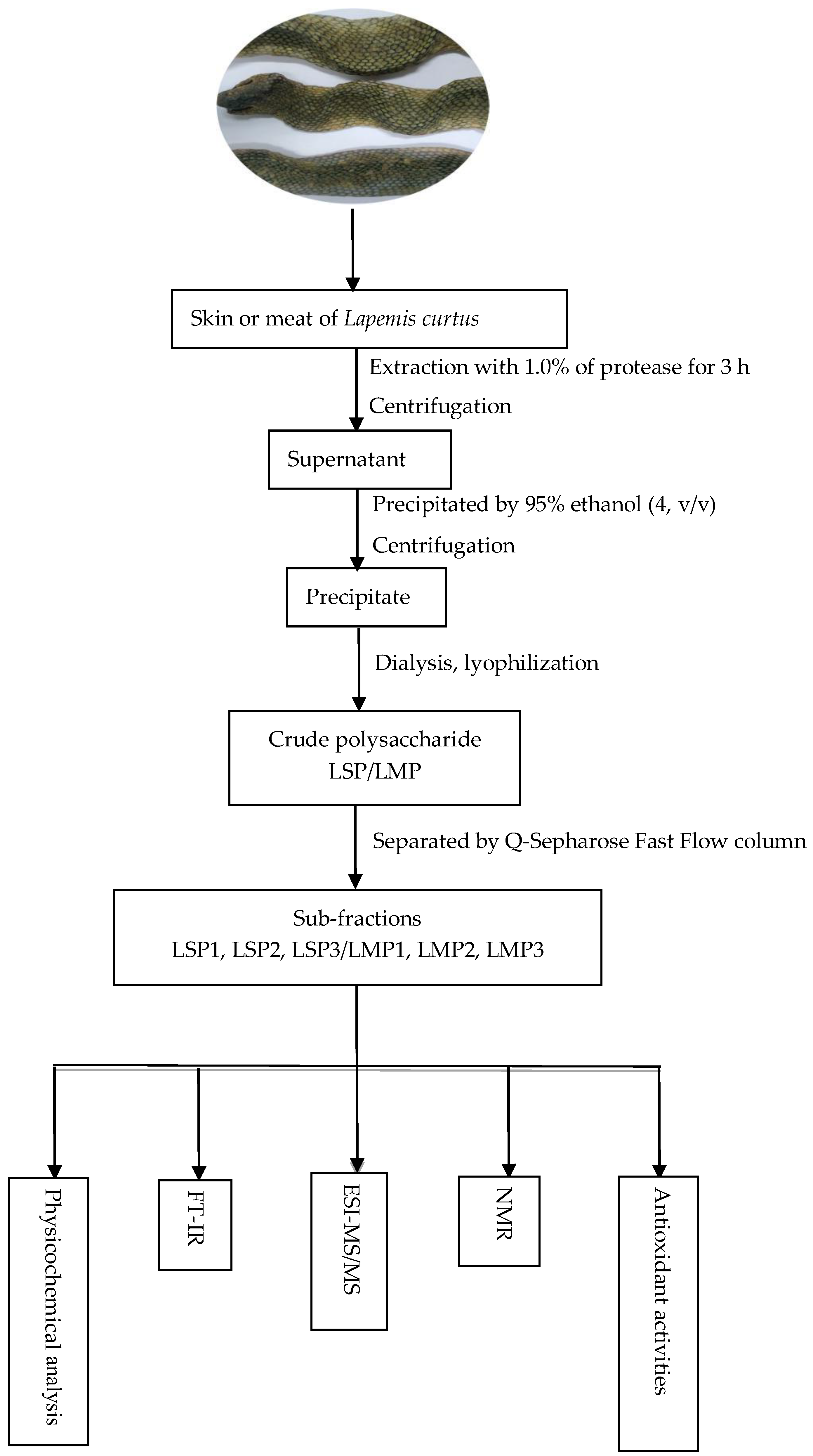
Figure 1. Experimental flowchart of Lapemis curtus polysacchloarides.
Marinedrugs 16 00170 g001
Figure 2. Isolation of the polysaccharides LSP and LMP.
Marinedrugs 16 00170 g002
Figure 3. Monosaccharide composition of LSP3 and LMP3. Chromatograms of the acid hydrolysates of LSP3 and LMP3, which were hydrolyzed in 3 mol/L TFA for 3 h at 110 °C to ensure a high response value of IdoA.
Marinedrugs 16 00170 g003
Figure 4. The IR spectra of LSP3 and LMP3.
Marinedrugs 16 00170 g004
Figure 5. The ESI-MS and MS/MS spectra of LSP3. The green lines are arrow marks from the software system.
Marinedrugs 16 00170 g005
Figure 6. The 13C-NMR spectra of LSP3 (a) and LMP3 (b), and the HSQC spectrum of LMP3 (c).
Marinedrugs 16 00170 g006
Figure 7. Schematic representation of the structure of LSP3, LMP3 and CS extracted from the cartilage of bovine trachea (BT), chicken sternum (Ch) and skate (Sk). Two commercial preparations were also used: C4S, from bovine trachea, and C6S, from shark cartilage [15].
Marinedrugs 16 00170 g007
Figure 8. Antioxidant properties of polysaccharide sub-fractions from Lapemis curtus. (a) DPPH radical scavenging activities, (b) iron chelating effect, (c) 2,2′-azino-bis(3-ethylbenzothiazoline-6-sulfonic acid) (ABTS) radical scavenging activities of different samples. LSP1, LSP2 and LSP3, extracted from the skin of Lapemis curtus. LMP1, LMP2 and LMP3 extracted from the meat of Lapemis curtus. BHT, EDTA and Vc were used as positive controls.
Marinedrugs 16 00170 g008aMarinedrugs 16 00170 g008b
Table 1. The chemical compositions of each fraction from Lapemis curtus. The total mole number of unsaturated disaccharides of each sample was taken as 100% in disaccharide compositions. “-” represents not detected.
CompositionPolysaccharides from SkinPolysaccharides from Meat
LSP1LSP2LSP3LMP1LMP2LMP3
Uronic acid (%)0.51.825.31.11.915.2
Total proteins (%)48.346.019.242.463.812.3
Sulfated groups (%)0.70.411.20.50.410.1
Molecular weight (kDa)3.73.182.02.12.779.0
Monosaccharide (molar ratio)
Mannose5.64.7-4.82.5-
N-acetyl Glucosamine12.617.21.818.113.82.0
Rhamnose------
Glucuronic acid-1.116.2--12.3
Galacturonic acid------
N-acetyl Galactosamine1.11.630.12.31.515.9
Glucose9.43.31.215.43.12.0
Galactose22.914.610.819.98.88.8
Xylose--1.3--1.5
Arabinose---1.11.6-
Fucose111111
Disaccharide (%)
ΔDi0S 5.0 1.6
ΔDi6S 42.2 20.0
ΔDi4S 47.9 70.5
ΔDi2,6S 4.9 8.0
Table 2. The 1H-NMR and 13C-NMR assignments of LSP3 and LMP3.
Signal/ppmH1H2H3H4H5H6Residue
(C1)(C2)(C3)(C4)(C5)(C6)
A→4)-β-GlcA-(1→4.353.273.473.663.57-→4)-β-GlcA-(1→3)-β-GalNAc4S-(1→
(103.35)(72.31)(73.46)(80.18)(76.42)(174.27)
B→3)-β-GalNAc4S-(1→4.443.913.904.633.713.69
(101.08)(51.43)(75.38)(76.28)(74.45)(60.83)
C→4)-β-IdoA-(1→4.773.423.783.974.60-→4)-β-IdoA-(1→3)-β-GalNAc4S-(1→
(103.13)(69.33)(71.46)(80.53)(69.72)(173.62)
D→3)-β-GalNAc4S-(1→4.563.933.844.553.713.69
(101.68)(51.76)(75.25)(76.09)(74.45)(60.83)
A′→4)-β-GlcA-(1→4.403.163.503.593.56-→4)-β-GlcA-(1→3)-β-GalNAc6S-(1→
(104.00)(72.29)(73.41)(81.20)(76.39)(174.27)
B′→3)-β-GalNAc6S-(1→4.423.913.744.003.864.10
(101.27)(51.41)(74.50)(67.71)(72.46)(67.51)
C′→4)-β-IdoA-(1→4.733.503.74NDND-→4)-β-IdoA-(1→3)-β-GalNAc6S-(1→
(102.98)(69.00)(70.85)NDND(173.62)
D′→3)-β-GalNAc6S-(1→4.503.933.754.03ND4.13
(101.68)(51.76)(80.21)(68.13)ND(67.90)