Article

Chloro-Furanocembranolides from Leptogorgia sp. Improve Pancreatic Beta-Cell Proliferation

by
Amalia B. Gallardo
1,2,
Ana R. Díaz-Marrero
1,†,
José M. De la Rosa
1,
Luis D’Croz
3,4,
Germán Perdomo
5,
Irene Cózar-Castellano
6,
José Darias
1 and
Mercedes Cueto
1,*
1
Instituto de Productos Naturales y Agrobiología (IPNA-CSIC), Avenida Astrofísico F. Sánchez, 3, 38206 La Laguna, Tenerife, Spain
2
Departamento de Química, Facultad de Ciencias, Universidad de Magallanes, Avenida Bulnes 01855, Punta Arenas 6200000, Chile
3
Departamento de Biología Marina y Limnología, Universidad de Panamá, Panama City P.O. Box 3366, Panama
4
Smithsonian Tropical Research Institute, STRI, Balboa P.O. Box 0843-03092, Panama
5
Facultad de Ciencias de la Salud, Universidad de Burgos, 09001 Burgos, Spain
6
Instituto de Biología y Genética Molecular, University of Valladolid-CSIC, 47005 Valladolid, Spain
*
Author to whom correspondence should be addressed.
Present address: University Institute of Bio-Organic Chemistry “Antonio González” (CIBICAN), University of La Laguna, 38206 Tenerife, Spain.
Mar. Drugs 2018, 16(2), 49; https://doi.org/10.3390/md16020049
Submission received: 20 December 2017 / Revised: 19 January 2018 / Accepted: 29 January 2018 / Published: 2 February 2018
(This article belongs to the Special Issue Progress on Marine Natural Products as Lead Compounds)

Abstract

:
Two new chloro-furanocembranolides (1, 2) and two new 1,4-diketo cembranolides (3, 4) were isolated from the crude extract of Leptogorgia sp. together with a new seco-furanocembranolide (5) and the known Z-deoxypukalide (6), rubifolide (7), scabrolide D (8) and epoxylophodione (9). Their structures were determined based on spectroscopic evidence. Four compounds: 1, 2, 7 and 8 were found to activate the proliferation of pancreatic insulin-producing (beta) cells.

1. Introduction

Octocorals of the genus Leptogorgia biosynthesize highly oxygenated diterpenoids based on the cembrane skeleton: (a) furanocembranolides into which a γ-lactone subunit is embedded; (b) 1,4-diketocembranoids produced by oxidative cleavage of the furan ring; and (c) nor-1,4-diketocembranolides that lack the C-18 isoprenic methyl group [1,2].
It has been suggested that activation of pancreatic beta-cell proliferation is a strategy to maintain functional beta-cell mass in diabetes mellitus [3]. Regarding this, we have shown that furanocembranoids such as epoxypukalide, pukalide, Z-deoxypukalide and leptolide improve beta-cell proliferation [4,5,6]. This prompted us to study a new extract of Leptogorgia sp. collected by SCUBA diving off Aleta (Panama), from which compounds 19 were obtained after flash chromatography followed by HPLC (Figure 1). The unusual halogenated compounds 1 and 2, together with the known Z-desoxypukalide (6) [1] and rubifolide (7) [7] belong to the furanocembranolide structural class. The remaining six compounds typify three different subclasses that involve several types of rearrangements from a furanocembranolide precursor: (a) 3, 4 and the previously isolated epoxylophodione (9) [8] are 1,4-diketocembranolides, 4 being the only haloderivative of the genus Leptogorgia of this subclass; (b) compound 5 is the first seco-furanocembranolide isolated from genus Leptogorgia, which showed a similar ring scission pattern to seco-bipinnatin J, the only seco-derivative [9] isolated from Pseudopterogorgia; and (c) scabrolide D (8) [10] is a nor-1,4-diketocembranolide which lacks the C-18 isoprenic methyl group.

2. Results

Compound 1 was obtained as an oil whose EIMS spectrum showed peaks at m/z [M − 1]+ 409/411, with relative intensities suggesting one chlorine atom. These peaks correspond to the molecular formula C20H23ClO7 (HREIMS) (m/z 409.1041 [M − 1]+, calcd. for C20H2235ClO7 409.1054). The diterpenic nature of 1 is corroborated by the 13C NMR spectrum (in Supplementary Materials), which displayed correlations in the HSQC spectrum indicative of seven quaternary carbons, six methines, six methylenes and one methyl (Table 1). Absorptions for a hydroxyl group at 3476 cm−1 and carbonyl groups at 1776 and 1676 cm−1 were observed in the IR spectrum.
Notable 1H and 13C NMR signals are: an aldehyde group [δH-18 9.89 (1H, s), δC-18 184.7], a trisubstituted furane ring [δH-5 6.70 (1H, s), δC-5 106.5; δC-3 161.3; δC-6 156.0; δC-4 123.4], a α,β-epoxy-γ-lactone ring [δH-11 3.73 (1H, s), δC-11 62.9; δC-20 172.1; δH-10 4.85 (1H, dd, J = 5.2, 11.3 Hz), δC-10 74.3 and δC-12 60.6] and a chloromethylene [δH-17 4.18 (2H, br s) y δC-17 46.6].
Connectivity information obtained from COSY, HSQC and HMBC experiments unambiguously determined the planar structure of compound 1 as a furanocembranolide, containing a C-18 oxidized to aldehyde, a vicinal diol at C-7–C-8, a C-10–C-20 α,β-epoxy-γ-lactone moiety and a chloroisopropenyl group at C-1.
1H-1H-COSY experiments established two spin systems: H2-2–H2-13 (fragment I) and H2-9–H-10 (fragment II) (Figure 2). The HMBC correlations H2-16/C-17, C-15, C-1 and H2-17/C-16, C-15, C-1 locate a chloroisopropenyl group at C-1 of fragment I, whereas the correlations H3-19/C-7, C-8 and C-9 allowed us lengthen fragment II by adding a vicinal dihydroxyl moiety bonded to C-9. The north end of both fragments II and I are connected together by insertion of a furane ring, in agreement with the HMBC correlations H-5/C-3, C-4, C-6 and H2-2 with C-4. An epoxy lactone binds the southern ends of fragments I and II between C-13 and C-10, as deduced from the HMBC correlations H-10/C-11, C-12, C-20; H2-13/C-20, C-11, C-12 and H2-14 with C-12. Thus, the structure of 1, with nine degrees of unsaturation, has been established.
Compound 2 was obtained as an oil whose EIMS spectrum showed a molecular ion at m/z [M]+ 394/396, with an isotopic pattern for a chlorine atom in the molecular formula C20H23ClO6 (HREIMS) (m/z 394.1181 [M]+, calcd. for C20H2335ClO6 394.1183). The 13C NMR spectrum and correlations in the HSQC spectrum indicated seven quaternary carbons, six methines, six methylenes and one methyl (Table 1). Absorptions for a hydroxyl group at 3558 cm−1 and carbonyl group at 1747 cm−1 were observed in its IR spectrum.
1H and 13C NMR data resemble those of 1. The principal differences lie in the chemical shifts of H-7, H2-9, C-9 and C-19, which were: δH-7 4.59 (1H, s), δH-9 1.89 (1H, m); 2.59 (1H, dd); δC-9 43.0 and δC-19 19.6, compared with those of 1 (Table 1). These shift values suggested that compounds 1 and 2 differ in the configuration of C-7 and C-8. Also, their molecular formulas diverge by 16 amu of oxygen. This suggests that the γ-lactone ring system is devoid of the epoxide ring on 2. This absence is confirmed by the new signals observed in the 1H and 13C NMR spectra for a proton (δH-11 5.86 (1H, s)) of a disubstituted olefin (δC-11 148.6; δC-12 136.2). Thus, the structure of 2 with nine degrees of unsaturation was established as shown in Figure 1 by COSY, HSQC and HMBC experiments.
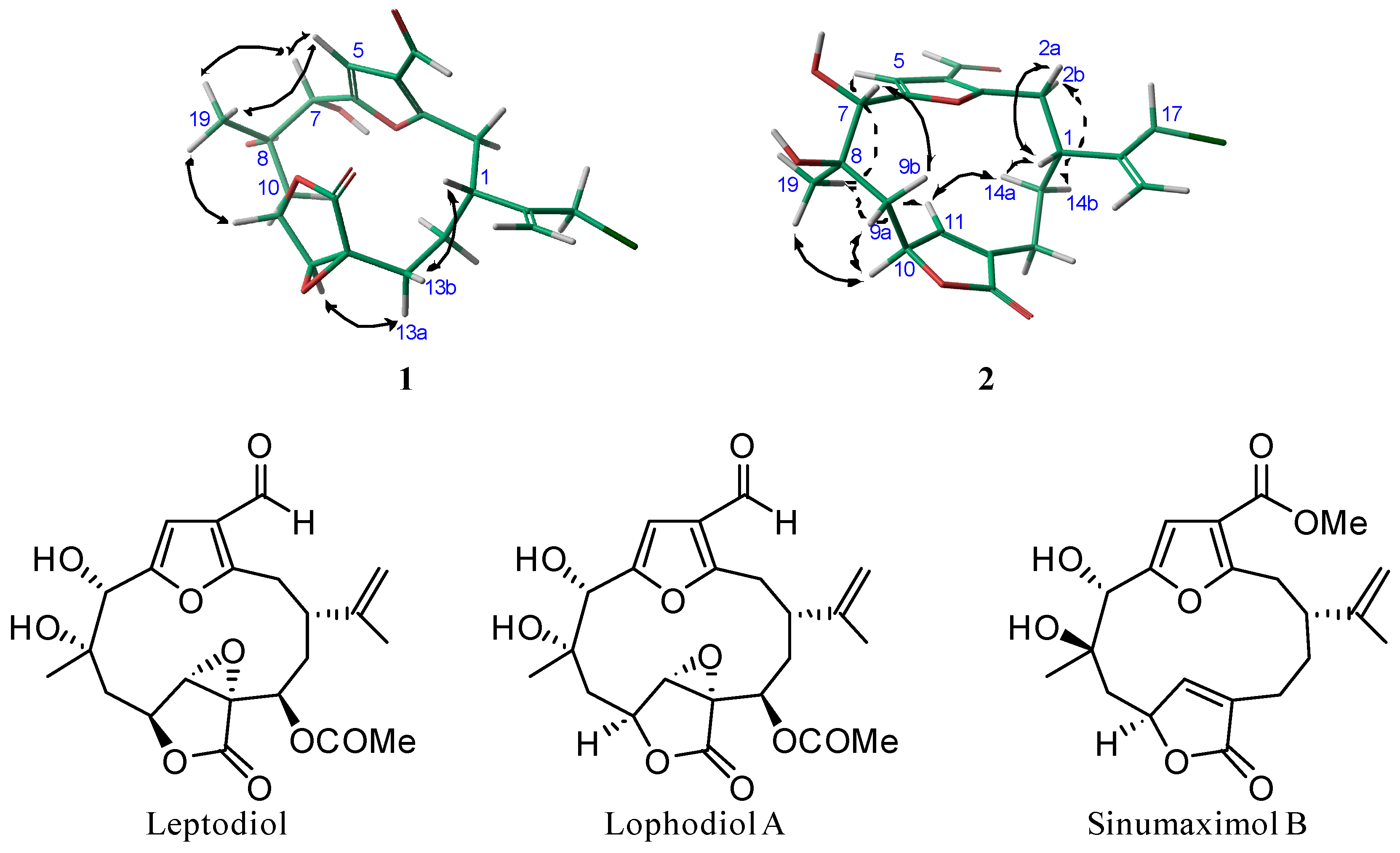
The relative configurations of compounds 1 and 2 were ascertained by NOESY experiments, molecular mechanics [11], chemical shift studies and comparison of their spectroscopic data with those of previously described cembranoids, leptodiol [1], lophodiol A [12] and sinumaximol B [13] (Figure 3).
In compound 1, the observed NOEs of H-5 with H-7 and H3-19, together with those of H-7 with H3-19 suggested that the adjacent hydroxyl groups at C-7–C-8 should be in a cis relationship. In compound 2, the observed NOEs of H3-19 with H-5 and H-10 indicate that these protons and Me-19 must be on same side of the molecule, whereas the NOEs of H3-19 with H-9a and of H-7 with H-9b indicates that H-7 and Me-19, so the vicinal diols on C-7–C-8, have a trans-relationship. Therefore, the relative configuration of C-8 is opposite to that on compound 1.
The configurations of C-7–C-8 vicinal diols were corroborated by comparison of the 1H and 13C NMR chemical shifts around the diol moiety C-7–C-8 of compounds 1 and 2 with those of the related diols leptodiol, lophodiol A and sinumaximol B, shown in Table 2. The chemical shifts of C-19 and H-7 of compounds 1 and 2 present strong differences (ΔδC-19 = 2.8 ppm and ΔδH-7 = 0.7). The chemical shifts of C-19 and H-7 of leptodiol (δC-19 22.7; δH-7 5.12) and lophodiol A (δC-19 22.7; δH-7 5.24), both with the C-7–C-8 diols in an α-cis-relationship, are very similar to those of compound 1. Whereas the chemical shifts of C-19 and H-7 of sinumaximol B (δC-19 19.8; δH-7 4.52), whose C-7–C-8 diols show a trans-relationship, present strong differences (upfield ΔδC-19 ≈ 2.6 ppm and downfield ΔδH-7 ≈ 0.8) which are very similar to those presented by compound 2 in comparison to compound 1.
In compound 1, a striking 1H NMR signal is the singlet observed for H-11 (δ 3.73, s) due to the roughly 90° dihedral angle formed between H-10 and H-11. This results in a small JH-10, H-11 that confirms the relative configuration of C-10 and C-11 as represented in the energetically favourable conformation shown in Figure 3.
Finally, in compound 1, the relative configuration of C-1 was secured by the NOESY correlation of H-13b with H-1 and H-13a with H-11, as shown in the 3D model in Figure 3. In compound 2, the observed NOEs of H-1 with H-2a and H-14a, as well as of H-14a with H-11 and of H-2b with H-14b, indicate that the isopropenyl group of 2 is situated on the alpha side of the molecule. Therefore, both compounds belong to the furanocembranolide of the α-series and their relative configurations are: 1R*, 7S*, 8S*, 10S*, 11S* and 12S* for compound 1 and 1R*, 7S*, 8R* and 10S* for compound 2.
Compound 3 was obtained as an oil whose EIMS spectrum showed a peak at m/z 362 [M]+, which corresponds to the molecular formula C20H26O6 (HREIMS) (m/z 362.1737 [M]+, calcd. for C20H26O6 362.1729). These data are in agreement with the 13C NMR spectrum, which displayed correlations in the HSQC spectrum indicative of seven quaternary carbons, four methines, six methylenes and three methyls. Absorptions for a hydroxyl group at 3475 cm−1 and carbonyl groups at 1751, 1721 and 1701 cm−1 were observed in their IR spectrum. According to the degree of unsaturation given by the 13C NMR data, 3 must be a tricyclic compound.
In the 1H and 13C NMR experiments (Table 3), signals were found for: α,β-unsaturated-γ-lactone ring [δH-11 6.76 (1H, s), δC-11 155.9; δC-20 170.6; δC-10 90.5]; isopropenyl group [δH-16 4.68 (1H, br s), δH-16 4.70 (1H, br s); δC-16 109.7] and [δH-17 1.69 (3H, s); δC-17 20.9]; methyl group on a quaternary carbon bonded to oxygen [δH-19 1.37 (CH3, s) and δC-8 74.3]; oximethine proton bonded to a secondary carbon [δH-5 3.31 (1H, d, 10.4 Hz) and δC-5 60.8] and two carbonyls [δC-3 215.4 and δC-6 203.5].
1H-1H-COSY experiments established two spin systems: H2-2–H2-13 (fragment I) and H3-18–H-5 (fragment II) (Figure 4). The HMBC correlations of H2-16/C-1, C-17 and H3-17/C-16, C-15, C-1 locates the isopropenyl group at C-1 in fragment I, whereas the HMBC correlations of H2-2 and H3-18 with C-3 connects fragments I and II through C-3. The HMBC correlations H3-19/ C-7, C-8, C-9, along with those of H-5 and H2-7 with C-6, allowed us to extend fragment II by connecting it with substructure III through C-6. Both ends of the fragment I and substructure III (C-6–C-9) are connected by inserting an α,β-unsaturated γ-lactone bonded to C-13 and C-9, respectively, due to the HMBC correlations H2-13/C-20, C-11, C-12 and those of H-11/C-10 and H2-9/C-10, C-11. The quaternary feature of C-10 comes from the oxygen linkage between C-5 and C-10 supported by an HMBC correlation of H-5 with C-10. Therefore, the tricyclic structure of 3 has been established as depicted in Figure 4.
Compound 4 was obtained as an oil whose EIMS spectrum showed peaks at m/z 396/398 [M]+, which correspond to the formula C20H25ClO6 (HREIMS) (m/z 396.1324 [M]+, calcd. for C20H25O635Cl, 396.1340). Considering the HSQC correlations, signals observed in the 13C NMR spectrum indicate seven quaternary carbons, four methines, seven methylenes and two methyls. Absorptions for a hydroxyl group at 3420 cm−1 and carbonyl groups at 1727, 1690 and 1647 cm−1 were observed in their IR spectrum.
1H and 13C NMR data (Table 3) resemble those of 3. The molecular formula of 4 showed that one proton of 3 is substituted by a chlorine atom in 4. Its corresponding data from both 1H and 13C NMR reveal that the substitution is situated on the isopropenyl appendage, where the methyl group of 3 changed to chloromethylene in 4. This substitution was confirmed by the fragment (m/z 321.1335 [M − C3H4Cl]+, calcd. for C17H21O6, 321.1338) observed in HREIMS. The planar structure of 4 was confirmed as the 17-chloro derivative of compound 3 by COSY, HSQC and HMBC experiments.
NOESY experiments, studies of coupling constants and molecular mechanics calculations suggest that 3 and 4 have the same relative stereochemistry (Figure 1). In both compounds, NOEs were observed for H-5 with H-9a and H-11 and for H3-19 with H-9a and H-9b, defining a relative configuration for C-5 and C-10 and establishing H3-19 on C-8 in a pseudo-equatorial disposition and therefore coplanar to H-5. Also, the large coupling constants of H-5 (J = 10.4 Hz) in 3 and H-5 (J = 10.3 Hz) in 4 indicate that H-5 and H-4 are trans-diaxial, as the observed NOE between H-5 and H3-18 corroborates. Finally, the NOE observed between H-11 and H-1 places the isopropenyl group on the opposite side to the Me-18. Therefore, the overall relative configuration for 3 and 4 should be 1R*, 4R*, 5S*, 8R* and 10S*.
Compound 5 was obtained as an oil with an EIMS spectrum peak at m/z 388 [M]+, which corresponds to the molecular formula C21H24O7 (HREIMS) (m/z 388.1524 [M]+, calcd. for C21H24O7, 388.1522). These data are in agreement with the 13C NMR spectrum, which displayed correlations in the HSQC spectrum indicating eight quaternary carbons, five methines, five methylenes and three methyls (Table 3). Absorptions for carbonyl groups at 1655, 1650 and 1638 cm−1 were noted in the IR spectrum.
In addition to the 1H and 13C NMR data registered for an isopropenyl group, a furan ring and a α,β-unsaturated-γ-lactone, other notable key signals were detected for the following functional groups: aldehyde [δH-7 9.54 (1H, s), δC-7 177.1]; methyl ketone [δH-19 2.21 (3H, s), δC-8 204.3] and methyl ester [δH-21 3.86 (3H, s), δC-18 162.9]. The 21 carbon atoms given by the molecular formula suggested that the isoprenic methyl group C-18 of a regular furanocembranolide is oxidized to a methyl ester and that the methyl-ketone and the aldehyde might come from the oxidative cleavage of the C-7–C-8 bond. According to the connectivity information from experiments COSY, HSQC and HMBC, compound 5 should be a seco-furanocembranolide.
1H-1H-COSY experiments established two spin systems: H2-2–H2-13 (fragment I) and H2-9–H-10 (fragment II) (Figure 5). The HMBC correlations of H2-16/C-1, C-17 and H3-17/C-16, C-15, C-1 locate the isopropenyl group at C-1 of fragment I and also those of H-5/C-3, C-4, C-6 and H2-2 with C-3 connected fragment I at the furan ring. The HMBC correlations of H2-13/C-11, C-12, C-20 and H-11/C-10 and H2-9/C-11 showed that fragments I and II are linked through an α,β-unsaturated-γ-lactone. HMBC correlations of H3-19/C-8, C-9 allowed us to place the methyl-ketone. Finally, the aldehyde must be located at C-6, in good agreement with the chemical shift observed for the aldehyde group of seco-bipinnatin J (δH-7 9.52 (1H, s), δC-7 177.5), the only seco-derivative [9] isolated from Pseudopterogorgia, which showed the same scission pattern.
Compound 5 is the first seco-furanocembranolide isolated from genus Leptogorgia. Z-deoxypukalide [1], also isolated in this work, can be considered a biogenetic precursor of 5 by oxidative cleavage of the corresponding Δ7,8. Since Z-deoxypukalide belongs to the α-cembranolide series, we assign the same relative configurations 1R*, 10S* to 5.
It should be expected that compounds 15 belong to the same enantiomeric series as Z-deoxypukalide, (6) whose absolute configuration we have previously determined using an NMR-based method using Pirkle’s reagent [1].

Activation of Pancreatic Beta-Cell Proliferation

Several strategies have been proposed to recover functional beta-cell mass loss in diabetes mellitus onset; one of them is to activate beta-cell proliferation [3]. In previous work, we showed that furanocembranolides such as epoxypukalide, pukalide, Z-deoxypukalide and leptolide augment beta-cell proliferation [4,5,6]. In order to acquire detailed knowledge of the proliferation effect induced by furanocembranolides, compounds 1, 2 and rubifolide (7) were used to treat synchronized INS-1 cells and proliferation was then measured. INS-1 cells were preincubated with 0.1 μM of each product and proliferation was measured by BrdU incorporation (Table 4), showing a 2–3-fold increase in proliferation. Although it is difficult to reach a conclusion regarding the functional groups that could modulate this proliferation activity, these results also support chloro-furanocembranolides being potential activators of pancreatic beta-cell proliferation.
Furthermore, synchronized INS-1 cells were treated with the nor-cembranolide, scabrolide D (8), (Table 4), showing a 2.8 ± 0.69-fold change above untreated cells (1.0). We therefore consider it of interest in searching for compounds of the furanocembranolide and nor-1,4-diketocembranolide families, in order to develop a new class of antidiabetic agents.

3. Experimental Section

3.1. General Experimental Procedures

Optical rotations were measured on a Perkin-Elmer model 343 Plus polarimeter (Perkin-Elmer, Rodgau, Germany) using a Na lamp at 20 °C. IR spectra were recorded on a Perkin-Elmer 1650/FTIR spectrometer (Perkin-Elmer, Rodgau, Germany). 1H NMR and 13C NMR, HSQC, HMBC and COSY spectra were measured employing a Bruker AMX 500 instrument (Bruker, Karlsruhe, Germany) operating at 500 MHz for 1H NMR and at 125 MHz for 13C NMR. All 13C and 1H NMR spectra were internally referenced to the residual solvent signal (CDCl3: δC 77.0 ppm, δH 7.25 ppm). Two-dimensional NMR spectra were obtained using the standard Bruker software (TOpSpin 2.1, Bruker, Karlsruhe, Germany). The EIMS data were obtained on a Waters Vg-Micromass spectrometer (Waters, Manchester, UK) model Zab 2F. HPLC separations were performed on an Agilent 1200 Series Quaternary LC system (Agilent Technologies, Waldbronn, Germany) apparatus equipped with a UV detector (DAD G1315D, Agilent Technologies, Waldbronn, Germany) and an Ascentis® C18 semi-preparative column (5 μm, 25 cm × 21.2 mm, Supelco, Bellefonte, PA, USA) eluted with CH3CN-H2O mixtures. Size-exclusion chromatography used Sephadex LH-20 as stationary phase and hexane-MeOH-CH2Cl2 (3:1:1) as solvent system. The spray reagent used to develop TLC plates was H2SO4-H2O-AcOH (1:4:20).

3.2. Collection, Extraction and Isolation

Leptogorgia sp. was collected by SCUBA diving off Aleta (Panama) at −10 m. A voucher specimen has been deposited at the Smithsonian Tropical Research Institute (Panama City, Panama) with code 200708. Specimens of Leptogorgia sp. (458.3 g) were extracted with acetone at room temperature and were concentrated to give a dark gum (10.1 g). C-18 reversed-phase flash chromatography of the crude extract gave fractions 4 (290.4 mg; 2:3 H2O/MeOH) and 5 (1119.1 mg; 1:4 H2O/MeOH) containing cembranolides, as indicated by their 1H NMR spectra. Fraction 4 was further chromatographed by molecular exclusion LH-20 to give two sub-fractions of interest, 41 (39.4 mg) and 45 (18.6 mg). C-18 reversed-phase HPLC of 41 using a gradient from H2O-CH3CN (7:3) to CH3CN (100%) afforded compound 1 (1.3 mg, tR 61 min) and scabrolide D (8) (6.5 mg, tR 64 min). From sub-fraction 45, compounds 2 (5.4 mg; tR 101.5 min) and 4 (0.5 mg; tR 83 min) were separated after C-18 reversed-phase HPLC using a gradient from H2O-CH3CN (7:3) to CH3CN (100%). Fraction 5 was chromatographed by molecular exclusion LH-20 to give five sub-fractions of interest: 51 (87.6 m), 52 (89.5 mg), 53 (82.9 mg), 54 (59.1 mg) and 55 (50.3 mg). All these fractions were chromatographed separately by C-18 reversed-phase HPLC using a gradient from H2O-CH3CN (7:3) to CH3CN (100%), to afford compounds 3 (1.6 mg; tR 83.1 min), 5 (2.1 mg; tR 52.3 min), Z-deoxypukalide (6) (5.7 mg; tR 111.2 min), rubifolide (7) (6.0 mg; tR 74 min) and epoxylophodione (9) (0.4 mg; tR 82.3 min).
Compound 1: Colourless oil; [ α ] D 20 −20.7 (c 0.03, CH2Cl2); IR (film) νmax 3476, 2932, 1776, 1676 cm−1; 1H (500 MHz, CDCl3) δ 1.35 (3H, s, H-19), 1.42 (1H, m, H-14a), 1.45 (1H, m, H-13a), 1.51 (1H, m, H-9), 1.87 (1H, dd, J = 5.2, 15.0 Hz, H-9), 2.04 (1H, m, H-14b), 2.44 (1H, dd, J = 11.5, 14.7 Hz, H-13b), 3.15 (2H, m, H-2), 3.39 (1H, m, H-1), 3.73 (1H, s, H-11), 4.18 (2H, br s, H-17), 4.85 (1H, dd, J = 5.2, 11.3 Hz, H-10), 5.29 (1H, s, H-7), 5.37 (1H, s, H-16a), 5.47 (1H, s, H-16b), 6.70 (1H, s, H-5), 9.89 (1H, s, H-18); 13C NMR (125 MHz CDCl3) δ 22.4 (CH3, C-19), 22.5 (CH2, C-13), 30.4 (CH2, C-14), 32.9 (CH2, C-2), 37.6 (CH, C-1), 40.5 (CH2, C-9), 46.6 (CH2, C-17), 60.6 (C, C-12), 62.9 (CH, C-11), 74.0 (CH, C-7), 74.3 (C, C-8), 74.3 (CH, C-10), 106.5 (CH, C-5), 118.0 (CH2, C-16), 123.4 (C, C-4), 144.2 (C, C-15), 156.0 (C, C-6), 161.3 (C, C-3), 172.0 (C, C-20), 184.7 (CH, C-18); EIMS m/z 409/411 [M − 1]+, 395/397 [M − CH3]+, 393/395 [M − OH]+, 355 [M − C3H4Cl]+; HREIMS m/z [M − 1]+ 409.1041 (calcd. for C20H2235ClO7 409.1054), 411.1027 (calcd. for C20H2237ClO7, 411.1025), 395.0887 (calcd. for C19H2035ClO7, 395.0898), 395.1069 (calcd. for C20H2237ClO6, 395.1075).
Compound 2: Colourless oil; [ α ] D 20 −11.0 (c 0.10, CH2Cl2); IR (film) νmax 3558, 2935, 1747, 1674 cm−1; 1H (500 MHz, CDCl3) δ 1.41 (3H, s, H-19), 1.60 (1H, ddd, J = 2.8, 2.8, 15.1 Hz, H-14a), 1.89 (1H, dd, J = 11.7, 14.8 Hz, H-9b), 2.02 (1H, m, H-14b), 2.14 (1H, m, H-13a), 2.35 (1H, ddd, J = 2.8, 11.9, 15.1 Hz, H-13b), 2.46 (1H, dddd, J = 2.2, 2.5, 9.5, 11.7 Hz, H-1), 2.59 (1H, dd, J = 4.1, 14.8 Hz, H-9a), 2.97 (1H, dd, J = 2.5, 14.8 Hz, H-2a), 3.19 (1H, dd, J = 11.9, 15.1 Hz, H-2b), 4.12 (2H, br s, H-17), 4.59 (1H, s, H-7), 4.97 (1H, m, H-10), 5.19 (1H, s, H-16), 5.36 (1H, s, H-16), 5.86 (1H, s, H-11), 6.80 (1H, s, H-5), 9.97 (1H, s, H-18); 13C NMR (125 MHz, CDCl3) δ 19.6 (CH3, C-19), 21.7 (CH2, C-13), 30.2 (CH2, C-14), 32.7 (CH2, C-2), 39.7 (CH, C-1), 43.0 (CH2, C-9), 47.2 (CH2, C-17), 73.6 (C, C-8), 75.6 (CH, C-7), 78.4 (CH, C-10), 106.5 (CH, C-5), 117.0 (CH2, C-16), 122.7 (C, C-4), 136.2 (C, C-12), 146.9 (C, C-15), 148.6 (CH, C-11), 154.3 (C, C-6), 162.1 (C, C-3), 173.5 (C, C-20), 184.3 (CH, C-18); EIMS m/z 394/396 [M]+, 377/379 [M − OH]+; HREIMS m/z [M]+ 394.1181 (calcd. for C20H23O635Cl, 394.1183), 396.1162 (calcd. for C20H23O637Cl, 396.1154), 377.1146 (calcd. for C20H22O535Cl, 377.1156), 379.1133 (calcd. for C20H22O537Cl, 379.1126).
Compound 3: Colourless oil; [ α ] D 20 +10.0 (c 0.16, CH2Cl2); IR (film) νmax 3475, 2933, 1751, 1721, 1701 cm−1; 1H (500 MHz, CDCl3) δ 1.04 (3H, d, J = 6.6 Hz, H-18), 1.37 (3H, s, H-19), 1.69 (3H, s, C-17), 1.89 (2H, m, H-14), 1.92 (1H, m, H-9b), 2.27 (2H, ddd, J = 5.4, 9.3, 14.3 Hz, H-13a), 2.34 (1H, d, J = 15.1 Hz, H-9a), 2.44 (1H, m, H-2a), 2.46 (1H, m, H-2b), 2.50 (1H, m, H-13b), 2.64 (1H, ddd, J = 4.2, 7.1, 7.1 Hz, H-1), 2.68 (2H, m, H-7), 2.89 (1H, ddd, J = 6.6, 6.6, 10.6 Hz, H-4), 3.31 (1H, d, J = 10.4 Hz, H-5), 4.68 (1H, br s, H-16a), 4.70 (1H, br s, H-16b), 6.76 (1H, s, H-11); 13C NMR (125 MHz, CDCl3) δ 17.2 (CH3, C-18), 20.9 (CH3, C-17), 24.0 (CH2, C-13), 30.1 (CH3, C-19), 31.7 (CH2, C-14), 39.7 (CH, C-1), 41.9 (CH, C-4), 45.6 (CH2, C-9), 49.7 (CH2, C-2), 55.9 (CH2, C-7), 60.8 (CH, C-5), 74.3 (C, C-8), 90.5 (C, C-10), 109.7 (CH2, C-16), 131.4 (C, C-12), 149.4 (C, C-15), 155.9 (CH, C-11), 170.5 (C, C-20), 203.5 (C, C-6), 215.4 (C, C-3); EIMS m/z 362 [M]+, 347 [M − CH3]+, 345 [M − OH]+; HREIMS m/z [M]+ 362.1737 (calcd. for C20H26O6, 362.1729), [M − CH3]+ 347.1509 (calcd. for C19H23O6, 347.1495), 345.1708 (calcd. for C20H25O5, 345.1702).
Compound 4: Colourless oil; [ α ] D 20 +66.0 (c 0.05, CH2Cl2); IR (film) νmax 3420, 2956, 1727, 1690, 1647 cm−1; 1H (500 MHz, CDCl3) δ 1.04 (3H, d, J = 6.6 Hz, H-18), 1.37 (3H, s, H-19), 1.90 (1H, dd, J = 2.6, 15.4 Hz H-9b), 2.00 (2H, m, H-14), 2.28 (1H, m, H-13a), 2.32 (1H, d, J = 15.1 Hz, H-9b), 2.49 (1H, m, H-13b), 2.53 (2H, m, H-2), 2.69 (2H, m, H-7), 2.90 (1H, m, H-1), 2.90 (1H, m, H-4), 3.31 (1H, d, J = 10.3 Hz, H-5), 4.03 (2H, m, H-17), 5.00 (1H, s, H-16b), 5.20 (1H, dd, J = 0.9, 0.9 Hz, H-16a), 6.79 (1H, s, H-11); 13C NMR (125 MHz, CDCl3) δ 17.3 (CH3, C-18), 22.8 (CH2, C-13), 30.1 (CH3, C-19), 31.5 (CH2, C-14), 35.4 (CH, C-1), 41.7 (CH, C-4), 45.6 (CH2, C-9), 47.6 (CH2, C-17), 49.9 (CH2, C-2), 55.9 (CH2, C-7), 60.9 (CH, C-5), 74.3 (C, C-8), 90.3 (C, C-10), 114.7 (CH2, C-16), 131.1 (C, C-12), 149.3 (C, C-15), 156.0 (CH, C-11), 171.6 (C, C-20), 205.8 (C, C-6), 215.3 (C, C-3); EIMS m/z 396/398 [M]+, 378/380 [M − H2O]+, 361 [M − Cl]+, 321 [M − C3H4Cl]+; HREIMS m/z [M]+ 396.1324 (calcd. for C20H25O635Cl, 396.1340), 398.1315 (calcd. for C20H25O637Cl, 398.1310), 361.1642 (calcd. for C20H25O6, 361.1651), 321.1335 (calcd. for C17H21O6, 321.1338).
Compound 5: Colourless oil; [ α ] D 20 −2.11 (c 0.21, CH2Cl2); IR (film) νmax 2100, 1655, 1650, 1638 cm−1; 1H (500 MHz, CDCl3) δ 1.64 (1H, m, H-14), 1.68 (3H, s, H-17), 1.72 (1H, m, H-14), 2.16 (1H, m, H-13a), 2.21 (3H, s, H-19), 2.25 (1H, m, H-13b), 2.63 (1H, dd, J = 7.3, 17.7 Hz, H-9a), 2.70 (1H, m, H-1), 2.99 (1H, dd, J = 6.7, 17.7 Hz, H-9b), 3.12 (1H, dd, J = 6.6, 14.2 Hz, H-2), 3.22 (1H, dd, J = 8.5, 14.2 Hz, H-2), 3.86 (3H, s, H-21), 4.64 (1H, br s, H-16a), 4.73 (1H, dd, J = 1.6, 1.6 Hz, H-16b), 5.27 (1H, m, H-10), 7.11 (1H, m, H-11), 7.44 (1H, s, H-5), 9.54 (1H, s, H-7); 13C NMR (125 MHz, CDCl3) δ 18.0 (CH3, C-17), 23.1 (CH2, C-13), 30.1 (CH2, C-14), 30.5 (CH3, C-19), 32.3 (CH2, C-2), 45.7 (CH, C-1), 46.5 (CH2, C-9), 51.8 (CH3, C-21), 76.6 (CH, C-10), 113.6 (CH2, C-16), 116.7 (C, C-4), 122.0 (CH, C-5), 134.3 (C, C-12), 144.8 (C, C-15), 147.7 (CH, C-11), 150.6 (C, C-6), 162.9 (C, C-18), 166.6 (C, C-3), 172.9 (C, C-20), 177.1 (CH, C-7), 204.3 (C, C-8); EIMS m/z 388 [M]+, 357 [M−OCH3]+; ESMS m/z [M]+ 388.1524 (calcd. for C21H24O7, 388.1522), 357.1341 (calcd. for C20H21O6, 357.1338).

3.3. Activation of Pancreatic Beta-Cell Proliferation

INS-1 cells (insulinoma rat cells) were seeded at a density of 20,000 cells per well in 96-well plates. Compounds 1, 2, rubifolide (7) and scabrolide (8) were assayed at a final concentration of 0.1 μM in culture medium supplemented with 5.5 mM glucose. Proliferation was measured after 24 h using the BrdU kit (Roche, Mannheim, Germany), following manufacturer’s instructions. At least three independent experiments in triplicate were preformed per compound.

Supplementary Materials

The following are available online at https://www.mdpi.com/1660-3397/16/2/49/s1, Figure S1: 1H NMR of 1 in CDCl3; Figure S2: 13C NMR spectrum of 1 in CDCl3; Figure S3: 1H NMR of 2 in CDCl3; Figure S4: 13C NMR spectrum of 2 in CDCl3; Figure S5: 1H NMR of 3 in CDCl3; Figure S6: 13C NMR spectrum of 3 in CDCl3; Figure S7: 1H NMR of 4 in CDCl3; Figure S8: 13C NMR spectrum of 4 in CDCl3; Figure S9: 1H NMR of 5 in CDCl3; Figure S10: 13C NMR spectrum of 5 in CDCl3.

Acknowledgments

This work was supported by the Ministerio de Ciencia e Innovación (SAF2009-0839 and RTA 2015-00010-C03-02). ARDM acknowledges funding from IMBRAIN project (FP7-REGPOT-2012-CT2012-31637-IMBRAIN) and from Cabildo de Tenerife (Agustín de Betancourt Programme). A.B.G. would like to thank Convenio Universidad de Magallanes (Chile) and CSIC, project 2009CL0031, for financial support.

Author Contributions

A.B.G., J.M.d.l.R., G.P. and I.C.-C. performed the experiments, analysed the data and contributed to discussion; L.D. contributed biological material; A.R.D.-M., J.D. and M.C. supervised experiments, analysed the data and wrote the manuscript. All authors approved the final version of this manuscript.

Conflicts of Interest

The authors declare no conflict of interest.

References

  1. Dorta, E.; Díaz-Marrero, A.R.; Brito, I.; Cueto, M.; D’Croz, L.; Darias, J. The oxidation profile at C-18 of furanocembranolides may provide a taxonomical marker for several genera of octocorals. Tetrahedron 2007, 63, 9057–9062. [Google Scholar] [CrossRef]
  2. Díaz-Marrero, A.R.; Porras, G.; Cueto, M.; D’Croz, L.; Lorenzo, M.; San-Martín, A.; Darias, J. Leptogorgolide, a biogenetically interesting 1,4-diketo-cembranoid that reinforces the oxidation profile of C-18 as a taxonomical marker for octocorals. Tetrahedron 2009, 65, 6029–6033. [Google Scholar] [CrossRef]
  3. Wang, P.; Fiaschi-Taesch, N.M.; Vasavada, R.C.; Scott, D.K.; García-Ocaña, A.; Stewart, A.F. Diabetes mellitus—Advances and challenges in human β-cell proliferation. Nat. Rev. Endocrinol. 2015, 11, 201–212. [Google Scholar] [CrossRef] [PubMed]
  4. Lopez-Acosta, J.F.; Moreno-Amador, J.L.; Jimenez-Palomares, M.; Diaz-Marrero, A.R.; Cueto, M.; Perdomo, G.; Cozar-Castellano, I. Epoxypukalide induces proliferation and protects against cytokine-mediated apoptosis in primary cultures of pancreatic beta-cells. PLoS ONE 2013, 8, e52862. [Google Scholar] [CrossRef] [PubMed]
  5. Lopez-Acosta, J.F.; Villa-Perez, P.; Fernandez-Diaz, C.M.; Roman Dde, L.; Diaz-Marrero, A.R.; Cueto, M.; Perdomo, G.; Cozar-Castellano, I. Protective effects of epoxypukalide on pancreatic beta-cells and glucose metabolism in STZ-induced diabetic mice. Islets 2015, 7, e1078053. [Google Scholar] [CrossRef] [PubMed]
  6. Villa-Perez, P.; Cueto, M.; Diaz-Marrero, A.R.; Lobatón, CD.; Moreno, A.; Perdomo, G.; Cozar-Castellano, I. Leptolide improves insulin resistance in diet-induced obese mice. Mar. Drugs 2017, 15, 289. [Google Scholar] [CrossRef] [PubMed]
  7. Williams, D.; Andersen, R.J.; Van Duyne, G.D.; Clardy, J. Cembrane and pseudopterane diterpenes from the soft coral Gersemia rubiformis. J. Org. Chem. 1987, 52, 332–335. [Google Scholar] [CrossRef]
  8. Bandurraga, M.M.; McKittrick, B.; Fenical, W.; Arnold, E.; Clardy, J. Diketone cembrenolides from the pacific gorgonian Lophogorgia alba. Tetrahedron 1982, 38, 305–310. [Google Scholar] [CrossRef]
  9. Rodríguez, A.D.; Shi, Y.-P. New Metabolites from the West Indian Sea Feather Pseudopterogorgia bipinnata. J. Nat. Prod. 2000, 63, 1548–1550. [Google Scholar] [CrossRef] [PubMed]
  10. Sheu, J.-H.; Ahmed, A.F.; Shiue, R.-T.; Dai, C.-F.; Kuo, Y.-H. Scabrolides A–D, Four New Norditerpenoids Isolated from the Soft Coral Sinularia scabra. J. Nat. Prod. 2002, 65, 1904–1908. [Google Scholar] [CrossRef] [PubMed]
  11. PCModel, version 9.2; Serena Software: Bloomington, IN, USA, 2007.
  12. Sánchez, C.M.; Ortega, M.J.; Zubía, E.; Carballo, J.L. Cembrane Diterpenes from the Gorgonian Lophogorgia peruana. J. Nat. Prod. 2006, 69, 1749–1755. [Google Scholar] [CrossRef] [PubMed]
  13. Thao, N.P.; Nam, N.H.; Cuong, N.X.; Quang, T.H.; Tung, P.T.; Tai, B.H.; Luyen, B.T.T.; Chae, D.; Kim, S.; Koh, Y.-S.; et al. Diterpenoids from the Soft Coral Sinularia maxima and Their Inhibitory Effects on Lipopolysaccharide-Stimulated Production of Pro-inflammatory Cytokines in Bone Marrow-Derived Dendritic Cells. Chem. Pharm. Bull. 2012, 60, 1581–1589. [Google Scholar] [CrossRef] [PubMed]
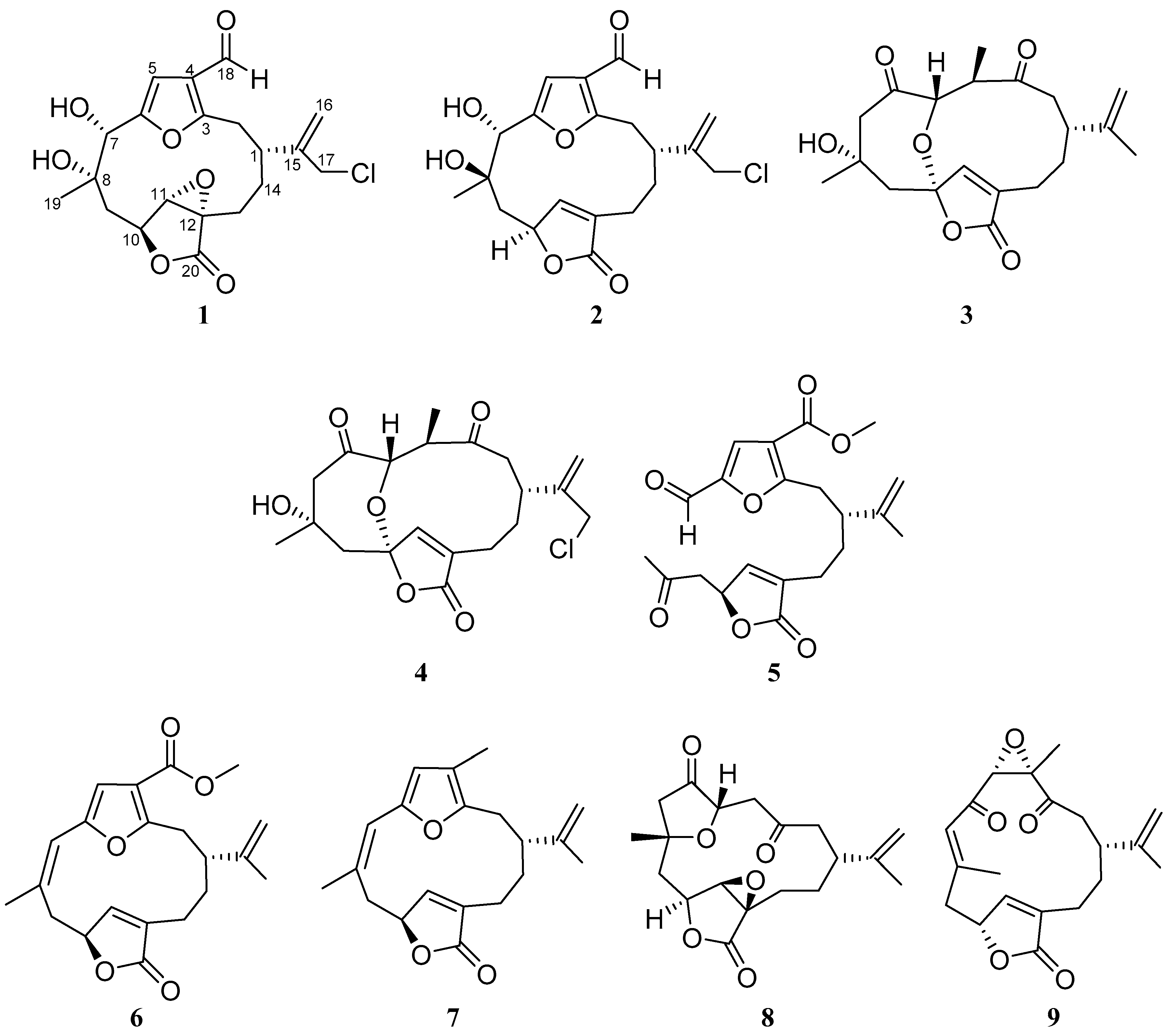
Figure 1. Novel chloro-furanocembranolides (1, 2), 1,4-diketocembranolides (3, 4), seco-furanocembranolide (5) and the known Z-deoxypukalide (6), rubifolide (7), scabrolide D (8) and epoxylophodione (9) from Leptogorgia sp.
Marinedrugs 16 00049 g001
Figure 2. 1H-1H-COSY (), HMBC (→) correlations of 1.
Marinedrugs 16 00049 g002
Figure 3. Selected NOE effects (↔) of 1 and 2 and leptodiol, lophodiol A and sinumaximol B.
Marinedrugs 16 00049 g003
Figure 4. 1H-1H-COSY (), HMBC (→) and selected NOE effects (↔) of 3.
Marinedrugs 16 00049 g004
Figure 5. 1H-1H-COSY (—) and HMBC (→) correlations of 5 and bipinnatin J.
Marinedrugs 16 00049 g005
Table 1. 1H and 13C NMR spectroscopic data [500 and 125 MHz, CDCl3] of compounds 1 and 2.
No.12
δC, TypeδH (J in Hz)δC, TypeδH (J in Hz)
137.6, CH3.39, m39.7, CH2.46, dddd (2.2, 2.5, 9.5, 11.7)
232.9, CH23.15, m32.7, CH2a: 2.97, dd (2.5, 14.8)
b: 3.19, dd (11.9, 15.1)
3161.3, C-162.1, C-
4123.4, C-122.7, C-
5106.5, CH6.70, s106.5, CH6.80, s
6156.0, C-154.3, C-
774.0, CH5.29, s75.6, CH4.59, s
874.3, C-73.6, C-
940.5, CH21.51, m
1.87, dd (5.2, 15.0)
43.0, CHb: 1.89, dd (11.7, 14.8)
a: 2.59, dd (4.2, 14.8)
1074.3, CH4.85, dd (5.2, 11.3)78.4, CH4.97, m
1162.9, CH3.73, s148.6, C5.86, s
1260.6, C-136.2, C-
1322.5, CH2a: 1.45, m
b: 2.44, dd (11.5, 14.7)
21.7, CH2a: 2.14, m
b: 2.35, ddd (2.8, 11.9, 15.1)
1430.4, CH21.42, m
2.04, m
30.2, CH21.60, ddd (2.8, 2.8, 15.1)
2.02, m
15144.2, C-146.9, C-
16118.0, CH25.37, s;
5.47, s
117.0, CH25.19, s;
5.36, s
1746.6, CH24.18, br s47.2, CH24.12, br s
18184.7, CH9.89, s184.3, CH9.97, s
1922.4, CH31.35, s19.6, CH31.41, s
20172.1, C-173.5, C-
Table 2. Selected 1H and 13C NMR data [CDCl3] of leptodiol, lophodiol A, 1, sinumaximol B and 2.
Marinedrugs 16 00049 i001 Marinedrugs 16 00049 i002
No.LeptodiolLophodiol A1Sinumaximol B2
δH-75.12, br s5.24, s5.29, s4.52, s4.59, s
δH-91.61, dd (8.8, 14.5)
1.68, dd (6.9, 14.5)
1.55, m
1.76, dd (6.4, 14.8)
1.55, m
1.76, dd (6.4, 14.8)
1.85, dd (11.5, 14.5)
2.55, dd (4.0, 14.5)
1.89, dd (11.7, 14.8)
2.59, dd (4.2, 14.8)
δH-191.38, s1.40 s1.40, s1.38, s1.41, s
δC-1922.722.722.419.819.6
δC-773.573.474.076.175.6
δC-941.140.940.543.243.0
Table 3. 1H and 13C NMR Spectroscopic data [500 and 125 MHz, CDCl3] of compounds 3, 4 and 5.
No.345
δC, Mult.δH (J in Hz)δC, Mult.δH (J in Hz)δC, Mult.δH (J in Hz)
139.7 CH2.64, ddd
(4.2, 7.1, 7.1)
35.4 CH2.90, m45.7, CH2.70, m
249.7, CH22.44, m
2.46, m
49.9, CH22.53 m
2.53, m
32.3, CH23.12, dd (6.6, 14.2)
3.22, dd (8.5, 14.2)
3215.4, C-215.3, C-166.6, C-
441.9, CH2.89, ddd
(6.6, 6.6, 10.6)
41.7, CH2.90, m116.7, C-
560.8, CH3.31, d (10.4)60.9, CH3.31, d (10.3)122.0, CH7.44, s
6203.5, C-205.8, C-150.6, C-
755.9, CH22.68, m55.9, CH22.69, m177.1, CH9.54, s
874.3, C-74.3, C-204.3, C-
945.6, CH2b: 1.92, m
a: 2.34, d (15.1)
45.6, CH2b: 1.90, dd (2.6, 15.4)
a: 2.32, d (15.1)
46.5, CH22.63, dd (7.3, 17.7)
2.99, dd (6.7, 17.7)
1090.5, C-90.3, C-76.6, CH5.27, m
11155.9, CH6.76, s 156.0, CH6.79, s147.7, CH7.11, m
12131.4, C-131.1, C-134.3, C-
1324.0, CH2a: 2.27, ddd (5.4, 9.3, 14.3)
b: 2.50, m
22.8, CH2a: 2.28, m
b: 2.49, m
23.1, CH22.16, m
2.25, m
1431.7, CH21.89, m31.5, CH22.00, m30.1, CH21.64, m
1.72, m
15149.4, CH-149.3, C-144.8, C-
16109.7, CH24.68, br s;
4.70, br s
114.7, CH25.00, s;
5.20, dd (0.9, 0.9)
113.6, CH24.64, br s
4.73, dd (1.6, 1.6)
1720.9, CH31.69, s47.6, CH24.03, m18.0, CH31.68, s
1817.2, CH31.04, d (6.6)17.3, CH31.04, d (6.6)162.9,C-
1930.1, CH31.37, s30.1, CH31.37, s30.5, CH32.21, s
20170.6, C-171.6, C-172.9, C-
OMe----51.8, CH33.86, s
Table 4. Beta-cell proliferation measurement after treating INS-1 beta-cells with each compound.
Compound (0.1 μM)Proliferation Ratio a
12.5 ± 0.65
22.0 ± 0.61
Rubifolide (7)3.3 ± 0.80 b
Scabrolide D (8)2.8 ± 0.69 b
a Fold change above untreated cells (1.0). A threshold over 1.5-fold was considered to increase proliferation. b p < 0.05 versus control.