Article

Bacillibactin and Bacillomycin Analogues with Cytotoxicities against Human Cancer Cell Lines from Marine Bacillus sp. PKU-MA00093 and PKU-MA00092

State Key Laboratory of Natural and Biomimetic Drugs, Department of Natural Medicines, School of Pharmaceutical Sciences, Peking University, 38 Xueyuan Road, Haidian District, Beijing 100191, China
*
Authors to whom correspondence should be addressed.
Mar. Drugs 2018, 16(1), 22; https://doi.org/10.3390/md16010022
Submission received: 27 November 2017 / Revised: 21 December 2017 / Accepted: 5 January 2018 / Published: 10 January 2018
(This article belongs to the Special Issue Genome Mining and Marine Microbial Natural Products)

Abstract

:
Nonribosomal peptides from marine Bacillus strains have received considerable attention for their complex structures and potent bioactivities. In this study, we carried out PCR-based genome mining for potential nonribosomal peptides producers from our marine bacterial library. Twenty-one “positive” strains were screened out from 180 marine bacterial strains, and subsequent small-scale fermentation, HPLC and phylogenetic analysis afforded Bacillus sp. PKU-MA00092 and PKU-MA00093 as two candidates for large-scale fermentation and isolation. Ten nonribosomal peptides, including four bacillibactin analogues (14) and six bacillomycin D analogues (510) were discovered from Bacillus sp. PKU-MA00093 and PKU-MA00092, respectively. Compounds 1 and 2 are two new compounds and the 1H NMR and 13C NMR data of compounds 7 and 9 is first provided. All compounds 110 were assayed for their cytotoxicities against human cancer cell lines HepG2 and MCF7, and the bacillomycin D analogues 710 showed moderate cytotoxicities with IC50 values from 2.9 ± 0.1 to 8.2 ± 0.2 µM. The discovery of 510 with different fatty acid moieties gave us the opportunity to reveal the structure-activity relationships of bacillomycin analogues against these human cancer cell lines. These results enrich the structural diversity and bioactivity properties of nonribosomal peptides from marine Bacillus strains.

Graphical Abstract

1. Introduction

Nonribosomal peptides produced by nonribosomal peptide synthetases (NRPS) from Bacillus strains are important sources of clinically used agents or bioactive molecules. Bacitracin, one mixture of nonribosomal peptides discovered from Bacillus subtilis, is used as an antibacterial drug, with the mode of action of interfering with cell wall and peptidoglycan synthesis [1]. Tyrocidines and gramicidins, nonribosomal peptides discovered from Bacillus brevis strains, show antibacterial activities [2,3], and their early biosynthetic research initiate the characterization of the functions of domains in NRPS assembly lines [4,5,6,7,8]. Bacillibactin, one nonribosomal peptide discovered from Bacillus subtilis, serves as a catecholic siderophore in iron acquisition that is essential to the host’s life [9,10]. Other important nonribosomal peptides produced by Bacillus strains include surfactin, fengycin, and iturin, which represent three families of lipopeptides showing antibacterial, antifungal, and hemolytic activities owing to their cell membrane-interacting properties [11,12]. In last two decades, marine Bacillus strains as nonribosomal peptide producers have received considerable attention [13]. The marine environments characterized by high salinity, high pressure, and low temperature hold the promise of producing new natural products structurally or bioactively distinct with those from terrestrial environments. Various Bacillus species have been isolated from marine sources with different isolation methods [14,15,16], and new nonribosomal peptides with varied bioactivities have been discovered. Gageopeptides that were discovered from Bacillus subtilis collected from Gageocho reef, Republic of Korea were linear lipopeptides showing potent antifungal and antibacterial activities [17]. Bogorol A discovered from Bacillus sp. collected from Papua New Guinea was first linear “cationic peptide antibiotics”, showing potent antibacterial activity [18]. Bacillistatins discovered from Bacillus silvestris collected from southern Chile ocean were cyclodepsipeptides showing potent antitumor activity [19]. More nonribosomal peptides with different bioactivities were summarized in Figure S1, depicting the potential of nonribosomal peptides from marine Bacillus strains as natural drug leads.
Genome mining targeting the NRPS genes is a useful tool for the discovery of new nonribosomal peptides. The investigation of sequenced genomic DNA based on bioinformatics analysis has been extensively applied to identify a specific nonribosomal peptide gene cluster and its corresponding natural product [20,21,22]. The PCR screening using degenerate primers with unsequenced genomic DNAs as the templates has been applied to discover 13 NRPS-containing Bacillus strains from 109 marine bacteria collection [14] and grisechelin-type nonribosomal peptides from an actinomycetes collection [23]. We here report the discovery of 10 nonribosomal peptides (110), including two new compounds (1 and 2) from our marine bacteria library (Figure 1), based on the PCR screening with degenerate primers designed from the conserved sequences of adenylation domains (A domain) in six nonribosomal peptides biosynthesis [24]. All the compounds 110 were assayed for their cytotoxicities against human cancer cell lines HepG2 and MCF-7, and compound 8 showed best cytotoxicities with IC50 values of 2.9 ± 0.1 µM. These results highlight marine Bacillus strains as the rich sources of nonribosomal peptides and the power of genome mining in natural product discovery.

2. Results and Discussion

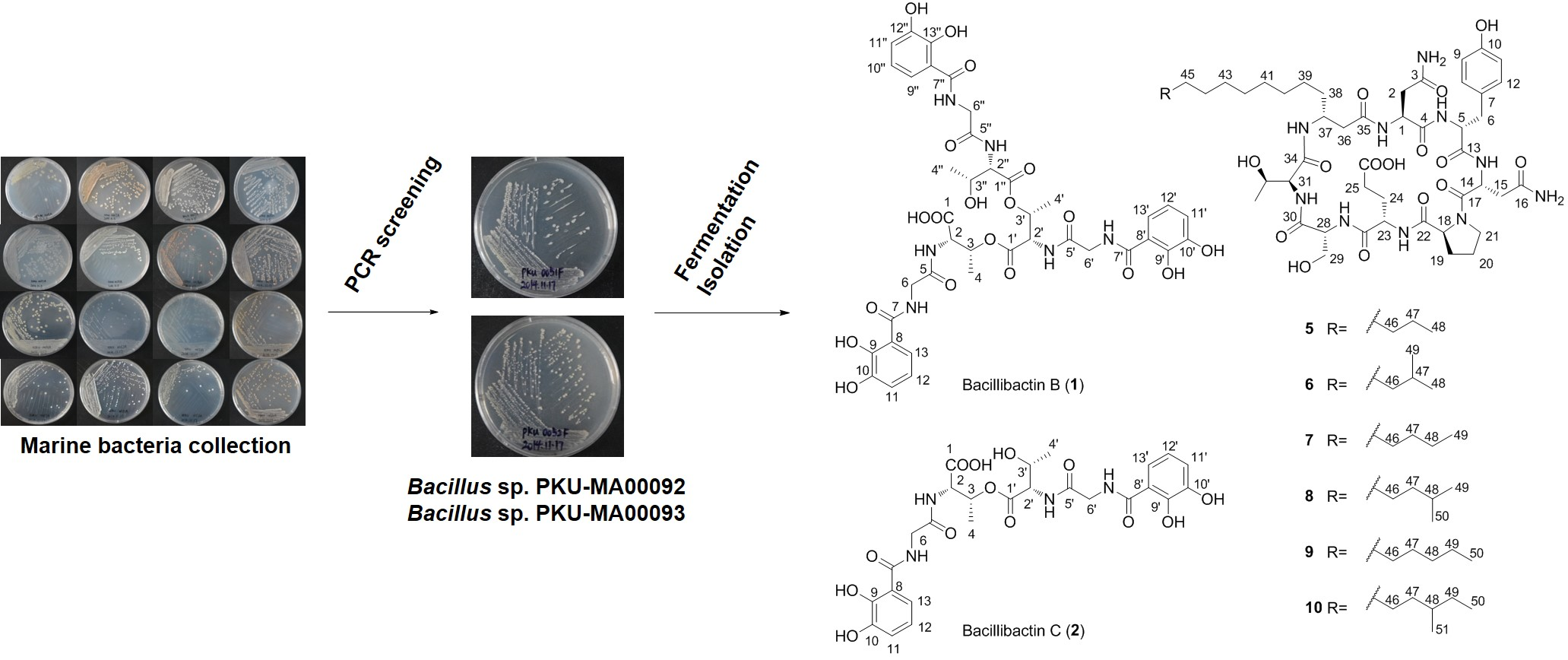
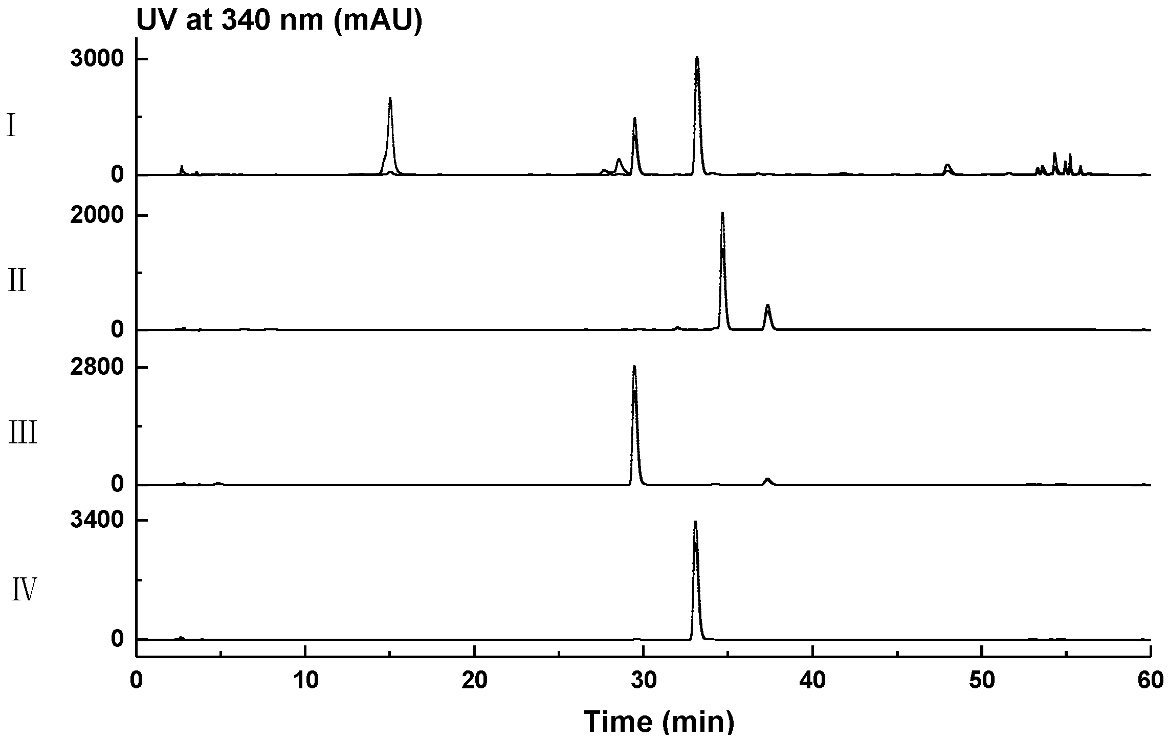
2.1. Genome Mining for Nonribosomal Peptides Discovery from Marine Bacteria Library Based on a PCR Screening Method

We have constructed a marine bacteria library containing strains isolated from sponges, corals and deposits collected from South China Sea and south coasts of China. To discover nonribosomal peptides from this library, we carried out a PCR screening targeting adenylation domains (A domain) in nonribosomal peptides biosynthesis with genomic DNAs of strains in the library as the templates, using the degenerate primers (A3F and A7R) that were designed from the conserved sequences of A domains in cephamycin, vancomycin, balhimycin, actinomycin, pristinamycin, and chloroeremomycin biosynthesis [24]. Totally 180 strains in the library were screened and 21 strains were identified as the “positive hits”, which gave clear PCR products with expected size of ~700 bp based on the agarose gel electrophoresis analysis (Figure S2). All 21 PCR products were sequenced and confirmed to encode A domains with high sequence identities to known NRPS homologues (Table S1). The high sequence identities (82–99%, Table S1) to known NRPS homologues confirmed our PCR screening’s accuracy, but excluded the possibility of new A domain sequences discovered. The same degenerate primers (A3F and A7R) have been used in the screening of NRPS genes from marine Actinobacteria strains [25,26,27]. These screening gave higher “positive” rates of 92% (22 out of 24) [25], 76% (19 out of 25) [26], and 63% (38 out of 60) [27], when compared to the lower “positive” rate of 12% (21 out of 180) in this screening. The degenerate primers (A3F and A7R) are specific for the screening of NRPS genes from Actinobacteria because they are designed from the conserved sequences of A domains in the biosynthesis of six nonribosomal peptides produced by Actinobacteria strains. We have amplified all the 16S rRNAs sequences of the 21 “positive” strains. The sequencing and BLAST results showed that the 21 “positive” strains consisted of 14 Firmicutes (14 from Bacillus) strains and seven Actinobacteria (four from Rhodococcus, one from Streptomyces, one from Nocardiopsis, and one from Mycobacterium) stains (Table S1). These results suggest that the majority of the 180 marine bacteria in this screening are not Actinobacteria, which may be the reason for the lower “positive” rate. Other PCR screening for NRPS genes from marine bacteria using different degenerate primers gave different phyla of “positive” strains with different “positive” rates (30% and 14%) [14,28], exemplifying the degenerate primers as another important factor affecting the screening result.
We then fermented the 21 strains in small-scale (50 mL) using four different media (see Materials and Methods), and analyzed the crude extracts by HPLC. The HPLC analysis showed that strains PKU-MA00092 and PKU-MA00093 provided abundant natural products in medium M2, under the UV detection of 210 nm. Thus, strains PKU-MA00092 and PKU-MA00093 were chosen for the large-scale fermentation in medium M2 and natural products isolation. Four bacillibactin analogues (14), including two new compounds (1 and 2) were isolated from a 4.5 L fermentation of PKU-MA00093, and six bacillomycin D analogues (510) were isolated from a 10.0 L fermentation of PKU-MA00092 (Figure 1). The structures of compounds 110 were elucidated based on comprehensive spectroscopic analyses. Bacillibactin and bacillomycin D have been previously isolated from different Bacillus strains [9,10,29]. To identify the genuses of strains PKU-MA00092 and PKU-MA00093, the housekeeping 16S rRNA genes of the two strains were amplified by PCR and sequenced. BLAST on the NCBI website showed that the two 16S rRNAs were highly homologous to 16S rRNAs from Bacillus strains. The phylogenetic analysis was carried out based on the sequence alignments of the two 16S rRNAs with different Bacillus homologues, identifying PKU-MA00092 and PKU-MA00093 as two Bacillus strains (Figure S3).

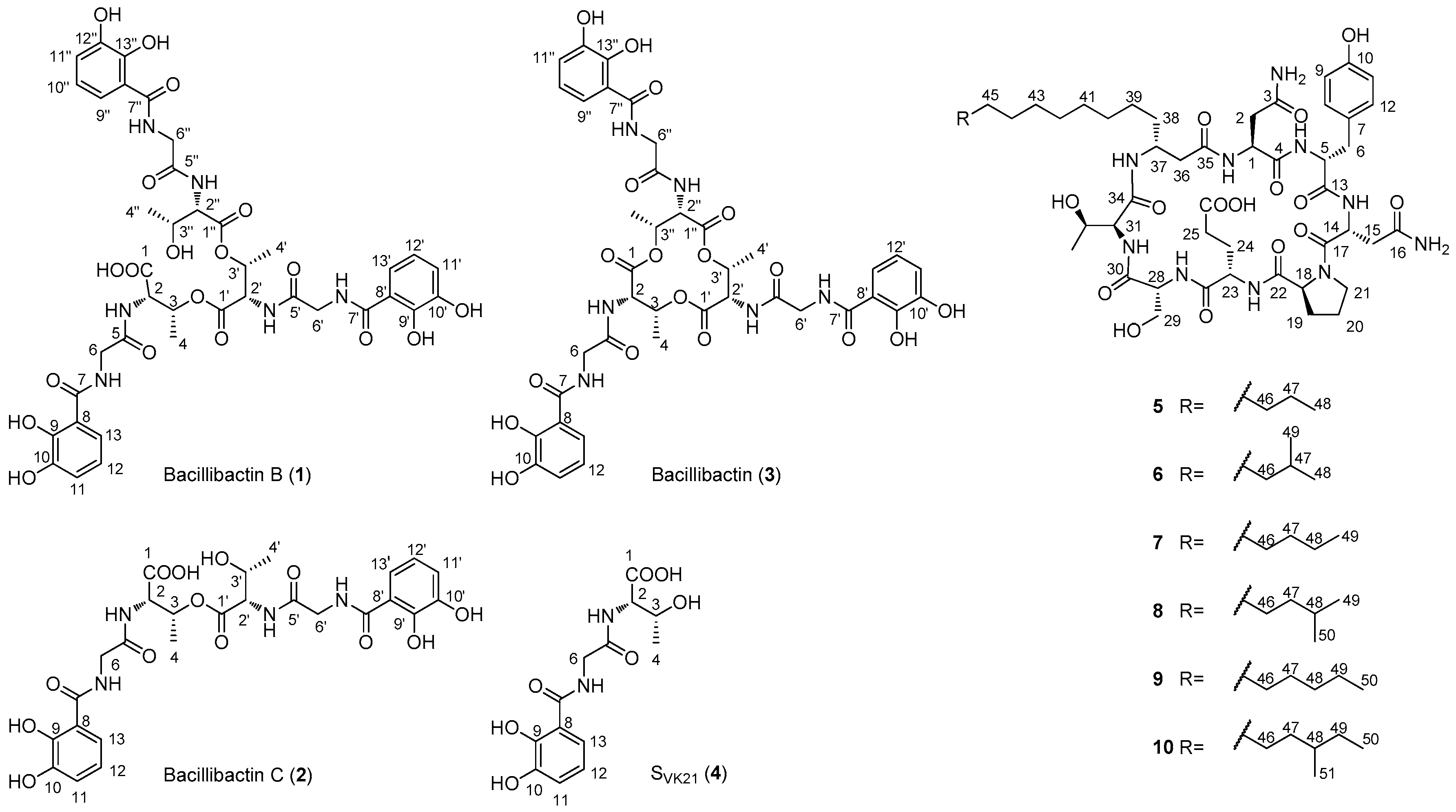
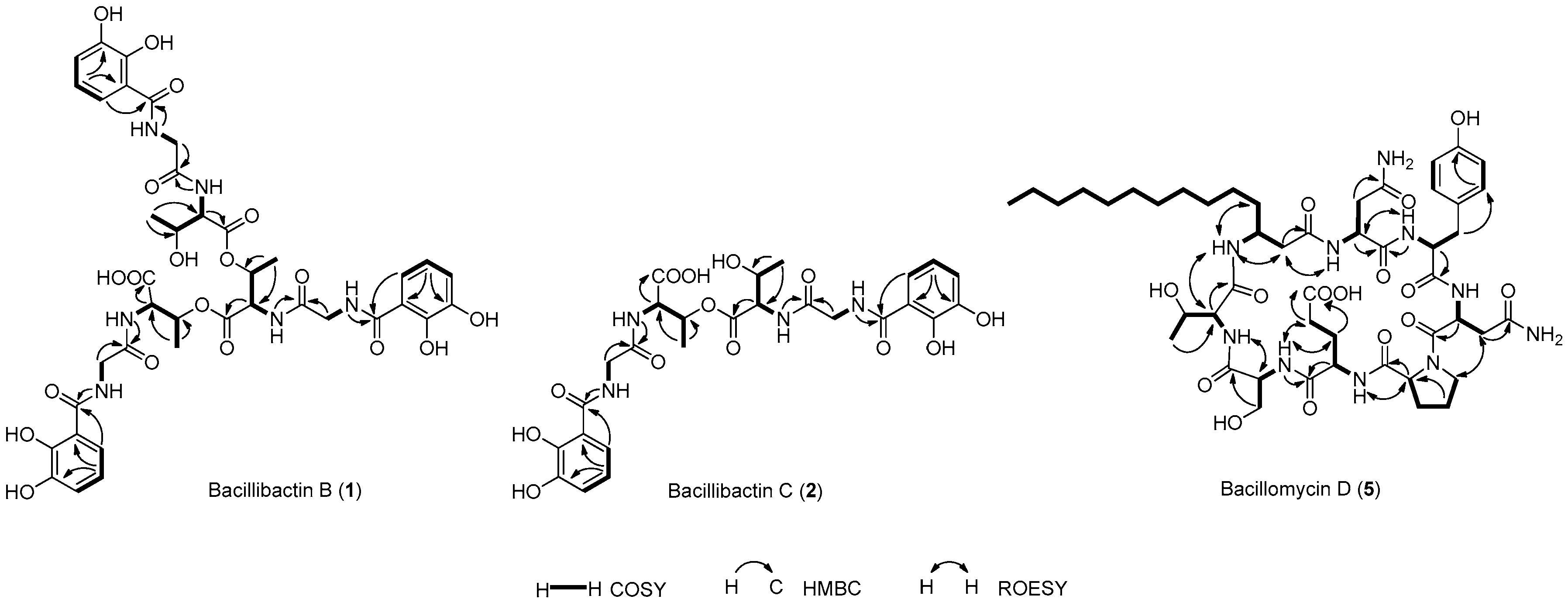
2.2. Structural Elucidation of Compounds 110

Compound 1 was obtained as a light green gum. HRESIMS analysis afforded an [M − H] ion at m/z 899.2584, giving the molecular formula of 1 as C39H44N6O19. The 1H NMR spectrum of 1 resembled that of bacillibactin (3) [30,31], except that the resonances at δH 5.31 (d, J = 6.8 Hz, H3-3/3′/3′′) in 3 split to δH 5.33 (br s, H-3), δH 5.38 (br s, H-3′) and δH 4.19 (br s, H-3′′) in 1, and the resonances at δH 4.59 (br s, H3-2/2′/2′′) in 3 split to δH 4.56 (br d, J = 8.0 Hz, H-2), δH 4.79 (br d, J = 8.0 Hz, H-2′), and δH 4.38 (br d, J = 8.0 Hz, H-2′′) in 1 (Figure S4, Table 1). The 13C NMR spectrum of 1 resembled that of bacillibactin (3) [30,31] except that the resonances at δC 70.8 (C-3/3′/3′′) in 3 split to δC 70.7 (C-3/3′) and δC 66.3 (C-3′′) in 1, the resonances at δC 56.6 (C-2/2′/2′′) in 3 split to δC 55.1 (C-2), δC 54.8 (C-2′) and δC 57.4 (C-2′′) in 1, the resonances at δC 16.5 (C-4/4′/4′′) in 3 split to δC 16.4 (C-4), δC 16.3 (C-4′) and δC 20.2 (C-4′′) in 1, and the resonance attributed to C-1 at δC 168.4 in 3 shifted downfield to δC 170.9 in 1 (Figure S6, Table 1). These differences in combination with the molecular formula of 1 suggested that 1 was the hydrolyzed product of 3, which was confirmed by the MS/MS analysis based on the observation of ions [a], [b], [c], and [d] at m/z 763.2365, 706.2209, 605.1732, and 311.0882, respectively (Figure S11). The linkage relations of 2,3-dihydroxybenzoic acid (DHBA), glycine and threonine in three units were confirmed by COSY, HSQC, and HMBC experiments (Figure 2, Figures S5, S7 and S8). To further establish the absolute configuration of 1, the Marfey’s analysis [32] was carried out. HPLC analysis of the FDAA derivatives of the amino acids in 1 unambiguously established that 1 only contained l-threonine (Figure 3). Thus, compound 1 was identified as the hydrolyzed product of bacillibactin (3), and named as bacillibactin B.
Compound 2 was obtained as a light green gum. HRESIMS analysis afforded an [M + Na]+ ion at m/z 629.1702, giving the molecular formula of 2 as C26H30N4O13. The 1H NMR spectrum of 2 resembled that of SVK21 (4) [33] except that the resonances at δH 4.20 (d, J = 6.0 Hz, H-3) in 4 split to δH 5.33 (m, H-3) and δH 4.21 (m, H-3′) in 2, the resonances at δH 4.10 (br s, H-2) in 4 split to δH 4.61 (dd, J = 8.8, 2.5 Hz, H-2) and δH 4.39 (dd, J = 8.5, 2.5 Hz, H-2′) in 2, the resonances at δH 1.04 (d, J = 6.3 Hz, H-4) in 4 split to δH 1.15 (d, J = 6.4 Hz, H-4) and δH 1.05 (d, J = 6.2 Hz, H-4′) in 2, and the resonances at δH 7.86 (d, J = 8.3 Hz, 2-NH) in 4 split to δH 8.36 (d, J = 8.8 Hz, 2-NH) and δH 8.06 (d, J = 8.5 Hz, 2′-NH) in 2 (Figure S12, Table 2). The 13C NMR spectrum of 2 resembled that of SVK21 (4) [33] except that the resonances at δC 66.2 (C-3) in 4 split to δC 70.8 (C-3) and δC 66.4 (C-3′) in 2, the resonances at δC 57.5 (C-2) in 4 split to δC 55.0 (C-2) and δC 57.4 (C-2′) in 2, and the resonances at δC 20.1 (C-4) in 4 split to δC 16.5 (C-4) and δC 20.1 (C-4′) in 2 (Figure S14, Table 2). These differences in combination with the molecular formula of 2 suggested that 2 was the esterification product of two SVK21 (4) units with the linkage between the two threonine residues. This was confirmed by the MS/MS analysis based on the observation of ions [a + H + Na]+, [b + H + Na]+, and [c + H + Na]+ at m/z 493.1541, 436.1345, and 335.0855, respectively (Figure S19). The linkage relations of 2,3-dihydroxybenzoic acid (DHBA), glycine, and threonine in the two units were confirmed by COSY, HSQC, and HMBC experiments (Figure 2, Figures S13, S15 and S16). The two l-threonine residues in 2 were established based on their similar chemical shifts, coupling constants and same biosynthetic pathway with that of 4. Thus, compound 2 was identified as the esterification product of two SVK21 (4) units and named as bacillibactin C.
Compounds 3 and 4 was identified as bacillibactin and SVK21, respectively, by comparison of their spectroscopic data with that of the authentic compounds [30,31,33]. The biological role of bacillibactin as the catecholic siderophore in iron acquisition has been characterized in details [10,34]. The biosynthetic research of bacillibactin has revealed how nature utilized NRPS machinery to synthesized this molecule. The standalone adenylation protein in the starter module, DhbE, activates 2,3-dihydroxybenzic acid (DHBA), and loaded this starter unit onto the first peptidyl carrier protein (PCP), initiating the downstream peptide elongation; the crystal structure of DhbE revealed the ten amino acids in the active sites determining the substrate selectivity [35]; the three-molular NRPS catalyzes the condensation between DHBA, glycine, and l-threonine to generate PCP-tethered linear tripeptides intermediate (thioester of compound 4), then the thioesterase domain (TE) in the last module catalyzes the polymerization of SVK21 units to generate the TE-tethered ester of compound 2, followed by the formation of TE-tethered ester of compound 1 and the intramolecular cyclization to generate bacillibactin (3) [35,36]. In this study, we isolated compounds 1, 2, and 4 whose structures are corresponding to the three biosynthetic intermediates in bacillibactin biosynthesis. The HPLC analysis of the crude extract of PKU-MA00093 using an increasing gradient of MeOH in H2O (containing no acids or bases) as the moble phase showed the co-occurrence of the peaks for compounds 14 (data not shown). Therefore, compounds 1, 2, and 4 may be isolated as the shunt products released from the NRPS machinery in the biosynthesis of bacillibactin (3).
Compound 5 was obtained as a white amorphous powder. HRESIMS analysis afforded an [M − H] ion at m/z 1029.5260, giving the molecular formula of 5 as C48H74N10O15. The 1H NMR and 13C NMR spectra (Figures S27 and S29) resembled that of bacillomycin D [29,37]. The COSY, HSQC, and HMBC experiments (Figure 2, Figures S28, S30 and S31) confirmed the presence of two asparagines, one tyrosine, one proline, one glutamate, one serine, one threonine, and one C14-fatty acid moieties in 5. The correlations of H-36 with 1-NH, H-1 with 5-NH, H-15 with H-21, H-18 with 23-NH, H-24 with 28-NH, H-25 with 28-NH, H-28 with 31-NH, H-31 with 37-NH, 37-NH with H-36, 37-NH with H-38 in the ROESY spectrum (Figure 2 and Figure S32) established the cyclo(Asn-Tyr-Asn-Pro-Glu-Ser-Thr-C14-fatty acid) structure of 5. To confirm that the amino acids in 5 have the same absolute configurations as that in bacillomycin D, the Marfey’s analysis was carried out. HPLC analysis of the FDAA derivatives of the amino acids established that 5 contained same l-asparagine, d-asparagine, d-tyrosine, l-proline, l-glutamate, d-serine, and l-threonine residues as bacillomycin D (Figure S49). Thus, compound 5 was identified as bacillomycin D.
The analyses of the 1H NMR and 13C NMR spectra of compounds 610 showed that they contained the same cyclopeptide skeleton as compound 5. However, their different molecular weights (see Section 3.6 part in Materials and Methods) determined by ESIMS analyses showed that they contained different fatty acid moieties. Compound 6 contained one C15-fatty acid moiety with a branch terminal, which was established by the HRESIMS analysis (Figure S36) and the resonances attributed to two methyl groups at δH 0.84 (d, J = 6.3 Hz, H3-48, and H3-49) in the 1H NMR spectrum (Figure S34, Table S4); Compound 7 contained one C15-fatty acid moiety with a linear terminal, which was established by the HRESIMS analysis (Figure S39) and the resonance attributed to the methyl group at δH 0.88 (t, J = 7.4 Hz, H3-49) in the 1H NMR spectrum (Figure S37, Table S5); Compound 8 contained one C16-fatty acid moiety with a branch terminal, which was established by the ESIMS analysis (Figure S42) and the resonances attributed to two methyl groups at δH 0.84 (d, J = 6.5 Hz, H3-49 and H3-50) in the 1H NMR spectrum (Figure S40, Table S6); Compound 9 contained one C16-fatty acid moiety with a linear terminal, which was established by the ESIMS analysis (Figure S45) and the resonance attributed to the methyl group at δH 0.87 (t, J = 6.4 Hz, H3-50) in the 1H NMR spectrum (Figure S43, Table S7); Compound 10 contained one C17-fatty acid moiety with a branch, which was established by the ESIMS analysis (Figure S48) and the resonances attributed to two methyl groups at δH 0.84 (t, J = 7.3 Hz, H3-50) and δH 0.83 (d, J = 6.3 Hz, H3-51) in the 1H NMR spectrum (Figure S46, Table S8), and the correlations of H3-51 with H-48, H-48 with H2-49, H2-49 with H3-50 in the COSY spectrum (Figure S47). Thus, compounds 6, 8, and 10 were identified as iso-C15 bacillomycin D [38], iso-C16 bacillomycin D [38], and anteiso-C17 bacillomycin D [38], respectively. Compounds 7 and 9 were identified as C15 bacillomycin D and C16 bacillomycin D, respectively. Compounds 7 and 9 were reported previously without NMR data provided [39,40], and we here report the structures of 7 and 9 with fully assigned 1H NMR and 13C NMR data for the first time.
The separation of compounds 510 is challenging because their structures are highly similar with only the fatty acid moieties different. Due to the difficulties in the separation, compounds 5 and 6 were purified as a mixture in the cytotoxicity assay against Ehrlich carcinoma tumor cell line and hemolytic activity assay [37]. The structural elucidation of compounds 510 is also challenging, because the large molecular weights and the complex structures of 510 require large amount of samples for 1D and 2D NMR analyses. Due to the little amount obtained, two bacillomycin D analogues with C14 and C15 fatty acid moieties [41] and five bacillomycin D analogues with C14 to C18 fatty acid moieties [42] were proposed only based on LC-MS analyses. The biosynthetic gene cluster of bacillomycin D has been cloned and sequenced [12,43]. The acyl-CoA ligase domain in the first module is responsible for the ligation of the fatty acid moiety to the polyketide synthase (PKS)-NRPS hybrid assembly line. The isolation of compounds 510 in this study suggested that the acyl-CoA ligase domain possessed substrate promiscuity, which allowed for the ligation of different length and branches of fatty acid moieties to the polyketide-nonribosomal peptide hybrid macrocycle. The substrate promiscuity of the acyl-CoA ligase domain can be employed in combinatorial biosynthesis for the generation of more bacillomycin D analogues.

2.3. Cytotoxicity Assays against Human Cancer Cell Lines

Compounds 110 were tested for their antitumor activities. Two human cancer cell lines, liver cancer cell line HepG2, and breast cancer cell line MCF7, were used in the assays. Compounds 710 showed moderate cytotoxicities with IC50 values from 2.9 ± 0.1 to 8.2 ± 0.2 µM and compounds 16 show no cytotoxicities (inhibition rates were below 20% at the concentration of 10 µM) (Table 3). The isolation of 510 allowed us to analyze the effections of different fatty acid moieties on the cytotoxicities. Compound 5 with a C14-fatty acid moiety showed no cytotoxicities (inhibition rates were below 20% at the concentration of 10 µM); both compounds 6 and 7 contain a C15-fatty acid moiety, however, compound 7 with a linear C15-fatty acid moiety showed moderate cytotoxicities and 6 with a branched C15-fatty acid moiety lost the cytotoxicities, suggesting that a linear C15-fatty acid moiety is important to the cytotoxicities; compounds 8 and 9 containing a C16-fatty acid moiety showed most potent cytotoxicities; compound 10 containing one more methyl group attached at C-49 when compared to 8 or attached at C-48 when compared to 9, showed decreased cytotoxicity against MCF7 and lost the cytotoxicity against HepG2, suggesting a C17-fatty acid moiety with terminal branch is disadvantageous to the cytotoxicities. In conclusion, bacillomycin D analogues with a C16-fatty acid moiety may possess best cytotoxicities against HepG2 and MCF7 cancer cell lines. The bacillomycin D analogues have been mostly assayed for their antifungal activities [44], especially against fungi of plant pathogens [38,40,45]. For their antitumor activities, a mixture of compounds 5 and 6’s moderate cytotoxicities have been reported against mice Ehrlich’s carcinoma cells [37]. In another report, one lipopeptide was proposed as bacillomycin D (5) only based on LC-MS analysis and showed inhibitory activities against three human cancer cell lines A549, A498 and HCT-15 [46]. The cytotoxicities against human cancer cell lines HepG2 and MCF7 and the structure-activity relationships that we first revealed here enriched our understanding of the diversity of bacillomycin D analogues’ bioactivities.

3. Materials and Methods

3.1. General Experimental Procedures

Optical rotations were measured on an Autopol III automatic polarimeter (Rudolph Research Analytical, Hackettstown, NJ, USA). UV spectra were collected on a Cary 300 spectrometer (VARIAN, Palo Alto, CA, USA). IR spectra were collected on a Nicolet Nexus 470 FT-IR spectrometer (Thermo Scientific, Waltham, MA, USA). 1H and 13C NMR spectra were collected on a Bruker Avance-400FT NMR spectrometer (Bruker Corporation, Billerica, MA, USA). HRESIMS and MS/MS spectra were collected on a Waters Xevo G2 Q-TOF spectrometer (Waters, Milford, MA, USA). ESIMS spectra were collected on a Waters SQD Single Quadruple Mass spectrometer (Waters, Milford, MA, USA). HPLC analysis was performed on an Agilent 1260 series (Agilent Technologies, Santa Clara, CA, USA) with a diode array detector (Agilent Technologies, Santa Clara, CA, USA) and a C18 RP-column (Eclipse XDBC18, 150 × 4.6 mm, 5 μm, Agilent Technologies, Santa Clara, CA, USA). Semi-preparative HPLC was performed on a SSI 23201 system (Scientific Systems Inc., State College, PA, USA) with a YMC-Pack ODS-1 column (250 × 10 mm, 5 μm, YMC Co., Ltd., Shimogyo-ku, Kyoto, Japan). MPLC was performed on a LC3000 series (Beijing Tong Heng Innovation Technology, Beijing, China) with a ClaricepTM Flash i-series C18 cartridge (20–35 μm, 40 g, Bonna-Agela, Wilmington, DE, USA). Size exclusion chromatography was carried out using a Sephadex LH-20 (Pharmacia Fine Chemicals, Piscataway, NJ, USA) column.

3.2. Marine Bacterial Strains Isolation

Total 15 Sponge samples were collected from Yongxin Island in the South China Sea and were deposited at the State Key Laboratory of Natural and Biomimetic Drugs, Peking University, China. The isolation of the marine bacterial strains was carried out according to the published procedures [47]. The sponge samples were thoroughly washed with sterilized water containing 3% sea salt for three times, and cut into pieces of about 1 cm3, then homogenized in a sterilized mortar with 9 mL sterilized water containing 3% sea salt. The homogenates were incubated in a water bath at 55 °C for 6 min, and put into biosafety cabinet for 30 min, and the supernatants were serially diluted by sterilized water containing 3% sea salt and plated in triplicate on agar plates for each sponge sample. Four media, M1 (yeast extract 1 g, peptone 5 g, beef extract 1 g, FePO4 0.01 g, agar 18 g, and sea salt 33 g in 1.0 L distilled water, pH 7.4), M2 (glycerol 6 mL, arginine 1 g, K2HPO4 1 g, MgSO4 0.5 g, agar 18 g, sea salt 33 g in 1.0 L distilled water, pH 7.2), M3 (soluble starch 20 g, KNO3 1 g, K2HPO4 0.5 g, MgSO4·7H2O 0.5 g, NaCl 0.5 g, FeSO4·7H2O 0.01 g, agar 18 g, sea salt 33 g in 1.0 L distilled water, pH 7.2), and M4 (sodium acetate trihydrate 5 g, peptone 0.5 g, yeast extract 0.5 g, glucose 0.5 g, sucrose 0.5 g, sodium citrate 0.05 g, malic acid 0.05 g, NH4NO3 1 g, NH4Cl 0.2 g, KH2PO4 0.5 g, agar 18 g, sea salt 33 g in 1.0 L distilled water, pH 7.6), were used for bacterial strains isolation. Each plate was supplemented with cycloheximide (50 μg/mL) and nalidixic acid (50 μg/mL) to suppress the growth of fungi and Gram-negative bacteria, and incubated at 28 °C for four weeks. Different colonies of strains were carefully selected and restreaked on agar plates with medium M1 for further proliferation and dereplication. Totally 180 bacterial strains were isolated and stored at −80 °C in 20% glycerol.

3.3. PCR Screening for Potential Producers of Nonribosomal Peptides

The genomic DNAs of all 180 strains were extracted following standard protocols [48]. The forward primer (5′-GCSTACSYSATSTACACSTCSGG-3′) and the reverse primer (5′-SASGTCVCCSGTSCGGTAS-3′), designed from the conserved sequences of adenylation domains (A domains) in the biosynthesis of cephamycin, vancomycin, balhimycin, actinomycin, pristinamycin, and chloroeremomycin, were used in the screening [24]. A 20 μL PCR system consisting of 10 µL Easy Taq Polymerase (Beijing TransGen Biotech, Beijing, China), 2 µL of forward and reverse primer mixture (each for 10 µM), 1 µL genomic DNA, and 7 µL sterilized water, were used. The PCR program was performed with an initial denaturation at 95 °C for 5 min, followed by 30 cycles of denaturation at 95 °C for 30 s, annealing at 57 °C for 1 min and extension at 72 °C for 1 min, followed by incubation at 72 °C for 10 min. The PCR products were analyzed by agarose gel electrophoresis (Figure S2), recovered with a gel purification kit (Beijing TransGen Biotech, Beijing, China) and sequenced to afford 21 “positive” strains.

3.4. Small-Scale Fermentation, HPLC

For each of the 21 “positive” strains, 50 µL of spore suspension was inoculated into 50 mL of seed medium (medium M1 broth), and incubated with a HYG-C shaker (Suzhou Peiying Laboratory Equipment, Suzhou, China) at 28 °C, 200 rpm for three days. Three milliliter of the resultant seed culture was inoculated into 50 mL of production media, and the fermentation continued at 28 °C, 200 rpm for seven days. Four different production media, media M1, M2, and M3 broth, and M5 (soluble starch 10 g, casein 0.3 g, K2HPO4 2 g, KNO3 2 g, MgSO4·7H2O 0.05 g, NaCl 2 g, FeSO4·7H2O 0.01 g, CaCO3 0.02 g, sea salt 33 g in 1.0 L distilled water, pH 7.2), were used in the small-scale fermentation. The Diaion HP20 (2 g/100 mL, Mitsubishi Chemical Corporation, Tokyo, Japan) and Amberlite XAD-16 (2 g/100 mL, Sigma-Aldrich, St. Louis, MO, USA) resins were added 10 h before the fermentation finished. The resins and cell mass were harvested by centrifugation and extracted with MeOH. The MeOH extracts were concentrated and analyzed by HPLC. The HPLC analysis was carried out with a flow rate of 1 mL/min with UV detection at 210 nm, using a gradient elution program from 5% MeOH in H2O to 100% MeOH over 45 min.

3.5. Phylogenetic Analysis

The phylogenetic analysis of strains PKU-MA00092 and PKU-MA00093 was carried out by sequence alignments of their 16S rRNAs with different Bacillus homologues. The primers 27F (5′-AGAGTTTGATCMTGGCTCAG-3′) and 1492R (5′-TACGGYTACCTTGTTACGACTT-3′) [49] were used to amplify the 16S rDNA genes (GeneBank accession number MF774821 for PKU-MA00092 and KY780192 for PKU-MA00093). Homologous genes were searched using BLAST on the NCBI website and the phylogenetic tree was generated with Mega 5.1 using the Neighbor-Joining algorithm.

3.6. Large-Scale Fermentation and Isolation

The large-scale fermentation of Bacillus sp. PKU-MA00093 and Bacillus sp. PKU-MA00092 in medium M2 broth were carried out with similar procedures used in the small-scale fermentation. For Bacillus sp. PKU-MA00092 or PKU-MA00093, 50 µL of spore suspension was inoculated into 50 mL of seed medium (medium M1 broth, pH 7.4) and incubated with HYG-C shakers at 28 °C, 200 rpm for three days. Twelve milliliter of the resultant seed culture was inoculated into 200 mL of medium M2 broth (pH 7.2) in 1 L Erlenmeyer flasks, and the fermentation continued at 28 °C, 200 rpm for seven days. The Diaion HP20 (2 g/100 mL), and Amberlite XAD-16 (2 g/100 mL) resins were added 10 h before the fermentation finished. The resins and cell mass were harvested by centrifugation and extracted with MeOH.
A 4.5 L fermentation of Bacillus sp. PKU-MA00093 gave a 4.3 g of MeOH extract. The MeOH extract was concentrated in a vacuum and suspended in deionized water, extracted with ethyl acetate for three times. The ethyl acetate extracts were combined (1.4 g) and loaded onto MPLC using step-gradient elution of MeOH in H2O (10%, 20%, 35%, 50%, 70%) to yield five fractions (F1–F5). Fraction F4 (147 mg) was purified by semi-preparative HPLC with MeOH/H2O (44/56, v/v) as the mobile phase to yield 1 (4.6 mg); fraction F2 (169 mg) was purified by semi-preparative HPLC with MeOH/H2O (37/63, v/v) as the mobile phase to yield compound 2 (7.8 mg) and 4 (1.4 mg); fraction F3 (231 mg) was purified by semi-preparative HPLC with MeOH/H2O (40/60, v/v) as the mobile phase to yield 3 (17.4 mg). All of the semi-preparative HPLCs were carried out at a flowrate of 2 mL/min and under the detection of 248 nm.
A 10.0 L fermentation of Bacillus sp. PKU-MA00092 gave a 1.5 g of MeOH extract. The MeOH extract was concentrated and loaded onto MPLC using step-gradient elution of MeOH in H2O (20%, 40%, 60%, 80%) to yield four fractions (F1–F4). Fraction F4 (368 mg) was purified repeatedly by semi-preparative HPLC with CH3CN/H2O (45/55 v/v) containing 0.01% TFA as the mobile phase, to afford compounds 5 (36.5 mg), 6 (15.7 mg), 7 (11.4 mg), 8 (13.3 mg), 9 (14.5 mg) and 10 (3.1 mg), at a flowrate of 2 mL/min and under the detection of 210 nm.
Bacillibactin B (1): light green gum; [ α ] D 25 +37 (c 0.23, MeOH); UV (MeOH) λmax (log ε): 207 (1.47), 248 (0.40), 312 (0.16) nm; IR (KBr) νmax 3317, 1740, 1642, 1541, 1266, 1023, 745 cm−1 (Figure S10); 1H NMR and 13C NMR data, Table 1; HRESIMS m/z 899.2584 [M − H] (calcd. for C39H43N6O19, 899.2580).
Bacillibactin C (2): light green gum; [ α ] D 25 +40 (c 0.27, MeOH); UV (MeOH) λmax (log ε): 206 (2.22), 249 (1.12), 312 (0.54) nm; IR (KBr) νmax 3320, 1738, 1644, 1540, 1264, 1022, 746 cm−1 (Figure S18); 1H NMR and 13C NMR data, Table 2; HRESIMS m/z 605.1740 [M − H] (calcd. for C26H29N4O13, 605.1731); m/z 629.1702 [M + Na]+ (calcd. for C26H30N4O13Na, 629.1705).
Bacillibactin (3): light green gum; 1H NMR and 13C NMR data, Supplementary Table S2; HRESIMS m/z 883.2621 [M + H]+ (calcd. for C39H43N6O18, 883.2631).
SVK21 (4): light green gum; 1H NMR and 13C NMR data, Supplementary Table S2; HRESIMS m/z 311.0878 [M − H] (calcd. for C13H15N2O7, 311.0878).
Bacillomycin D (5): white amorphous powder; 1H NMR and 13C NMR data, Supplementary Table S3; HRESIMS m/z 1029.5260 [M − H] (calcd. for C48H73N10O15, 1029.5254).
iso-C15 Bacillomycin (6): white amorphous powder; 1H NMR and 13C NMR data, Supplementary Table S4; HRESIMS m/z 1043.5421 [M − H] (calcd. for C49H75N10O15, 1043.5411).
C15 Bacillomycin (7): white amorphous powder; 1H NMR and 13C NMR data, Supplementary Table S5; HRESIMS m/z 1043.5411 [M − H] (calcd. for C49H75N10O15, 1043.5411).
iso-C16 Bacillomycin (8): white amorphous powder; 1H NMR and 13C NMR data, Supplementary Table S6; ESIMS m/z 1057.34 [M − H].
C16 Bacillomycin (9): white amorphous powder; 1H NMR and 13C NMR data, Supplementary Table S7; ESIMS m/z 1057.43 [M − H].
anteiso-C17 Bacillomycin (10): white amorphous powder; 1H NMR data, Supplementary Table S8; ESIMS m/z 1071.45 [M − H].

3.7. Marfey’s Analysis for Compounds 2 and 5

The Marfey’s analysis of compounds 2 and 5 was carried out with same procedure [32,50]. The peptide (1.0 mg) was hydrolyzed with 500 μL of 6 N HCl in a sealed glass vial (1.5 mL) at 110 °C for 18 h, and dried with N2 gas. The hydrolysate was dissolved in 50 μL of H2O, followed by the addition of 100 μL FDAA (1-fluoro-2,4-dinitrophenyl-5-l-alanine amide, 1% in acetone, 3.7 µmol) and 20 μL of 1 M NaHCO3. The mixture was heated at 45 °C for 1 h, followed by the addition of 10 μL of 2 M HCl to terminate the reaction. The FDAA-derivatives were dried with N2 gas and dissolved in 1 mL of 50% CH3CN in H2O for HPLC analysis. The HPLC was carried out with a C18 column (50 × 4.6 mm, 5 µm, Grace Corporate, Columbia, MD, USA) and with a program of linear gradient of 10% CH3CN in H2O containing 0.1% TFA to 40% CH3CN in H2O containing 0.1% TFA over 40 min, at a flow rate of 1.0 mL/min, and under the detection of 340 nm. The FDAA-derivatives of standard l or d-amino acids were used as comparison.

3.8. Cytotoxicity Assays for Compounds 110

HepG2 and MCF7 cell lines were purchased from Peking Union Medical College, Cell Bank (Beijing, China). In this study, the cells were grown in Dulbecco’s modified Eagle’s medium (Hyclone, Waltham, MA, USA), supplemented with 10% fetal bovine serum (FBS, PAN Biotech, Aidenbach, Germany), penicillin (100 U/mL), and streptomycin (100 µg/mL) in a humidified incubator containing 95% air and 5% CO2 at 37 °C. Cell viability was detected by MTT colorimetric assay. Briefly, the cells were treated with different concentrations of compounds (in DMSO stock) for 48 h, with taxol as the positive control. Then, culture supernatants were replaced with medium containing 0.5 mg/mL MTT for 4 h. The supernatant was removed and 100 μL of dimethyl sulfoxide was added. Absorbance was detected at 550 nm and cell viability was expressed as the mean percentage of the absorbance in treated vs. control cells. Different concentrations of compounds ranging from 0.1 to 40 µM were tested and each of the concentration was tested in parallel triplicate. Six concentration points in the slope region were used in the calculation of IC50 values. The IC50 values were calculated from the concentration–response curves that were generated by nonlinear regression with Graph Pad Prism 5 software (Graph Pad Software, Inc., San Diego, CA, USA).

Supplementary Materials

The following are available online at www.mdpi.com/1660-3397/16/1/22/s1, Figure S1: nonribosomal peptides from marine-derived Bacillus species, Figure S2: the agarose gel electrophoresis analysis of the 21 “positive” PCR products, Figure S3: the phylogenetic analysis of strains PKU-MA00092 and PKU-MA00093, Figures S4–S11: the NMR, MS and IR spectra of compound 1, Figures S12–S19: the NMR, MS and IR spectra of compound 2, Figures S20–S23: the NMR and MS spectra of compound 3, Figures S24–S26: the NMR and MS spectra of compound 4, Figures S27–S33: the NMR and MS spectra of compound 5, Figures S34–S36: the NMR and MS spectra of compound 6, Figures S37–S39: the NMR and MS spectra of compound 7, Figures S40–S42: the NMR and MS spectra of compound 8, Figures S43–S45: the NMR and MS spectra of compound 9, Figures S46–S48: the NMR and MS spectra of compound 10, Figure S49: the Marfey’s analysis of compound 5. Table S1: the homologues of the 21 “positive” strains and their PCR products, Tables S2–S8: the 1H NMR and 13C NMR data of compounds 310.

Acknowledgments

This research was financially supported by the National Natural Science Foundation of China (grant number 81573326 and grant number 81673332).

Author Contributions

M.Z. and F.L. contributed equally; M.M. and D.Y. conceived and designed the experiments; M.Z., F.L., X.Y., J.J. and X.D. performed the experiments; K.-W.Z., D.L., Y.Z., M.M. and D.Y. analyzed the data; M.M. wrote the manuscript.

Conflicts of Interest

The authors declare no conflict of interest.

References

  1. Johnson, B.A.; Anker, H.; Meleney, F.L. Bacitracin: A new antibiotic produced by a member of the B. subtilis group. Science 1945, 102, 376–377. [Google Scholar] [CrossRef] [PubMed]
  2. Danders, W.; Marahiel, M.A.; Krause, M.; Kosui, N.; Kato, T.; Izumiya, N.; Kleinkauf, H. Antibacterial action of gramicidin S and tyrocidines in relation to active transport, in vitro transcription, and spore outgrowth. Antimicrob. Agents Chemother. 1982, 22, 785–790. [Google Scholar] [CrossRef] [PubMed]
  3. Liou, J.W.; Hung, Y.J.; Yang, C.H.; Chen, Y.C. The antimicrobial activity of gramicidin A is associated with hydroxyl radical formation. PLoS ONE 2015, 10, e0117065. [Google Scholar] [CrossRef] [PubMed]
  4. Felnagle, E.A.; Jackson, E.E.; Chan, Y.A.; Podevels, A.M.; Berti, A.D.; McMahon, M.D.; Thomas, M.G. Nonribosomal peptide synthetases involved in the production of medically relevant natural products. Mol. Pharm. 2008, 5, 191–211. [Google Scholar] [CrossRef] [PubMed]
  5. Mootz, H.D.; Marahiel, M.A. The tyrocidine biosynthesis operon of Bacillus brevis: Complete nucleotide sequence and biochemical characterization of functional internal adenylation domains. J. Bacteriol. 1997, 179, 6843–6850. [Google Scholar] [CrossRef] [PubMed]
  6. Linne, U.; Stein, D.B.; Mootz, H.D.; Marahiel, M.A. Systematic and quantitative analysis of protein-protein recognition between nonribosomal peptide synthetases investigated in the tyrocidine biosynthetic template. Biochemistry 2003, 42, 5114–5124. [Google Scholar] [CrossRef] [PubMed]
  7. Krätzschmar, J.; Krause, M.; Marahiel, M.A. Gramicidin S biosynthesis operon containing the structural genes grsA and grsB has an open reading frame encoding a protein homologous to fatty acid thioesterases. J. Bacteriol. 1989, 171, 5422–5429. [Google Scholar] [CrossRef] [PubMed]
  8. Conti, E.; Stachelhaus, T.; Marahiel, M.A.; Brick, P. Structural basis for the activation of phenylalanine in the non-ribosomal biosynthesis of gramicidin S. EMBO J. 1997, 16, 4174–4183. [Google Scholar] [CrossRef] [PubMed]
  9. May, J.J.; Wendrich, T.M.; Marahiel, M.A. The dhb operon of Bacillus subtilis encodes the biosynthetic template for the catecholic siderophore 2,3-dihydroxybenzoate-glycine-threonine trimeric ester bacillibactin. J. Biol. Chem. 2001, 276, 7209–7217. [Google Scholar] [CrossRef] [PubMed]
  10. Segond, D.; Abi Khalil, E.; Buisson, C.; Daou, N.; Kallassy, M.; Lereclus, D.; Arosio, P.; Bou-Abdallah, F.; Nielsen Le Roux, C. Iron acquisition in Bacillus cereus: The roles of IlsA and bacillibactin in exogenous ferritin iron mobilization. PLoS Pathog. 2014, 10, e1003935. [Google Scholar] [CrossRef] [PubMed] [Green Version]
  11. Inès, M.; Dhouha, G. Lipopeptide surfactants: Production, recovery and pore forming capacity. Peptides 2015, 71, 100–112. [Google Scholar] [CrossRef] [PubMed]
  12. Roongsawang, N.; Washio, K.; Morikawa, M. Diversity of nonribosomal peptide synthetases ivolved in the biosynthesis of lipopeptide biosurfactants. Int. J. Mol. Sci. 2011, 12, 141–172. [Google Scholar] [CrossRef] [PubMed]
  13. Mondol, M.A.M.; Shin, H.J.; Islam, M.T. Diversity of secondary metabolites from marine Bacillus species: Chemistry and biological activity. Mar. Drugs 2013, 11, 2846–2872. [Google Scholar] [CrossRef] [PubMed]
  14. Zhang, W.; Li, Z.; Miao, X.; Zhang, F. The screening of antimicrobial bacteria with diverse novel nonribosomal peptide synthetase (NRPS) genes from South China Sea sponges. Mar. Biotechnol. 2009, 11, 346–355. [Google Scholar] [CrossRef] [PubMed]
  15. Toledo, G.; Green, W.; Gonzalez, R.A.; Christoffersen, L.; Podar, M.; Chang, H.W.; Hemscheidt, T.; Trapido-Rosenthal, H.G.; Short, J.M.; Bidigare, R.R.; et al. High throughput cultivation for isolation of novel marine microorganisms. Oceanography 2006, 19, 120–125. [Google Scholar] [CrossRef]
  16. Margassery, L.M.; Kennedy, J.; O′Gara, F.; Dobson, A.D.; Morrissey, J.P. Diversity and antibacterial activity of bacteria isolated from the coastal marine sponges Amphilectus fucorum and Eurypon major. Lett. Appl. Microbiol. 2012, 55, 2–8. [Google Scholar] [CrossRef] [PubMed]
  17. Tareq, F.S.; Lee, M.A.; Lee, H.S.; Lee, Y.J.; Lee, J.S.; Hasan, C.M.; Islam, M.T.; Shin, H.J. Non-cytotoxic antifungal agents: Isolation and structures of gageopeptides A-D from a Bacillus strain 109GGC020. J. Agric. Food Chem. 2014, 62, 5565–5572. [Google Scholar] [CrossRef] [PubMed]
  18. Barsby, T.; Kelly, M.T.; Gagné, S.M.; Andersen, R.J. Bogorol A produced in culture by a marine Bacillus sp. reveals a novel template for cationic peptide antibiotics. Org. Lett. 2001, 3, 437–440. [Google Scholar] [CrossRef] [PubMed]
  19. Pettit, G.R.; Knight, J.C.; Herald, D.L.; Pettit, R.K.; Hogan, F.; Mukku, V.J.R.V.; Hamblin, J.S.; Dodson, M.J., II; Chapuis, J.C. Isolation and structure elucidation of bacillistatins 1 and 2 from a marine Bacillus silvestris. J. Nat. Prod. 2009, 72, 366–371. [Google Scholar] [CrossRef] [PubMed]
  20. Lautru, S.; Deeth, R.J.; Bailey, L.M.; Challis, G.L. Discovery of a new peptide natural product by Streptomyces coelicolor genome mining. Nat. Chem. Biol. 2005, 1, 265–269. [Google Scholar] [CrossRef] [PubMed]
  21. Gross, H.; Stockwell, V.O.; Henkels, M.D.; Nowak-Thompson, B.; Loper, J.E.; Gerwick, W.H. The genomisotopic approach: A systematic method to isolate products of orphan biosynthetic gene clusters. Chem. Biol. 2007, 14, 53–63. [Google Scholar] [CrossRef] [PubMed]
  22. Wyatt, M.A.; Wang, W.; Roux, C.M.; Beasley, F.C.; Heinrichs, D.E.; Dunman, P.M.; Magarvey, N.A. Staphylococcus aureus nonribosomal peptide secondary metabolites regulate virulence. Science 2010, 329, 294–296. [Google Scholar] [CrossRef] [PubMed]
  23. Xie, P.; Ma, M.; Rateb, M.E.; Shaaban, K.A.; Yu, Z.; Huang, S.X.; Zhao, L.X.; Zhu, X.; Yan, Y.; Peterson, R.M.; et al. Biosynthetic potential-based strain prioritization for natural product discovery: A showcase for diterpenoid-producing actinomycetes. J. Nat. Prod. 2014, 77, 377–387. [Google Scholar] [CrossRef] [PubMed]
  24. Ayuso-Sacido, A.; Genilloud, O. New PCR primers for the screening of NRPS and PKS-I systems in actinomycetes: Detection and distribution of these biosynthetic gene sequences in major taxonomic groups. Microb. Ecol. 2005, 49, 10–24. [Google Scholar] [CrossRef] [PubMed]
  25. Jiang, S.; Sun, W.; Chen, M.; Dai, S.; Zhang, L.; Liu, Y.; Lee, K.J.; Li, X. Diversity of culturable actinobacteria isolated from marine sponge Haliclona sp. Antonie Van Leeuwenhoek 2007, 92, 405–416. [Google Scholar] [CrossRef] [PubMed]
  26. Abdelmohsen, U.R.; Yang, C.; Horn, H.; Hajjar, D.; Ravasi, T.; Hentschel, U. Actinomycetes from Red Sea sponges: Sources for chemical and phylogenetic diversity. Mar. Drugs 2014, 12, 2771–2789. [Google Scholar] [CrossRef] [PubMed] [Green Version]
  27. Gontang, E.A.; Gaudêncio, S.P.; Fenical, W.; Jensen, P.R. Sequence-based analysis of secondary-metabolite biosynthesis in marine actinobacteria. Appl. Environ. Microbiol. 2010, 76, 2487–2499. [Google Scholar] [CrossRef] [PubMed]
  28. Tambadou, F.; Lanneluc, I.; Sablé, S.; Klein, G.L.; Doghri, I.; Sopéna, V.; Didelot, S.; Barthélémy, C.; Thiéry, V.; Chevrot, R. Novel nonribosomal peptide synthetase (NRPS) genes sequenced from intertidal mudflat bacteria. FEMS Microbiol. Lett. 2014, 357, 123–130. [Google Scholar] [CrossRef] [PubMed]
  29. Oleinikova, G.K.; Kuznetsova, T.A.; Huth, F.; Laatsch, H.; Isakov, V.V.; Shevchenko, L.S.; Elyakov, G.B. Cyclic lipopetides with fungicidal activity from the sea isolate of the bacterium Bacillus subtilis. Russ. Chem. Bull. 2001, 50, 2231–2235. [Google Scholar] [CrossRef]
  30. Budzikiewicz, H.; Bössenkamp, A.; Taraz, K.; Pandey, A.; Meyer, J.M. Corynebactin, a cyclic catecholate siderophore from Corynebacterium glutamicum ATCC 14067 (Brevibacterium sp. DSM 20411). Z. Naturforschung 1997, 52, 551–554. [Google Scholar] [CrossRef]
  31. Li, J.; Liu, S.; Jiang, Z.; Sun, C. Catechol amide iron chelators produced by a mangrove-derived Bacillus subtilis. Tetrahedron 2017, 73, 5245–5252. [Google Scholar] [CrossRef]
  32. Marfey, P. Determination of D-amino acids. II. Use of a bifunctional reagent, 1,5-difluoro-2,4-dinitroenzene. Carlsberg Res. Commun. 1984, 49, 591–596. [Google Scholar] [CrossRef]
  33. Temirov, Y.V.; Esikova, T.Z.; Kashparov, I.A.; Balashova, T.A.; Vinokurov, L.M.; Alakhov, Y.B. A catecholic siderophore produced by the thermoresistant Bacillus licheniformis VK21 Strain. Russ. J. Bioorg. Chem. 2003, 29, 542–549. [Google Scholar] [CrossRef]
  34. Dertz, E.A.; Xu, J.; Stintzi, A.; Raymond, K.N. Bacillibactin-mediated iron transport in Bacillus subtilis. J. Am. Chem. Soc. 2006, 128, 22–23. [Google Scholar] [CrossRef] [PubMed]
  35. May, J.J.; Kessler, N.; Marahiel, M.A.; Stubbs, M.T. Crystal structure of DhbE, an archetype for aryl acid activating domains of modular nonribosomal peptide synthetases. Proc. Natl. Acad. Sci. USA 2002, 99, 12120–12125. [Google Scholar] [CrossRef] [PubMed]
  36. Raza, W.; Wu, H.; Shah, M.A.A.; Shen, Q. A catechol type siderophore, bacillibactin: Biosynthesis, regulation and transport in Bacillus subtilis. J. Basic Microbiol. 2008, 48, 1–12. [Google Scholar] [CrossRef] [PubMed]
  37. Oleinikova, G.K.; Dmitrenok, A.S.; Voinov, V.G.; Chaikina, E.L.; Shevchenko, L.S.; Kuznetsova, T.A. Bacillomycin D from the Marine Isolate of Bacillus subtilis KMM 1922. Chem. Nat. Compd. 2005, 41, 461–464. [Google Scholar] [CrossRef]
  38. Tanaka, K.; Ishihara, A.; Nakajima, H. Isolation of anteiso-C17, iso-C17, iso-C16, and iso-C15 bacillomycin D from Bacillus amyloliquefaciens SD-32 and their antifungal activities against plant pathogens. J. Agric. Food Chem. 2014, 62, 1469–1476. [Google Scholar] [CrossRef] [PubMed]
  39. Birgit, K.; Andrea, O.; Birgit, H.; Peter, B.; Jewgenij, M.; Winfried, E. Peptides, Their Production and Use and Producing Microorganism. Patent PCT/ DE1964/1213 A1, 16 April 1998. [Google Scholar]
  40. Kim, J.D.; Jeon, B.J.; Han, J.W.; Park, M.Y.; Kang, S.A.; Kim, B.S. Evaluation of the endophytic nature of Bacillus amyloliquefaciens strain GYL4 and its efficacy in the control of anthracnose. Pest Manag. Sci. 2016, 72, 1529–1536. [Google Scholar] [CrossRef] [PubMed]
  41. Yuan, J.; Li, B.; Zhang, N.; Waseem, R.; Shen, Q.; Huang, Q. Production of bacillomycin- and macrolactin-type antibiotics by Bacillus amyloliquefaciens NJN-6 for suppressing soilborne plant pathogens. J. Agric. Food Chem. 2012, 60, 2976–2981. [Google Scholar] [CrossRef] [PubMed]
  42. Jemil, N.; Manresa, A.; Rabanal, F.; Ben Ayed, H.; Hmidet, N.; Nasri, M. Structural characterization and identification of cyclic lipopeptides produced by Bacillus methylotrophicus DCS1 strain. J. Chromatogr. B 2017, 1060, 374–386. [Google Scholar] [CrossRef] [PubMed]
  43. Zhao, P.; Quan, C.; Jin, L.; Wang, L.; Guo, X.; Fan, S. Sequence characterization and computational analysis of the non-ribosomal peptide synthetases controlling biosynthesis of lipopeptides, fengycins and bacillomycin D, from Bacillus amyloliquefaciens Q-426. Biotechnol. Lett. 2013, 35, 2155–2163. [Google Scholar] [CrossRef] [PubMed]
  44. Tabbene, O.; Azaiez, S.; Grazia, A.D.; Karkouch, I.; Slimene, I.B.; Elkahoui, S.; Alfeddy, M.N.; Casciaro, B.; Luca, V.; Limam, F.; et al. Bacillomycin D and its combination with amphotericin B: Promising antifungal compounds with powerful antibiofilm activity and wound-healing potency. J. Appl. Microbiol. 2016, 120, 289–300. [Google Scholar] [CrossRef] [PubMed]
  45. Moyne, A.L.; Shelby, R.; Cleveland, T.E.; Tuzun, S. Bacillomycin D: An iturin with antifungal activity against Aspergillus flavus. J. Appl. Microbiol. 2001, 90, 622–629. [Google Scholar] [CrossRef] [PubMed]
  46. Hajare, S.N.; Subramanian, M.; Gautam, S.; Sharma, A. Induction of apoptosis in human cancer cells by a Bacillus lipopeptide bacillomycin D. Biochimie 2013, 95, 1722–1731. [Google Scholar] [CrossRef] [PubMed]
  47. Xi, L.; Ruan, J.; Huang, Y. Diversity and biosynthetic potential of culturable actinomycetes associated with marine sponges in the China Seas. Int. J. Mol. Sci. 2012, 13, 5917–5932. [Google Scholar] [CrossRef] [PubMed]
  48. Kieser, T.; Bibb, M.J.; Buttner, M.J.; Chater, K.F.; Hopwood, D.A. Practical Streptomyces Genetics; The John Innes Foundation: Norwich, UK, 2000. [Google Scholar]
  49. Heuer, H.; Krsek, M.; Baker, P.; Smalla, K.; Wellington, E.M.H. Analysis of actinomycete communities by specific amplification of genes encoding 16S rRNA and gel-electrophoretic separation in denaturing gradients. Appl. Environ. Microbiol. 1997, 63, 3233–3241. [Google Scholar] [PubMed]
  50. Daletos, G.; Kalscheuer, R.; Koliwer-Brandl, H.; Hartmann, R.; de Voogd, N.J.; Wray, V.; Lin, W.; Proksch, P. Callyaerins from the marine sponge Callyspongia aerizusa: Cyclic peptides with antitubercular activity. J. Nat. Prod. 2015, 78, 1910–1925. [Google Scholar] [CrossRef] [PubMed]
Figure 1. The structures of bacillibactin analogues (14) and bacillomycin D analogues (510) discovered from Bacillus sp. PKU-MA00093 and PKU-MA00092, respectively.
Marinedrugs 16 00022 g001
Figure 2. The key COSY, HMBC, and ROESY correlations of compounds 1, 2, and 5.
Marinedrugs 16 00022 g002
Figure 3. The Marfey’s analysis for the determination of absolute configuration of threonine moiety in compound 1. (I) The HPLC analysis for the FDAA derivative of hydrolysates of compound 1, showing the peaks for FDAA derivatives of threonine and glycine between 28 and 35 min. (II) The HPLC analysis of FDAA derivative of d-threonine. (III) The HPLC analysis of FDAA derivative of l-threonine. (IV) The HPLC analysis of FDAA derivative of glycine.
Marinedrugs 16 00022 g003
Table 1. The 1H (400 MHZ) and 13C NMR (100 MHZ) data of compound 1 in DMSO-d6.
PositionδH (J in Hz)δC, TypePositionδH (J in Hz)δC, TypePositionδH (J in Hz)δC, Type
1 170.9, C1′ 168.0, C1′′ 169.3 b, C
24.56, br d (8.0)55.1, CH2′4.79, br d (8.0)54.8, CH2′′4.38, br d (7.9)57.4, CH
35.33, br s70.7, CH3′5.38, br s70.7, CH3′′4.19, br s66.3, CH
41.09, d (6.0)16.4 a, CH34′1.16, d (6.1)16.3 a, CH34′′1.06, d (5.9)20.2, CH3
5 168.9 b, C5′ 169.1 b, C5′′ 169.2 b, C
64.09, m41.8 c, CH26′4.09, m41.9 c, CH26′′4.03, m42.2 c, CH2
7 169.49 b, C7′ 169.52 b, C7′′ 169.7 b, C
8 115.33 d, C8′ 115.2 d, C8′′ 115.26 d, C
9 149.05 e, C9′ 149.14 e, C9′′ 149.2 e, C
10 146.1, C10′ 146.1, C10′′ 146.1, C
116.93, m118.8, CH11′6.93, m118.8, CH11′′6.93, m118.8, CH
126.69, m118.1, CH12′6.69, m118.1, CH12′′6.69, m118.1, CH
137.31, m117.7 f, CH13′7.31, m117.6 f, CH13′′7.31, m117.6 f, CH
2-NH8.33, br s 2′-NH8.47, br s 2′′-NH8.14, br s
6-NH9.04, d (5.1) 6′-NH9.04, d (5.1) 6′′-NH9.13, m
9-OH12.31, br s 9′-OH12.31, br s 9′′-OH12.31, br s
10-OH9.23, br s 10′-OH9.23, br s 10′′-OH9.23, br s
a–f Assignments may be interchanged.
Table 2. The 1H (400 MHZ) and 13C NMR (100 MHZ) data of compound 2 in DMSO-d6.
PositionδH (J in Hz)δC, TypePositionδH (J in Hz)δC, Type
1 170.7, C1′ 169.4 a, C
24.61, dd (2.5, 8.8)55.0, CH2′4.39, dd (2.5, 8.5)57.4, CH
35.33, m70.8, CH3′4.21, m66.4, CH
41.15, d (6.4)16.5, CH34′1.05, d (6.2)20.1, CH3
5 168.9 a, C5′ 169.0 a, C
64.06, m41.9 b, CH26′4.06, m42.1 b, CH2
7 169.5 a, C7′ 169.6 a, C
8 115.3, C8′ 115.3, C
9 149.2, C9′ 149.2, C
10 146.1, C10′ 146.1, C
116.93, d (7.6)118.8, CH11′6.93, d (7.6)118.8, CH
126.70, t (7.6)118.1, CH12′6.70, t (7.6)118.1, CH
137.32, d (7.6)117.7, CH13′7.32, d (7.6)117.7, CH
2-NH8.36, d (8.8) 2′-NH8.06, d (8.5)
6-NH9.06, br s 6′-NH9.06, br s
9-OH12.36, br s 9′-OH12.36, br s
10-OH9.27, br s 10′-OH9.27, br s
a,b Assignments may be interchanged.
Table 3. The cytotoxicity assays of compounds 710.
Cytotoxicity (IC50 Values in µM)
Cell Lines78910Taxol
HepG28.2 ± 0.25.1 ± 0.24.9 ± 0.2>100.075 ± 0.004
MCF74.2 ± 0.12.9 ± 0.13.3 ± 0.17.2 ± 0.20.043 ± 0.003

