Article

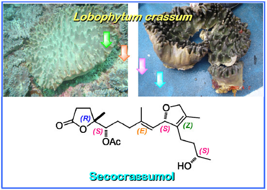
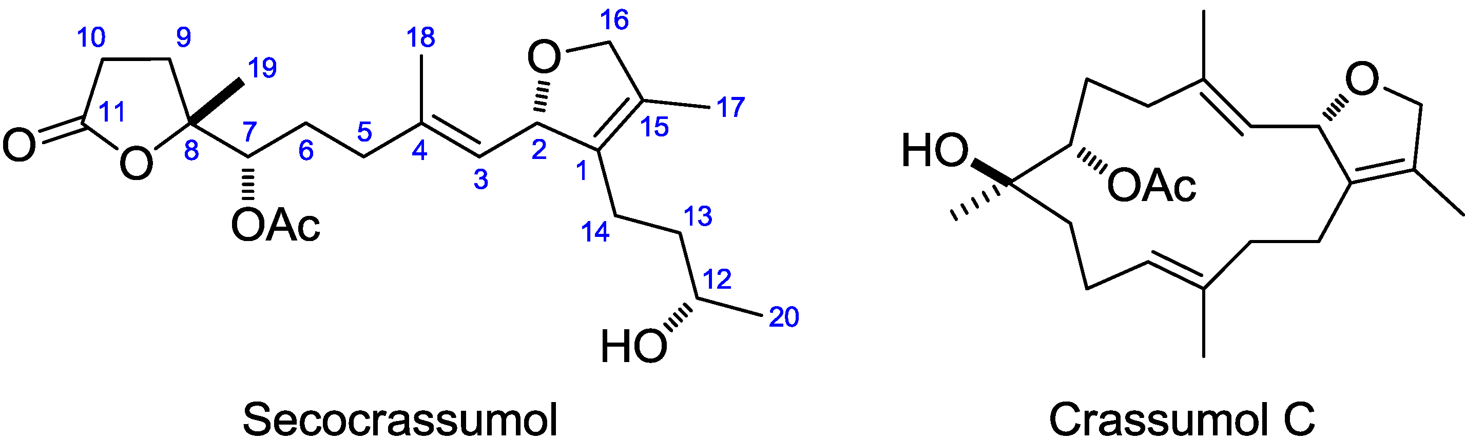
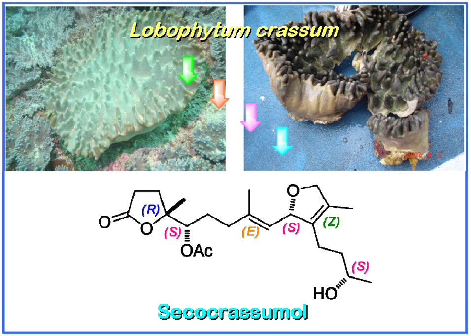
Secocrassumol, a seco-Cembranoid from the Dongsha Atoll Soft Coral Lobophytum crassum

1
Department of Life Sciences, National University of Kaohsiung, Kaohsiung 811, Taiwan
2
Department of Microbiology, Kaohsiung Medical University, Kaohsiung 807, Taiwan
3
Department of Marine Biotechnology and Resources, National Sun Yat-Sen University, Kaohsiung 804, Taiwan
4
Graduate Institute of Natural Products, Kaohsiung Medical University, Kaohsiung 807, Taiwan
*
Author to whom correspondence should be addressed.
Mar. Drugs 2014, 12(12), 6028-6037; https://doi.org/10.3390/md12126028
Submission received: 16 October 2014 / Revised: 7 November 2014 / Accepted: 28 November 2014 / Published: 17 December 2014

Abstract

:
Chemical investigations on the Dongsha Atoll soft coral Lobophytum crassum led to the purification of a new seco-cembranoid, secocrassumol. The structural elucidation was established by extensive NMR, HRESIMS and CD data. The absolute configuration at C-12 was determined as S using a modified Mosher’s acylation. Secocrassumol differs from previously known marine seco-cembranoid in that it possesses an unprecedented skeleton functionalized at C11-C12 bond cleavage. Secocrassumol showed antiviral activity against human cytomegalovirus (HCMV) with an IC50 value of 5.0 μg/mL.

Graphical Abstract

1. Introduction

Marine soft corals have evolved characteristic metabolic and physiological capabilities to produce secondary metabolites that may function in defense, food capture, interference competition, and even possibly the acquisition and selection of symbiotic zooxanthellae [1]. The first cembrane-type diterpenoid was obtained in 1951 from the oleoresin of Pinus albicaulis [2]. Marine cembranoids are the representative compounds from soft corals, having been first discovered from gorgonians by the Ciereszko lab in 1960 [3]. For more than 60 years, hundreds of cembranoids possessing almost every structural modification have been reported from virtually all alcyonarians and gorgonians [4]. Prior studies have shown that members of the genus Lobophytum produce a rich harvest of cembranoids endowed with diversified macrocyclic skeletons [4,5,6,7,8,9,10,11,12,13,14,15,16,17,18,19,20,21,22,23,24,25]. Previous bioassay results of these metabolites have been shown to exhibit diverse biological activities such as cytotoxicity [11,13,15,16,17,18,19,23], anti-inflammatory properties [20,21,22], antimicrobial activities [20], and HIV-inhibitory activities [17]. During the course of our initial investigation of bioactive metabolites from the soft coral L. crassum (von Marenzeller, 1886), six cembranoids (lobocrassolide, lobocrasol, crassumols A–C and 13-acetoxysarcophytoxide) and two á-tocopherols (crassumtocopherols A and B) were discovered and some of these have been shown to possess cytotoxic properties [18,23,24,25]. Our continuing chemical investigations of this organism led to the isolation of secocrassumol (Figure 1). Secocrassumol apparently derives from a cembranoid precursor through cleavage of the C11-C12 bond. The plausible biosynthetic pathway for formation of secocrassumol is postulated in Scheme 1. Furthermore, it was evaluated in vitro for cytotoxicity against A-459 (human lung carcinoma), P-388 (mouse lymphocytic leukemia), and HT-29 (human colon adenocarcinoma) cancer cell lines as well as antiviral activity against HCMV (human cytomegalovirus) cells.
Figure 1. The structure of secocrassumol and crassumol C.
Marinedrugs 12 06028 g001
Scheme 1. The plausible biosynthetic pathway for formation of secocrassumol.
Marinedrugs 12 06028 g005

2. Results and Discussion
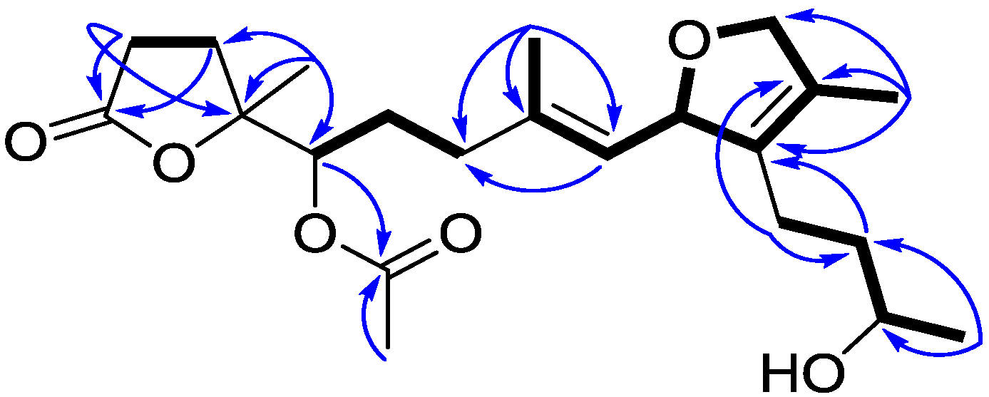
The chromatographic separation of the EtOAc extract (20 g) of the soft coral L. crassum using Si-60 and ODS gel columns in combination with semi-preparative reversed-phase C18 HPLC resulted in the purification of secocrassumol (see Experimental Section), which was obtained as a colorless oil. The HRESIMS exhibited a pseudo molecular ion peak at m/z 417.2256 [M + Na]+ (calcd. for C22H34O6Na, 417.2253), consistent with the molecular formula of C22H34O6, requiring six degrees of unsaturation. The IR spectrum demonstrated a broad absorption band at 3434 cm−1 (OH stretching) diagnostic of a secondary hydroxy group, which was associated to C-12 on the basis of the HMBC correlations from Me-20 to C-12 and C-13 (Figure 2). The IR spectrum revealed the presence of an ester (1742 cm−1) moiety, which was further identified by the 1H·NMR signals at δH 2.09 (3H, s) and 13C NMR signals at δC 170.4 (qC) and 20.9 (CH3) (Table 1). The existence of two quaternary carbons [δC 133.0 (qC, C-1) and 128.4 (qC, C-15)], an oxygenated methine [δH 5.09 (dd, 1H, J = 9.2, 0.8 Hz) and δC 84.4 (C-2)] and an oxygenated methylene [δH 4.54 (dd, 1H, J = 11.6, 5.6 Hz) and 4.45 (br d, 1H, J = 11.6 Hz); δC 78.1 CH2], as well as the long-range COSY correlations between H-2 and H2-16 exhibited the presence of a 2,5-dihydrofuran ring (Figure 2). In addition, the 13C NMR signals at δC 126.0 (CH, C-3) and 138.2 (qC, C-4) assigned a trisubstituted double bond. Although there were no direct HMBC correlations available, the remaining one unsaturation indicated that an oxygen bridge is probably present between a lactone carbonyl carbon [δC 176.2 (qC, C-11)] and an oxygenated quaternary carbon [δC 82.6 (qC, C-8)]. This assumption was further confirmed by a strong IR absorption at 1771 cm−1 [26]. Comparison to the NMR data reported for crassumol C [24] permitted us to propose the 11,12-secocembranoid structure for secocrassumol.
Figure 2. Selected 1H–1H COSY (▬) and HMBC () correlations of secocrassumol.
Marinedrugs 12 06028 g002
The geometry of the trisubstituted olefin was assigned as E based on the γ-effect of the olefinic methyl signals for C-18 (16.4 ppm) [27] and the NOESY correlations between H-3 and H2-5, and H-2 and H3-18. The relative configurations of C2, C7 and C8 could not be determined due to the absence of decided NOESY correlations available. Additionally, the failure to crystallize, the limitation of material, and scarcity of sample source make secocrassumol inaccessible to X-ray crystallographic analysis for confirmation of the relative configurations of the aforementioned carbons. Based on biogenic considerations, a 2S, 7S configuration of secocrassumol was assumed to be identical with that of crassumol C [24]. The circular dichroism (CD) spectrum exhibited a positive Cotton effect around λmax 214 nm due to the γ-lactone (Figure 3). The absolute configuration at C-8 was deduced to be R based on its CD data comparable to that of some C11-C12 secocembranoids from the leaves of air-cured Burley tobaccos [26]. The appropriate stereochemistry of secocrassumol was identified by Mosher’s esterification for absolute configuration determination of chiral alcohols [28]. Analysis of the ΔδSR values according to the Mosher model pointed to an S configuration for C-12, because H2-13 and H2-14 of the (S)-MTPA ester were less shielded by the phenyl ring of MTPA products (Figure 4). Accordingly, the structure of secocrassumol was elucidated unambiguously.
Table 1. NMR spectroscopic data of secocrassumol a.
#13C1H1H–1H COSYHMBC (H→C)
1133.0 (qC) b
284.4 (CH)5.42 mH-3, H-16, H-17
3126.0 (CH)5.09 br·d (9.2, 0.8) cH-2, H-18C-5, C-18
4138.2 (qC)
535.8 (CH2)2.04 mH-6
627.6 (CH2)a: 1.94 mH-5, H-7
b: 1.80 m
775.9 (CH)5.01 dd (10.4, 2.4)H-6C-8, C-9, 7-OAc
882.6 (qC)
929.9 (CH2)a: 2.22 mH-10C-10, C-11, C-19
b: 1.91 m
1028.7 (CH2)2.61 mH-9C-8, C-9, C-11
11176.2 (qC)
1268.0 (CH)3.76 mH-13, H-20
1337.5 (CH2)1.49 mH-12, H-14C-1, C-14, C-20
1421.1 (CH2)a: 2.24 mH-13C-1, C-2, C-13
b: 1.89 m
15128.4 (qC)
1678.1 (CH2)a: 4.54 dd (11.6, 5.6)H-2, H-17C-1
b: 4.45 br·d (11.6)
1710.0 (CH3)1.66 sH-2, H-16C-1, C-15, C-16
1816.4 (CH3)1.76 sH-3C-3, C-4, C-5
1922.5 (CH3)1.40 s C-7, C-8, C-9
2023.3 (CH3)1.19 d (6.0)H-12C-12, C-13
7-OAc20.9 (CH3)2.09 s 7-OAc
170.4 (qC)
a Spectra were measured in CDCl3 (1H, 400 MHz and 13C, 100 MHz). b Multiplicities are deduced by HSQC and DEPT experiments. c J values (in Hz) are in parentheses.
Figure 3. The CD spectrum of secocrassumol.
Marinedrugs 12 06028 g003
Figure 4. Selected 1H·NMR ΔδSR values in ppm for the S- and R-MTPA esters of secocrassumol in CDCl3.
Marinedrugs 12 06028 g004
Apparently, the co-occurrence of crassumol C within the same organism raises the probability that secocrassumol results from crassumol C. The biosynthetic precursor is transformed into intermediate I by oxidative C11-C12 cleavage. The intermediate I would undergo an enzymatic lactonization to yield intermediate II, which was converted into secocrassumol by reduction (Scheme 1). Purification of secocembranoids is intriguing, especially in light of the previous isolation and characterization of the related metabolites [29,30,31,32,33], suggesting that marine soft corals possess a biodegradable capacity to modify parent cembranoids through simple ring cleavage. The few examples of secocembranoids from soft corals include two C12-C13 analogues from Sinularia mayi [29], a C1-C14 secocembranoid from Eunicea succinea [30], a C8-C9 secocembranoid (seco-sethukarailin) from Sinularia dissecta [31], two C2-C3 secocembranoids (caucanolides E and F) from Pseudopterogorgia bipinnata [32], two C9-C10 secocembranoids from Nephthea sp. [33], and a C11-C12 secocembranoid from Sinularia flexibilis [34]. Among these marine secocembranoids already reported in the literature, secocrassumol also represents a secocembrane skeleton functionalized at the C11-C12 bond cleavage.
Preliminary cytotoxic screening revealed that secocrassumol exhibited no discernible cytotoxicity against mouse lymphocytic leukemia (P-388), human lung carcinoma (A-459) as well as human colon adenocarcinoma (HT-29) (ED50 > 50 μM). Similarly, the biosynthetic precursor crassumol C was not cytotoxic to P-388, A-549, and HT-29 cells [24]. The anticancer agent mithramycin was used as the positive control and exhibited EC50 values of 0.05, 0.06 and 0.07 μM against P-388, A-549 and HT-29 cells, respectively. It was noteworthy to mention that crassumol C did not show anti-HCMV activity (IC50 > 50 μg/mL), but secocrassumol exhibited moderate antiviral activity against HCMV cells with an IC50 value of 5.0 μg/mL.

3. Experimental Section

3.1. General Experimental Procedures

Optical rotations were recorded on a JASCO P1020 polarimeter (Tokyo, Japan). CD analysis was performed on a JASCO J-815 spectropolarimeter (Tokyo, Japan). IR and UV spectra were measured on JASCO FT/IR-4100 (Tokyo, Japan) and JASCO V-650 spectrophotometers (Tokyo, Japan), respectively. The NMR spectra were recorded on a Varian 400 MR NMR spectrometer (Santa Clara, CA, USA) at 400 MHz for 1H and 100 MHz for 13C, respectively. Chemical shifts are expressed in δ (ppm) referring to the solvent peaks δH 7.27 and δC 77.0 for CDCl3, respectively, and coupling constants are expressed in Hz. ESIMS spectra were recorded by ESI FT-MS on a Bruker APEX II mass spectrometer (Bruker, Bremen, Germany). Silica gel 60 (Merck, Darmstadt, Germany, 230–400 mesh), LiChroprep RP-18 (Merck, Darmstadt, Germany, 40–63 μm) and Sephadex LH-20 (Amersham Pharmacia Biotech., Piscataway, NJ, USA) were used for column chromatography. Precoated silica gel plates (Merck, Darmstadt, Germany, Kieselgel 60 F254, 0.25 mm) and precoated RP-18 F254s plates (Merck, Darmstadt, Germany) were used for analytical thin-layer chromatography (TLC) analyses. High-performance liquid chromatography (HPLC) was performed on a Hitachi L-7100 pump (Tokyo, Japan) equipped with a Hitachi L-7400 UV detector (Tokyo, Japan) at 220 nm and an ODS column (Merck, Darmstadt, Germany, Hibar Purospher RP-18e, 5 μm, 250 × 10 mm). S-(+)- and R-(–)-α-methoxy-α-trifluoromethylphenylacetyl chloride were obtained from ACROS Organics (Geel, Belgium).

3.2. Animal Material

Specimens of L. crassum, identified by Professor Chang-Feng Dai of the Institute of Oceanography, National Taiwan University (Taipei, Taiwan), were collected from coral reefs offshore from the Dongsha Atoll off Taiwan in April 2007, at a depth of 8 m, and were immediately frozen at −20 °C until further processed for extraction in the laboratory. A voucher specimen (TS-11) has been deposited at the Department of Marine Biotechnology and Resources, National Sun Yat-sen University (Kaohsiung, Taiwan).

3.3. Extraction and Isolation

The sliced bodies of L. crassum were exhaustively extracted with acetone. The combined extracts were concentrated in vacuo (under 35 °C) to obtain a dry crude extract (25 g), which was suspended in water and extracted with EtOAc. The EtOAc-soluble portion was evaporated to dryness in vacuo to give a dark brown residue (20 g). The resulting EtOAc residue was subjected to a silica gel chromatography using a stepwise gradient mixture of n-hexane–EtOAc–MeOH as elution and separated into 40 fractions. Fraction 20 (223 mg) eluted with n-hexane/EtOAc (1:10) was submitted to repeated chromatography over Si-60 gel column using n-hexane–EtOAc mixtures of increasing polarity as eluent. Altogether, three subfractions were obtained, of which subfraction 20-3 (142 mg) was followed by column chromatography on ODS column using 53% MeOH in H2O to yield a mixture (25 mg). In turn, the mixture was further purified by RP-18 HPLC using an isocratic solvent system of 65% MeOH in H2O to give secocrassumol (2 mg).
Secocrassumol: Colorless oil; [ a ] D 25 −192 (c 0.1, CHCl3); IR (KBr) νmax 3434, 2965, 2924, 2857, 1771, 1742, 1649, 1557, 1375, 1232, 1036 cm−1; CD (4.80 × 10⁴ M, MeOH) λmax (Δε) 214 (+12.24) nm; 1H·NMR (CDCl3, 400 MHz) and 13C·NMR (CDCl3, 100 MHz) data, see Table 1; ESIMS m/z 417 [M + Na]+; HRESIMS m/z 417.2256 [M + Na]+ (calcd. for C22H34O6Na, 417.2253) (Supplementary Figures S1–S7).

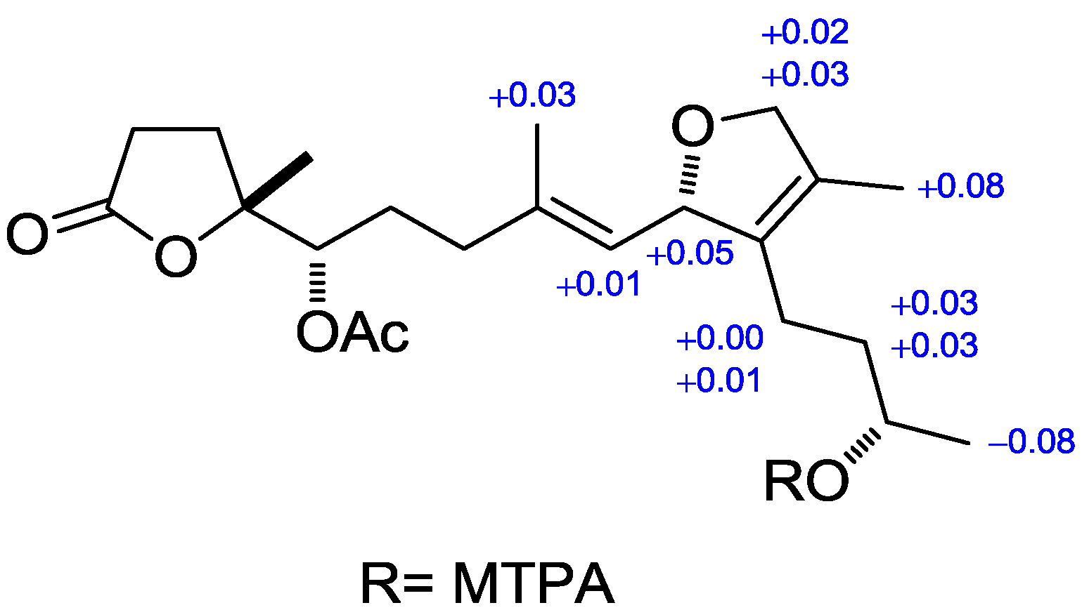
3.4. Preparation of (R)- and (S)-MTPA Esters of Secocrassumol

Two secocrassumol samples (0.5 mg) were dissolved in pyridine-d5 (0.6 mL) and allowed to react overnight at room temperature with (R)- and (S)-MTPA chloride (one drop), affording the (S)-MTPA ester (S) and (R)-MTPA ester (R), respectively.
Selected 1H-NMR (pyridine-d5, 400 MHz) of S: δH 7.88–7.61 (5H, m, Ph), 5.70 (1H, m, H-2), 5.35 (1H, d, J = 9.2 Hz, H-3), 5.29 (1H, m, H-12), 4.65 (1H, dd, J = 11.6, 4.8 Hz, 16a), 4.56 (1H, d, J = 11.6 Hz, 16b), 2.70 (1H, t, J = 8.8 Hz, H-14a), 2.30 (1H, m, H-14b), 1.87 (1H, m, H-13a), 1.76 (1H, m, H-13b), 2.10 (3H, s, 7-OAc), 1.76 (3H, s, H-18), 1.59 (3H, s, H-17), 1.36 (3H, s, H-19), 1.29 (3H, d, J = 6.4 Hz, H-20).
Selected 1H-NMR (pyridine-d5, 400 MHz) of R: δH 7.88–7.61 (5H, m, ph), 5.65 (1H, m, H-2), 5.34 (1H, d, J = 9.2 Hz, H-3), 5.30 (1H, m, H-12), 4.63 (1H, dd, J = 11.6, 4.8 Hz, 16a), 4.53 (1H, d, J = 11.6 Hz, 16b), 2.70 (1H, t, J = 8.8 Hz, H-14a), 2.29 (1H, m, H-14b), 1.84 (1H, m, H-13a), 1.73 (1H, m, H-13b), 2.10 (3H, s, 7-OAc), 1.73 (3H, s, H-18), 1.51 (3H, s, H-17), 1.36 (3H, s, H-19), 1.37 (3H, d, J = 6.4 Hz, H-20).

3.5. Cytotoxicity Assay

Cytotoxicity was determined against P-388 (mouse lymphocytic leukemia), HT-29 (human colon adenocarcinoma), as well as A-549 (human lung epithelial carcinoma) tumor cells using a modification of the MTT colorimetric method according to a previously described procedure [35,36]. The P-388 cell line was kindly provided by John M. Pezzuto, formerly of the Department of Medicinal Chemistry and Pharmacognosy, University of Illinois at Chicago. Additionally, HT-29 and A-549 cell lines were purchased from the American Type Culture Collection (Manassas, VA, USA).

3.6. Anti-HCMV Assay

To determine the effects of natural products upon HCMV cytopathic effect (CPE), confluent human embryonic lung (HEL) cells grown in 24-well plates were incubated for 1 h in the presence or absence of various concentrations of tested natural products. Then, cells were infected with HCMV at an input of 1000 pfu (plaque forming units) per well of 24-well dish. Antiviral activity was expressed as IC50 (50% inhibitory concentration), or compound concentration required to reduce virus-induced CPE by 50% after seven days, as compared with the untreated control. To monitor the cell growth upon treatment with natural products, an MTT-colorimetric assay was employed [37].

4. Conclusions

A new seco-cembranoid, designated as secocrassumol, was isolated from the Dongsha Atoll soft coral Lobophytum crassum. Secocrassumol differs from the previously known marine seco-cembranoid in that it possesses an unprecedented skeleton functionalized at the C11-C12 bond cleavage. Preliminary cytotoxic screening revealed that secocrassumol are not cytotoxic to P-388, A-549, and HT-29 cells. However, secocrassumol showed antiviral activity against HCMV with an IC50 value of 5.0 μg/mL.

Supplementary Files

Supplementary File 1

Acknowledgments

Financial support was provided by the National Science Council (NSC102-2320-B-110-003-MY3 and NSC102-2113-M-390-001) of Taiwan and NSYSUKMU Joint Project (NSYSUKMU 103-P016).

Author Contributions

Conceived and designed the experiments: Chang-Yih Duh; Performed the experiments: Shi-Yie Cheng, Shang-Kwei Wang; Analyzed the data: Chang-Yih Duh, Shi-Yie Cheng.

Conflicts of Interest

The authors declare no conflict of interest.

References

  1. Coll, J.C. The chemistry and chemical ecology of octocorals (Coelenterata, Anthozoa, Octocorallia). Chem. Rev. 1992, 92, 613–631. [Google Scholar] [CrossRef]
  2. Haagen-Smit, A.J.; Wang, T.H.; Mirov, N.T. Composition of gum turpentines of pines. XIII. A report on Pinus albicaulis. J. Am. Pharm. Assoc. Sci. Ed. 1951, 40, 557–559. [Google Scholar] [CrossRef]
  3. Ciereszko, L.S.; Sifford, D.H.; Weinheimer, A.J. Chemistry of coelenterates. I. Occurrence of terpenoid compounds in gorgonians. Ann. N.Y. Acad. Sci. 1960, 90, 917–919. [Google Scholar] [CrossRef] [PubMed]
  4. Blunt, J.W.; Copp, B.R.; Keyzers, R.A.; Munro, M.H.G.; Prinsep, M.R. Marine natural products. Nat. Prod. Rep. 2013, 30, 237–323, and references cited therein. [Google Scholar] [CrossRef] [PubMed]
  5. Kashman, Y.; Groweiss, A. Lobolide: A new epoxy cembranolide from marine origin. Tetrahedron Lett. 1977, 13, 1159–1160. [Google Scholar] [CrossRef]
  6. Kashman, Y.; Carmely, S.; Groweiss, A. Further cembranoid derivatives from the Red Sea soft corals Alcyonium flaccidum and Lobophytum crassum. J. Org. Chem. 1981, 46, 3592–3596. [Google Scholar] [CrossRef]
  7. Kinamoni, Z.; Groweiss, A.; Carmely, S.; Kashman, Y. Several new cembranoid diterpenes from three soft corals of the Red Sea. Tetrahedron 1983, 39, 1643–1648. [Google Scholar] [CrossRef]
  8. Bowden, B.F.; Coll, J.C.; Tapiolas, D.M. Studies of Australian soft corals. XXXIII. New cembranoid diterpenes from a Lobophytum species. Aust. J. Chem. 1983, 36, 2289–2295. [Google Scholar] [CrossRef]
  9. Bowden, B.F.; Coll, J.C.; Decosta, M.S.L.; Desilva, E.D.; Mackay, M.F.; Mahendran, M.; Willis, R.H. The structure determination of a new cembranolide diterpene from the soft coral Lobophytum cristigalli (Coelenterata, Octocorallia, Alcyonacea). Aust. J. Chem. 1984, 34, 545–552. [Google Scholar] [CrossRef]
  10. Bowdon, B.F.; Coll, J.C.; Heaton, A.; Konig, G.; Bruck, M.A.; Cramer, R.E.; Klein, D.M.; Scheuer, P.J. The structures of four isomeric dihydrofuran-containing cembranoid diterpenes from several species of soft coral. J. Nat. Prod. 1987, 50, 650–659. [Google Scholar] [CrossRef]
  11. Wang, S.-K.; Duh, C.-Y.; Wu, Y.-C.; Wang, Y.; Cheng, M.-C.; Soong, K.; Fang, L.-S. Cytotoxic cembranolides from the soft coral Lobophytum michaelae. J. Nat. Prod. 1992, 55, 1430–1435. [Google Scholar] [CrossRef]
  12. Subrahmanyam, C.; Rao, C.V.; Anjaneyulu, V.; Satyanarayana, P.; Rao, P.V.S. New diterpenes from a new species of Lobophytum soft coral of the South Andaman coast. Tetrahedron 1992, 48, 3111–3120. [Google Scholar] [CrossRef]
  13. Coval, S.J.; Patton, R.W.; Petrin, J.M.; James, L.; Rothofsky, M.L.; Lin, S.L.; Patel, M.; Reed, J.K.; McPhil, A.T.; Bishop, W.R. A cembranolide diterpene farnesyl protein transferase inhibitor from the marine soft coral Lobophytum cristagalli. Bioorg. Med. Chem. Lett. 1996, 6, 909–912. [Google Scholar] [CrossRef]
  14. Yamada, K.; Ryu, K.; Miyamoto, T.; Higuchi, R. Bioactive terpenoids from octocorallia. 4. Three new cembrane-type diterpenoids from the soft coral Lobophytum schoedei. J. Nat. Prod. 1997, 60, 798–801. [Google Scholar] [CrossRef]
  15. Higuchi, R.; Miyamoto, T.; Yamada, K.; Komori, T. Cytotoxic and ichthyotoxic compounds from marine opisthobranchia and soft coral. Toxicon 1998, 36, 1703–1705. [Google Scholar] [CrossRef]
  16. Matthee, G.F.; Konig, G.M.; Wright, A.D. Three new diterpenes from the marine soft coral Lobophytum crassum. J. Nat. Prod. 1998, 61, 237–240. [Google Scholar] [CrossRef] [PubMed]
  17. Rashid, M.A.; Gustafson, K.R.; Boyd, M.R. HIV-inhibitory cembrane derivatives from a Philippines collection of the soft coral Lobophytum species. J. Nat. Prod. 2000, 63, 531–533. [Google Scholar] [CrossRef]
  18. Duh, C.-Y.; Wang, S.-K.; Huang, B.-T.; Dai, C.-F. Cytotoxic cembrenolide diterpenes from the Formosan soft coral Lobophytum crassum. J. Nat. Prod. 2000, 63, 884–885. [Google Scholar] [CrossRef] [PubMed]
  19. Zhang, W.; Krohn, K.; Ding, J.; Miao, Z.-H.; Zhou, X.-H.; Chen, S.-H.; Pescitelli, G.; Salvadori, P.; Kurtan, T.; Guo, Y.-W. Structural and stereochemical studies of α-methylene-γ-lactone-bearing cembrane diterpenoids from a South China sea soft coral Lobophytum crassum. J. Nat. Prod. 2008, 71, 961–966. [Google Scholar] [CrossRef] [PubMed]
  20. Cheng, S.-Y.; Wen, Z.-H.; Chiou, S.-F.; Wang, S.-K.; Hsu, C.-H.; Dai, C.-F.; Chiang, M.Y.; Duh, C.-Y. Durumolides A–E, Anti-inflammatory and antibacterial cembranolides from the soft coral Lobophytum durum. Tetrahedron 2008, 64, 9698–9704. [Google Scholar] [CrossRef]
  21. Cheng, S.-Y.; Wen, Z.-H.; Wang, S.-K.; Chiou, S.-F.; Hsu, C.-H.; Dai, C.-F.; Duh, C.-Y. Anti-inflammatory cembranolides from the soft coral Lobophytum durum. Bioorg. Med. Chem. 2009, 17, 3763–3769. [Google Scholar] [CrossRef] [PubMed]
  22. Cheng, S.-Y.; Wen, Z.-H.; Chiou, S.-F.; Wang, S.-K.; Hsu, C.-H.; Dai, C.-F.; Chiang, M.Y.; Duh, C.-Y. Unprecedented hemiketal cembranolides with anti-inflammatory activity from the soft coral Lobophytum durum. J. Nat. Prod. 2009, 72, 152–155. [Google Scholar] [CrossRef] [PubMed]
  23. Lin, S.-T.; Wang, S.-K.; Cheng, S.-Y.; Duh, C.-Y. Lobocrasol, a new diterpenoid from the soft coral Lobophytum crassum. Org. Lett. 2009, 14, 3012–3014. [Google Scholar] [CrossRef]
  24. Lin, S.-T.; Wang, S.-K.; Duh, C.-Y. Cembranoids from the Dongsha Atoll soft coral Lobophytum crassum. Mar. Drugs. 2011, 9, 2705–2716. [Google Scholar] [CrossRef]
  25. Cheng, S.-Y.; Lin, S.-T.; Wang, S.-K.; Duh, C.-Y. α-Tocopherols from the Formosan soft coral Lobophytum crassum. Bull. Chem. Soc. Jpn. 2011, 7, 783–787. [Google Scholar] [CrossRef]
  26. Wahlberg, I.; Arndt, R.; Nishida, T.; Enzell, C.R. Tobacco chemistry. 63. Syntheses and stereostructures of six tobacco seco-cembranoids. Acta. Chem. Scand. 1986, 40B, 123–134. [Google Scholar] [CrossRef]
  27. Iwashima, M.; Matsumoto, Y.; Takenaka, Y.; Iguchi, K.; Yamori, T. New marine diterpenoids from the Okinawan soft coral Clavularia koellikeri. J. Nat. Prod. 2002, 65, 1441–1446. [Google Scholar] [CrossRef] [PubMed]
  28. Ohtani, I.; Kusumi, T.; Kashman, Y.; Kakisawa, H. High-field FT NMR application of Mosher’s method. The absolute configurations of marine terpenoids. J. Am. Chem. Soc. 1991, 113, 4092–4096. [Google Scholar] [CrossRef]
  29. Kobayashi, M. Marine terpenes and terpenoids. IV. Isolation of new cembranoid and secocembranoid lactones from the soft coral Sinularia mayi. Chem. Pharm. Bull. 1988, 36, 488–494. [Google Scholar] [CrossRef]
  30. Rodríguez, A.D.; Acosta, A.L. New cembranoid diterpenes and a geranylgeraniol derivative from the common Caribbean sea whip Eunicea succinea. J. Nat. Prod. 1997, 60, 1134–1138. [Google Scholar] [CrossRef] [PubMed]
  31. Srinivasa Reddy, N.; Venkateshwar Goud, T.; Venkateswarlu, Y. Seco-sethukarailin, a novel diterpenoid from the soft coral Sinularia dissecta. J. Nat. Prod. 2002, 65, 1059–1060. [Google Scholar] [CrossRef] [PubMed]
  32. Ospina, C.A.; Rodríguez, A.D.; Sánchez, J.A.; Ortega-Barria, E.; Capson, T.L.; Mayer, A.M.S. Caucanolides A‒F, unusual antiplasmodial constituents from a Colombian collection of the gorgonian coral Pseudopterogorgia bipinnata. J. Nat. Prod. 2005, 68, 1519–1526. [Google Scholar] [CrossRef] [PubMed]
  33. Gross, H.; Kehraus, S.; Nett, M.; König, G.M.; Beil, W.; Wright, A.D. New cytotoxic cembrane based diterpenes from the soft corals Sarcophyton cherbonnieri and Nephthea sp. Org. Biomol. Chem. 2003, 1, 944–949. [Google Scholar] [CrossRef] [PubMed]
  34. Shih, H.-J.; Tseng, Y.-J.; Huang, C.-Y.; Wen, Z.-H.; Dai, C.-F.; Sheu, J.-H. Cytotoxic and anti-inflammatory diterpenoids from the Dongsha Atoll soft coral Sinularia flexibilis. Tetrahedron 2012, 68, 244–249. [Google Scholar] [CrossRef]
  35. Geran, R.I.; Greenberg, N.H.; MacDonald, M.M.; Schumacher, A.M.; Abbott, B.J. Protocols for screening chemical agents and natural products against animal tumors and other biological syatems. Cancer Chemother. Rep. 1972, 3, 1–91. [Google Scholar]
  36. Hou, R.-S.; Duh, C.-Y.; Chiang, M.Y.; Lin, C.-N. Sinugibberol, a new cytotoxic cembranoid diterpene from the soft coral Sinularia gibberosa. J. Nat. Prod. 1995, 58, 1126–1130. [Google Scholar] [CrossRef] [PubMed]
  37. Stevens, M.; Balzarini, J.; Tabarrini, O.; Andrei, G.; Snoeck, R.; Cecchetti, V.; Fravolini, A.; de Clercq, E.; Pannecouque, C. Cell-dependent interference of a series of new 6-aminoquinolone derivatives with viral (HIV/CMV) transactivation. J. Antimicrob. Chemother. 2005, 56, 847–855. [Google Scholar] [CrossRef] [PubMed]

