Article

Early Allograft Dysfunction After Liver Transplantation: Impact on Clinical Outcomes and Associated Risk Factors

1
Department of Internal Medicine, College of Medicine, Chung-Ang University, Seoul 06973, Republic of Korea
2
Department of Surgery, College of Medicine, Chung-Ang University, Seoul 06793, Republic of Korea
*
Author to whom correspondence should be addressed.
Medicina 2025, 61(9), 1710; https://doi.org/10.3390/medicina61091710
Submission received: 31 July 2025 / Revised: 7 September 2025 / Accepted: 18 September 2025 / Published: 19 September 2025
(This article belongs to the Special Issue Advances in Liver Surgery)

Abstract

Background and Objectives: Early allograft dysfunction (EAD), defined as suboptimal initial graft function following liver transplantation (LT), is a serious complication associated with increased post-LT morbidity and mortality. This study aimed to evaluate the impact of EAD on clinical outcomes and to identify associated risk factors. Materials and Methods: Ninety-three patients who underwent LT between July 2015 and August 2024 were retrospectively analyzed. EAD was defined by the presence of one or more of the following criteria: total bilirubin ≥ 10 mg/dL or international normalized ratio ≥ 1.6 on postoperative day 7, and alanine or aspartate aminotransferase levels > 2000 IU/L within the first 7 days. Results: EAD occurred in 20 patients (21.5%). Patients with EAD exhibited significantly lower graft survival (p < 0.01) and patient survival (p = 0.03) compared with those without EAD. EAD was an in-dependent risk factor for both graft survival (p = 0.021) and patient survival (p = 0.027). Acute liver failure (odds ratio [OR], 6.228; 95% confidence interval [CI], 1.179–32.906; p = 0.031), donor age (OR, 1.051; 95% CI, 1.008–1.096; p = 0.020), and warm ischemic time (OR, 1.048; 95% CI, 1.001–1.098; p = 0.046) were identified as significant predictors of EAD development. Conclusions: EAD adversely affects both graft and patient survival following LT. Recipient clinical status, donor age, and intraoperative conditions should be carefully considered to minimize the risk of EAD.

1. Introduction

Liver transplantation (LT) remains the definitive treatment for patients with end-stage liver disease and selected cases of hepatocellular carcinoma (HCC) in cirrhotic livers [1]. Despite improvements in surgical techniques and perioperative care, graft insufficiency and failure continue to pose significant clinical challenges [2]. Early allograft dysfunction (EAD) refers to impaired initial graft function occurring shortly after LT [3,4,5]. The most widely accepted definition of EAD, proposed by Olthoff et al. [6] in a model for end-stage liver disease (MELD), is based on the presence of one or more of the following criteria: serum total bilirubin ≥ 10 mg/dL on postoperative day 7, international normalized ratio (INR) ≥ 1.6 on day 7, or aspartate aminotransferase (AST) or alanine ami-notransferase (ALT) > 2000 U/L within the first 7 days.
EAD is a relatively common complication, with an incidence rate ranging from 20% to 44%, and is associated with increased postoperative morbidity and mortality [7,8]. Although graft function may recover in some cases, approximately 7% of patients progress to graft failure, necessitating re-transplantation [9,10]. However, the feasibility of re-transplantation is limited by organ shortages, particularly in certain regions, thereby contributing to elevated mortality rates. Consequently, identifying risk factors for EAD is critical for guiding clinical decision-making during both donor selection and intraoperative management. Risk stratification may also facilitate individualized postoperative care and timely planning for potential re-transplantation, ultimately improving survival outcomes.
Several risk factors for EAD have been reported, including donor age, graft steatosis, donation after cardiac death (DCD), and perioperative variables such as cold ischemic time (CIT), warm ischemic time (WIT), and MELD score [5,11,12]. However, most of these studies have focused exclusively on deceased donor LT (DDLT). In contrast, living donor LT (LDLT) allows for the selection of healthier donors and grafts with reduced ischemia–reperfusion injury (IRI), achieved by minimizing CIT and WIT. Therefore, investigating EAD risk factors in a cohort that includes both DDLT and LDLT is warranted. Evaluating the impact of perioperative variables across a broader range of values may yield a more nuanced understanding of their roles in EAD development.
The objective of this study was to assess the impact of EAD on graft and patient survival and to identify associated risk factors in patients undergoing LT.

2. Patients and Methods

2.1. Patients

This retrospective study included 93 patients who underwent LT at our institution between July 2015 and August 2024, comprising 32 DDLTs and 61 LDLTs. Exclusion criteria were recipient age < 18 years, re-transplantation, and incomplete clinical data. EAD was defined by the presence of one or more of the following criteria: total bilirubin (TB) ≥ 10 mg/dL or INR ≥ 1.6 on postoperative day 7, and AST or ALT > 2000 IU/L within the first 7 days. Patients were stratified into two groups based on the presence of EAD: Group A (EAD, n = 20) and Group B (non-EAD, n = 73). Clinico-demographic characteristics and survival outcomes were compared between groups, and the effects of EAD on graft and patient survival were analyzed. In addition, perioperative risk factors associated with EAD development were assessed.
The study was approved by the Institutional Review Board of our institution (IRB No. 2407-001-19529) and was exempt from the requirement for informed consent due to the retrospective nature of the analysis and the absence of patient-identifiable data.

2.2. Data Collection

The following clinico-demographic and perioperative variables were collected: recipient age, sex, body mass index (BMI), presence of diabetes mellitus or hypertension, primary liver disease, MELD score, donor age, ABO incompatibility (ABOi), donor type (LDLT or DDLT), degree of hepatic macrosteatosis, operative time, CIT, WIT, and postoperative hospital stay. Laboratory values, including TB, INR, AST, and ALT, were recorded from admission through postoperative day 7. Graft failure was defined as re-transplantation or death. Follow-up duration was calculated from the date of LT to the last clinical visit or death.

2.3. Statistical Analysis

Normality of data distribution was assessed using the Shapiro–Wilk test. Normally distributed variables were expressed as means ± standard deviations and compared be-tween groups using Student’s t-test or the Kruskal–Wallis test, as appropriate. Categorical variables were compared using the χ2 test or Fisher’s exact test. Graft and patient survival were analyzed using the Kaplan–Meier method and compared with the log-rank test. Risk factors for graft and overall survival were assessed by multivariate analysis using the Cox proportional hazards model. Risk factors for EAD development were evaluated using multivariate logistic regression. Continuous variables identified as significant predictors were further analyzed using fractional polynomial regression. Statistical analyses were performed using SPSS Statistics for Windows, version 19.0 (IBM Corp., Armonk, NY, USA).

3. Results

3.1. Clinico-Pathologic Characteristics of Patients

The clinico-pathologic characteristics of the patients are summarized in Table 1. No significant differences were observed between groups in terms of age (53.6 ± 8.8 vs. 54.1 ± 11.9 years, p = 0.852), sex (65.0% vs. 68.5%, p = 0.767), prevalence of diabetes mellitus (70.0% vs. 69.9%, p = 0.991) or hypertension (80.0% vs. 78.1%, p = 0.853), ABOi transplantation (10.0% vs. 11.0%, p = 0.902), DDLT (50.0% vs. 30.1%, p = 0.098), hepatic steatosis > 10% (10.0% vs. 8.2%, p = 0.801), operative time (460 [225—815] vs. 475 [285—830] min, p = 0.523), or postoperative hospital stay (52.2 ± 73.2 vs. 38.8 ± 43.5 days, p = 0.305). Regarding primary liver disease, group A showed higher proportions of hepatitis B virus infection (25.0% vs. 13.7%), alcohol-related liver disease (45.0% vs. 42.5%), and acute liver failure (ALF; 25.0% vs. 4.1%), whereas group B showed higher proportions of hepatitis C virus infection (0% vs. 4.1%), hepatocellular carcinoma (5.0% vs. 27.4%), and cholestatic disease (0% vs. 8.2%). These distributions significantly differed between groups (p = 0.010). Laboratory MELD scores (29.5 [6—40] vs. 15 [6—40], p = 0.027), donor age (49.6 ± 16.5 vs. 39.2 ± 13.3 years, p = 0.005), CIT (3.5 ± 2.7 vs. 2.3 ± 2.0 h, p = 0.030), and WIT (48.6 ± 31.8 vs. 37.3 ± 8.8 min, p = 0.008) were significantly higher in group A than in group B. The peak levels of AST (3614 ± 5192 vs. 424 ± 336, p = 0.000) and ALT (1847 ± 2179 vs. 357 ± 274, p = 0.000) within the first 7days after LT were both significantly higher in group A than in group B. In general, the peak AST (91.8%) and ALT (75.0%) levels were recorded on the first day after LT.

3.2. Cumulative Probability of Graft and Overall Survival

The median follow-up duration was 4.1 (range, 0–8.8) years. The cumulative probability of graft survival significantly differed between groups based on the presence of EAD. In group A, 1-, 3-, and 5-year graft survival rates were 65.0%, 65.0%, and 57.8%, respectively, compared with 94.5%, 91.5%, and 82.0% in group B (p = 0.003; Figure 1A). Similarly, overall survival at 1, 3, and 5 years was 64.2%, 64.2%, and 64.2% in group A, versus 94.5%, 91.4%, and 80.0% in group B, with a statistically significant difference (p = 0.003; Figure 1B).

3.3. Cumulative Probability of Graft and Overall Survival in LDLT and DDLT

Subgroup analysis of the cumulative probability of graft and overall survival in LDLT and DDLT was performed. Overall, graft survival was significantly influenced by EAD in DDLT (p = 0.012; Figure 2B) but not in LDLT (p = 0.158; Figure 2A). The overall survival significantly differed between patients with and without EAD in both LDLT (p = 0.026; Figure 3A) and DDLT (p = 0.033; Figure 3B) groups.

3.4. Risk Factor Analysis for Graft and Overall Survival

Univariate analysis identified WIT (hazard ratio [HR], 1.024; 95% confidence interval [CI], 1.006–1.042; p = 0.010) and EAD (HR, 3.388; 95% CI, 1.457–7.881; p = 0.005) as significant risk factors for graft survival. In multivariate analysis, EAD remained an independent predictor (HR, 2.866; 95% CI, 1.169–7.025; p = 0.021) (Table 2). For overall survival, univariate analysis revealed WIT (HR, 1.025; 95% CI, 1.009–1.041; p = 0.002) and EAD (HR, 3.329; 95% CI, 1.450–7.645; p = 0.005) as significant predictors. Both WIT (HR, 1.018; 95% CI, 1.001–1.035; p = 0.038) and EAD (HR, 2.728; 95% CI, 1.119–6.654; p = 0.027) remained significant in multivariate analysis (Table 3).

3.5. Risk Factor Analysis for EAD

Univariate analysis identified ALF (odds ratio [OR], 7.778; 95% CI, 1.674–36.141; p = 0.009), laboratory MELD score (OR, 1.045; 95% CI, 1.004–1.088; p = 0.031), donor age (OR, 1.053; 95% CI, 1.014–1.093; p = 0.008), and CIT (OR, 1.257; 95% CI, 1.037–1.557; p = 0.037) as significant risk factors for EAD. In multivariate analysis, ALF (OR, 6.228; 95% CI, 1.179–32.906; p = 0.031), donor age (OR, 1.051; 95% CI, 1.008–1.096; p = 0.020), and WIT (OR, 1.048; 95% CI, 1.001–1.098; p = 0.046) remained significant (Table 4). The associations of donor age (Figure 4A) and WIT (Figure 4B) with EAD are shown graphically.

4. Discussion

This study demonstrated that EAD was associated with significantly reduced graft and overall survival following LT, and it was identified as an independent risk factor for both outcomes. ALF, donor age, and WIT emerged as significant predictors of EAD.
EAD is a relatively common complication, with an incidence of 21.5% in this cohort. Its development has a profound impact on graft function and patient survival, often necessitating re-transplantation [8,13]. Patients who develop EAD have higher rates of sepsis, bleeding, and need for reoperation/re-transplantation. In our institution, intensive postoperative supportive management strategies are performed in this patient group, including optimizing hemodynamics with fluid resuscitation, correcting coagulopathy, administering empirical antibiotics for infections, and providing organ support including vasopressors or continuous renal replacement therapy if clinically indicated. Re-transplantation is indicated if patients have a high risk of early allograft failure. Therapeutic plasma exchange has been employed in some cases to enhance liver function by reducing bilirubin levels and improving coagulation parameters [14]. However, in severe cases, EAD is associated with high mortality unless graft recovery or re-transplantation is achieved, highlighting the importance of identifying risk factors to guide perioperative management and improve post-LT outcomes.
Donor characteristics substantially influence the risk of EAD. Previous studies have consistently shown that older donor age and higher BMI are associated with increased EAD risk [6,12,15,16,17]. In line with these findings, our results identified donor age as a significant risk factor. One study specifically reported a notable increase in EAD incidence with donor age exceeding 45 years [6], likely due to reduced hepatic progenitor cell populations and diminished regenerative capacity in older livers [12]. Although higher BMI has been linked to increased susceptibility to ischemia–reperfusion injury, often due to hepatic steatosis [17], our study did not find a significant association between BMI and EAD. This may be attributable to our institutional policy of excluding grafts with >30% macrovesicular steatosis from DDLT. Extended criteria donors, including donation after circulatory death or those with pre-existing hepatic injury, are also at elevated risk for EAD [18]. In LDLT, smaller graft size and low graft-to-recipient weight ratio (GRWR) are significant predictors of EAD, with elevated portal pressure implicated as a contributing factor [11,12]. In the present study, EAD development was not statistically significant in patients with a GRWR < 0.8, compared to that in others undergoing LDLT (22.2% vs. 15.4%, p = 0.609). All donors with a GRWR of <0.8 were young adults without liver steatosis. In our institution, we carefully assess the graft condition in marginal donors before proceeding with LDLT. This may explain the relatively better survival outcomes of LDLT in this study. The recipient’s preoperative condition is another critical determinant of EAD risk. A high MELD score has previously been identified as an independent predictor of EAD [19,20], reflecting the severity of underlying liver disease and associated systemic complications that may impair early graft function. Although MELD score was not significant in our analysis, ALF was a notable risk factor. ALF is marked by rapid hepatic decompensation, often accompanied by systemic inflammation and multi-organ failure. The persistence of this inflammatory milieu after LT likely contributes to the development of multi-organ failure, resulting in a need for EAD [21]. Additionally, a high visceral fat area has been reported as an independent risk factor for EAD, likely due to its pro-inflammatory and metabolically dysregulating effects [22].
Intraoperative variables also play a role in EAD development. CIT, when prolonged, is strongly associated with IRI, which compromises early graft function and elevates EAD risk [5,23]. However, in our cohort, all grafts had CIT < 6 h, and no significant association was observed. Recent studies have shown that normothermic machine perfusion improves graft viability and reduces EAD incidence compared to static cold storage [13]. WIT, by contrast, was significantly associated with EAD in our study. WIT contributes to direct hepatocellular injury from IRI, particularly under metabolically demanding warm conditions. This injury may be further exacerbated by increased intraoperative blood loss and transfusion requirements. Prior research has identified WIT > 45 min as a significant risk factor for EAD [11]. Moreover, surgical technique can influence WIT: the piggyback technique for hepatic vein reconstruction has been associated with shorter WIT compared to the bicaval technique [24].
Subgroup analysis revealed that EAD status resulted in a significant difference in graft survival in patients who underwent DDLT and those who underwent LDLT. When performing LDLT, we were able to select the donor in consideration of the graft condition and medical risks and benefits of the procedure for both the living donor and recipient. Donor age > 60 years, a remnant liver volume of <30%, or a GRWR of <0.8 were all considered as extended criteria resulting in further assessment of the graft condition. Living donors with >10% hepatic steatosis were administered a scheduled protein-rich diet and advised to perform exercises; after several weeks of body weight reduction, we re-analyzed the patients to confirm an improvement in their hepatic steatosis before surgery. These processes ensured that the graft quality of LDLT was generally superior to that of DDLT, which may have influenced the differences in graft failure in patients undergoing EAD after LT. Two patients who underwent LDLT and four who underwent DDLT experienced graft failure within 30 days after transplantation in this study. The two patients who underwent LDLT ultimately died due to sepsis and could not undergo re-transplantation. In the DDLT group, four patients had acute liver failure, which was identified as a risk factor for EAD in this study; of these patients, only one underwent re-transplantation and survived. It is important to consider donor age and graft quality to prevent EAD; normothermic machine perfusion can be used in selective cases. Further, the veno-veno bypass for the piggyback technique under stable vital signs during operation was considered to shorten the WIT. Re-transplantation would also be required in patients with a high risk of EAD after LT. Overall survival between patients with and without EAD was significantly for both LDLT and DDLT groups, indicating that the prevention of EAD is still important.
There are several limitations to this study. Its retrospective design means that the ac-curacy of the analyses depended on the completeness of hospital medical records. Additionally, the relatively small sample size limits the generalizability of the findings; larger, prospective studies are warranted to better elucidate the impact of EAD on clinical out-comes and to validate associated risk factors.

5. Conclusions

EAD was associated with significantly reduced graft and patient survival following LT. Acute liver failure, advanced donor age, and prolonged warm ischemic time were significant risk factors for EAD development. Patients presenting with these risk factors should be carefully managed before and during LT, with consideration given to early intervention strategies, including timely preparation for re-transplantation, to optimize clinical outcomes.

Author Contributions

J.S. (investigation, resources, data curation, writing—original, review and editing, and visualization); S.-W.S. (conceptualization, methodology, formal analysis, investigation, resources, data curation, writing—original, review and editing, and visualization). All authors have read and agreed to the published version of the manuscript.

Funding

This research received no external funding.

Institutional Review Board Statement

This study was approved by the Institutional Review Board of Chung-Ang University hospital (IRB No. 2508-003-19587; decision date: 13 August 2025).

Informed Consent Statement

This study was exempt from the requirement to obtain informed consent because accrual patient records were analyzed and no patient identification data were used.

Data Availability Statement

Data are available from the authors upon request because of our institutional ethical restrictions.

Conflicts of Interest

The authors declare no conflicts of interest.

Abbreviations

The following abbreviations are used in this manuscript: ABOi, ABO-incompatible transplantation; ALF, acute liver failure; ASTS, American Society of Transplant Surgeons; CIT, cold ischemic time; DCD, donation after circulatory death; DDLT, deceased donor liver transplantation; EAD, early allograft dysfunction; GRWR, graft-to-recipient weight ratio; IRI, ischemia–reperfusion injury; LDLT, living donor liver transplantation; LT, liver transplantation; MELD, model for end-stage liver disease; TB, total bilirubin; WIT, warm ischemic time.

References

  1. Fayek, S.A.; Quintini, C.; Chavin, K.D.; Marsh, C.L. The Current State of Liver Transplantation in the United States: Perspective From American Society of Transplant Surgeons (ASTS) Scientific Studies Committee and Endorsed by ASTS Council. Am. J. Transpl. 2016, 16, 3093–3104. [Google Scholar] [CrossRef]
  2. Brea-Gomez, E.; Villar-Quintana, R.; Plata-Illescas, C.; Zambudio-Carroll, N.; Lopez-Garrido, M.A.; Nogueras-Lopez, F. Analysis of the Predictive Ability for Graft Loss and Mortality of Two Criteria for Early Allograft Dysfunction After Liver Transplantation. Transpl. Proc. 2018, 50, 605–609. [Google Scholar] [CrossRef]
  3. Agopian, V.G.; Harlander-Locke, M.P.; Markovic, D.; Dumronggittigule, W.; Xia, V.; Kaldas, F.M. Evaluation of Early Allograft Function Using the Liver Graft Assessment Following Transplantation Risk Score Model. JAMA Surg. 2018, 153, 436–444. [Google Scholar] [CrossRef]
  4. Ali, J.M.; Davies, S.E.; Brais, R.J.; Randle, L.V.; Klinck, J.R.; Allison, M.E. Analysis of ischemia/reperfusion injury in time-zero biopsies predicts liver allograft outcomes. Liver Transpl. 2015, 21, 487–499. [Google Scholar] [CrossRef]
  5. Ito, T.; Naini, B.V.; Markovic, D.; Aziz, A.; Younan, S.; Lu, M. Ischemia-reperfusion injury and its relationship with early allograft dysfunction in liver transplant patients. Am. J. Transpl. 2021, 21, 614–625. [Google Scholar] [CrossRef]
  6. Olthoff, K.M.; Kulik, L.; Samstein, B.; Kaminski, M.; Abecassis, M.; Emond, J. Validation of a current definition of early allograft dysfunction in liver transplant recipients and analysis of risk factors. Liver Transpl. 2010, 16, 943–949. [Google Scholar] [CrossRef] [PubMed]
  7. Golse, N.; Guglielmo, N.; El Metni, A.; Frosio, F.; Cosse, C.; Naili, S. Arterial Lactate Concentration at the End of Liver Transplantation Is an Early Predictor of Primary Graft Dysfunction. Ann. Surg. 2019, 270, 131–138. [Google Scholar] [CrossRef] [PubMed]
  8. Yang, L.; Zhu, L.; Qi, B.; Zhang, Y.; Ni, C.; Zhang, Y. Dexmedetomidine use during orthotopic liver transplantation surgery on early allograft dysfunction: A randomized controlled trial. Int. J. Surg. 2024, 110, 5518–5526. [Google Scholar] [CrossRef] [PubMed]
  9. Al-Freah, M.A.B.; McPhail, M.J.W.; Dionigi, E.; Foxton, M.R.; Auzinger, G.; Rela, M. Improving the Diagnostic Criteria for Primary Liver Graft Nonfunction in Adults Utilizing Standard and Transportable Laboratory Parameters: An Outcome-Based Analysis. Am. J. Transpl. 2017, 17, 1255–1266. [Google Scholar] [CrossRef]
  10. Frick, K.; Beller, E.A.; Kalisvaart, M.; Dutkowski, P.; Schupbach, R.A.; Klinzing, S. Procalcitonin in early allograft dysfunction after orthotopic liver transplantation: A retrospective single center study. BMC Gastroenterol. 2022, 22, 404. [Google Scholar] [CrossRef]
  11. Agrawal, D.; Saigal, S. Early allograft dysfunction after living donor liver transplantation-current concepts and future directions. Liver Transpl. 2023, 29, 871–884. [Google Scholar] [CrossRef]
  12. Pomposelli, J.J.; Goodrich, N.P.; Emond, J.C.; Humar, A.; Baker, T.B.; Grant, D.R. Patterns of Early Allograft Dysfunction in Adult Live Donor Liver Transplantation: The A2ALL Experience. Transplantation 2016, 100, 1490–1499. [Google Scholar] [CrossRef]
  13. Masior, L.; Grat, M. Primary Nonfunction and Early Allograft Dysfunction after Liver Transplantation. Dig. Dis. 2022, 40, 766–776. [Google Scholar] [CrossRef]
  14. Choe, W.; Kwon, S.W.; Kim, S.S.; Hwang, S.; Song, G.W.; Lee, S.G. Effects of therapeutic plasma exchange on early allograft dysfunction after liver transplantation. J. Clin. Apher. 2017, 32, 147–153. [Google Scholar] [CrossRef]
  15. Kulik, U.; Lehner, F.; Klempnauer, J.; Borlak, J. Primary non-function is frequently associated with fatty liver allografts and high mortality after re-transplantation. Liver Int. 2017, 37, 1219–1228. [Google Scholar] [CrossRef]
  16. Kanneganti, M.; Olthoff, K.M.; Bittermann, T. Impact of Older Donor Age on Recipient and Graft Survival After LDLT: The US Experience. Transplantation 2023, 107, 162–171. [Google Scholar] [CrossRef] [PubMed]
  17. Croome, K.P.; Lee, D.D.; Croome, S.; Chadha, R.; Livingston, D.; Abader, P. The impact of postreperfusion syndrome during liver transplantation using livers with significant macrosteatosis. Am. J. Transpl. 2019, 19, 2550–2559. [Google Scholar] [CrossRef] [PubMed]
  18. Lee, D.D.; Singh, A.; Burns, J.M.; Perry, D.K.; Nguyen, J.H.; Taner, C.B. Early allograft dysfunction in liver transplantation with donation after cardiac death donors results in inferior survival. Liver Transpl. 2014, 20, 1447–1453. [Google Scholar] [CrossRef]
  19. Agopian, V.G.; Markovic, D.; Klintmalm, G.B.; Saracino, G.; Chapman, W.C.; Vachharajani, N. Multicenter validation of the liver graft assessment following transplantation (L-GrAFT) score for assessment of early allograft dysfunction. J. Hepatol. 2021, 74, 881–892. [Google Scholar] [CrossRef] [PubMed]
  20. Roll, G.R.; Spiro, M.; Raptis, D.A.; Jalal, A.; Yan, C.T.; Olthoff, K.M. Which recipient pretransplant factors, such as MELD, renal function, sarcopenia, and recent sepsis influence suitability for and outcome after living donor liver transplantation? A systematic review of the literature and expert panel recommendations. Clin. Transpl. 2022, 36, e14656. [Google Scholar] [CrossRef]
  21. Fu, Z.; Cheng, P.; Jian, Q.; Wang, H.; Ma, Y. High Systemic Immune-Inflammation Index, Predicting Early Allograft Dysfunction, Indicates High 90-Day Mortality for Acute-On-Chronic Liver Failure after Liver Transplantation. Dig. Dis. 2023, 41, 938–945. [Google Scholar] [CrossRef]
  22. Yuan, G.; Li, S.; Liang, P.; Chen, G.; Luo, Y.; Shen, Y. High visceral adipose tissue area is independently associated with early allograft dysfunction in liver transplantation recipients: A propensity score analysis. Insights Imaging 2022, 13, 165. [Google Scholar] [CrossRef]
  23. Zhou, J.; Chen, J.; Wei, Q.; Saeb-Parsy, K.; Xu, X. The Role of Ischemia/Reperfusion Injury in Early Hepatic Allograft Dysfunction. Liver Transpl. 2020, 26, 1034–1048. [Google Scholar] [CrossRef]
  24. Al-Kurd, A.; Kitajima, T.; Delvecchio, K.; Tayseer Shamaa, M.; Ivanics, T.; Yeddula, S. Short recipient warm ischemia time improves outcomes in deceased donor liver transplantation. Transpl. Int. 2021, 34, 1422–1432. [Google Scholar] [CrossRef]
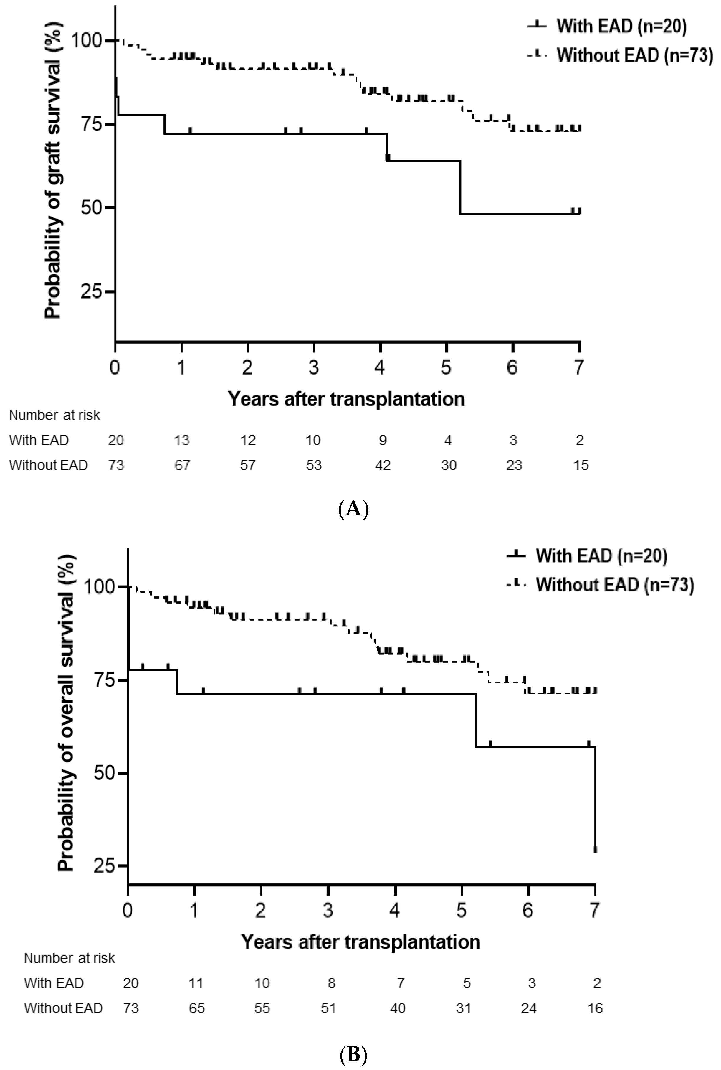
Figure 1. Cumulative probability of graft survival (A) and overall survival (B). Graft survival (p = 0.003) and overall survival (p = 0.003) were significantly lower in patients with early allograft dysfunction (EAD) compared with those without EAD. EAD, early allograft dysfunction.
Medicina 61 01710 g001
Figure 2. Cumulative probability of graft survival in LDLT (A) and DDLT (B). Graft survival was significantly lower in patients with EAD than in those without EAD in DDLT (p = 0.012) but not LDLT (p = 0.158). LDLT, living donor liver transplantation; DDLT, deceased donor liver transplantation; EAD, early allograft dysfunction.
Medicina 61 01710 g002
Figure 3. Cumulative probability of overall survival in LDLT (A) and DDLT (B). Overall survival in LDLT (p = 0.026) and DDLT (p = 0.033) was significantly lower in patients with EAD than in those without EAD. LDLT, living donor liver transplantation; DDLT, deceased donor liver transplantation; EAD, early allograft dysfunction.
Medicina 61 01710 g003
Figure 4. Association of donor age (A) and warm ischemic time (WIT) (B) with the risk of early allograft dysfunction (EAD). Curves were fitted using fractional polynomial regression; 95% confidence intervals are shown as gray shading.
Medicina 61 01710 g004
Table 1. Clinico-demographic findings.
With EAD (n = 20)Without EAD (n = 73)p
Age, years53.6 ± 8.854.1 ± 11.90.852
Sex (male)13 (65.0%)50 (68.5%)0.767
BMI24.5 ± 4.223.6 ± 4.00.358
Diabetes mellitus14 (70.0%)51 (69.9%)0.991
Hypertension16 (80.0%)57 (78.1%)0.853
Original liver disease
HBV5 (25.0%)10 (13.7%)
HCV03 (4.1%)
Alcoholic9 (45.0%)31 (42.5%)
HCC1 (5.0%)20 (27.4%)
Cholestatic06 (8.2%)
Acute liver failure5 (25.0%)3 (4.1%)0.010
Laboratory MELD29.5 (6–40)15 (6–40)0.027
Donor age, years49.6 ± 16.539.2 ± 13.30.005
ABOi2 (10.0%)8 (11.0%)0.902
DDLT10 (50.0%)22 (30.1%)0.098
GRWR < 0.82 (10.0%)7 (9.6%)0.956
Hepatic steatosis > 10%2 (10.0%)6 (8.2%)0.801
Operative time, min460 (225–815)475 (285–830)0.523
CIT, h3.5 ± 2.72.3 ± 2.00.030
WIT, min48.6 ± 31.837.3 ± 8.80.008
Peak AST within 7 days, U/L 3614 ± 5192424 ± 3360.000
Peak ALT within 7 days, U/L1847 ± 2179357 ± 2740.000
Postoperative hospital stay, days52.2 ± 73.238.8 ± 43.50.305
Table 2. Risk factors analysis for graft survival.
UnivariateMultivariate
HR95% CIpHR95% CIp
Age, years0.9940.956−1.0340.763
Sex (male)1.5800.584−4.2710.367
BMI0.9820.885−1.0910.740
DM1.5410.571−4.1600.393
HTN1.7680.524−5.9640.358
Primary liver disease1.0640.823−1.3750.636
Acute hepatic failure2.2730.671−7.7010.187
Hepatocellular carcinoma0.6530.221−1.9280.441
Laboratory MELD1.0170.983−1.0530.334
Donor age, years1.0170.989−1.0470.242
ABOi0.8140.191−3.4760.782
DDLT1.1300.479−2.6670.781
Hepatic steatosis > 10%0.4260.057−3.1580.403
Operative time, min0.9990.996−1.0030.716
CIT, h1.1320.946−1.3560.177
WIT, min1.0241.006−1.0420.010
EAD3.3881.457−7.8810.0052.8661.169−7.0250.021
Table 3. Risk factors analysis for overall survival.
UnivariateMultivariate
HR95% CIpHR95% CIp
Age, years1.0160.976−1.0580.433
Sex (male)2.1820.745−6.3920.155
BMI1.0330.938−1.1380.512
DM0.9800.405−2.3720.964
HTN1.2350.421−3.6260.701
Primary liver disease0.9580.709−1.2950.781
Acute hepatic failure2.7850.821−9.4440.100
Hepatocellular carcinoma1.0180.403−2.5730.969
Laboratory MELD1.0060.971−1.0410.755
Donor age, years1.0170.989−1.0450.232
ABOi0.8920.209−3.8020.877
DDLT0.8530.353−2.0610.724
Hepatic steatosis > 10%0.8560.200−3.6550.834
Operative time, min1.0010.998−1.0050.406
CIT, h1.0330.862−1.2370.726
WIT, min1.0251.009−1.0410.0021.0181.001−1.0350.038
EAD3.3291.450−7.6450.0052.7281.119−6.6510.027
Table 4. Risk factor analysis for EAD.
UnivariateMultivariate
HR95% CIpHR95% CIp
Age, years0.9960.953−1.0410.850
Sex (male)0.8540.301−2.4250.767
BMI1.0590.938−1.1960.355
DM1.0070.342−2.9610.991
HTN1.1230.329−3.8340.853
Acute hepatic failure7.7781.674−36.1410.0096.2281.179−32.9060.031
Hepatocellular carcinoma0.1390.018−1.1120.063
Laboratory MELD1.0451.004−1.0880.031
Donor age, years1.0531.014−1.0930.0081.0511.008−1.0960.020
ABOi0.9030.176−4.6310.903
DDLT2.3180.845−6.3590.102
Hepatic steatosis > 10%1.2410.231−6.6760.802
Operative time, min1.0020.997−1.0060.476
CIT, h1.2570.037−1.5570.037
WIT, min1.0401.000−1.0820.0511.0481.001−1.0980.046
Disclaimer/Publisher’s Note: The statements, opinions and data contained in all publications are solely those of the individual author(s) and contributor(s) and not of MDPI and/or the editor(s). MDPI and/or the editor(s) disclaim responsibility for any injury to people or property resulting from any ideas, methods, instructions or products referred to in the content.