Systematic Review

Does the Uterine Injection Site Matter for the Pelvic Sentinel Lymph Node Mapping? A Systematic Review and Meta-Analysis

by
Pier Carlo Zorzato
1,†,
Simone Garzon
1,†,
Mariachiara Bosco
1,
Filippo Ferrari
1,
Francesca Magni
1,
Rosa Maria Laterza
2,
Antonio Simone Laganà
3,
Francesco Fanfani
4,5 and
Stefano Uccella
1,*
1
Unit of Obstetrics and Gynecology, Department of Surgery, Dentistry, Pediatrics, and Gynecology, AOUI Verona, University of Verona, 37125 Verona, Italy
2
Division of General Gynecology and Gynecologic Oncology, Department of Obstetrics and Gynecology, Medical University of Vienna, 1090 Wien, Austria; Karl Landsteiner Society for Special Gynecology and Obstetrics, 3100 St. Pölten, Austria
3
Unit of Obstetrics and Gynecology, Department of Health Promotion, Mother and Child Care, Internal Medicine and Medical Specialties (PROMISE), “Paolo Giaccone” Hospital, University of Palermo, 90127 Palermo, Italy
4
Gynecologic Oncology Unit, Department of Women, Children and Public Health Sciences, Fondazione Policlinico Universitario Agostino Gemelli IRCCS, 00168 Rome, Italy
5
Catholic University of the Sacred Heart, 00168 Rome, Italy
*
Author to whom correspondence should be addressed.
These authors contributed equally to this work.
Medicina 2025, 61(4), 699; https://doi.org/10.3390/medicina61040699
Submission received: 9 March 2025 / Revised: 24 March 2025 / Accepted: 6 April 2025 / Published: 10 April 2025
(This article belongs to the Section Obstetrics and Gynecology)

Abstract

Background and Objectives: To summarize the evidence on in vivo uterine pelvic lymphatic drainage. Materials and Methods: A literature search was performed in multiple electronic databases from inception to December 2024. We included all the studies that compared two different uterine injection sites in the mapping of pelvic sentinel lymph nodes by injecting two different tracers into two distinct injection sites. The primary outcomes included the concordance and discordance rates in the mapped pelvic sentinel lymph nodes between the pairs of injection sites. The secondary outcomes were the detection rates per injection site and tracer. Four reviewers independently reviewed the records for inclusion, assessed the risk of bias, and extracted the data. Pooled concordance, discordance, and detection rates with 95% confidence intervals (CIs) were estimated using the random effects model. Heterogeneity was quantified using the I2 tests. Results: Out of 2512 records, we included 4 studies (172 patients and 344 hemipelves). Three studies injected the cervix with the technetium-99m and the uterine corpus with methylene blue; one study injected the cervix with indocyanine green and the utero-ovarian ligament with methylene blue. Both tracers/injection sites successfully identified a sentinel lymph node in 132 hemipelves (132/344; 38.4%), identifying the same sentinel lymph node in 116 cases (116/132; 87.9%). The pooled concordance rate per hemipelvis was 91.8% (95% CI 0.665–1.000; I2 = 92%; chi2 p-value < 0.01). Two different sentinel lymph nodes were identified in the remaining 16 hemipelves, with a pooled hemipelvis discordance rate of 8.2% (95% CI 0.000–0.335; I2 = 92%; chi2 p-value < 0.01). The cervix and technetium-99m were the injection site and tracer with the highest pooled detection rate. Conclusions: Different uterine injection sites appear to share a common pelvic lymphatic pathway and sentinel lymph node in most cases, consistent with the current practice in endometrial cancer. Future research will confirm whether cervical injections might be proposed for pelvic sentinel lymph node mapping in all gynecological cancers.

1. Introduction

Sentinel lymph node mapping algorithms have been integrated into the staging of most gynecological cancers [1,2]. Only in apparent early-stage ovarian neoplasm is this technique still debated [3,4,5].
Sentinel lymph node mapping algorithms are based on the anatomy of the lymphatic system and the expected drainage pathways from the genital organs [6,7], directly affecting the choice of anatomical sites for tracer injection. However, evidence regarding the actual in vivo lymphatic drainage is limited [8,9]. Specifically, whether different anatomic sites of tracer injection drain differently or equally in vivo, identifying a different versus the same sentinel lymph node is not clear for all possible alternative injection sites.
Testing whether two different tracer injection sites map to the same or a different sentinel lymph node is relevant when the most intuitive method employs a more complex and less reliable technique. In endometrial cancer, the cervical injection has been compared with and favored over hysteroscopic peritumoral or uterine corpus tracer injections because they were equally effective yet easier [10]. A similar issue has been raised for sentinel lymph node mapping in apparent early-stage ovarian cancer. Observed poor pelvic detection rate raises concerns about the suitability of the utero-ovarian ligament as an appropriate uterine injection site for pelvic sentinel lymph node mapping [4,11]. Technical issues and possible inefficient pelvic lymphatic drainage, particularly in delayed staging with previous adnexectomy [5], highlights the need to use other injection sites [4].
Based on this, an in-depth understanding of the uterine pelvic lymphatic drainage system in vivo is paramount for pelvic sentinel lymph node mapping in apparent early-stage ovarian cancer [4]. Therefore, we conducted a systematic review and meta-analysis of the studies that have investigated uterine pelvic lymphatic drainage by injecting two different tracers into two distinct uterine injection sites. The use of two tracers in two distinct uterine injection sites enables a direct comparison in the same patient and clarifies whether the identified pelvic SLN is the same or different. This evidence may be crucial in supporting or disputing the use of different injection sites than the specific tumor location for pelvic SLN mapping in gynecological cancers (i.e., the cervix in endometrial cancer and potentially in ovarian cancer).

2. Materials and Methods

The systematic review and meta-analysis was planned before beginning the online search and conducted in accordance with the Cochrane Handbook for Systematic Reviews of Diagnostic Test Accuracy [12]. The findings were reported following the Preferred Reporting Items for Systematic Reviews and Meta-Analyses of Diagnostic Test Accuracy (PRISMA-DTA) [13]. The protocol has been registered on PROSPERO (CRD 42024612435).

2.1. Search Strategy and Eligibility Criteria

A certified professional librarian (Biblioteca Meneghetti—Verona University) performed a literature search, from database inception to December 2024, in the following electronic databases: EMBASE, Scopus, PubMed, Web of Science, and the Cochrane Library. The search strategy included combinations of the following keywords: “ovarian cancer”, “endometrial cancer”, “cervical cancer”, “Indocyanine green”, “Methylene blue”, “Technetium 99”, “Lymph Node Metastasis”, “sentinel lymph node”, and “injection”. The detailed search strategy is available in the Supplementary Materials. The references of all included records were systematically revised.
We included all the studies meeting the predetermined inclusion criteria. The population comprised patients with gynecological cancer undergoing pelvic sentinel lymph node mapping. For the index test, pelvic sentinel lymph node mapping was performed by injecting a tracer into a specific site along the female genital tract; for the comparator test, a different tracer was injected into a second different site along the female genital tract. The primary outcomes were the concordance and discordance rates in the pelvic sentinel lymph nodes mapped per hemipelvis between the pairs of injection sites; the secondary outcomes were injection-site-specific and tracer-specific detection rates. We excluded any records that did not present original study data. No language exclusion criterion was applied; non-English records were translated with Google Translator [14].

2.2. Study Selection and Data Extraction

After duplicate removal, two reviewers independently screened the titles and abstracts of the identified records (PCZ, FFA). Two other reviewers (SG, FM) retrieved and independently assessed the full text of potentially eligible publications. Any disagreement was resolved with a further reviewer (SU). Two reviewers (PCZ, SG) developed and used a standardized form to extract the following data from the selected studies: authors, publication year, research setting, study design, patient population (i.e., age, number of patients, and types, histotype, and stages of cancer), sites of injection, injection techniques, types of tracers, adverse events, outcome measures (hemipelvis level data), and items for quality evaluation.

2.3. Assessment of Risk of Bias

Two reviewers (FFA and PCZ) independently assessed the risk of bias in the selected studies following a modified version of the Quality Assessment of Diagnostic Accuracy Studies tool 2 (QUADAS-2) (Supplementary Materials). Any disagreement was resolved with a further reviewer (SG) [15].

2.4. Data Synthesis

The concordance rate was defined as the number of hemipelves with the same sentinel lymph node mapped by both tracers out of the total number of hemipelves with double successful mapping. The discordance rate was defined as the number of hemipelves with two different sentinel lymph nodes mapped by the two tracers out of the total number of hemipelves with double successful mapping. The concordance and discordance rates were calculated per pair of injection sites. The detection rate was defined as the number of hemipelves in which at least one sentinel lymph node was successfully identified out of the total number of hemipelves, according to the injection site and tracer.
Proportion meta-analyses were conducted using the random effects model to estimate the pooled proportions with 95% confidence intervals (CIs) for the concordance and discordance rates per pair of injection sites and detection rates per injection site and tracer. Heterogeneity was quantified using the I² tests; an I² less than 25% was considered low, and an I² more than 75% was considered high. When ten or more studies were identified, publication bias was assessed by funnel plot and Egger test. All analyses were two-tailed with a statistical significance threshold of p < 0.05. R Statistical Software (v4.1.2; R Core Team 2024, Vienna, Austria) was used.

3. Results

3.1. Study Selection

The initial literature search retrieved 2512 records, including a cross-reference review. After duplicate removal, 1491 records underwent title and abstract screening, with 34 articles that underwent a full-text assessment. Thirty studies were excluded for two reasons. Most studies were excluded because they investigated a single injection site or tracer. When two uterine injection sites were analyzed, the same tracer was used, or two different tracers were injected into the same uterine site. A minority did not provide the outcome of interest; although two different tracers were injected into two different uterine sites, the authors did not provide the concordance or discordance rate. Therefore, four records regarding four different studies were finally included (Table 1) [16,17,18,19]. The PRISMA Flowchart illustrates the study selection process (Figure S1).

3.2. Study Characteristics

The four studies encompassed 172 patients who underwent pelvic sentinel lymph node mapping for endometrial cancer (Table 1). Three studies injected the cervix with technetium-99m and the uterine corpus with methylene blue [17,18,19]; one study injected the cervix with indocyanine green and the utero-ovarian ligament with methylene blue [16]. In all four studies, the cervical injection was compared with an alternative injection site. The data on sentinel lymph node mapping are reported in Table 2.

3.3. Risk of Bias Assessment

Two studies were considered at high risk of bias in the patient selection domain because of the exclusion of cases with suspicious lymph nodes on preoperative imaging [18,19]. One study failed to provide details regarding the patient recruitment method, so the selection domain was considered unclear [17]. One study was deemed unclear in the reference standard domain because of missing detailed clinical data [18] (Figure S2, Table S1).

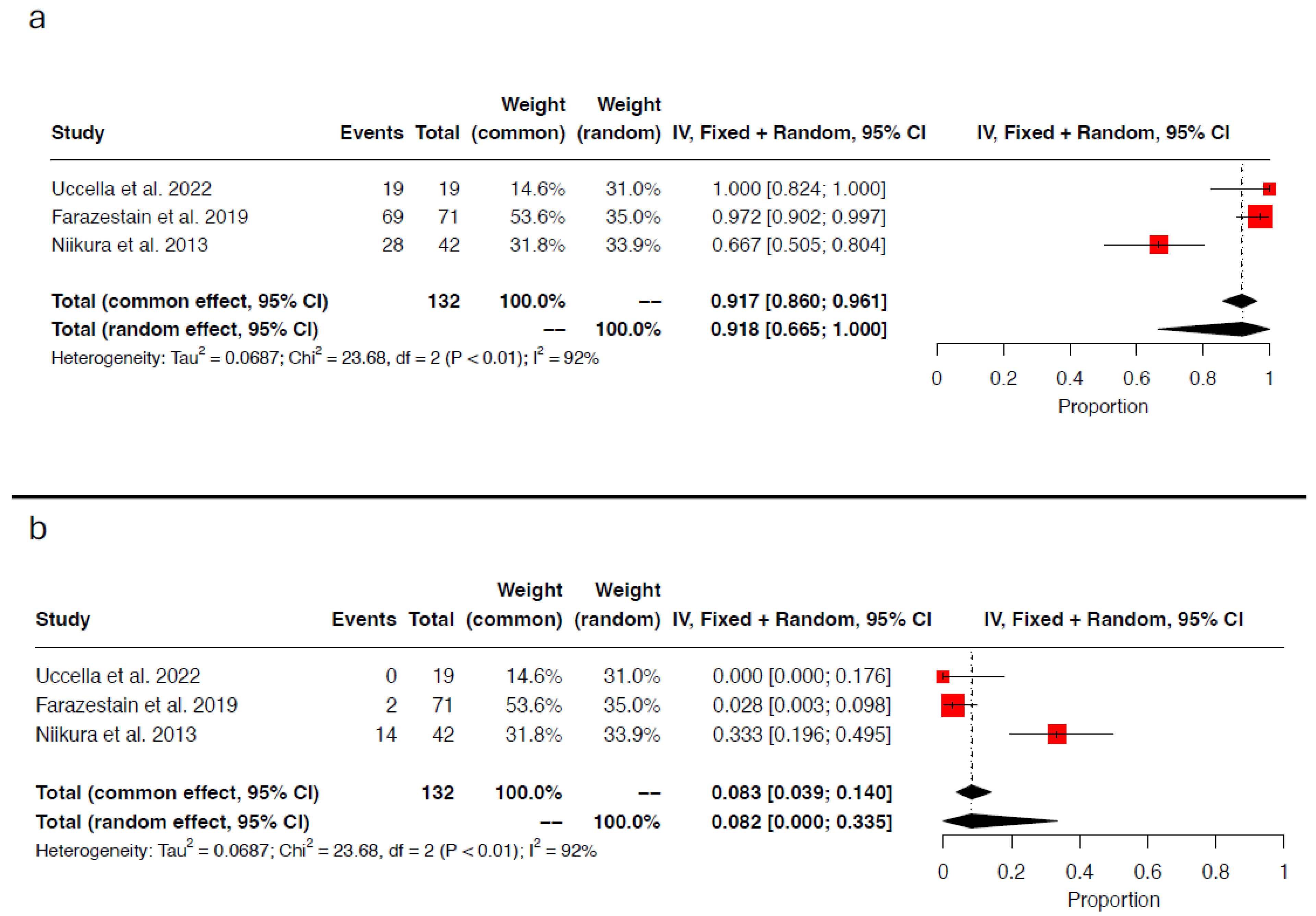
3.4. Concordance and Discordance Rates

Both tracers/injection sites successfully identified of at least one sentinel lymph node in 132 hemipelves (132/344; 38.4%) and the same sentinel lymph node in 116 out of 132 hemipelvis (116/132; 87.9%). The pooled concordance rate per hemipelvis was 91.8% (95% CI 0.665–1.000; I2 = 92%; chi2 p-value < 0.01) (Figure 1a). Two different sentinel lymph nodes were detected in the remaining 16 hemipelves, leading to a pooled hemipelvis discordance rate of 8.2% (95% CI 0.000–0.335; I2 = 92%; chi2 p-value < 0.01) (Figure 1b).

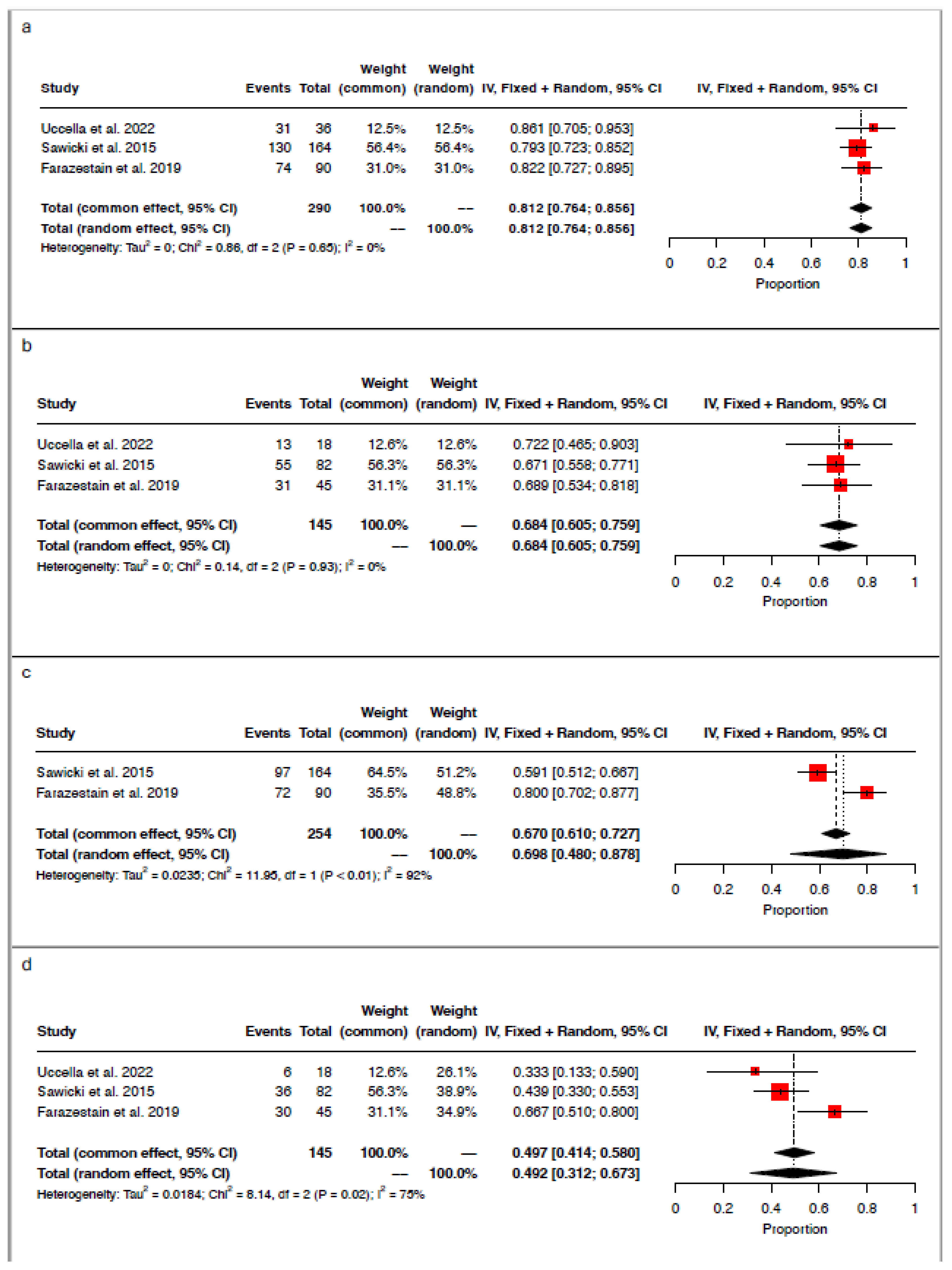
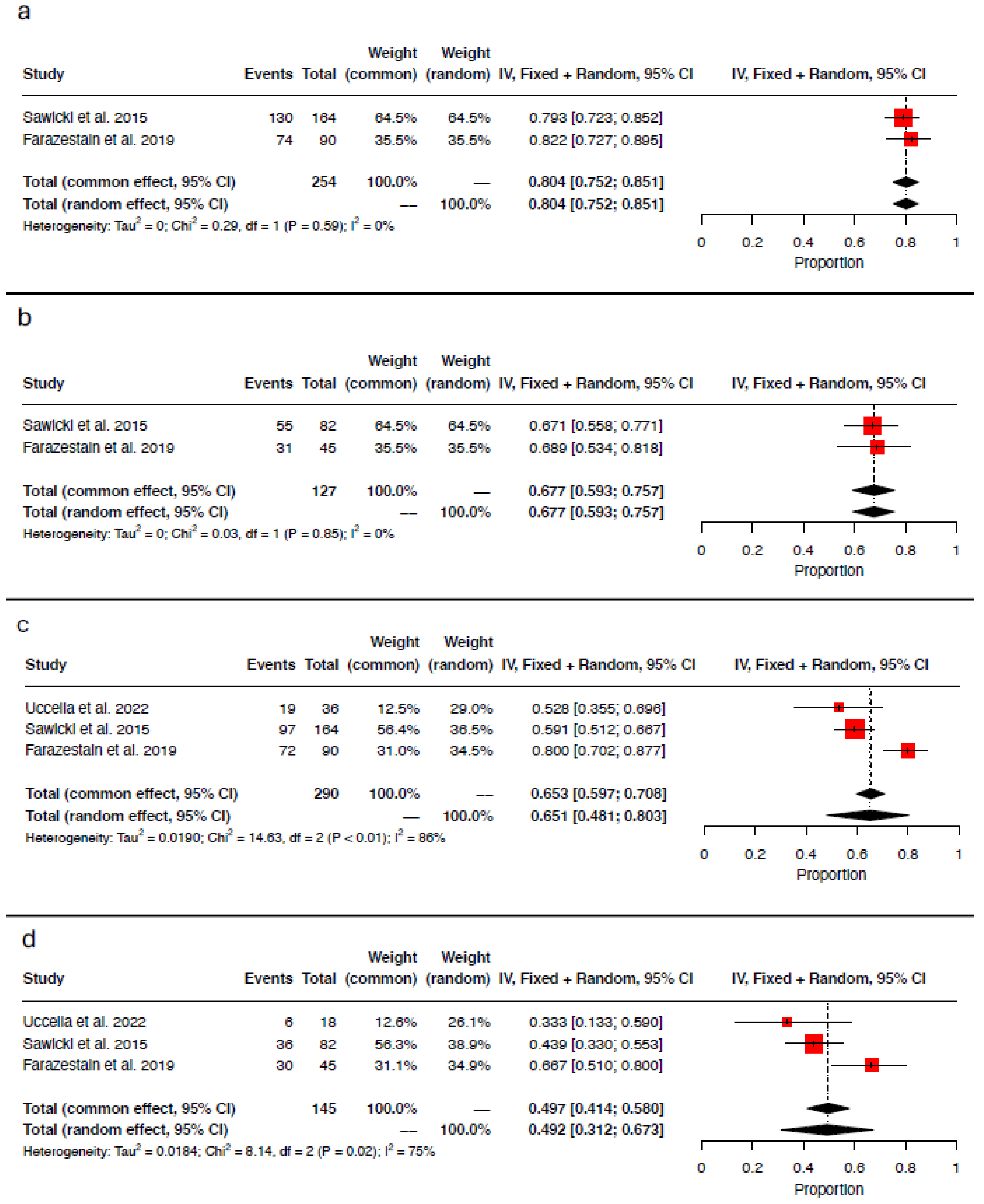
3.5. Detection Rate per Injection Site

The pooled detection rate for the cervical injection (technetium-99m or indocyanine green) was 81.2% (95% CI 0.764–0.856; I2 = 0%; chi2 p-value = 0.65), with 235/290 hemipelves with one identified sentinel lymph node (Figure 2a). Bilateral mapping was achieved in 99/145 patients, leading to a pooled bilateral detection rate for cervical injection of 68.4% (95% CI 0.605–0.759; I2 = 0%; chi2 p-value = 0.93; Figure 2b).
Regarding the uterine corpus injection (methylene blue), the pooled hemipelvis detection rate was 69.8% (95% CI 0.480–0.878; I2 = 92%; chi2 p-value < 0.01; Figure 2c), with 169/254 hemipelves having an identified sentinel lymph node. Bilateral mapping was achieved in 72/145 cases, with a pooled bilateral detection rate of 49.2% (95% CI 0.312–0.673-; I2 = 75%; chi2 p-value = 0.02; Figure 2d).
Only 18 patients from the study by Uccella et al. [16] underwent a tracer injection into the utero-ovarian ligament (methylene blue). At least one sentinel lymph node was identified in 19 out of 36 hemipelves, with a detection rate of 52.8% and a bilateral detection rate of 33.3% [16].

3.6. Detection Rate per Tracer

Only two authors reported results on technetium-99m alone (cervix) [17,19]. According to these findings, 204/254 hemipelves had at least one detected sentinel lymph node (pooled detection rate 80.4%; 95% CI 0.752–0.851; I2 = 0%; chi2 p-value = 0.59; Figure 3a). Bilateral mapping was achieved in 86/127 patients, with a pooled Technetium-99m bilateral detection rate of 67.7% (95% CI 0.593–0.757; I2 = 0%; chi2 p-value = 0.85; Figure 3b). Data regarding the injection of methylene blue alone (uterine corpus or utero-ovarian ligament) were available from three authors [16,17,19]. At least one sentinel lymph node was detected in 188/290 hemipelves (pooled methylene blue detection rate 65.1%; 95% CI 0.481–0.803; I2 = 86%; chi2 p-value <0.01; Figure 3c). Bilateral mapping was accomplished in 72/145 individuals, resulting in a pooled methylene blue bilateral detection rate of 49.2% (95% CI 0.312–0.673; I2 = 75%; chi2 p value = 0.02; Figure 3d). Indocyanine green (cervix) was employed exclusively in the study by Uccella et al. [16], with a detection rate of 86.1% per hemipelvis and a bilateral detection rate of 72.2%.

4. Discussion

All studies that investigated pelvic sentinel lymph node mapping by injecting two different tracers into two distinct injection sites compared the cervical injection with the uterine corpus or the utero-ovarian ligament, and the pooled concordance rate in the identified pelvic sentinel lymph node was high. The cervix and technetium-99m reported the highest detection rates.
The studies investigating uterine corpus/perilesional versus cervical injections for pelvic sentinel lymph node mapping in endometrial cancer were based on the assumption that different injection sites would determine different pelvic sentinel lymph nodes. An injection site distant from the tumor may be less valid, not reflecting the tumoral lymphatic drainage [20,21,22,23]. In opposition to this assumption, the pooled high concordance rate suggests that a dominant uterine lymphatic drainage pathway and associated sentinel lymph node may be present in most female pelves. In most cases, the two tracers identified the same pelvic sentinel lymph node regardless of the injection site. This is consistent with the current practice and available evidence in endometrial cancer [24]. The studies that compared the cervical versus hysteroscopic peritumoral or uterine corpus injections in endometrial cancer did not observe differences in the anatomical distribution of the pelvic sentinel lymph nodes [10,20,21]. Furthermore, the pelvic sentinel lymph node location was not observed to be associated with the endometrial cancer location in the uterine cavity [25].
Nevertheless, in a minority of cases, the pelvic sentinel lymph node identified differed between the two injection sites, and only one of the two would have been identified with the standard practice, resulting in possible suboptimal staging. This observation potentially questions the use of an injection site different from the tumor site for all gynecological cancers. However, caution is needed when interpreting the pooled discordance rate. Two different homolateral pelvic sentinel lymph nodes for the uterus do not imply suboptimal staging. Identifying two pelvic sentinel lymph nodes on the same side is common; more efficient tracers with adequate waiting time may identify both sentinel lymph nodes. In the FILM trial, a mean number of 3.2 (1.6) pelvic sentinel lymph nodes per patient was obtained [26]. Additionally, the pooled discordance rate was characterized by high heterogeneity. The study by Niikura et al. provided a discordance rate higher than the other studies, raising concerns on whether study-specific factors may have increased the discordance rate. A sensitivity analysis excluding Niikura et al. showed higher concordance (98.5%; 95% CI 0.94–1.000; I2 = 0%) and lower discordance (2.8%; 95% CI 0.001–0.079; I2 = 0%) rates with low heterogeneity. The high concordance and low discordance rates support the hypothesis that two different uterine injection sites may detect the same pelvic SLN, allowing the choice of the injection site to be based on other factors. These observations are consistent with the current use of the cervical injection as a substitute for the more precise injection at the tumor level in endometrial cancer [20,21,24].
The pooled detection rate was higher for technetium-99m than methylene blue. Indocyanine green was only used in one study, and meta-analysis was not feasible. These results are consistent with the current practice and available evidence showing methylene blue as the tracer with the lowest performance [26,27,28,29]. In this regard, differences between the tracers may explain the discrepancies between injection sites, as methylene blue was constantly used for the injection site alternative to the cervix.
However, the advantage of using a cervical injection in mapping the pelvis over other uterine injection sites has already been observed. In most investigations, the pelvic sentinel lymph node detection rate was higher with the cervical injection of indocyanine green than with a perilesional hysteroscopic/uterine corpus injection [10,20]. The risk of tracer spillage and the lower injected volume associated with a hysteroscopic injection are technical issues that may increase the risk of failure and the need for experienced surgeons [10,20]. Technical issues and a lower pelvic lymphatic drainage efficiency may explain the similarly low pelvic sentinel lymph node detection rate observed for the utero-ovarian ligament. The most comprehensive meta-analysis on sentinel lymph node mapping in apparent early-stage ovarian cancer reported a pelvic detection rate of 59.4% (66/111) [4]. Notably, Rey et al. estimated the pelvic detection rate, by utero-ovarian ligament injection, only among 111 out of 239 total cases (46.4%). Most cases were excluded due to protocol violations in the utero-ovarian ligament injection, which were reported in up to 69.8% of cases in the SELLY trial [4,11,30]. Laven et al., who focused on patients who underwent restaging after adnexectomy, provided a pelvic detection rate of 27% [31]. On these bases, the question of whether cervical injections may be equal to but more efficient and straightforward than utero-ovarian ligament injections for sentinel lymph node mapping in apparent early-stage ovarian cancer merits further investigation [4]. Unlike other studies [11,31], Lago et al. reported a higher detection rate of 93% [32]. This difference may be attributed to the deeper injection of the tracer near the dorsal/lateral parametrium rather than in the utero-ovarian stump [11].
This is the first systematic review and meta-analysis that specifically investigated whether using two different injection sites of the uterus would map the same or a different pelvic SLN. The rigorous and systematic approach with strict adherence to the predetermined criteria strengthens the study results. The selective inclusion of studies in which two different uterine injection sites were investigated using two different tracers in the same patient allowed for an in vivo anatomical investigation of the uterine lymphatic drainage system with a direct comparison between the injection site pairs. However, identifying only four studies involving 172 patients limits the study results. Moreover, only studies enrolling patients with endometrial cancer were included; therefore, caution is needed when inferring hypotheses on other gynecological malignancies. Additionally, indocyanine green was underrepresented. Considering indocyanine green as the standard, the results provided can be regarded as outdated. However, the tracer and underlying cancer are unlikely to represent confounders for the concordance rate. Furthermore, none of the trials randomized the tracer to the two injection sites, and only one study was designed for the anatomical investigation of uterine lymphatic drainage [16]. These characteristics determine that the results are often secondary outcomes, and that each injection site was linked to a specific tracer, thereby connecting the two performances. Moreover, the inclusion of two studies with a high risk of bias in the patient selection domain, due to the exclusion of cases with suspicious lymph nodes on preoperative imaging, raises concerns about generalizability. Finally, the identification of only four studies also impeded investigations into the presence of publication bias.
Understanding the pelvic lymphatic drainage of the uterus impacts the choice of the tracer injection site for pelvic sentinel lymph node mapping [6,7]. Clarifying whether two different uterine sites for tracer injection drain differently or equally in the pelvis becomes essential, especially given that the more intuitive peritumoral injection is a more complex technique with a high failure rate and low detection rate. Our results suggest that, in most patients, a main pelvic lymphatic drainage pathway and a common sentinel lymph node are present regardless of the injection site. These observations are consistent with the available evidence and support the current practice in endometrial cancer [24]. Moreover, our results may have implications for pelvic sentinel lymph node mapping in apparent early-stage ovarian cancer, supporting further investigations into whether the cervical injection may be equal but more efficient than the utero-ovarian ligament injection.

5. Conclusions

Our findings support the hypothesis that different uterine pelvic lymphatic pathways share a common pelvic sentinel lymph node in most cases. These observations support the current clinical practice in endometrial cancer and have significant implications on sentinel lymph node mapping in apparent early-stage ovarian cancer. The cervical injection of indocyanine green, with advantages in terms of accessibility and reliability, might be proposed in future investigations into pelvic sentinel lymph node mapping in apparent early-stage ovarian cancer.

Supplementary Materials

The following supporting information can be downloaded at: https://www.mdpi.com/article/10.3390/medicina61040699/s1, Figure S1: PRISMA flowchart of study selection; Figure S2: Risk of Bias assessment with the Modified Quality Assessment of Diagnostic Accuracy Studies tool 2 (QUADAS-2); Table S1: Risk of bias assessment for each included study based on the Modified Quality Assessment of Diagnostic Accuracy Studies tool 2 (QUADAS-2).

Author Contributions

Conceptualization: P.C.Z., S.G., F.F. (Filippo Ferrari) and S.U.; Data Curation: P.C.Z., F.F. (Filippo Ferrari), F.M. and S.G.; Formal Analysis: S.G. and P.C.Z.; Methodology: S.G., S.U., F.F. (Francesco Fanfani) and F.M.; Supervision: S.U. and F.F. (Francesco Fanfani); Validation: R.M.L. and A.S.L.; Writing—original draft: P.C.Z., S.G., M.B. and S.U.; Writing—review and editing: All authors. All authors have read and agreed to the published version of the manuscript.

Funding

This research received no external funding.

Conflicts of Interest

The authors declare no conflicts of interest.

References

  1. Concin, N.; Matias-Guiu, X.; Vergote, I.; Cibula, D.; Mirza, M.R.; Marnitz, S.; Ledermann, J.; Bosse, T.; Chargari, C.; Fagotti, A.; et al. ESGO/ESTRO/ESP Guidelines for the Management of Patients with Endometrial Carcinoma. Int. J. Gynecol. Cancer Off. J. Int. Gynecol. Cancer Soc. 2021, 31, 12–39. [Google Scholar] [CrossRef] [PubMed]
  2. Cibula, D.; Raspollini, M.R.; Planchamp, F.; Centeno, C.; Chargari, C.; Felix, A.; Fischerová, D.; Jahnn-Kuch, D.; Joly, F.; Kohler, C.; et al. ESGO/ESTRO/ESP Guidelines for the Management of Patients with Cervical Cancer-Update 2023. Int. J. Gynecol. Cancer Off. J. Int. Gynecol. Cancer Soc. 2023, 33, 649–666. [Google Scholar] [CrossRef] [PubMed]
  3. Agusti, N.; Viveros-Carreño, D.; Grillo-Ardila, C.; Izquierdo, N.; Paredes, P.; Vidal-Sicart, S.; Torne, A.; Díaz-Feijoo, B. Sentinel Lymph Node Detection in Early-Stage Ovarian Cancer: A Systematic Review and Meta-Analysis. Int. J. Gynecol. Cancer Off. J. Int. Gynecol. Cancer Soc. 2023, 33, 1493–1501. [Google Scholar] [CrossRef] [PubMed]
  4. Rey, I.; Lago, V.; Arnáez, M.; Bizzarri, N.; Agustí, N.; Nero, C.; Díaz-Feijoo, B.; Padilla-Iserte, P.; Domingo, S. Key Issues in Diagnostic Accuracy of Sentinel Lymph Node Biopsy in Early-Stage Ovarian Cancer: Systematic Review and Meta-Analysis. Int. J. Gynecol. Cancer Off. J. Int. Gynecol. Cancer Soc. 2024, 34, 1787–1794. [Google Scholar] [CrossRef]
  5. Uccella, S.; Zorzato, P.C.; Lanzo, G.; Fagotti, A.; Cianci, S.; Gallina, D.; Gueli Alletti, S.; Monterossi, G.; Franchi, M.; Ghezzi, F.; et al. The Role of Sentinel Node in Early Ovarian Cancer: A Systematic Review. Minerva Med. 2019, 110, 358–366. [Google Scholar] [CrossRef]
  6. Kleppe, M.; Kraima, A.C.; Kruitwagen, R.F.P.M.; Van Gorp, T.; Smit, N.N.; Van Munsteren, J.C.; DeRuiter, M.C. Understanding Lymphatic Drainage Pathways of the Ovaries to Predict Sites for Sentinel Nodes in Ovarian Cancer. Int. J. Gynecol. Cancer 2015, 25, 1405–1414. [Google Scholar] [CrossRef]
  7. Ercoli, A.; Delmas, V.; Iannone, V.; Fagotti, A.; Fanfani, F.; Corrado, G.; Ferrandina, G.; Scambia, G. The Lymphatic Drainage of the Uterine Cervix in Adult Fresh Cadavers: Anatomy and Surgical Implications. Eur. J. Surg. Oncol. J. Eur. Soc. Surg. Oncol. Br. Assoc. Surg. Oncol. 2010, 36, 298–303. [Google Scholar] [CrossRef]
  8. Geppert, B.; Lönnerfors, C.; Bollino, M.; Arechvo, A.; Persson, J. A Study on Uterine Lymphatic Anatomy for Standardization of Pelvic Sentinel Lymph Node Detection in Endometrial Cancer. Gynecol. Oncol. 2017, 145, 256–261. [Google Scholar] [CrossRef]
  9. Vanneuville, G.; Mestas, D.; Le Bouedec, G.; Veyre, A.; Dauplat, J.; Escande, G.; Guillot, M. The Lymphatic Drainage of the Human Ovary in Vivo Investigated by Isotopic Lymphography before and after the Menopause. Surg. Radiol. Anat. SRA 1991, 13, 221–226. [Google Scholar] [CrossRef]
  10. Ditto, A.; Casarin, J.; Pinelli, C.; Perrone, A.M.; Scollo, P.; Martinelli, F.; Bogani, G.; Leone Roberti Maggiore, U.; Signorelli, M.; Chiappa, V.; et al. Hysteroscopic versus Cervical Injection for Sentinel Node Detection in Endometrial Cancer: A Multicenter Prospective Randomised Controlled Trial from the Multicenter Italian Trials in Ovarian Cancer (MITO) Study Group. Eur. J. Cancer 2020, 140, 1–10. [Google Scholar] [CrossRef]
  11. Nero, C.; Bizzarri, N.; Di Berardino, S.; Sillano, F.; Vizzielli, G.; Cosentino, F.; Vargiu, V.; De Iaco, P.; Perrone, A.M.; Vizza, E.; et al. Sentinel-Node Biopsy in Apparent Early Stage Ovarian Cancer: Final Results of a Prospective Multicentre Study (SELLY). Eur. J. Cancer 2024, 196, 113435. [Google Scholar] [CrossRef] [PubMed]
  12. Deeks, J.; Bossuyt, P.; Leeflang, M.; Takwoingi, Y. Cochrane Handbook for Systematic Reviews of Diagnostic Test Accuracy, 2nd ed.; John Wiley & Sons: Hoboken, NJ, USA, 2023. [Google Scholar]
  13. McInnes, M.D.F.; Moher, D.; Thombs, B.D.; McGrath, T.A.; Bossuyt, P.M.; the PRISMA-DTA Group. Preferred Reporting Items for a Systematic Review and Meta-Analysis of Diagnostic Test Accuracy Studies: The PRISMA-DTA Statement. JAMA 2018, 319, 388–396. [Google Scholar] [CrossRef] [PubMed]
  14. Balk, E.M.; Chung, M.; Chen, M.L.; Trikalinos, T.A.; Kong Win Chang, L. Assessing the Accuracy of Google Translate to Allow Data Extraction From Trials Published in Non-English Languages; AHRQ Methods for Effective Health Care; Agency for Healthcare Research and Quality: Rockville, MD, USA, 2013.
  15. Whiting, P.F.; Rutjes, A.W.S.; Westwood, M.E.; Mallett, S.; Deeks, J.J.; Reitsma, J.B.; Leeflang, M.M.G.; Sterne, J.A.C.; Bossuyt, P.M.M.; QUADAS-2 Group. QUADAS-2: A Revised Tool for the Quality Assessment of Diagnostic Accuracy Studies. Ann. Intern. Med. 2011, 155, 529–536. [Google Scholar] [CrossRef] [PubMed]
  16. Uccella, S.; Garzon, S.; Bosco, M.; Porcari, I.; Lanzo, G.; Laganà, A.S.; Chiantera, V.; Cliby, W.A.; Mariani, A.; Franchi, M.; et al. Cervical versus Utero-Ovarian Ligament Injection of the Tracer for the Pelvic Sentinel Lymph Node Mapping in Gynecologic Oncology: A Prospective Observational Study. Gynecol. Obstet. Investig. 2022, 87, 242–247. [Google Scholar] [CrossRef]
  17. Farazestanian, M.; Yousefi, Z.; Zarifmahmoudi, L.; Hasanzadeh Mofrad, M.; Kadkhodayan, S.; Sadeghi, R. Concordance Between Intracervical and Fundal Injections for Sentinel Node Mapping in Patients With Endometrial Cancer?: A Study Using Intracervical Radiotracer and Fundal Blue Dye Injections. Clin. Nucl. Med. 2019, 44, e123–e127. [Google Scholar] [CrossRef]
  18. Niikura, H.; Kaiho-Sakuma, M.; Tokunaga, H.; Toyoshima, M.; Utsunomiya, H.; Nagase, S.; Takano, T.; Watanabe, M.; Ito, K.; Yaegashi, N. Tracer Injection Sites and Combinations for Sentinel Lymph Node Detection in Patients with Endometrial Cancer. Gynecol. Oncol. 2013, 131, 299–303. [Google Scholar] [CrossRef]
  19. Sawicki, S.; Lass, P.; Wydra, D. Sentinel Lymph Node Biopsy in Endometrial Cancer-Comparison of 2 Detection Methods. Int. J. Gynecol. Cancer Off. J. Int. Gynecol. Cancer Soc. 2015, 25, 1044–1050. [Google Scholar] [CrossRef]
  20. Rossi, E.C.; Jackson, A.; Ivanova, A.; Boggess, J.F. Detection of Sentinel Nodes for Endometrial Cancer with Robotic Assisted Fluorescence Imaging: Cervical versus Hysteroscopic Injection. Int. J. Gynecol. Cancer Off. J. Int. Gynecol. Cancer Soc. 2013, 23, 1704–1711. [Google Scholar] [CrossRef]
  21. Gezer, Ş.; Duman Öztürk, S.; Hekimsoy, T.; Vural, Ç.; İşgören, S.; Yücesoy, İ.; Çorakçı, A. Cervical versus Endometrial Injection for Sentinel Lymph Node Detection in Endometrial Cancer: A Randomized Clinical Trial. Int. J. Gynecol. Cancer Off. J. Int. Gynecol. Cancer Soc. 2020, 30, 325–331. [Google Scholar] [CrossRef]
  22. Martinelli, F.; Ditto, A.; Signorelli, M.; Bogani, G.; Chiappa, V.; Lorusso, D.; Scaffa, C.; Recalcati, D.; Perotto, S.; Haeusler, E.; et al. Sentinel Node Mapping in Endometrial Cancer Following Hysteroscopic Injection of Tracers: A Single Center Evaluation over 200 Cases. Gynecol. Oncol. 2017, 146, 525–530. [Google Scholar] [CrossRef]
  23. Bogani, G.; Murgia, F.; Ditto, A.; Raspagliesi, F. Sentinel Node Mapping vs. Lymphadenectomy in Endometrial Cancer: A Systematic Review and Meta-Analysis. Gynecol. Oncol. 2019, 153, 676–683. [Google Scholar] [CrossRef] [PubMed]
  24. About the NCCN Clinical Practice Guidelines in Oncology (NCCN Guidelines®). Available online: https://www.nccn.org/professionals/default.aspx (accessed on 23 June 2020).
  25. Capozzi, V.A.; Armano, G.; Maglietta, G.; Rosati, A.; Vargiu, V.; Scarpelli, E.; Sozzi, G.; Chiantera, V.; Cosentino, F.; Gioè, A.; et al. Hysteroscopic Endometrial Tumor Localization and Sentinel Lymph Node Mapping. An Upgrade of the Hysteroscopic Role in Endometrial Cancer Patients. Eur. J. Surg. Oncol. J. Eur. Soc. Surg. Oncol. Br. Assoc. Surg. Oncol. 2023, 49, 106952. [Google Scholar] [CrossRef] [PubMed]
  26. Frumovitz, M.; Plante, M.; Lee, P.S.; Sandadi, S.; Lilja, J.F.; Escobar, P.F.; Gien, L.T.; Urbauer, D.L.; Abu-Rustum, N.R. Near-Infrared Fluorescence for Detection of Sentinel Lymph Nodes in Women with Cervical and Uterine Cancers (FILM): A Randomised, Phase 3, Multicentre, Non-Inferiority Trial. Lancet Oncol. 2018, 19, 1394–1403. [Google Scholar] [CrossRef] [PubMed]
  27. Ruscito, I.; Gasparri, M.L.; Braicu, E.I.; Bellati, F.; Raio, L.; Sehouli, J.; Mueller, M.D.; Panici, P.B.; Papadia, A. Sentinel Node Mapping in Cervical and Endometrial Cancer: Indocyanine Green Versus Other Conventional Dyes-A Meta-Analysis. Ann. Surg. Oncol. 2016, 23, 3749–3756. [Google Scholar] [CrossRef]
  28. Papadia, A.; Zapardiel, I.; Bussi, B.; Ghezzi, F.; Ceccaroni, M.; De Ponti, E.; Elisei, F.; Imboden, S.; de la Noval, B.D.; Gasparri, M.L.; et al. Sentinel Lymph Node Mapping in Patients with Stage I Endometrial Carcinoma: A Focus on Bilateral Mapping Identification by Comparing Radiotracer Tc99m with Blue Dye versus Indocyanine Green Fluorescent Dye. J. Cancer Res. Clin. Oncol. 2017, 143, 475–480. [Google Scholar] [CrossRef]
  29. Persson, J.; Salehi, S.; Bollino, M.; Lönnerfors, C.; Falconer, H.; Geppert, B. Pelvic Sentinel Lymph Node Detection in High-Risk Endometrial Cancer (SHREC-Trial)-the Final Step towards a Paradigm Shift in Surgical Staging. Eur. J. Cancer Oxf. Engl. 1990 2019, 116, 77–85. [Google Scholar] [CrossRef]
  30. Uccella, S.; Nero, C.; Vizza, E.; Vargiu, V.; Corrado, G.; Bizzarri, N.; Ghezzi, F.; Cosentino, F.; Turco, L.C.; Fagotti, A.; et al. Sentinel-Node Biopsy in Early-Stage Ovarian Cancer: Preliminary Results of a Prospective Multicentre Study (SELLY). Am. J. Obstet. Gynecol. 2019, 221, 324.e1–324.e10. [Google Scholar] [CrossRef]
  31. Laven, P.; Kruitwagen, R.; Zusterzeel, P.; Slangen, B.; Van Gorp, T.; Van Der Pol, J.; Lambrechts, S. Sentinel Lymph Node Identification in Early Stage Ovarian Cancer: Is It Still Possible after Prior Tumor Resection? J. Ovarian Res. 2021, 14, 132. [Google Scholar] [CrossRef]
  32. Lago, V.; Bello, P.; Montero, B.; Matute, L.; Padilla-Iserte, P.; Lopez, S.; Marina, T.; Agudelo, M.; Domingo, S. Sentinel Lymph Node Technique in Early-Stage Ovarian Cancer (SENTOV): A Phase II Clinical Trial. Int. J. Gynecol. Cancer 2020, 30, 1390–1396. [Google Scholar] [CrossRef]
Figure 1. Forest plot of proportional meta-analysis for (a) concordance rate per hemipelvis and (b) discordance rate per hemipelvis [16,17,18].
Medicina 61 00699 g001
Figure 2. Forest plot of proportional meta-analysis for (a) hemipelvis detection rate for cervical injection, (b) bilateral detection rate for cervical injection, (c) hemipelvis detection rate for uterine corpus injection, and (d) bilateral detection rate for uterine corpus injection [16,17,19].
Medicina 61 00699 g002
Figure 3. Forest plot of proportional meta-analysis for (a) hemipelvis detection rate for technetium-99m, (b) bilateral detection rate for technetium-99m, (c) hemipelvis detection rate for methylene blue, and (d) bilateral detection rate for methylene blue [16,17,19].
Medicina 61 00699 g003
Table 1. Characteristics of studies included.
Author, Year (Country)DesignN of PatientsHistotypeStage (FIGO)Lymphovascular InvasionSurgical ApproachSite of InjectionTracerTechniqueTime Tracer Injection—SLN Evaluation
Niikura et al., 2013 [18]
(Japan)
Prospective27NANANAOpen SurgeryCervix99mTcPre-operative lymphoscintigraphy by injecting Tc99m-labeled phytate in the cervix at 3, 6, 9, and 12 o’clock positions one day before surgery. At the time of surgery, a gamma-detecting probe was used to locate radioactive SLN.NA
CorpusMBInjection into five different sites of the subserosal endometrium.
Sawicki et al., 2015 [19]
(Poland)
Retrospective8296.4% endometrioid
1.2% clear cell
2.4% serous
IA: 47.6%
IB: 23.2%
II: 17.1%
IIIA: 2.4%
IIIB: 2.4%
IIIC1: 7.3%
Yes: 12.2%
No: 87.8%
Open SurgeryCervix99mTcInjection of the cervix with Tc99m-labeled nanocolloid and the SLN was subsequently located using a handheld gamma probe.NA
CorpusMB4 mL in the uterine fundus.
Farazestanian et al., 2019 [17]
(Iran)
Prospective4586.7% endometrioid
8.9% clear cell
4.4% serous
NAYes: 15.6%
No: 84.4%
Open SurgeryCervix99mTcTwo injections of Tc99m-phytate into the cervix at the 6- and 12-o’clock positions the day before surgery. Planar lymphoscintigraphy was conducted for each patient 8 to 18 h after injections, and surgery was scheduled 18 to 24 h after the radiotracer injection. Hot SLN were identified using a handheld gamma probe.15 min
CorpusMB2 mL at subserosal fundal midline locations
Uccella et al., 2022 [16]
(Italy)
Prospective1894.5% endometrioid
5.5% non endometrioid
IA: 38.8%
IB: 44.4%
II: 11.1%
IIIC: 5.6%
Yes: 55.5%
No: 44.5%
Conventional LaparoscopyCervixICG2 mL ICG at 3 and 9 o’clock positions, 1 mL deeply at 1.5–2.5 cm into the stroma, and 1 mL superficially into the submucosal tissue.15 min
UOLMB2 mL bilateral injections into the utero-ovarian ligaments trans-abdominally under laparoscopic vision. Nodes were detected using a near-infrared high-intensity light source.
SLN: Sentinel lymph node; N: Number; UOL: Utero-ovarian ligament; 99mTc: Technetium-99m; MB: Methylene blue; ICG: Indocianine green; NA: Not available; FIGO: International Federation of Gynecology and Obstetrics.
Table 2. Concordance and detection rates in included studies.
Author, YearN of Patients/HemipelvisSite of InjectionTracerDetection RateHemipelvis with Double MappingConcordance RateDiscordance Rate
Mapped HemipelvisPatients with at Least One Mapped HemipelvisPatients with Bilateral Pelvic Mapping
Niikura,
2013 [18]
27/54Cervix99mTcNANANA4228/42 (66.7%)14/42 (33.3%)
CorpusMBNANANA
Cervix + Corpus 42/54 (77.8%)27/27 (100%)26/27 (96.3%)
Sawicki, 2015 [19] 82/164Cervix99mTc130/164 (79.3%)75/82 (91.5%)55/82 (67.1%)NANANA
CorpusMB97/164 (59.1%)61/82 (74.4%)36/82 (43.9%)
Cervix + Corpus 140/164 (85.4%)78/82 (95.1%)62/82 (75.6%)
Farazestanian,
2019 [17]
45/90Cervix99mTc74/90 (82.2%)42/45 (93.3%)31/45 (68.8%)7169/71 (97.2%)2/71 (2.8%)
CorpusMB72/90 (80.0%)42/45 (93.3%)30/45 (66.6%)
Cervix + Corpus 76/90 (84.4%)42/45 (93.3%)43/45 (95.5%)
Uccella,
2022 [16]
18/36CervixICG31/36 (86.1%)18/18 (100%)13/18 (72.2%)1919/19 (100%)0/19 (0%)
UOLMB19/36 (52.8%)13/18 (72.2%)6/18 (33.3%)
Cervix + UOL 31/36 (86.1%)18/10 (100%)13/18 (72.2%)
N: number; DR: detection rate; UOL: utero-ovaria ligament; 99mTc: Technetium-99m; MB: methylene blue; NA: not available.
Disclaimer/Publisher’s Note: The statements, opinions and data contained in all publications are solely those of the individual author(s) and contributor(s) and not of MDPI and/or the editor(s). MDPI and/or the editor(s) disclaim responsibility for any injury to people or property resulting from any ideas, methods, instructions or products referred to in the content.

