Article

Molecular Subtypes and Survival Patterns in Female Breast Cancer: Insights from a 12-Year Cohort

by
Ionut Marcel Cobec
1,2,
Ingolf Juhasz-Böss
1,
Peter Seropian
2,
Sarah Huwer
1,
Vlad Bogdan Varzaru
3,4,5,*,
Andreas Rempen
5 and
Aurica Elisabeta Moatar
3,6
1
Department of Obstetrics and Gynecology, Faculty of Medicine, Medical Center-University of Freiburg, 79106 Freiburg, Germany
2
Clinic of Obstetrics and Gynecology, Klinikum Freudenstadt, 72250 Freudenstadt, Germany
3
ANAPATMOL Research Center, Faculty of Medicine, “Victor Babes” University of Medicine and Pharmacy Timisoara, 300041 Timisoara, Romania
4
Doctoral School, Faculty of Medicine, “Victor Babes” University of Medicine and Pharmacy Timisoara, 300041 Timisoara, Romania
5
Clinic of Obstetrics and Gynecology, Diakoneo Diak Klinikum, 74523 Schwäbisch Hall, Germany
6
Clinic of Internal Medicine-Cardiology, Klinikum Freudenstadt, 72250 Freudenstadt, Germany
*
Author to whom correspondence should be addressed.
Medicina 2025, 61(10), 1858; https://doi.org/10.3390/medicina61101858
Submission received: 17 September 2025 / Revised: 9 October 2025 / Accepted: 14 October 2025 / Published: 16 October 2025
(This article belongs to the Special Issue New Developments in Diagnosis and Management of Breast Cancer)

Abstract

Background and Objectives: Breast cancer is one of the most common cancers in women and the most common cause of cancer death. Hormone receptors, specifically the estrogen receptor (ER) and progesterone receptor (PR), as well as human epidermal growth factor receptor-2 (Her2), are tumor-specific markers used to guide breast cancer therapy. The purpose of this study is to evaluate the impact of tumor biology, including ER, PR, and Her2 expression, on survival in female breast cancer. Materials and Methods: This retrospective cohort study represents an analysis of 2016 female breast cancer cases using anonymized data. We reviewed cases of female breast cancer diagnosed from 1 January 2010 to 31 December 2021, in the Clinic of Obstetrics and Gynecology, Diakoneo Diak Klinikum Schwäbisch Hall, Germany. Data on clinical, pathology, immunohistochemistry, and follow-up characteristics were retrieved from the clinic’s database. To interpret the data, we used the software IBM SPSS Statistics 20, and, to account for multiple comparisons, we used a Bonferroni-adjusted significance level of 0.004. In the survival analysis, the Kaplan–Meier method and the log-rank test of equality of survival distributions were applied. Results: Among 2016 female breast cancer cases, 84.5% (1703/2016) were hormone receptor (HR)-positive. The 5-year overall survival was 0.873 (95% CI (0.851, 0.895); 99.6% CI (0.841, 0.905)) for HR-positive patients and 0.760 (95% CI (0.713, 0.807); 99.6% CI (0.691, 0.829)) for HR-negative patients (p < 0.001). Statistically significant differences were observed among HR+/HER2+, HR+/HER2−, HR−/HER2+, and triple-negative subtypes (p = 0.003). When comparing survival distributions based solely on HER2 expression (positive vs. negative), no statistically significant difference was observed (p = 0.29). Conclusions: Statistically significant differences in unadjusted overall survival distributions were observed among breast cancer molecular subtypes. HR-positive breast cancers demonstrated better overall survival than HR-negative cancers, while no statistically significant difference in unadjusted survival was observed between HER2-positive and HER2-negative groups.

1. Introduction

Breast cancer is the most frequent cancer diagnosed among women, being responsible for the highest number of cancer-related deaths among women all over the world [1,2].
According to the current medical guidelines, the standard therapy for breast cancer includes four main therapy options—surgery, radiotherapy, chemotherapy, and endocrine therapy [3]. The current standard of care for the treatment of breast cancer is neoadjuvant chemotherapy, which frequently uses targeted drugs, conservative surgery, adjuvant radiotherapy with or without adjuvant chemotherapy, and/or endocrine therapy [4].
Hormone receptors, specifically the estrogen receptor (ER) and progesterone receptor (PR), as well as human epidermal growth factor receptor-2 (Her2), are tumor-specific markers extensively used to guide breast cancer therapy.
According to the St. Gallen International Breast Cancer Conferences 2011 and 2013, it was agreed that the different breast cancer intrinsic subtypes can be defined not only by genetic array testing but also by an approximation to this classification that can be made by immunohistochemistry, and a definition of intrinsic subtypes of breast cancer was suggested: luminal A (ER+ and/or PR+, Ki67 low and Her2−), luminal B (ER+ and/or PR+, Ki67 high and/or Her2+), Her2-positive (ER−, PR− and Her2+), and triple-negative (ER−, PR−, Her2−) [5,6,7]. The hormone-receptor status and Her2 should be assessed in every diagnosed breast carcinoma in order to provide further information on mortality risk, therapy response, and decision-making [8]. Ki-67 is known as a marker of proliferative activity, and an increased index is associated with increased mitotic activity and less expression of ER and PR [9]. The immunohistochemical subtypes of breast cancer are a close reflection of the molecular breast cancer subtypes determined by microarray techniques [10].
In 3–4% of all women with breast cancer, germline mutations in BRCA1 or BRCA2 genes can be found, which are also involved in ovarian cancer [11,12,13,14]. Tumor immune microenvironment in ER+ breast cancer is influenced by PIK3CA mutation status [15]. The estrogen positivity in lobular, compared to ductal breast, carcinoma activates distinct signaling pathways including PI3K/Akt/mTOR signaling, in part through the actions of WNT4 [16].
The molecular characteristics of each molecular subtype and biological features that determine each therapeutic approach and clinical outcome specific to the molecular subtype are global health concerns [17]. Breast cancer genomic profiling could contribute to better disease management, monitoring, and therapy response [17,18].
The Nottingham Prognostic Index can be applied in prognostication, using tumor size, grade, lymph node status, and ER, PR, and Her2 (with or without Ki67, a marker of proliferation) [19,20].
This observational study aimed to uncover underlying patterns between survival outcome and tumor biology—specifically ER, PR, and Her2 expression.

2. Materials and Methods

2.1. Study Design and Population

This retrospective cohort study analyzed 2016 female breast cancer cases using anonymized data collected at the Clinic of Obstetrics and Gynecology, Diakoneo Diak Klinikum Schwäbisch Hall, Germany. All consecutive cases of female breast cancer diagnosed between 1 January 2010 and 31 December 2021 that met the inclusion and exclusion criteria were reviewed.
As a retrospective full-cohort analysis, no formal sample size calculation was performed. Instead, the entire eligible population from the institutional database was included to ensure comprehensive coverage and maximize statistical power.

2.2. Inclusion and Exclusion Criteria

The study included only primary, non-metastatic invasive breast cancers. The following exclusion criteria were applied:
1.
Male patients with breast cancer;
2.
Relapses or metastases of previously diagnosed tumors (including de novo stage IV disease);
3.
Breast carcinoma in situ;
4.
Cases with unknown pathology results;
5.
Patients with synchronous or metachronous bilateral primary breast cancers during the observed period;
6.
Cases lacking follow-up data.
These criteria ensured that only patients with primary, non-metastatic invasive disease were analyzed.

2.3. Clinicopathologic Data

Data on clinical, pathology, immunohistochemistry, and follow-up characteristics were retrieved from the clinic’s database. The follow-up period was measured in days, from the date of diagnosis to the date of death or last contact. Patients were treated according to the German Breast Cancer Guidelines applicable during their diagnostic period, which included surgery, radiotherapy, chemotherapy, and endocrine therapy. However, due to anonymization, individual treatment regimens (chemotherapy, endocrine therapy, anti-HER2 therapy) and adherence data were unavailable for analysis.

2.4. Pathology and Immunohistochemistry

Estrogen receptor (ER) and progesterone receptor (PR) expression were evaluated using immunohistochemistry and classified as positive when ≥1% of tumor nuclei showed specific staining, following contemporaneous ASCO/CAP recommendations. Cases with <1% nuclear staining were considered negative. The category of ER-low (1–9%), introduced in more recent guidelines, was not separately reported during most of the study period.
HER2 expression was scored by immunohistochemistry (IHC) as 0, 1+, 2+, or 3+. Scores of 0 and 1+ were considered negative, and 3+ was considered positive. For borderline (2+) cases, fluorescence in situ hybridization (FISH) was performed when available to determine HER2 amplification status. Among 163 tumors with a 2+ IHC score, FISH results were available for 38 cases (10 positive, 28 negative). The remaining 125 HER2 2+ cases lacked FISH data due to archival limitations and were excluded from comparative analyses to avoid potential misclassification bias.
Data on Ki-67 were not included in the statistical analysis because of inconsistency in reporting practices over the 12-year period. Earlier reports described Ki-67 qualitatively, while later ones used variable numerical cut-offs; thus, standardization across all cases was not feasible.

2.5. Statistical Analysis

All statistical analyses were performed using IBM SPSS Statistics version 20 (IBM Corp., Armonk, NY, USA). Continuous variables were summarized as medians and interquartile ranges (IQRs), while categorical variables were expressed as frequencies and percentages.
The primary endpoint was overall survival (OS), defined as the time (in years) from diagnosis to death from any cause or last follow-up. Patients alive at last contact were treated as censored observations.
Survival functions were estimated using the Kaplan–Meier method, and differences between groups were assessed with the log-rank test for equality of survival distributions. To account for multiple comparisons, the significance threshold (α = 0.05) was adjusted using the Bonferroni correction, resulting in an effective alpha of 0.004 (rounded to three decimal places). Accordingly, both 95% confidence intervals (CIs) and Bonferroni-adjusted 99.6% CIs are reported for survival probabilities.
Because the dataset was anonymized and lacked detailed clinicopathologic and treatment variables, multivariable adjustment (e.g., Cox proportional hazards modeling) could not be performed. All results therefore represent unadjusted, exploratory comparisons of survival distributions across molecular subtypes.
Statistical significance was defined as a two-tailed p < 0.004 after Bonferroni adjustment.

3. Results

Baseline characteristics of the study cohort are summarized in Table 1. The median age at diagnosis was 63 years (interquartile range: 53–74 years). Most tumors were hormone receptor-positive (84.5%), and HER2 positivity (IHC 3+ or 2+/FISH+) was observed in approximately 16% of evaluable cases. The distribution of molecular subtypes was as follows: HR+/HER2− (69.7%), HR+/HER2+ (9.3%), HR−/HER2+ (5.8%), and triple-negative (9.1%).
Because the dataset was anonymized, detailed variables such as tumor grade, size, lymph node status, and comorbidities were unavailable for analysis.
The median age at diagnosis was 63 years (interquartile range: 53–74 years). The median follow-up time was 32.198 months, with a minimum of 1 day and a maximum of 4597 days, or 151.03 months or 12.59 years.
ER-positive tumors represented 83.5% (1683 tumors), while ER-negative tumors represented 16.5% (333 tumors). PR-positive tumors represented 73.9% (1490 tumors), while PR-negative represented 26.1% (526 tumors). Of these, 84.5% of the tumors were hormone receptor (HR)-positive. The majority of HR+ breast cancers were double positive, i.e., ER+/PR+ (1470 double positive to 233 single positive, or 83% of HR+ tumors). Her2neu immunohistochemical scores identified in our studied cohort were as follows: 0—783 cases (38.8%), 1—775 cases (38.4%), 2—163 cases (8.1%), and 3—295 cases (14.6%). FISH for Her2 amplification was performed for 38 out of 163 tumors with a Her2 score of 2: 10 were Her2-positive, and 28 were Her2 negative. Of these, 83.9% of tumors were Her2neu-negative, excluding the 125 tumors with undefined borderline Her2neu. Of the 2002 cases, 9.1% were triple-negative, while 14 cases were excluded from the analysis due to an undefined borderline Her2neu in the absence of both ER and PR (Figure 1 and Figure 2, Table 2 and Table 3).
In order to compare survival between negative and positive ER, PR, and Her2 breast cancers, we conducted a Kaplan–Meier survival analysis and the log-rank test of equality of survival distributions. The outcome variable was all-cause mortality. The time was measured in years, calculated as the number of days of follow-up divided by 365.24. Patients lost to follow-up were censored. The median survival time could not be computed, since the survival curve did not reach 0.5.
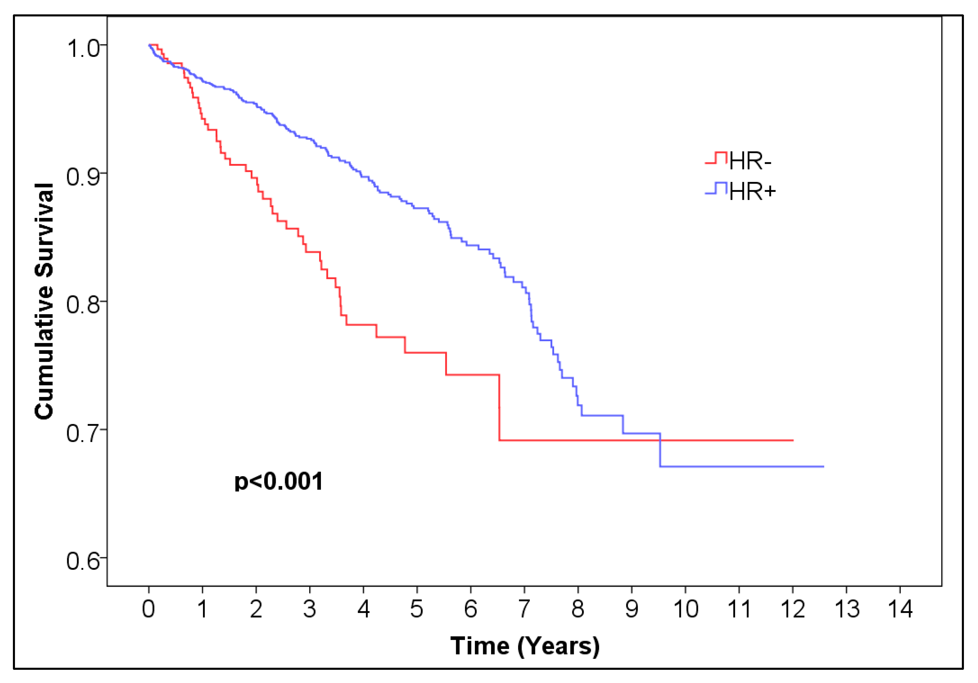
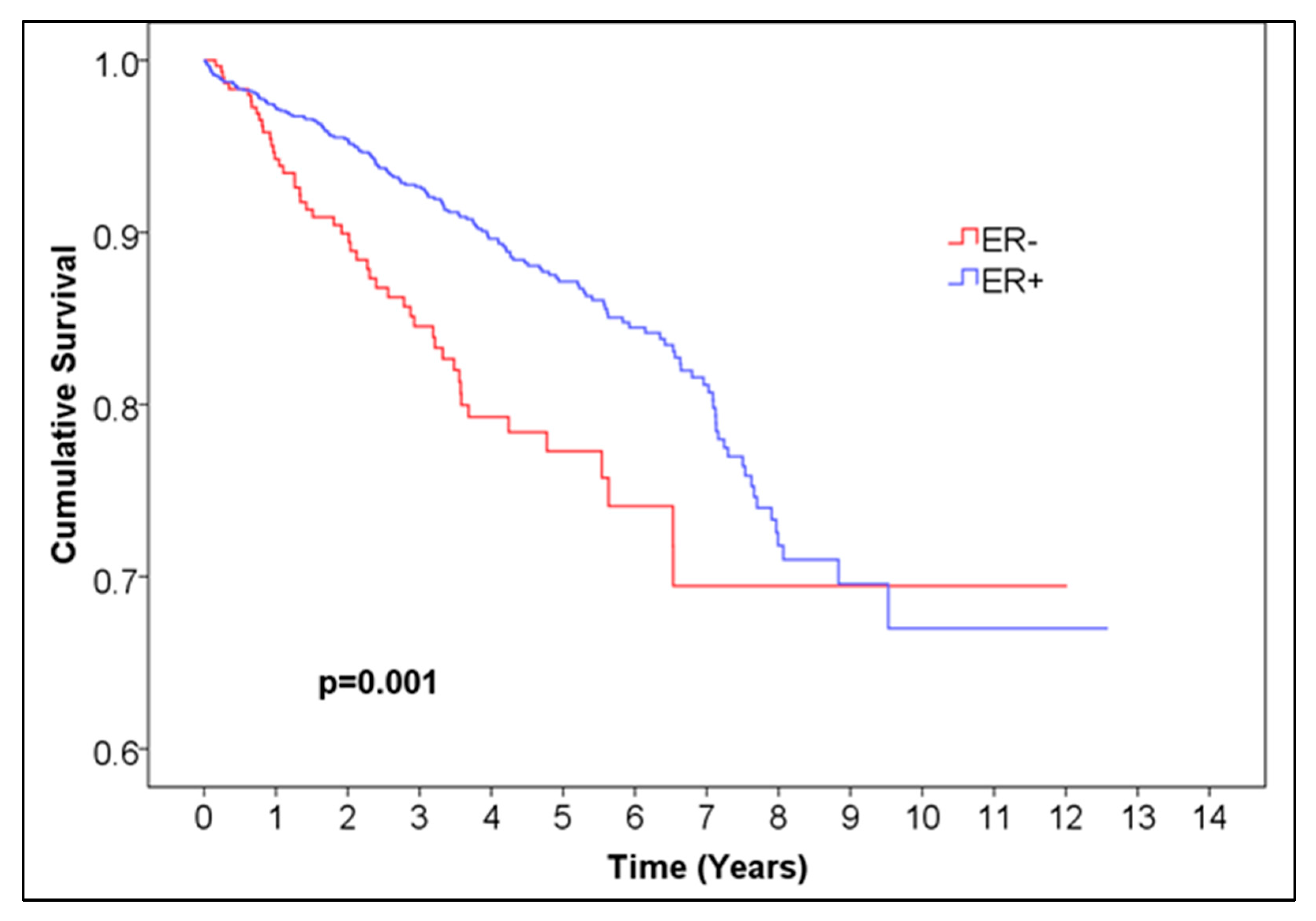
For ER, the 5-year overall survival was 0.872 for ER-positive patients, with 95% CI (0.85044, 0.89356) and 99.6% CI, corresponding to alpha = 0.004 (0.84034, 0.90366), and 0.773 (95% CI (0.71028, 0.83572), 99.6% CI (0.6809, 0.8651)) for ER-negative patients, p = 0.001 (Figure 1, Table 1). For PR, the 5-year overall survival was 0.876 (95% CI (0.85248, 0.89952), 99.6% CI (0.84146, 0.91054)) for PR-positive patients and 0.794 (95% CI (0.745, 0.843), 99.6% CI (0.72205, 0.86595)) for negative patients, p < 0.001 (Figure 2, Table 2). For HR, the 5-year overall survival was 0.873 (95% CI (0.85144, 0.89456), 99.6% CI (0.84134, 0.90466)) for HR-positive patients and 0.760 (95% CI (0.71296, 0.80704), 99.6% CI (0.69092, 0.82908)) for HR-negative patients, p < 0.001 (Figure 3, Table 4).
We compared overall survival distributions of single HR-positive breast cancer (n = 233) and double HR-positive breast cancer (n = 1470) and found no statistically significant difference (p = 0.169) (Figure 4, Table 5).
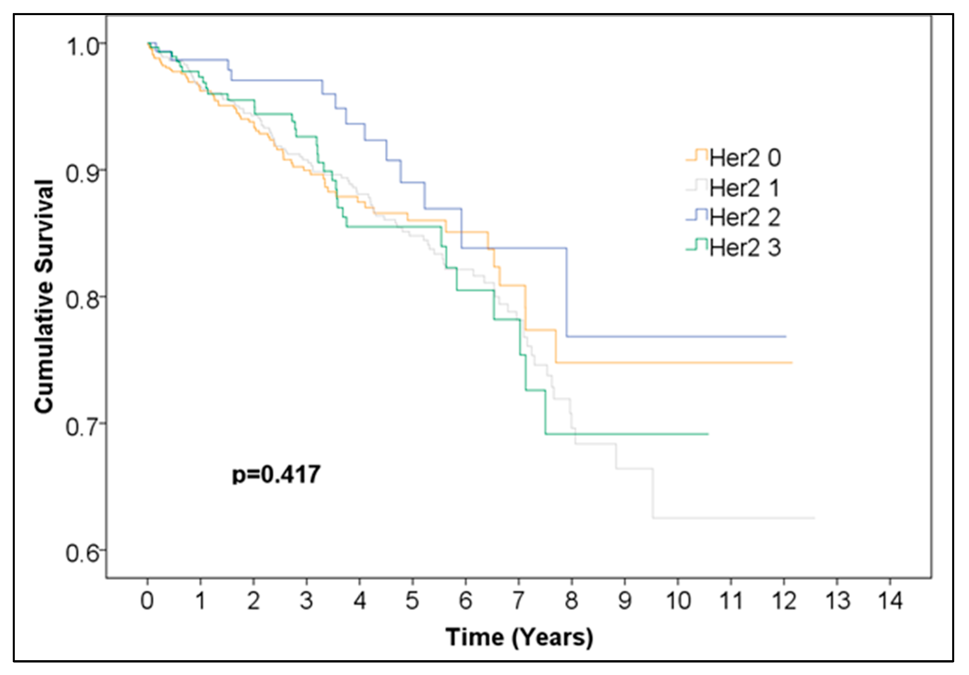
Overall survival was not significantly different between Her2 groups (0, 1, 2, 3) (Figure 5, Table 6). Additionally, we performed a Kaplan–Meier analysis for positive vs. negative Her2neu expression excluding the 125 cases with undefined borderline Her2neu scores and found no statistically significant difference between the survival curves (Figure 6, Table 7). This finding should be interpreted as indicating no statistically significant difference in observed survival distributions rather than as evidence of a lack of biological or prognostic influence.
Statistically significant differences in overall survival distributions of HR+/Her2+ (188 cases), HR+/Her2− (1404 cases), HR−/Her2+ (117 cases), and triple-negative (182 cases) breast cancer were observed (p = 0.003) (Figure 7, Table 8). When comparing across categories, HR+/Her2+ vs. HR+/Her2− (p = 0.288) and HR−/Her+ vs. HR−/Her2− (triple-negative) (p = 0.725) indicated no statistically significant difference between the survival curves over time. Two comparisons produced p-values above the Bonferroni-adjusted alpha but below the uncorrected threshold of 0.05, namely HR−/Her2+ vs. HR+/Her2− (p = 0.036), and HR+/Her2+ vs. HR−/Her2− (triple-negative) (p = 0.006). The difference in overall survival between HR−/Her2− (triple-negative) and HR+/Her2− was statistically significant (p = 0.004).
The 5-year survival rates for each group were as follows:
-
The 5-year survival HR−Her2− (triple-negative): 0.762 (95% CI (0.67772, 0.84628), 99.6% CI (0.63824, 0.88576));
-
The 5-year survival HR−Her2+: 0.758 (95% CI (0.6502, 0.8658), 99.6% CI (0.5997, 0.9163));
-
The 5-year survival HR+Her2−: 0.865 (95% CI (0.83952, 0.89048), 99.6% CI (0.82758, 0.90242));
-
The 5-year survival HR+Her2+: 0.919 (95% CI (0.86804, 0.96996), 99.6% CI (0.84417, 0.99383)).

4. Discussion

The observed differences in survival between hormone receptor-positive (HR+) and hormone receptor-negative (HR−) breast cancers likely reflect both biological behavior and treatment-related effects. Because the analysis was unadjusted for tumor stage, grade, or therapy, these findings should be interpreted as descriptive associations, not independent prognostic effects.
In this retrospective, unadjusted analysis, HER2 expression did not correspond to a statistically significant difference in overall survival distributions. However, this finding should not be interpreted as implying that HER2 status has no prognostic relevance. Rather, it likely reflects the impact of effective anti-HER2 therapies and the incomplete adjudication of HER2 2+ cases. The widespread clinical adoption of targeted therapy during the study period (2010–2021) may have mitigated survival differences traditionally associated with HER2-positive disease [21].
HR+ breast cancers demonstrated better overall survival than HR− cancers, consistent with prior literature [22,23]. This difference is likely explained by endocrine sensitivity and favorable responses to hormonal therapy. We did not observe a statistically significant difference between single HR-positive and double HR-positive tumors, a finding that differs from some reports suggesting marginally better outcomes for single-positive cases [24].
When evaluating combined HR/HER2 subtypes, both HR+/HER2− and HR+/HER2+ cancers showed better survival than HR−/HER2+ and triple-negative cancers. The HR+/HER2+ subtype had the highest 5-year survival rate, which may reflect the dual benefit of hormonal and targeted biological therapies. In contrast, triple-negative and HR−/HER2+ tumors demonstrated less favorable survival, consistent with their known aggressive biology. Although these group differences reached significance at the overall level (p = 0.003), pairwise comparisons often fell between the nominal 0.05 threshold and the Bonferroni-adjusted α = 0.004, suggesting these results should be interpreted with caution.
Incomplete adjudication of HER2 IHC 2+ cases represents another limitation. FISH data were available for only 38 of 163 borderline cases; therefore, the remaining 125 were excluded to maintain internal validity. This may have slightly influenced the estimated HER2-positive prevalence and survival comparisons. Future analyses with complete reflex testing for all 2+ cases are warranted.
During the study period, HER2 assessment followed contemporaneous ASCO/CAP guidelines, which did not yet define the HER2-low category. Consequently, IHC 1+ and 2+/ISH– cases were not distinguished from HER2-zero tumors in a standardized manner. Given the emerging therapeutic relevance of HER2-low disease, future analyses with updated classifications are needed to better characterize this subgroup.
Although our introduction referenced St. Gallen surrogate definitions for luminal A-like and luminal B-like subtypes, consistent Ki-67 data were unavailable due to reporting variability. Earlier pathology records described Ki-67 qualitatively, while later ones used heterogeneous numerical thresholds. As a result, luminal reclassification could not be performed. Future studies with standardized Ki-67 quantification would be essential to accurately analyze luminal subtype survival.
The triple-negative breast cancer pattern was, as expected, associated with the least favorable outcomes [25]. This aligns with prior studies reporting distinct metastatic patterns by subtype: HR−/HER2+ tumors more frequently metastasize to the liver, HR+/HER2− to the lungs, and triple-negative tumors to the brain [25,26]. Bone metastases are more common in ER-positive cases, while visceral metastases are more frequent in ER-negative disease [25]. Such biological heterogeneity likely contributes to observed survival differences.
Knowledge of ER, PR, and HER2 status remains essential for optimizing systemic therapy. By assessing endocrine and HER2 sensitivity, clinicians can tailor treatment strategies and better predict therapeutic response [27]. Even tumors with <10% ER positivity may behave biologically as ER-positive and benefit from adjuvant endocrine therapy [27,28]. Adjuvant tamoxifen therapy, for example, reduces breast cancer mortality by roughly 31% at five years, with cumulative benefits extending to 15 years post-diagnosis [29]. HER2-positive and triple-negative cancers generally exhibit higher recurrence rates and lower disease-free survival, whereas HR-positive tumors have more favorable long-term outcomes [30,31,32,33,34,35].
The results of this study have direct practical relevance for clinical and pathology workflows. By confirming the significant survival advantage of hormone receptor-positive disease and the comparable survival of HER2-positive and HER2-negative subtypes under modern treatment conditions, our data underscore the ongoing importance of accurate ER, PR, and HER2 determination for therapeutic decision-making. These findings support the use of standardized immunohistochemical assessment and reporting especially in regional centers to ensure consistent identification of molecular subtypes that guide systemic therapy. Furthermore, the observed survival patterns provide a valuable reference for institutional benchmarking and the design of future multicenter outcome studies integrating treatment and molecular data.
Finally, de novo stage IV cases were excluded, thereby restricting the cohort to primary, non-metastatic invasive breast cancers. This design choice was intended to minimize survival confounding from advanced-stage disease.

Limitations

This study has several limitations. Because some statistical tests were not fully independent, the Bonferroni correction may have been overly conservative. To address this, both the original and Bonferroni-adjusted confidence intervals were reported. The retrospective nature of the study and its reliance on an anonymized dataset mean that the analysis is exploratory rather than confirmatory.
A major limitation is the absence of clinicopathologic and treatment covariates, which precluded multivariable Cox regression analysis. Potential confounding by stage at diagnosis, tumor grade, and treatment type cannot therefore be excluded. Incomplete HER2 adjudication (particularly among IHC 2+ cases) may also have influenced classification accuracy. Because HER2-low and ER-low categories were not defined during much of the study period, these subgroups could not be analyzed separately. Inconsistent Ki-67 reporting prevented accurate luminal A/B-like classification.
Data on systemic therapy, including anti-HER2 treatment, chemotherapy, and endocrine therapy adherence, were not available. Consequently, the observed survival outcomes may reflect both biological and therapeutic factors rather than pure biomarker effects. Finally, because stage IV cases were excluded, the findings apply primarily to early and locally advanced breast cancers and do not reflect outcomes in metastatic disease.
5. Conclusions

Statistically significant differences were observed between the overall survival distributions of different breast cancer molecular subtypes. Hormone receptor-positive (HR+) breast cancers showed significantly better overall survival compared with hormone receptor-negative (HR−) tumors. In contrast, no statistically significant difference was found between HER2-positive and HER2-negative survival distributions in this unadjusted cohort.
These results demonstrate significant differences in unadjusted overall survival across breast cancer molecular patterns, primarily driven by hormone receptor status. However, they should be interpreted with caution, as therapy effects, treatment-era influences (including the introduction of targeted therapies for HER2-positive disease), and incomplete HER2 adjudication may have affected the observed outcomes. Future studies incorporating detailed clinicopathologic and treatment data are warranted to confirm the independent prognostic impact of these molecular markers.

Author Contributions

Conceptualization, I.M.C., A.E.M. and V.B.V.; methodology, I.M.C., A.E.M. and V.B.V.; software, V.B.V., S.H., P.S. and I.M.C.; validation, I.J.-B., P.S., A.E.M., S.H. and A.R.; formal analysis, V.B.V., A.R. and I.M.C.; data curation, V.B.V., I.M.C. and A.R.; writing—original draft preparation, A.E.M., I.M.C., P.S., S.H. and V.B.V.; writing—review and editing, I.M.C., I.J.-B., P.S., A.E.M. and A.R.; visualization, A.E.M., I.J.-B., P.S. and S.H.; supervision, I.M.C.; project administration, I.M.C. All authors have read and agreed to the published version of the manuscript.

Funding

This research received no external funding.

Institutional Review Board Statement

The study was approved by the Ethics Committee Landesärztekammer Baden-Württemberg, Germany, approval number: F-2023-067/30.05.2023.

Informed Consent Statement

Not applicable. This study represents a retrospective and anonymized data analysis.

Data Availability Statement

Further information concerning the present study is available from the corresponding author upon reasonable request.

Conflicts of Interest

The authors declare no conflicts of interest.

References

  1. World Health Organisation. Breast Cancer. Available online: http://www.who.int/cancer/prevention/diagnosis-screening/breast-cancer/en/ (accessed on 13 May 2020).
  2. Cobec, I.M.; Moleriu, L.; Moatar, A.E.; Rempen, A. First clinical experience with CDK4/6 inhibitors in breast cancer therapy. Exp. Ther. Med. 2021, 21, 522. [Google Scholar] [CrossRef]
  3. Gucalp, A.; Traina, T.A.; Eisner, J.R.; Parker, J.S.; Selitsky, S.R.; Park, B.H.; Elias, A.D.; Baskin-Bey, E.S.; Cardoso, F. Male breast cancer: A disease distinct from female breast cancer. Breast Cancer Res. Treat. 2019, 173, 37–48. [Google Scholar] [CrossRef]
  4. Hennequin, C.; Belkacémi, Y.; Bourgier, C.; Cowen, D.; Cutuli, B.; Fourquet, A.; Hannoun-Lévi, J.-M.; Pasquier, D.; Racadot, S.; Rivera, S. Radiotherapy of breast cancer. Cancer Radiother. 2022, 26, 221–230. [Google Scholar] [CrossRef] [PubMed]
  5. Goldhirsch, A.; Wood, W.C.; Coates, A.S.; Gelber, R.D.; Thürlimann, B.; Senn, H.J. Strategies for subtypes--dealing with the diversity of breast cancer: Highlights of the St. Gallen International Expert Consensus on the Primary Therapy of Early Breast Cancer 2011. Ann Oncol. 2011, 22, 1736–1747. [Google Scholar] [CrossRef]
  6. Goldhirsch, A.; Winer, E.P.; Coates, A.S.; Gelber, R.D.; Piccart-Gebhart, M.; Thürlimann, B.; Senn, H.-J. Personalizing the treatment of women with early breast cancer: Highlights of the St Gallen International Expert Consensus on the Primary Therapy of Early Breast Cancer 2013. Ann. Oncol. 2013, 24, 2206–2223. [Google Scholar] [CrossRef] [PubMed]
  7. Dai, X.; Li, T.; Bai, Z.; Yang, Y.; Liu, X.; Zhan, J.; Shi, B. Breast cancer intrinsic subtype classification, clinical use and future trends. Am. J. Cancer Res. 2015, 5, 2929–2943. [Google Scholar]
  8. Dunnwald, L.K.; Rossing, M.A.; Li, C.I. Hormone receptor status, tumor characteristics, and prognosis: A prospective cohort of breast cancer patients. Breast Cancer Res. 2007, 9, R6. [Google Scholar] [CrossRef] [PubMed]
  9. Reddy, P.; Mithraa, D.S. Correlation of ER, PR, Her2neu and Ki67 with other prognostic factors in breast carcinoma. Trop. J. Pathol. Microbiol. 2020, 6, 350. [Google Scholar] [CrossRef]
  10. Carey, L.A.; Perou, C.M.; Livasy, C.A.; Dressler, L.G.; Cowan, D.; Conway, K.; Karaca, G.; Troester, M.A.; Tse, C.K.; Edmiston, S.; et al. Race, breast cancer subtypes, and survival in the Carolina Breast Cancer Study. JAMA 2006, 295, 2492–2502. [Google Scholar] [CrossRef]
  11. Ford, D.; Easton, D.F.; Peto, J. Estimates of the gene frequency of BRCA1 and its contribution to breast and ovarian cancer incidence. Am. J. Hum. Genet. 1995, 57, 1457–1462. [Google Scholar]
  12. Cancer Genome Atlas Network. Comprehensive molecular portraits of human breast tumours. Nature 2012, 490, 61–70. [Google Scholar] [CrossRef]
  13. Fasching, P.A.; Hu, C.; Hart, S.N.; Ruebner, M.; Polley, E.C.; Gnanaolivu, R.D.; Hartkopf, A.D.; Huebner, H.; Janni, W.; Hadji, P.; et al. Susceptibility gene mutations in germline and tumors of patients with HER2-negative advanced breast cancer. NPJ Breast Cancer 2024, 10, 57. [Google Scholar] [CrossRef]
  14. Cobec, I.M.; Sas, I.; Moatar, A.E.; Moleriu, L.; Rempen, A. Ovarian cancer health politics in Romania and Germany: A comparative study. Exp. Ther. Med. 2021, 22, 1217. [Google Scholar] [CrossRef]
  15. Sobral-Leite, M.; Salomon, I.; Opdam, M.; Kruger, D.T.; Beelen, K.J.; van der Noort, V.; van Vlierberghe, R.L.P.; Blok, E.J.; Giardiello, D.; Sanders, J.; et al. Cancer-immune interactions in ER-positive breast cancers: PI3K pathway alterations and tumor-infiltrating lymphocytes. Breast Cancer Res. 2019, 21, 90. [Google Scholar] [CrossRef]
  16. Sikora, M.J.; Jacobsen, B.M.; Levine, K.; Chen, J.; Davidson, N.E.; Lee, A.V.; Alexander, C.M. WNT4 mediates estrogen receptor signaling and endocrine resistance in invasive lobular carcinoma cell lines. Breast Cancer Res. 2016, 18, 92. [Google Scholar] [CrossRef] [PubMed]
  17. Sarhangi, N.; Hajjari, S.; Heydari, S.F.; Ganjizadeh, M.; Rouhollah, F.; Hasanzad, M. Breast cancer in the era of precision medicine. Mol. Biol. Rep. 2022, 49, 10023–10037. [Google Scholar] [CrossRef]
  18. Ultimescu, F.; Hudita, A.; Popa, D.E.; Olinca, M.; Muresean, H.A.; Ceausu, M.; Stanciu, D.I.; Ginghina, O.; Galateanu, B. Impact of Molecular Profiling on Therapy Management in Breast Cancer. J. Clin. Med. 2024, 13, 4995. [Google Scholar] [CrossRef] [PubMed]
  19. Galea, M.H.; Blamey, R.W.; Elston, C.E.; Ellis, I.O. The Nottingham Prognostic Index in primary breast cancer. Breast Cancer Res. Treat. 1992, 22, 207–219. [Google Scholar] [CrossRef] [PubMed]
  20. Cuzick, J.; Dowsett, M.; Pineda, S.; Wale, C.; Salter, J.; Quinn, E.; Zabaglo, L.; Mallon, E.; Green, A.R.; Ellis, I.O.; et al. Prognostic value of a combined estrogen receptor, progesterone receptor, Ki-67, and human epidermal growth factor receptor 2 immunohistochemical score and comparison with the Genomic Health recurrence score in early breast cancer. J. Clin. Oncol. 2011, 29, 4273–4278. [Google Scholar] [CrossRef]
  21. Patel, A.; Unni, N.; Peng, Y. The Changing Paradigm for the Treatment of HER2-Positive Breast Cancer. Cancers 2020, 12, 2081. [Google Scholar] [CrossRef]
  22. Howlader, N.; Cronin, K.A.; Kurian, A.W.; Andridge, R. Differences in Breast Cancer Survival by Molecular Subtypes in the United States. Cancer Epidemiol. Biomark. Prev. 2018, 27, 619–626. [Google Scholar] [CrossRef] [PubMed]
  23. Li, X.; Yang, J.; Peng, L.; Sahin, A.A.; Huo, L.; Ward, K.C.; O’Regan, R.; Torres, M.A.; Meisel, J.L. Triple-negative breast cancer has worse overall survival and cause-specific survival than non-triple-negative breast cancer. Breast Cancer Res. Treat. 2017, 161, 279–287. [Google Scholar] [CrossRef]
  24. Li, Y.; Yang, D.; Yin, X.; Zhang, X.; Huang, J.; Wu, Y.; Wang, M.; Yi, Z.; Li, H.; Li, H.; et al. Clinicopathological Characteristics and Breast Cancer-Specific Survival of Patients with Single Hormone Receptor-Positive Breast Cancer. JAMA Netw. Open 2020, 3, e1918160. [Google Scholar] [CrossRef] [PubMed]
  25. Wang, R.; Zhu, Y.; Liu, X.; Liao, X.; He, J.; Niu, L. The Clinicopathological features and survival outcomes of patients with different metastatic sites in stage IV breast cancer. BMC Cancer 2019, 19, 1091. [Google Scholar] [CrossRef]
  26. Martin, A.M.; Cagney, D.N.; Catalano, P.J.; Warren, L.E.; Bellon, J.R.; Punglia, R.S.; Claus, E.B.; Lee, E.Q.; Wen, P.Y.; Haas-Kogan, D.A.; et al. Brain metastases in newly diagnosed breast Cancer: A population-based study. JAMA Oncol. 2017, 3, 1069–1077. [Google Scholar] [CrossRef] [PubMed]
  27. García Fernández, A.; Giménez, N.; Fraile, M.; González, S.; Chabrera, C.; Torras, M.; González, C.; Salas, A.; Barco, I.; Cirera, L.; et al. Survival and clinicopathological characteristics of breast cancer patient according to different tumour subtypes as determined by hormone receptor and Her2 immunohistochemistry. a single institution survey spanning 1998 to 2010. Breast 2012, 21, 366–373. [Google Scholar] [CrossRef]
  28. Iwamoto, T.; Booser, D.; Valero, V.; Murray, J.L.; Koenig, K.; Esteva, F.J.; Ueno, N.T.; Zhang, J.; Shi, W.; Qi, Y.; et al. Estrogen receptor (ER) mRNA and ER-related gene expression in breast cancers that are 1% to 10% ER-positive by immunohistochemistry. J. Clin. Oncol. 2012, 30, 729–734. [Google Scholar] [CrossRef]
  29. Early Breast Cancer Trialists’ Collaborative Group (EBCTCG). Effects of chemotherapy and hormonal therapy for early breast cancer on recurrence and 15-year survival: An overview of the randomised trials. Lancet 2005, 365, 1687–1717. [Google Scholar] [CrossRef]
  30. Pal, S.K.; Childs, B.H.; Pegram, M. Triple negative breast cancer: Unmet medical needs. Breast Cancer Res. Treat. 2011, 125, 627–636. [Google Scholar] [CrossRef]
  31. Howell, A.; Cuzick, J.; Baum, M.; Buzdar, A.; Dowsett, M.; Forbes, J.F.; Adams, J.; Bianco, A.R.; Cella, D.; Coibion, M.; et al. ATAC Trialists’ Group. Results of the ATAC (arimidex, tamoxifen, alone or in combination) trial after completion of 5 years’ adjuvant treatment for breast cancer. Lancet 2005, 365, 60–62. [Google Scholar]
  32. Goldstein, N.S.; Ferkowicz, M.; Odish, E.; Mani, A.; Hastah, F. Minimum formalin fixation time for consistent estrogen receptor immunohistochemical staining of invasive breast carcinoma. Am. J. Clin. Pathol. 2003, 120, 86–92. [Google Scholar] [CrossRef] [PubMed]
  33. Grann, V.R.; Troxel, A.B.; Zojwalla, N.J.; Jacobson, J.S.; Hershman, D.; Neugut, A.I. Hormone receptor status and survival in a population-based cohort of patients with breast carcinoma. Cancer 2005, 103, 2241–2251. [Google Scholar] [CrossRef]
  34. Goldhirsch, A.; Glick, J.H.; Gelber, R.D.; Coates, A.S.; Thurlimann, B.; Senn, H.J. Meeting highlights: International expert consensus on the primary therapy of early breast cancer 2005. Ann. Oncol. 2005, 16, 1569–1583. [Google Scholar] [CrossRef] [PubMed]
  35. Kang, Y.J.; Oh, S.J.; Bae, S.Y.; Kim, E.K.; Lee, Y.J.; Park, E.H.; Jeong, J.; Park, H.K.; Suh, Y.J.; Kim, Y.S. Predictive biological factors for late survival in patients with HER2-positive breast cancer. Sci. Rep. 2023, 13, 11008. [Google Scholar] [CrossRef] [PubMed]
Figure 1. Kaplan–Meier curve of ER− vs. ER+ female breast cancer.
Medicina 61 01858 g001
Figure 2. Kaplan–Meier curve of PR− vs. PR+ female breast cancer.
Medicina 61 01858 g002
Figure 3. Kaplan–Meier curve of HR− vs. HR+ female breast cancer.
Medicina 61 01858 g003
Figure 4. Kaplan–Meier curve of single vs. double HR+ female breast cancer.
Medicina 61 01858 g004
Figure 5. Kaplan–Meier curve of different Her2 scores in female breast cancer.
Medicina 61 01858 g005
Figure 6. Kaplan–Meier curve of Her2− vs. Her2+ female breast cancer.
Medicina 61 01858 g006
Figure 7. Kaplan–Meier curve of triple-negative vs. HR+/Her2− vs. HR−/Her2+ vs. HR+/Her2+ female breast cancer.
Medicina 61 01858 g007
Table 1. Baseline characteristics of the study population (n = 2016). Abbreviations: HR = hormone receptor (estrogen and/or progesterone receptor); HER2 = human epidermal growth factor receptor 2; IQR = interquartile range.
VariableTotal (n, %)HR+/HER2−HR+/HER2+HR−/HER2+Triple-Negative (HR−/HER2−)
Number of cases2016 (100%)1404 (69.7%)188 (9.3%)117 (5.8%)182 (9.1%)
Median age (years)63 (IQR 53–74)63626159
Table 2. Number of patients at risk of ER− vs. ER+ female breast cancer. Abbreviations: ER = estrogen receptor.
Time (Years)012345678910111213
Number at risk ER−333240179143986436271452110
Number at risk ER+168312589998046124482801879541181070
Table 3. Number of patients at risk of PR− vs. PR+ female breast cancer. Abbreviations: PR = progesterone receptor.
Time (Years)012345678910111213
Number at risk PR−52637527221714710263442085320
Number at risk PR+14901123906730563410253170893815860
Table 4. Number of patients at risk of HR− vs. HR+ female breast cancer. Abbreviations: HR = hormone receptor.
Time (Years)012345678910111213
Number at risk HR−313225167132895732251242110
Number at risk HR+1703127310118156214552841899742181070
Table 5. Number of patients at risk of single vs. double HR+ female breast cancer. Abbreviations: HR = hormone receptor; HR+ = estrogen and/or progesterone receptor–positive.
Time (Years)012345678910111213
Number at risk Single HR+ breast cancer23316511796675235211053210
Number at risk Double HR+ breast cancer14701108894719554403249168873715860
Table 6. Number of patients at risk of different Her2 scores in female breast cancer. Abbreviations: HER2 = human epidermal growth factor receptor 2; IHC = immunohistochemistry.
Time (Years)012345678910111213
Number at risk Her2 0783541402307204139744824125310
Number at risk Her2 1775598491400333256172119602812760
Number at risk Her2 216313511098734826181011110
Number at risk Her2 32952241751421006944291552000
Table 7. Number of patients at risk of Her2− vs. Her2+ female breast cancer. Abbreviations: HER2 = human epidermal growth factor receptor 2.
Time (Years)012345678910111213
Number at risk Her2−158611569027155393952461678440171070
Number at risk Her2+3052301781441006844291552000
Table 8. Number of patients at risk of triple-negative vs. HR+/Her2− vs. HR−/Her2+ vs. HR+/Her2+ female breast cancer. Abbreviations: HR = hormone receptor (estrogen and/or progesterone receptor); HER2 = human epidermal growth factor receptor 2.
Time (Years)012345678910111213
Number at risk
Triple-negative breast cancer
182126886646311815521110
Number at risk HR+/Her2−14041030814649493364228152793816960
Number at risk HR−/Her2+1178767543320117521000
Number at risk HR+/Her2+18814311190674933221031000
Disclaimer/Publisher’s Note: The statements, opinions and data contained in all publications are solely those of the individual author(s) and contributor(s) and not of MDPI and/or the editor(s). MDPI and/or the editor(s) disclaim responsibility for any injury to people or property resulting from any ideas, methods, instructions or products referred to in the content.