Diagnostic Challenges in Inflammatory Choroidal Neovascularization
Abstract
1. Introduction
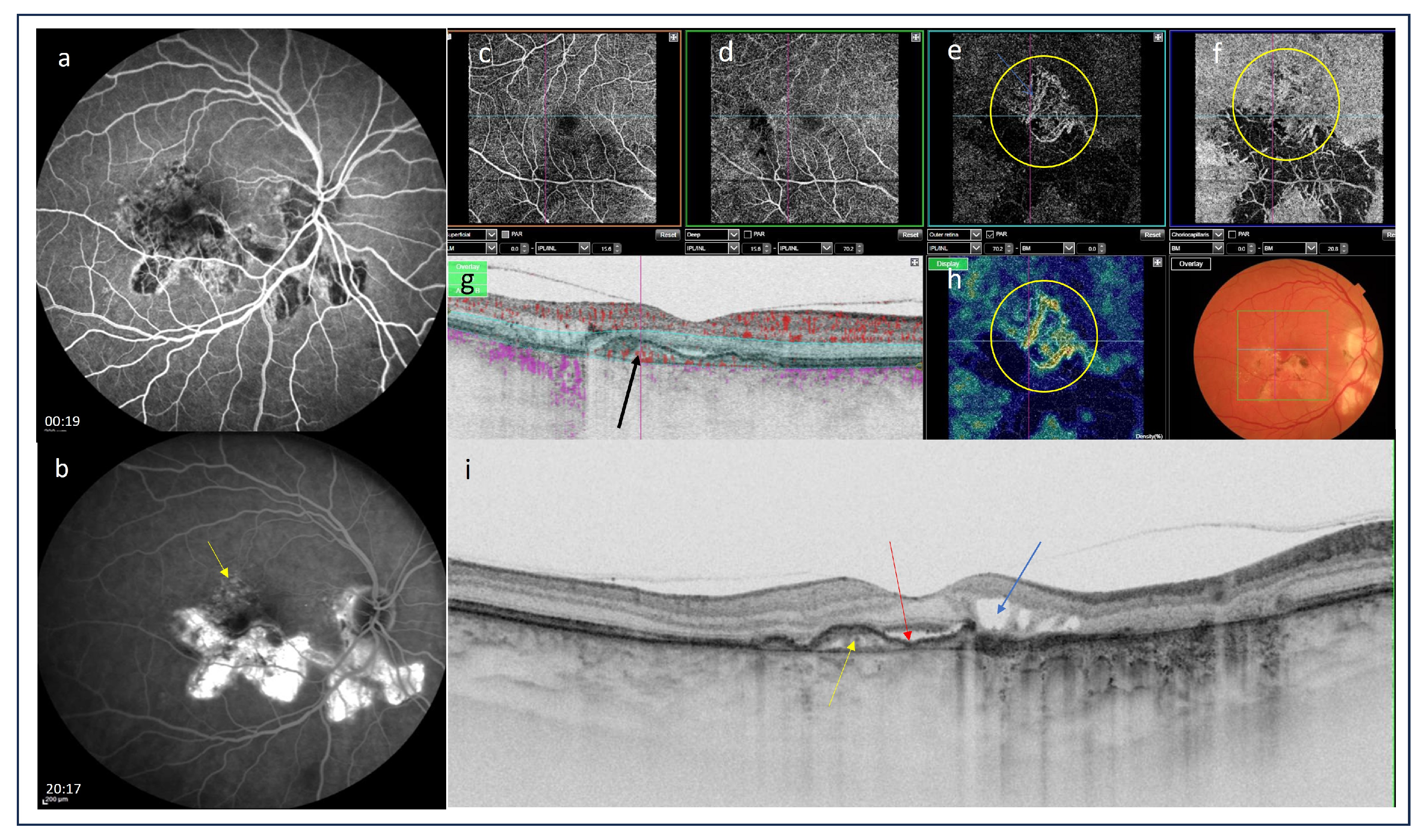
2. Imaging Tools for the Detection of iCNV
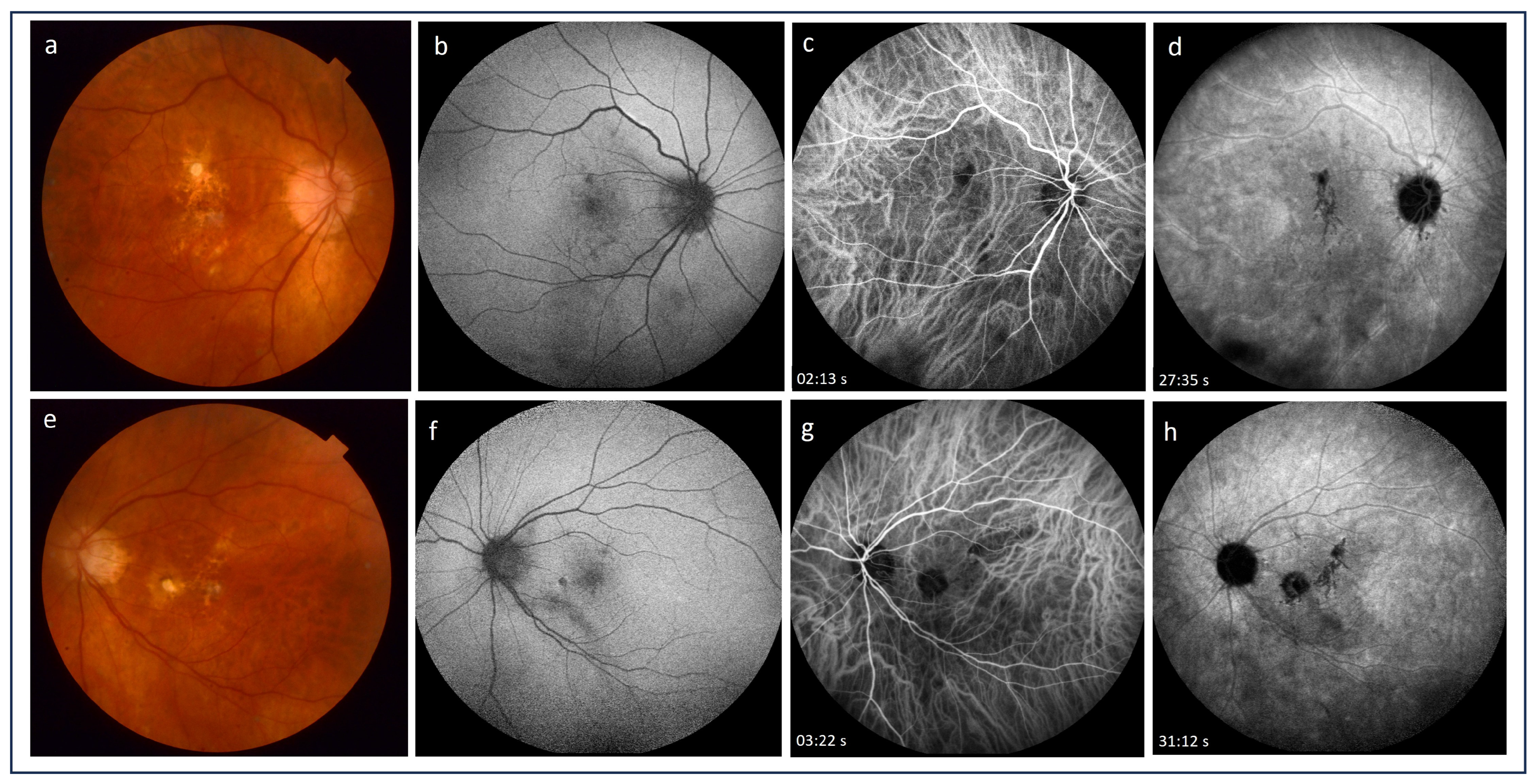
2.1. Fluorescein Angiography
2.2. Indocyanine Green Angiography
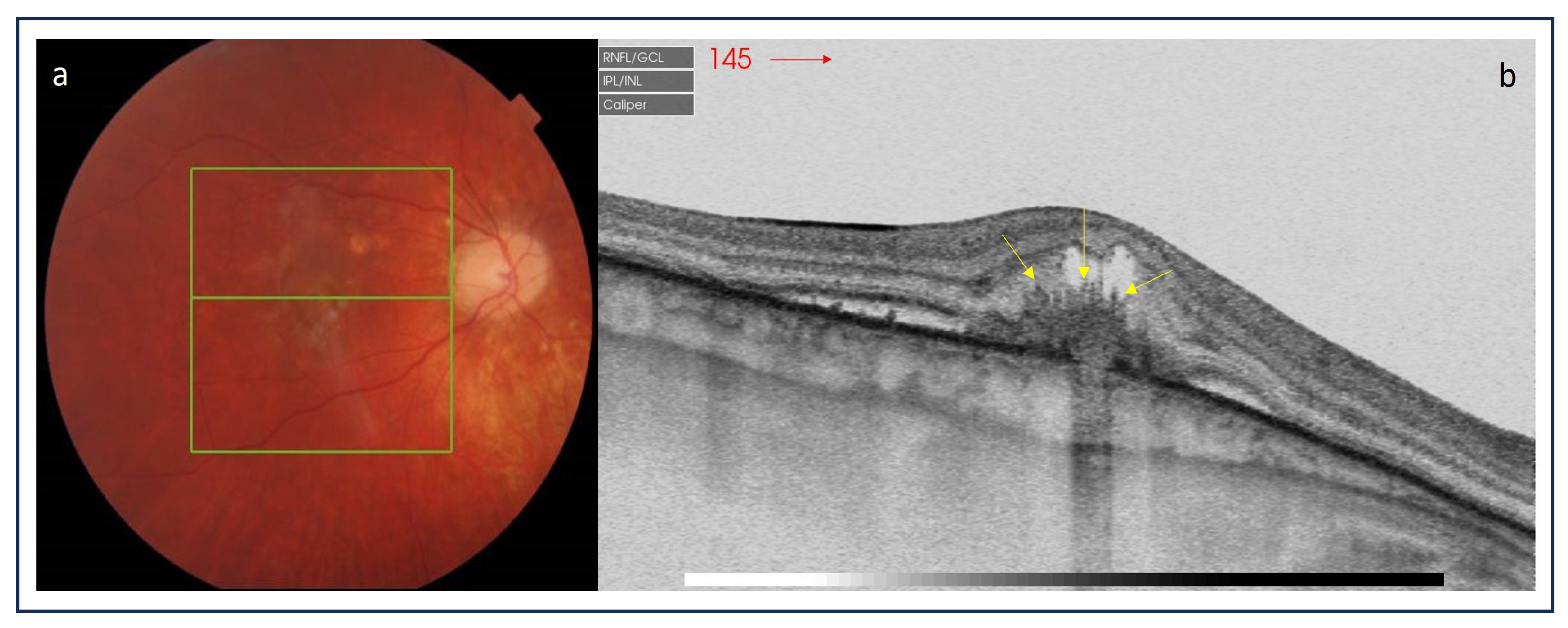
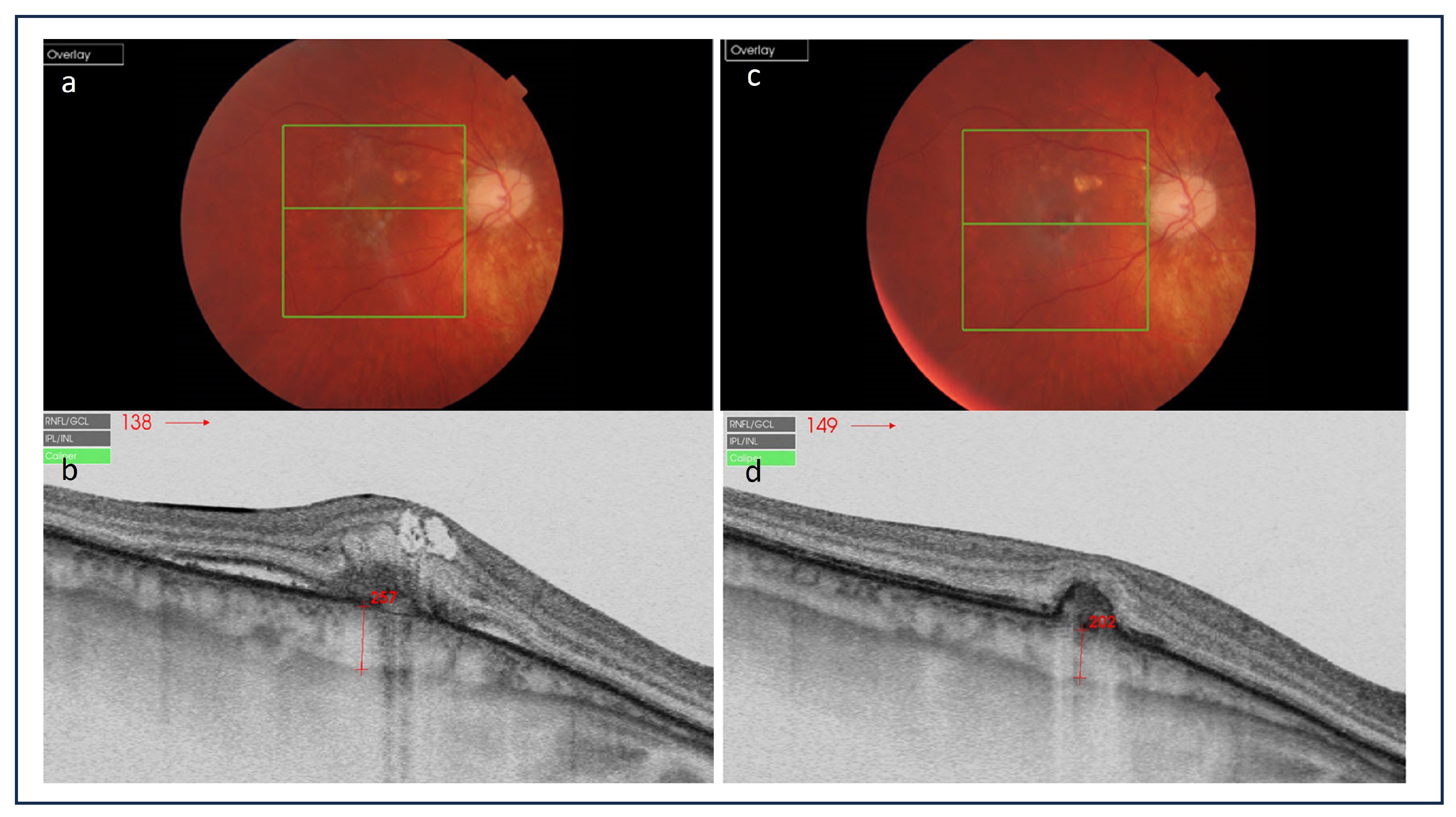
2.3. Optical Coherence Tomography
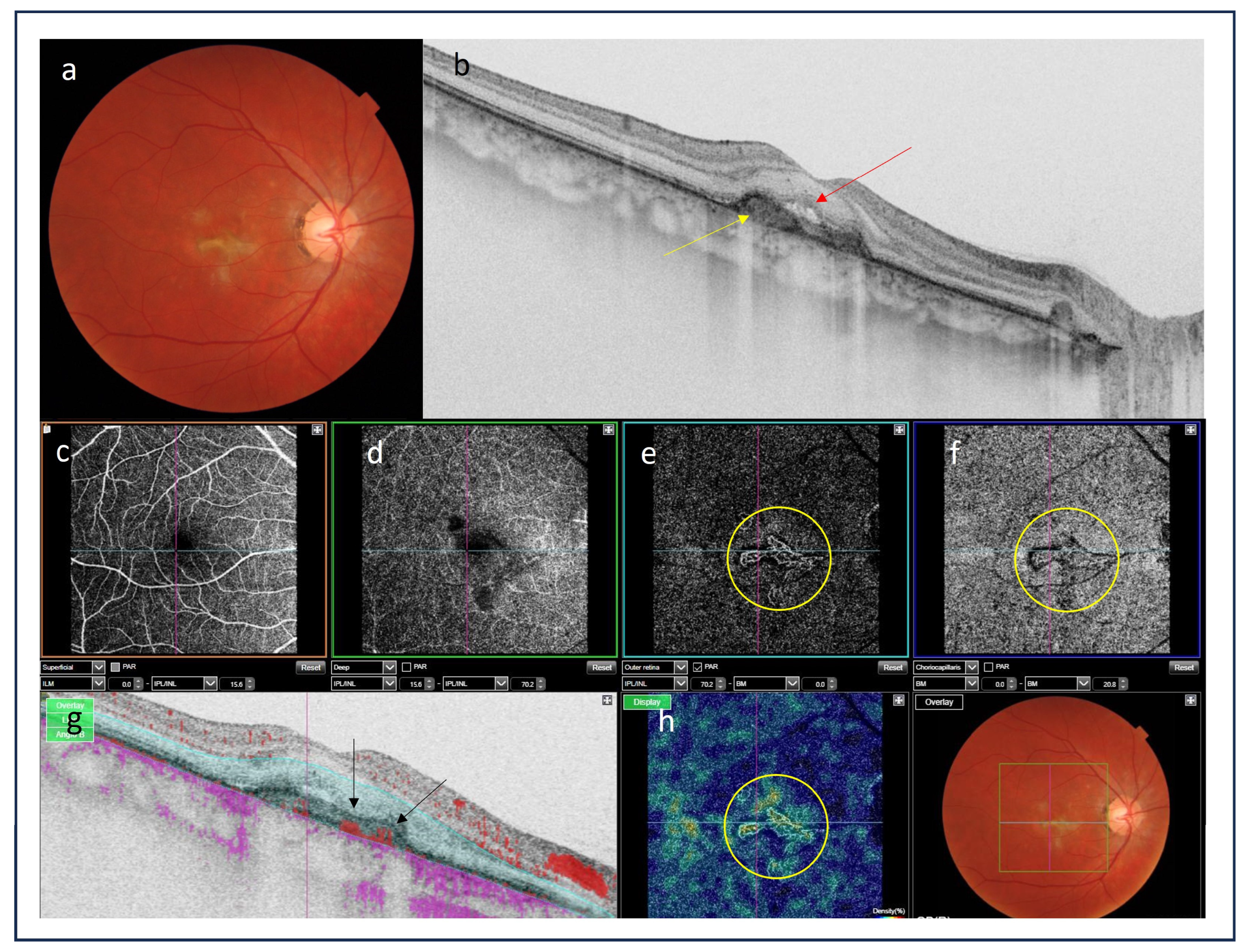
2.4. Optical Coherence Tomography Angiography
2.5. Near-Infrared Autofluorescence Imaging
3. Conclusions
Author Contributions
Funding
Institutional Review Board Statement
Informed Consent Statement
Data Availability Statement
Conflicts of Interest
References
- Agarwal, A.; Invernizzi, A.; Singh, R.B.; Foulsham, W.; Aggarwal, K.; Handa, S.; Agrawal, R.; Pavesio, C.; Gupta, V. An update on inflammatory choroidal neovascularization: Epidemiology, multimodal imaging, and management. J. Ophthalmic Inflamm. Infect. 2018, 8, 13. [Google Scholar] [CrossRef]
- Dhingra, N.; Kelly, S.; Majid, M.A.; Bailey, C.B.; Dick, A.D. Inflammatory choroidal neovascular membrane in posterior uveitis-pathogenesis and treatment. Indian. J. Ophthalmol. 2010, 58, 3–10. [Google Scholar] [CrossRef] [PubMed]
- Neri, P.; Lettieri, M.; Fortuna, C.; Manoni, M.; Giovannini, A. Inflammatory choroidal neovascularization. Middle East. Afr. J. Ophthalmol. 2009, 16, 245–251. [Google Scholar] [CrossRef] [PubMed]
- Baxter, S.L.; Pistilli, M.; Pujari, S.S.; Liesegang, T.L.; Suhler, E.B.; Thorne, J.E.; Foster, C.S.; Jabs, D.A.; Levy-Clarke, G.A.; Nussenblatt, R.B.; et al. Risk of choroidal neovascularization among the uveitides. Am. J. Ophthalmol. 2013, 156, 468–477. [Google Scholar] [CrossRef] [PubMed]
- Dreyer, R.F.; Gass, D.J. Multifocal choroiditis and panuveitis. A syndrome that mimics ocular histoplasmosis. Arch. Ophthalmol. 1984, 102, 1776–1784. [Google Scholar] [CrossRef] [PubMed]
- Morgan, C.M.; Schatz, H. Recurrent Multifocal Choroiditis. Ophthalmology 1986, 93, 1138–1147. [Google Scholar] [CrossRef] [PubMed]
- Brown, J., Jr.; Folk, J.C.; Reddy, C.V.; Kimura, A.E. Visual prognosis of multifocal choroiditis, punctate inner choroidopathy, and the diffuse subretinal fibrosis syndrome. Ophthalmology 1996, 103, 1100–1105. [Google Scholar] [CrossRef] [PubMed]
- Ahnood, D.; Madhusudhan, S.; Tsaloumas, M.D.; Waheed, N.K.; Keane, P.A.; Denniston, A.K. Punctate inner choroidopathy: A review. Surv. Ophthalmol. 2017, 62, 113–126. [Google Scholar] [CrossRef] [PubMed]
- Kuo, I.C.; Cunningham, E.T., Jr. Ocular neovascularization in patients with uveitis. Int. Ophthalmol. Clin. 2000, 40, 111–126. [Google Scholar] [CrossRef]
- Saatci, A.O.; Ayhan, Z.; Engin Durmaz, C.; Takes, O. Simultaneous Single Dexamethasone Implant and Ranibizumab Injection in a Case with Active Serpiginous Choroiditis and Choroidal Neovascular Membrane. Case Rep. Ophthalmol. 2015, 6, 408–414. [Google Scholar] [CrossRef]
- Kępka, M.; Brydak-Godowska, J.; Ciszewska, J.; Turczyńska, M.; Ciszek, M.; Kęcik, D. Clinical features, diagnosis and management of serpiginuos choroiditis. Ophthalmol. J. 2022, 7, 127–135. [Google Scholar] [CrossRef]
- Rao, N.A.; Gupta, A.; Dustin, L.; Chee, S.P.; Okada, A.A.; Khairallah, M.; Bodaghi, B.; Lehoang, P.; Accorinti, M.; Mochizuki, M.; et al. Frequency of distinguishing clinical features in Vogt Koyanagi-Harada disease. Ophthalmology 2010, 117, 591–599. [Google Scholar] [CrossRef]
- Brucker, A.J.; Deglin, E.A.; Bene, C.; Hoffman, M.E. Subretinal Choroidal Neovascularization in Birdshot Retinochoroidopathy. Am. J. Ophthalmol. 1985, 99, 40–44. [Google Scholar] [CrossRef] [PubMed]
- Mehta, S.; Hariharan, L.; Ho, A.C.; Kempen, J.H. Peripapillary choroidal neovascularization in pars planitis. J. Ophthalmic Inflamm. Infect. 2013, 3, 13. [Google Scholar] [CrossRef]
- Nageeb, M.R. Intermediate Uveitis Complicated by Peripapillary Choroidal Neovascularization. Cureus 2022, 14, e31040. [Google Scholar] [CrossRef] [PubMed]
- Inagaki, M.; Harada, T.; Kiribuchi, T.; Ohashi, T.; Majima, J. Subfoveal choroidal neovascularization in uveitis. Ophthalmologica 1996, 210, 229–233. [Google Scholar] [CrossRef] [PubMed]
- Pagliarini, S.; Piguet, B.; Ffytche, T.J.; Bird, A.C. Foveal involvement and lack of visual recovery in APMPPE associated with uncommon features. Eye 1995, 9, 42–47. [Google Scholar] [CrossRef]
- Bowie, E.M.; Sletten, K.R.; Kayser, D.L.; Folk, J.C.; James, C. Acute Posterior Multifocal Placoid Pigment Epitheliopathy And Choroidal Neovascularization. Retina 2005, 25, 362–364. [Google Scholar] [CrossRef]
- Saatci, A.O.; Ayhan, Z.; Ipek, S.C.; Soylev Bajin, M. Intravitreal Aflibercept as an Adjunct to Systemic Therapy in a Case of Choroidal Neovascular Membrane Associated with Sympathetic Ophthalmia. Turk. J. Ophthalmol. 2018, 48, 209–211. [Google Scholar] [CrossRef]
- Rouvas, A.A.; Ladas, I.D.; Papakostas, T.D.; Moschos, M.M.; Vergados, I. Intravitreal ranibizumab in a patient with choroidal neovascularization secondary to multiple evanescent white dot syndrome. Eur. J. Ophthalmol. 2007, 17, 996–999. [Google Scholar] [CrossRef]
- Burova, M.; Stepanov, A.; Almesmary, B.; Jiraskova, N. Choroidal neovascularization in a patient after resolution of multiple evanescent white dot syndrome: A case report. Clin. Case Rep. 2022, 10, e05802. [Google Scholar] [CrossRef]
- Liu, T.Y.A.; Zhang, A.Y.; Wenick, A. Evolution of Choroidal Neovascularization due to Presumed Ocular Histoplasmosis Syndrome on Multimodal Imaging including Optical Coherence Tomography Angiography. Case Rep. Ophthalmol. Med. 2018, 2018, 4098419. [Google Scholar] [CrossRef]
- Hu, J.; Hoang, Q.V.; Chau, F.Y.; Blair, M.P.; Lim, J.I. Intravitreal anti-vascular endothelial growth factor for choroidal neovascularization in ocular histoplasmosis. Retin. Cases Brief. Rep. 2014, 8, 24–29. [Google Scholar] [CrossRef]
- Nielsen, J.S.; Fick, T.A.; Saggau, D.D.; Barnes, C.H. Intravitreal anti-vascular endothelial growth factor therapy for choroidal neovascularization secondary to ocular histoplasmosis syndrome. Retina 2012, 32, 468–472. [Google Scholar] [CrossRef] [PubMed]
- Walia, H.S.; Shah, G.K.; Blinder, K.J. Treatment of CNV secondary to presumed ocular histoplasmosis with intravitreal aflibercept 2.0 mg injection. Can. J. Ophthalmol. 2016, 51, 91–96. [Google Scholar] [CrossRef]
- Zonnevylle, K.E.; Stanescu-Segall, D. Intravitreal Bevacizumab for Treatment of Bilateral Candida Chorioretinitis Complicated with Choroidal Neovascularization. Ocul. Immunol. Inflamm. 2020, 28, 39–42. [Google Scholar] [CrossRef] [PubMed]
- Jampol, L.M.; Sung, J.; Walker, J.D.; Folk, J.C.; Townsend-Pico, W.A.; Lowder, C.Y.; Dodds, E.M.; Westrich, D.; Terry, J. Choroidal neovascularization secondary to Candida albicans chorioretinitis. Am. J. Ophthalmol. 1996, 121, 643–649. [Google Scholar] [CrossRef] [PubMed]
- Makragiannis, G.; Vahdani, K.; Carreño, E.; Lee, R.W.J.; Dick, A.D.; Ross, A.H. Bevacizumab for treatment of choroidal neovascularization secondary to candida chorioretinitis. Int. Ophthalmol. 2018, 38, 781–785. [Google Scholar] [CrossRef] [PubMed]
- Fine, S.L.; Owens, S.L.; Haller, J.A.; Knox, D.L.; Patz, A. Choroidal neovascularization as a late complication of ocular toxoplasmosis. Am. J. Ophthalmol. 1981, 91, 318–322. [Google Scholar] [CrossRef] [PubMed]
- Willerson, D.; Aaberg, T.M.; Reeser, F.; Meredith, T.A. Unusual ocular presentation of acute toxoplasmosis. Br. J. Ophthalmol. 1977, 61, 693–698. [Google Scholar] [CrossRef] [PubMed]
- Benevento, J.D.; Jager, R.D.; Noble, A.G.; Latkany, P.; Mieler, W.F.; Sautter, M.; Meyers, S.; Mets, M.; Grassi, M.A.; Rabiah, P.; et al. Toxoplasmosis Study Group. Toxoplasmosis-associated neovascular lesions treated successfully with ranibizumab and antiparasitic therapy. Arch. Ophthalmol. 2008, 126, 1152–1156. [Google Scholar] [CrossRef] [PubMed]
- Sienicka, P. Eye toxoplasmosis complicated by choroidal neovascularization effectively treated with the anti-vascular endothelial growth factor aflibercept. Klin. Ocz. 2022, 124, 114–119. [Google Scholar] [CrossRef]
- Lampariello, D.A.; Primo, S.A. Ocular toxocariasis: A rare presentation of a posterior pole granuloma with an associated choroidal neovascular membrane. J. Am. Optom. Assoc. 1999, 70, 245–252. [Google Scholar] [PubMed]
- Lyall, D.; Hutchison, B.; Gaskell, A.; Varikkara, M. Intravitreal Ranibizumab in the treatment of choroidal neovascularisation secondary to ocular toxocariasis in a 13-year-old boy. Eye 2010, 24, 1730–1731. [Google Scholar] [CrossRef]
- Yoon, D.Y.; Woo, S.J. Intravitreal Administration of Ranibizumab and Bevacizumab for Choroidal Neovascularization Secondary to Ocular Toxocariasis: A Case Report. Ocular Immunol. Inflamm. 2018, 26, 639–641. [Google Scholar] [CrossRef] [PubMed]
- Chung, Y.M.; Yeh, T.S.; Sheu, S.J.; Liu, J.H. Macular subretinal neovascularization in choroidal tuberculosis. Ann. Ophthalmol. 1989, 21, 225–229. [Google Scholar] [PubMed]
- Lee Kim, E.; Rodger, D.C.; Rao, N.A. Choroidal neovascularization secondary to tuberculosis: Presentation and management. Am. J. Ophthalmol. Case Rep. 2017, 6, 124–129. [Google Scholar] [CrossRef]
- Zhang, Y.K.; Fu, H.Y.; Guan, Y.; Li, Y.J.; Bai, H.Z. Concurrent tuberculous chorioretinitis with choroidal neovascularization and tuberculous meningitis: A case report. BMC Ophthalmol. 2020, 20, 227. [Google Scholar] [CrossRef]
- Interlandi, E.; Pellegrini, F.; Pavesio, C.; De Luca, M.; De Marco, R.; Papayannis, A.; Mandarà, E.; Cuna, A.; Cirone, D.; Ciabattoni, C.; et al. Intraocular Tuberculosis: A Challenging Case Mimicking Wet Age-Related Macular Degeneration. Case Rep. Ophthalmol. 2021, 12, 519–524. [Google Scholar] [CrossRef]
- Deutman, A.F.; Grizzard, W.S. Rubella retinopathy and subretinal neovascularization. Am. J. Ophthalmol. 1978, 85, 82–87. [Google Scholar] [CrossRef]
- Parodi, M.B.; Romano, F.; Montagna, M.; Bandello, F. Large choroidal excavation in a patient with rubella retinopathy. Eur. J. Ophthalmol. 2018, 28, 251–252. [Google Scholar] [CrossRef] [PubMed]
- Dewan, L.; Hasan, N.; Aron, N.; Chawla, R.; Sundar, D. Rubella retinopathy with choroidal neovascular membrane in a 7-year-old. Indian J. Ophthalmol. 2020, 68, 1176–1177. [Google Scholar] [CrossRef] [PubMed]
- Hirano, K.; Tanikawa, A.; Miyake, Y. Neovascular maculopathy associated with rubella retinopathy. Jpn. J. Ophthalmol. 2000, 44, 697. [Google Scholar] [CrossRef] [PubMed]
- Khairallah, M.; Ben Yahia, S.; Attia, S.; Jelliti, B.; Zaouali, S.; Ladjimi, A. Severe ischemic maculopathy in a patient with West Nile virus infection. Ophthalmic Surg. Lasers Imaging 2006, 37, 240–242. [Google Scholar] [CrossRef] [PubMed]
- Seth, R.K.; Stoessel, K.M.; Adelman, R.A. Choroidal neovascularization associated with West Nile virus chorioretinitis. Semin. Ophthalmol. 2007, 22, 81–84. [Google Scholar] [CrossRef] [PubMed]
- Zito, R.; Micelli Ferrari, T.; Di Pilato, L.; Lorusso, M.; Ferretta, A.; Ferrari, L.M.; Accorinti, M. Clinical course of choroidal neovascular membrane in West Nile virus chorioretinitis: A case report. J. Med. Case Rep. 2021, 15, 206. [Google Scholar] [CrossRef] [PubMed]
- Campa, C.; Costagliola, C.; Incorvaia, C.; Sheridan, C.; Semeraro, F.; De Nadai, K.; Sebastiani, A.; Parmeggiani, F. Inflammatory mediators and angiogenic factors in choroidal neovascularization: Pathogenetic interactions and therapeutic implications. Mediators Inflamm. 2010, 2010, 546826. [Google Scholar] [CrossRef]
- D’Ambrosio, E.; Tortorella, P.; Iannetti, L. Management of uveitis-related choroidal neovascularization: From the pathogenesis to the therapy. J. Ophthalmol. 2014, 2014, 450428. [Google Scholar] [CrossRef]
- Cerquaglia, A.; Fardeau, C.; Cagini, C.; Fiore, T.; LeHoang, P. Inflammatory Choroidal Neovascularization: Beyond the Intravitreal Approach. Ocul. Immunol. Inflamm. 2018, 26, 1047–1052. [Google Scholar] [CrossRef]
- Sato, K.; Takeda, A.; Hasegawa, E.; Jo, Y.; Arima, M.; Oshima, Y.; Ryoji, Y.; Nakazawa, T.; Yuzawa, M.; Nakashizuka, H.; et al. Interleukin-6 plays a crucial role in the development of subretinal fibrosis in a mouse model. Immunol. Med. 2018, 41, 23–29. [Google Scholar] [CrossRef]
- Tatar, O.; Adam, A.; Shinoda, K.; Stalmans, P.; Eckardt, C.; Lüke, M.; Bartz-Schmidt, K.U.; Grisanti, S. Expression of VEGF and PEDF in Choroidal Neovascular Membranes Following Verteporfin Photodynamic Therapy. Am. J. Ophthalmol. 2006, 142, 95–104. [Google Scholar] [CrossRef] [PubMed]
- Cunningham, E.T., Jr.; Pichi, F.; Dolz-Marco, R.; Freund, K.B.; Zierhut, M. Inflammatory Choroidal Neovascularization. Ocul. Immunol. Inflamm. 2020, 28, 2–6. [Google Scholar] [CrossRef] [PubMed]
- Bou Ghanem, G.; Neri, P.; Dolz-Marco, R.; Albini, T.; Fawzi, A. Review for Diagnostics of the Year: Inflammatory Choroidal Neovascularization-Imaging Update. Ocul. Immunol. Inflamm. 2023, 31, 819–825. [Google Scholar] [CrossRef] [PubMed]
- Machida, S.; Fujiwara, T.; Murai, K.; Kubo, M.; Kurosaka, D. Idiopathic choroidal neovascularization as an early manifestation of inflammatory chorioretinal diseases. Retina 2008, 28, 703–710. [Google Scholar] [CrossRef]
- Bansal, R.; Bansal, P.; Gupta, A.; Gupta, V.; Dogra, M.R.; Singh, R.; Katoch, D. Diagnostic challenges in inflammatory choroidal neovascular membranes. Ocul. Immunol. Inflamm. 2017, 25, 554–562. [Google Scholar] [CrossRef]
- Atmaca, L.S.; Batioğlu, F.; Atmaca, P. Evaluation of choroidal neovascularization in age-related macular degeneration with fluorescein and indocyanine green videoangiography. Ophthalmologica 1996, 210, 148–151. [Google Scholar] [CrossRef]
- Kotsolis, A.I.; Killian, F.A.; Ladas, I.D.; Yannuzzi, L.A. Fluorescein angiography and optical coherence tomography concordance for choroidal neovascularisation in multifocal choroidtis. Br. J. Ophthalmol. 2010, 94, 1506–1508. [Google Scholar] [CrossRef]
- Spaide, R.F.; Goldberg, N.; Freund, K.B. Redefining multifocal choroiditis and panuveitis and punctate inner choroidopathy through multimodal imaging. Retina 2013, 33, 1315–1324. [Google Scholar] [CrossRef]
- Perentes, Y.; Van Tran, T.; Sickenberg, M.; Herbort, C.P. Subretinal neovascular membranes complicating uveitis: Frequency, treatments, and visual outcome. Ocul. Immunol. Inflamm. 2005, 13, 219–224. [Google Scholar] [CrossRef]
- Bischoff, P.M.; Flower, R.W. Ten years’ experience with choroidal angiography using indocyanine green dye: A new routine examination or an epilogue? Doc. Ophthalmol. 1985, 60, 235–291. [Google Scholar] [CrossRef]
- Agrawal, R.V.; Biswas, J.; Gunasekaran, D. Indocyanine green angiography in posterior uveitis. Indian J. Ophthalmol. 2013, 61, 148–159. [Google Scholar] [CrossRef] [PubMed]
- Herbort, C.P. Fluorescein and indocyanine green angiography for uveitis. Middle East. Afr. J. Ophthalmol. 2009, 16, 168–187. [Google Scholar] [CrossRef] [PubMed]
- Sulzbacher, F.; Kiss, C.; Munk, M.; Deak, G.; Sacu, S.; Schmidt-Erfurth, U. Diagnostic evaluation of type 2 (classic) choroidal neovascularization: Optical coherence tomography, indocyanine green angiography, and fluorescein angiography. Am. J. Ophthalmol. 2011, 152, 799–806.e1. [Google Scholar] [CrossRef] [PubMed]
- Rush, R.B.; Rush, S.W. Evaluation of idiopathic choroidal neovascularization with indocyanine green angiography in patients undergoing bevacizumab therapy. J. Ophthalmol. 2015, 2015, 642624. [Google Scholar] [CrossRef] [PubMed]
- Mrejen, S.; Spaide, R.F. Optical coherence tomography: Imaging of the choroid and beyond. Surv. Ophthalmol. 2013, 58, 387–429. [Google Scholar] [CrossRef] [PubMed]
- Amer, R.; Priel, E.; Kramer, M. Spectral-domain optical coherence tomographic features of choroidal neovascular membranes in multifocal choroiditis and punctate inner choroidopathy. Graefes Arch. Clin. Exp. Ophthalmol. 2015, 253, 949–957. [Google Scholar] [CrossRef]
- Hoang, Q.V.; Cunningham, E.T., Jr.; Sorenson, J.A.; Freund, K.B. The “pitchfork sign” a distinctive optical coherence tomography finding in inflammatory choroidal neovascularization. Retina 2013, 33, 1049–1055. [Google Scholar] [CrossRef] [PubMed]
- Christakopoulos, C.; Munch, I. The ‘pitchfork sign’ on optical coherence tomography in a case of acute syphilitic posterior placoid chorioretinitis. Acta Ophthalmol. 2019, 97, e942–e943. [Google Scholar] [CrossRef]
- Rajabian, F.; Arrigo, A.; Grazioli, A.; Sperti, A.; Bandello, F.; Battaglia Parodi, M. Focal choroidal excavation and pitchfork sign in choroidal neovascularization associated with choroidal osteoma. Eur. J. Ophthalmol. 2021, 31, NP67–NP70. [Google Scholar] [CrossRef]
- Berensztejn, P.; Brasil, O.F. Re: The ’pitchfork sign’ a distinctive optical coherence tomography finding in inflammatory choroidal neovascularization. Retina 2015, 35, e23–e24. [Google Scholar]
- Falavarjani, K.G.; Au, A.; Anvari, P.; Molaei, S.; Ghasemizadeh, S.; Verma, A.; Tsui, I.; Sadda, S.; Sarraf, D. En face OCT of Type 2 neovascularization: A reappraisal of the pitchfork sign. Ophthalmic Surg. Lasers Imaging Retin. 2019, 50, 719–725. [Google Scholar] [CrossRef]
- Roy, R.; Saurabh, K.; Bansal, A.; Kumar, A.; Majumdar, A.K.; Paul, S.S. Inflammatory choroidal neovascularization in Indian eyes: Etiology, clinical features, and outcomes to anti-vascular endothelial growth factor. Indian J. Ophthalmol. 2017, 65, 295–300. [Google Scholar] [CrossRef] [PubMed]
- Mansour, A.M.; Mackensen, F.; Arevalo, J.F.; Ziemssen, F.; Mahendradas, P.; Mehio-Sibai, A.; Hrisomalos, N.; Lai, T.Y.; Dodwell, D.; Chan, W.M.; et al. Intravitreal bevacizumab in inflammatory ocular neovascularization. Am. J. Ophthalmol. 2008, 146, 410–416. [Google Scholar] [CrossRef] [PubMed]
- Karti, O.; Ipek, S.C.; Ates, Y.; Saatci, A.O. Inflammatory Choroidal Neovascular Membranes in Patients with Noninfectious Uveitis: The Place of Intravitreal Anti-VEGF Therapy. Med. Hypothesis Discov. Innov. Ophthalmol. 2020, 9, 118–126. [Google Scholar]
- Mansour, A.M.; Arevalo, J.F.; Ziemssen, F.; Mehio-Sibai, A.; Mackensen, F.; Adan, A.; Chan, W.-M.; Ness, T.; Banker, A.S.; Dodwell, D.; et al. Long-term visual outcomes of intravitreal bevacizumab in inflammatory ocular neovascularization. Am. J. Ophthalmol. 2009, 148, 310–316.e2. [Google Scholar] [CrossRef] [PubMed]
- Giuffrè, C.; Marchese, A.; Fogliato, G.; Miserocchi, E.; Modorati, G.M.; Sacconi, R.; Cicinelli, M.V.; Miere, A.; Amoroso, F.; Capuano, V.; et al. The “Sponge sign”: A novel feature of inflammatory choroidal neovascularization. Eur. J. Ophthalmol. 2021, 31, 1240–1247. [Google Scholar] [CrossRef] [PubMed]
- Invernizzi, A.; Pichi, F.; Symes, R.; Zagora, S.; Agarwal, A.K.; Nguyen, P.; Erba, S.; Xhepa, A.; De Simone, L.; Cimino, L.; et al. Twenty-four-month outcomes of inflammatory choroidal neovascularisation treated with intravitreal anti vascular endothelial growth factors: A comparison between two treatment regimens. Br. J. Ophthalmol. 2020, 104, 1052–1056. [Google Scholar] [CrossRef] [PubMed]
- Wu, K.; Zhang, X.; Su, Y.; Ji, Y.; Zuo, C.; Li, M.; Wen, F. Clinical characteristics of inflammatory choroidal neovascularization in a Chinese population. Ocul. Immunol. Inflamm. 2016, 24, 261–267. [Google Scholar] [CrossRef] [PubMed]
- Cheng, L.; Chen, X.; Weng, S.; Mao, L.; Gong, Y.; Yu, S.; Xu, X. Spectral-domain optical coherence tomography angiography findings in multifocal choroiditis with active lesions. Am. J. Ophthalmol. 2016, 169, 145–161. [Google Scholar] [CrossRef]
- Zahid, S.; Chen, K.C.; Jung, J.J.; Balaratnasingam, C.; Ghadiali, Q.; Sorenson, J.; Rofagha, S.; Freund, K.B.; Yannuzzi, L.A. Optical coherence tomography angiography of chorioretinal lesions due to idiopathic multifocal choroiditis. Retina 2017, 37, 1451–1463. [Google Scholar] [CrossRef]
- Yee, H.Y.; Keane, P.A.; Ho, S.L.; Agrawal, R. Optical coherence tomography angiography of choroidal neovascularization associated with tuberculous serpiginous-like choroiditis. Ocul. Immunol. Inflamm. 2016, 24, 699–701. [Google Scholar] [CrossRef] [PubMed]
- Aggarwal, K.; Agarwal, A.; Sharma, A.; Sharma, K.; Gupta, V. OCTA Study Group. Detection Of Type 1 Choroidal Neovascular Membranes Using Optical Coherence Tomography Angiography In Tubercular Posterior Uveitis. Retina 2019, 39, 1595–1606. [Google Scholar] [CrossRef] [PubMed]
- Astroz, P.; Miere, A.; Mrejen, S.; Sekfali, R.; Souied, E.H.; Jung, C.; Nghiem-Buffet, S.; Cohen, S.Y. Optical coherence tomography angiography to distinguish choroidal neovascularization from macular inflammatory lesionns in multifocal choroiditis. Retina 2018, 38, 299–309. [Google Scholar] [CrossRef] [PubMed]
- Theelen, T.; Berendschot, T.T.J.M.; Hoyng, C.B.; Boon, C.J.F.; Klevering, B.J. Near-infrared reflectance imaging of neovascular age-related macular degeneration. Graefes Arch. Clin. Exp. Ophthalmol. 2009, 247, 1625–1633. [Google Scholar] [CrossRef]
- Toju, R.; Iida, T.; Sekiryu, T.; Saito, M.; Maruko, I.; Kano, M. Near-infrared autofluorescence in patients with idiopathic submacular choroidal neovascularization. Am. J. Ophthalmol. 2012, 153, 314–319. [Google Scholar] [CrossRef]
- Samy, A.; Lightman, S.; Ismetova, F.; Talat, L.; Tomkins-Netzer, O. Role of autofluorescence in inflammatory/infective diseases of the retina and choroid. J. Ophthalmol. 2014, 2014, 418193. [Google Scholar] [CrossRef]



| Type of Uveitis | Prevalence of iCNV [References] |
|---|---|
| Non-infectious | |
| Multifocal choroiditis | 32–50% [4,5,6,7] |
| Punctate inner choroidopathy | 17–40% [1,4,8,9] |
| Serpiginous choroidopathy | 10–25% [4,10,11] |
| Vogt–Koyanagi–Harada disease | 9–15% [4,7,9,12] |
| Birdshot chorioretinitis | 5% [4,13] |
| Intermediate uveitis | single cases reported [14,15] |
| Behçet disease | very rare [4,16] |
| Acute posterior multifocal placoid pigment epitheliopathy | single cases reported [4,16,17,18] |
| Sympathetic ophthalmia | single cases reported [16,19] |
| Sarcoidosis | single cases reported [4,9,16] |
| Multiple evanescent white dot syndrome | single cases reported [4,20,21] |
| Infectious | |
| Histoplasmosis | 5–17.4% [4,9,16,22,23,24,25] |
| Candidiasis | rare; prevalence unknown [26,27,28] |
| Toxoplasmosis | 0.3–19% [29,30,31,32] |
| Toxocariasis | single cases reported [33,34,35] |
| Tuberculosis | single cases reported [36,37,38,39] |
| Rubella retinopathy | single cases reported [40,41,42,43] |
| West Nile virus | single cases reported [44,45,46] |
Disclaimer/Publisher’s Note: The statements, opinions and data contained in all publications are solely those of the individual author(s) and contributor(s) and not of MDPI and/or the editor(s). MDPI and/or the editor(s) disclaim responsibility for any injury to people or property resulting from any ideas, methods, instructions or products referred to in the content. |
© 2024 by the authors. Licensee MDPI, Basel, Switzerland. This article is an open access article distributed under the terms and conditions of the Creative Commons Attribution (CC BY) license (https://creativecommons.org/licenses/by/4.0/).
Share and Cite
Karska-Basta, I.; Pociej-Marciak, W.; Żuber-Łaskawiec, K.; Markiewicz, A.; Chrząszcz, M.; Romanowska-Dixon, B.; Kubicka-Trząska, A. Diagnostic Challenges in Inflammatory Choroidal Neovascularization. Medicina 2024, 60, 465. https://doi.org/10.3390/medicina60030465
Karska-Basta I, Pociej-Marciak W, Żuber-Łaskawiec K, Markiewicz A, Chrząszcz M, Romanowska-Dixon B, Kubicka-Trząska A. Diagnostic Challenges in Inflammatory Choroidal Neovascularization. Medicina. 2024; 60(3):465. https://doi.org/10.3390/medicina60030465
Chicago/Turabian StyleKarska-Basta, Izabella, Weronika Pociej-Marciak, Katarzyna Żuber-Łaskawiec, Anna Markiewicz, Michał Chrząszcz, Bożena Romanowska-Dixon, and Agnieszka Kubicka-Trząska. 2024. "Diagnostic Challenges in Inflammatory Choroidal Neovascularization" Medicina 60, no. 3: 465. https://doi.org/10.3390/medicina60030465
APA StyleKarska-Basta, I., Pociej-Marciak, W., Żuber-Łaskawiec, K., Markiewicz, A., Chrząszcz, M., Romanowska-Dixon, B., & Kubicka-Trząska, A. (2024). Diagnostic Challenges in Inflammatory Choroidal Neovascularization. Medicina, 60(3), 465. https://doi.org/10.3390/medicina60030465


