Article

Spine Metastasis Is Associated with the Development of Brain Metastasis in Non-Small-Cell Lung Cancer Patients

1
Department of Pulmonology, Internal Medicine, Inha University Hospital, Inha University College of Medicine, Incheon 22332, Republic of Korea
2
Department of Radiology, Inha University Hospital, Inha University College of Medicine, Incheon 22332, Republic of Korea
*
Authors to whom correspondence should be addressed.
Medicina 2024, 60(1), 152; https://doi.org/10.3390/medicina60010152
Submission received: 27 November 2023 / Revised: 3 January 2024 / Accepted: 11 January 2024 / Published: 14 January 2024
(This article belongs to the Section Oncology)

Abstract

:
Background and Objectives: The mechanisms involved in the development of brain metastasis (BM) remain elusive. Here, we investigated whether BM is associated with spine involvement in patients with non-small-cell lung cancer (NSCLC). Materials and Methods: A consecutive 902 patients with metastatic NSCLC were included from the Inha Lung Cancer Cohort. Patients with BM at diagnosis or subsequent BM development were evaluated for both spine involvement in NSCLC and anatomic proximity of BM to the cerebrospinal fluid (CSF) space. Results: At diagnosis, BM was found in 238 patients (26.4%) and bone metastasis was found in 393 patients (43.6%). In patients with bone metastasis, spine involvement was present in 280 patients. BM subsequently developed in 82 (28.9%) of 284 patients without BM at diagnosis. The presence of spine metastasis was associated with BM at diagnosis and subsequent BM development (adjusted odd ratios and 95% confidence intervals = 2.42 and 1.74–3.37, p < 0.001; 1.94 and 1.19–3.18, p = 0.008, respectively). Most patients with spine metastasis, either with BM at diagnosis or subsequent BM, showed BM lesions located adjacent (within 5mm) to the CSF space (93.8% of BM at the diagnosis, 100% of subsequent BM). Conclusions: These findings suggest that the presence of spine involvement is a risk factor for BM development in NSCLC patients with bone metastasis.

1. Introduction

Brain metastasis (BM) occurs in 20% to 40% of patients with advanced non-small-cell lung cancer (NSCLC) [1,2]. The early detection of BM is of clinical interest as its presence often confers a dismal prognosis with a median survival of 6 months and a deterioration of quality of life in NSCLC patients [3]. Imaging to detect BM is routinely recommended for newly diagnosed NSCLC patients, regardless of symptoms. Yet, there is no clear guidance on BM surveillance in patients without BM at diagnosis. The identification of high-risk groups for BM is an unmet clinical need.
The most common organs to which NSCLC metastasizes are bone and brain [4]. In particular, the spine is commonly involved in NSCLC patients with bone metastasis. Several mechanisms for the development of BM have been suggested. Commonly, BM develops within the brain parenchyma, especially in watershed areas through hematogenous spread and disruption of the blood–brain barrier [5]. In other cases, BM can arise from the infiltration of cancer cells into the cerebrospinal fluid (CSF) through direct invasion from adjacent tumors or metastatic lesions in the spine [6]. Furthermore, cancer cells within the CSF could then invade the brain parenchyma through the disruption of the blood–CSF barrier in patients with leptomeningeal metastasis (LM) [7].
In a previous study on patients with breast cancer, patients with bone metastases showed a high frequency of subsequent brain metastases [8]. In addition, a recent study showed that there were groups of patients diagnosed with incidental LM by CSF tapping among patients with solid tumors receiving spinal stereotactic radiotherapy for spine metastases [9]. However, there is little clinical evidence to support BM through the CSF pathway from the spine. There has been speculation that metastases in the spine could potentially enter the CSF through the retrograde spread of cancer cells along the valveless venous plexus encircling the vertebral column, but this potential metastatic route remains hypothetical [10,11].
Therefore, we hypothesized that patients with metastatic involvement of the spine could have an increased risk for BM. We investigated whether the presence of spine involvement was associated with BM in NSCLC patients.

2. Materials and Methods

2.1. Study Population

A total of 902 consecutive patients diagnosed with stage IV NSCLC between January 2005 and December 2018 at Inha University Hospital (Incheon, Republic of Korea) were initially considered for this study. All patients underwent computed tomography (CT) of the chest and upper abdomen, 18F-fluorodeoxyglucose positron emission tomography/computed tomography (FDG-PET/CT) scan, and brain magnetic resonance imaging (MRI) at diagnosis and follow-up. Information such as gender, smoking history, Eastern Cooperative Oncology Group (ECOG) performance status, histology, mutational status of the epidermal growth factor receptor (EGFR) gene, T category, N category, and organs of metastasis were analyzed. The stage of all patients was estimated according to the eighth edition of the TNM classification system [12]. All information was collected prospectively from the Inha Lung Cancer Cohort (ILCC) [13]. This study was approved by the Institutional Review Board of Inha University Hospital (2020-03-018) and informed consent was obtained from patients.

2.2. Identification of Brain or Bone Metastasis and Spine Involvement

BM was identified based on brain MRI. In patients without BM at diagnosis, subsequent BM development was evaluated. In patients with BM at diagnosis, the presence of metastatic lesions within 5 mm of the CSF space was investigated [14]. The presence of intracranial LM was also evaluated. All imaging was reviewed by a radiologist. Bone metastasis was identified based on FDG-PET/CT scan results. All PET images were corrected for attenuation using the acquired CT data. The presence of abnormal FDG uptake was indicated when the accumulation of the radiotracer moderately-to-markedly increased relative to the expected uptake in normal structures or surrounding tissue, with the exclusion of physiologic bowel and urinary activity. Bone metastases were classified into spine and non-spine involvement by their location. Spinal canal invasion on spine MRIs was evaluated.

2.3. Statistical Analysis

The association between BM and clinical variables was assessed using chi-square tests. Univariate and multivariate binary logistic regression analyses were performed to identify the association of spine metastasis with BM at diagnosis along with odds ratios (ORs) and 95% confidence intervals (CIs). To assess the effect of spine metastasis on subsequent BM, we performed univariate and multivariate analyses using the Cox proportional hazards model. Variables that were found to have a value of p ≤ 0.1 in univariate analysis were included in a multivariate Cox proportional hazards model. Statistical significance was considered as two-sided p values of ≤0.05. All statistical analyses were performed using a statistical software package (SPSS, version 19.0, Chicago, IL, USA).

3. Results

3.1. Patient Characteristics by BM at Diagnosis or Subsequent BM

The median age of the 902 patients was 69 years (range, 34–96) (Table 1). At diagnosis, 238 patients (26.4%) had BM and 393 patients (43.6%) had bone metastasis. The distributions of age, ECOG performance status, T category, and N category were not different between patients with and without BM. However, BM was common in female patients (p = 0.015), those who had never smoked (p < 0.001), and patients with adenocarcinoma histology (p = 0.003), EGFR activating mutations (p = 0.013), and bone metastasis (p < 0.001). Among the patients with bone metastasis, metastatic involvement of the spine was present in 280 patients (71.2%) and common in those with BM (p < 0.001).
Two hundred and eighty-four patients without BM at diagnosis were followed up for subsequent BM development with serial brain MRIs (interval of follow-up, median and 95% CI = 6.6 months and 4.1–8.3 months) (Table 2). Of these, subsequent BM was observed in 82 patients (28.9%). Subsequent BM was more common in female patients (p = 0.003), patients with an age ≤ 69 (p = 0.034), and those who had never smoked (p = 0.008). Furthermore, subsequent BM was more common in patients with adenocarcinoma histology (p = 0.001), EGFR-activating mutations (p < 0.001), and bone metastases at diagnosis, especially with spine involvement (p < 0.001).

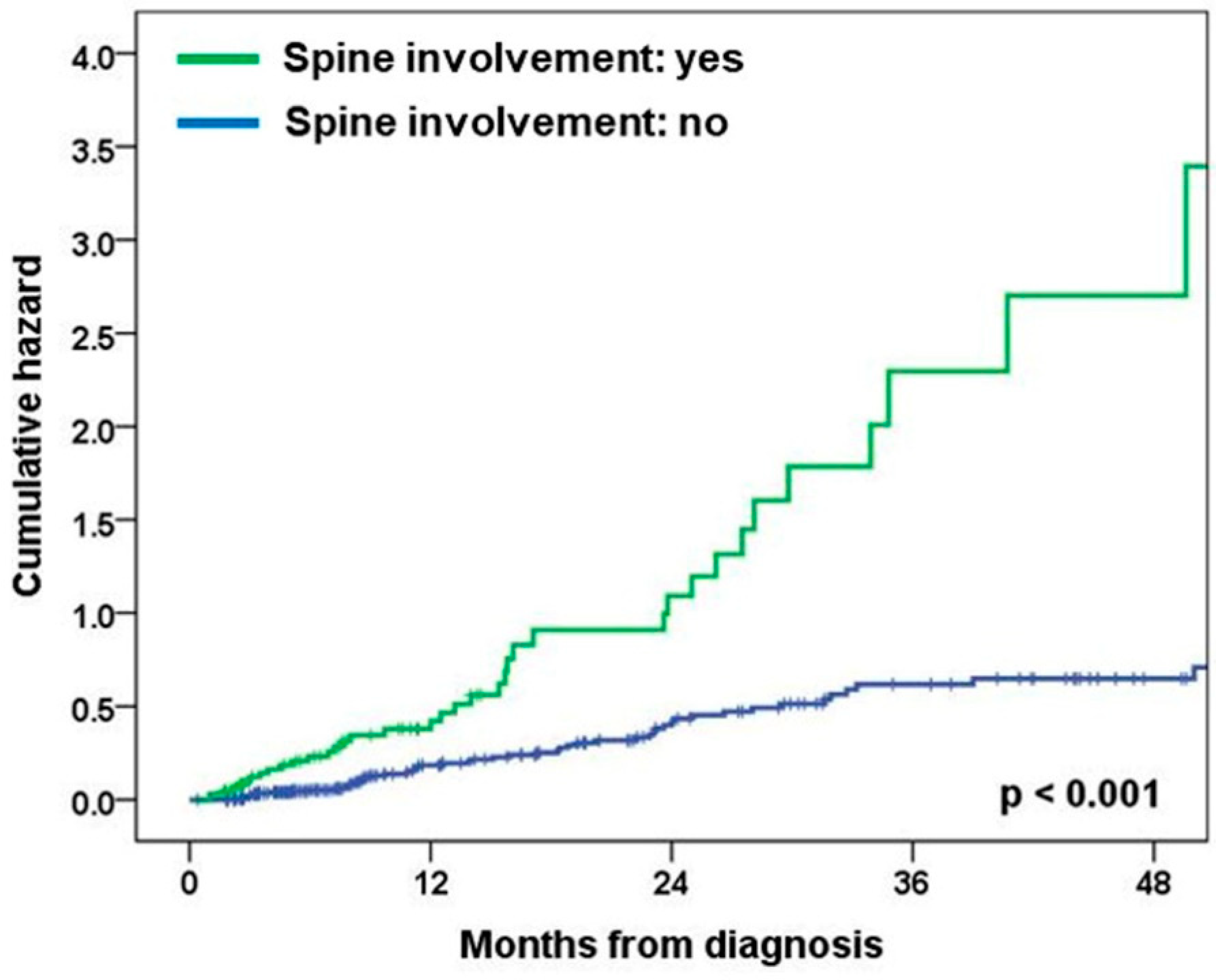
3.2. Association of Spine Metastasis with BM at Diagnosis or Subsequent BM

Female gender (OR and 95% CI = 1.46 and 1.08–1.98, p = 0.015), never having smoked (1.77 and 1.30–2.40, p < 0.001), adenocarcinoma histology (1.91 and 1.27–2.87, p = 0.002), higher N category (1.55 and 1.06–2.29, p = 0.026), and bone metastasis (2.31 and 1.71–3.12, p < 0.001) were significantly associated with an increased risk of BM at diagnosis (Table 3 and Table S1). Bone metastasis showed a significant association with BM after adjustment for potential confounding by other clinical variables (2.07 and 1.50–2.84, p < 0.001). Spine involvement was significantly associated with the risk of BM in multivariate logistic regression analysis (2.42 and 1.74–3.37, p < 0.001), but non-spine involvement was not (0.89 and 0.55– 1.42, p = 0.615) (Table S2).
Higher N category (2.47 and 1.45–4.21, p = 0.001) and spine involvement (2.46 and 1.53–3.94, p < 0.001) were significantly associated with an increased risk of subsequent BM development (Table 4). Spine involvement showed a significant association with subsequent BM development after adjustment for potential confounding by other clinical variables (1.94 and 1.19–3.18, p = 0.008) (Figure 1).

3.3. Anatomic Proximity of BM Lesions to CSF Space in Patients with Spine Metastasis

BM lesions adjacent (within 5 mm) to the CSF space were observed in 105 (93.8%) of 112 patients with both spine metastasis and BM at diagnosis and in 26 (100%) of 26 patients with spine metastasis at diagnosis and subsequent BM development. In addition, intracranial LM on brain MRI was observed in 35.7% of patients with spine metastasis and BM at diagnosis and in 61.5% of patients with spine metastasis at diagnosis and subsequent BM development. Of the 33 patients who underwent spine MRI, spinal canal invasion was noted in 17 (65.4%) of 26 patients with BM at diagnosis, and 5 (71.4%) of 7 patients with subsequent BM development.

4. Discussion

This study, for the first time, demonstrated an association between spine involvement in bone metastasis with BM, either present at diagnosis or developed subsequently. Interestingly, the results suggest a sequential causal relationship through the anatomic proximities of the brain, spine, and CSF. In this study, the presence of spine involvement in bone metastasis was significantly associated with BM at diagnosis or subsequent BM development in the univariate analysis. These effects on BM were maintained after adjustments for potential confounders.
BM lesions adjacent to the CSF space were observed in most patients with spine metastasis and BM at diagnosis or subsequent BM [15]. In addition, spinal canal invasion on spine MRI was observed in a significant proportion of patients with spine metastasis and BM at the time of diagnosis. Furthermore, intracranial LM on brain MRI was commonly observed in these patients. These findings support the fact that spine metastasis is present with BM at diagnosis or with subsequent BM development. Taken together, the data suggest that cancer cells in the spine could metastasize to the brain via the CSF [6]. Our study suggests the need for prophylactic spine radiotherapy before spinal canal involvement and subsequent LM and BM occur due to spinal metastatic lesions. A study with this design has never been conducted before, and clinical trials regarding this topic are needed in the future.
In a previous study with 592 NSCLC patients, 59 of 102 patients (57.8%) with LM had concurrent BM at diagnosis. In addition, patients with LM had a significantly high rate of bone metastases compared to patients with only BM or no CNS metastases, which is in line with our study. In another study of 125 non-small-cell lung cancer patients with LM, 102 (82%) patients had brain metastases [16]. Consistent with prior studies, a significant proportion of patients in our study had coexisting BM and LM. In addition, spinal canal invasion was observed by spinal MRI in a significant proportion of patients with small-cell lung cancer in a study with 163 solid tumor patients with LM [17]. This supports the results of our study, which emphasized the mechanism of LM development through tumor invasion of the spinal canal.
Circulating tumor cells (CTCs) present in the CSF and leptomeninges pose significant challenges to therapeutic interventions and have the potential to initiate metastasis in the brain and spine [18]. A considerable proportion of CNS metastases affecting the leptomeninges originate from breast and lung tumors. Exploiting the nutrient-poor microenvironment of the CSF, tumor cells adapt to enhance their survival. A prior investigation revealed that invasive tumor cells release complement C3 into the CSF, which then interacts with the C3aR receptor on choroid plexus cells. This interaction disrupts the blood–CSF barrier (BCSFB), enabling the unimpeded entry of nutrients and growth factors into the CSF. Further exploration is warranted to determine whether BCSFB disruption facilitates the invasion of tumors into the brain parenchyma.
It is important to measure the impact that brain metastases have on quality of life (QoL) for a complete picture of the disease burden [19]. Symptoms of BM include headaches, cognitive deficits, ataxia, seizures, and visual and speech problems, which can impact patient’s QoL in addition to the symptoms from their primary tumor. Furthermore, the side effects associated with the treatment for BM can seriously impact a patient’s QoL by limiting their ability to perform everyday activities and by altering neurocognitive processes, especially if the treatment involves surgery or radiation to the brain. Therefore, it is important to identify high-risk groups for BM in advance, and this study provides useful information in this regard.
Patients that were EGFR-positive or ALK-positive had higher rates of BM than those with wild-type tumors, which is supported by previous studies [20]. The blood–brain barrier shields the CNS from harmful substances, yet simultaneously hinders the majority of therapeutic agents from reaching the brain parenchyma and leptomeningeal space [21]. The influx and efflux mechanisms collectively determine the entry of drugs into the CSF. Active efflux transport systems act as barriers, impeding the delivery of drugs to the CNS. Export proteins, like P-glycoprotein and the breast cancer resistance protein, situated in the luminal membrane of the brain capillary endothelium, are the primary obstacles to efficient drug transport into the brain and leptomeningeal space. While various anticancer treatments, including tyrosine kinase inhibitors and chemotherapeutic agents, serve as substrates for these efflux transporters, their penetration into the CSF varies due to the opposing interplay of influx and efflux mechanisms. For instance, despite being a substrate for efflux transporters, osimertinib, a third-generation EGFR tyrosine kinase inhibitor, exhibits adequate permeability to counteract efflux [22]. Alectinib, a second-generation inhibitor targeting ALK and known for its remarkable ability to penetrate the CNS, demonstrates notable effectiveness in both systemic and CNS outcomes for individuals with ALK-rearranged NSCLC [23,24]. The landscape of NSCLC treatment has been significantly altered by the advent of immunotherapy. Despite their substantial impact, programmed death-1/PD-ligand 1 pathway inhibitors face challenges in penetrating the blood–brain barrier due to their high molecular weight [25]. Instead, they exert their effects by systemically activating immune cells. Potential avenues for access to the CSF compartment and brain tissues include the choroid plexus and CSF, enabling peripheral immune cells and large molecules to enter. AZD3759, a recently developed EGFR tyrosine kinase inhibitor of the new generation, exhibits encouraging efficacy in an EGFR-mutant mouse model with LM originating from NSCLC [22]. Lorlatinib, a next-generation ALK inhibitor, was specifically designed to reduce drug efflux facilitated by P-glycoprotein [26].
A recent study demonstrated that vertebral skeletal stem cells (vSSC) are distinct from other skeletal stem cells and mediate the unique physiology and pathology of vertebrae, including contributing to the high rate of vertebral metastasis [27]. In particular, human vSSCs secreting MFGE8 were more likely to interact with cancer cells than were those vSSCs that were not secreting the protein. Considering our results, we suggest that treatment targeting vSSC and MFGE8 has the potential to prevent the development of not only spinal metastases but also brain metastases.
There are several limitations in this study. First, the recruitment of patients from only a single center challenges the generalizability of these results and necessitates external confirmatory studies. However, clinical information was prospectively obtained from the ILCC and imaging studies were extensively evaluated by an experienced neuro-radiologist. Second, treatments given to NSCLC patients were not considered in this study and their effects on the development of BM remain elusive. Finally, the presence of LM was not confirmed with cytological examination of CSF in most patients due to the difficulty of CSF tapping. Alternatively, the proportion of patients with LM on brain MRI was analyzed. Within the limits of the study, the results suggest CSF is a potential pathway of spine involvement in bone metastasis to BM.

5. Conclusions

In conclusion, this study suggests that spine metastases at diagnosis is a risk factor for baseline and subsequent BM development in patients with NSCLC. As such, clinicians should carefully monitor subsequent BM with brain MRI in NSCLC patients with spine involvement.

Supplementary Materials

The following supporting information can be downloaded at: https://www.mdpi.com/article/10.3390/medicina60010152/s1, Table S1: Association of bone metastasis with BM at diagnosis; Table S2: Association of BM at diagnosis in patients with non-spine bone metastasis.

Author Contributions

Conceptualization: J.-S.R., J.-H.L.; data curation: H.-K.C., J.-S.R., J.-H.L.; formal analysis: H.-K.C., J.-S.R., J.-H.L.; investigation: H.-K.C., W.-K.R., H.-Y.L., H.-J.K., J.-S.R., J.-H.L.; methodology: H.-K.C., H.-Y.L., J.-S.R., J.-H.L.; project administration: J.-S.R., J.-H.L.; supervision: J.-S.R., J.-H.L.; validation: H.-K.C., J.-S.R., J.-H.L.; writing—original draft: H.-K.C., W.-K.R., H.-Y.L., H.-J.K., J.-S.R., J.-H.L.; writing—review and editing: H.-K.C., W.-K.R., H.-Y.L., H.-J.K., J.-S.R., J.-H.L. All authors have read and agreed to the published version of the manuscript.

Funding

This work was supported by an Inha University Research Grant (Inha University, 66376-1).

Institutional Review Board Statement

The study was conducted in accordance with the Declaration of Helsinki, and approved by the Institutional Review Board of Inha University Hospital (protocol code: 2020-03-018 and date of approval: 30 March 2020).

Informed Consent Statement

Informed consent was obtained from all subjects involved in the study.

Data Availability Statement

The data presented in this study are available in the manuscript. Additional raw data are available on request from the corresponding author.

Conflicts of Interest

The authors declare no conflict of interest.

References

  1. Barnholtz-Sloan, J.S.; Sloan, A.E.; Davis, F.G.; Vigneau, F.D.; Lai, P.; Sawaya, R.E. Incidence proportions of brain metastases in patients diagnosed (1973 to 2001) in the Metropolitan Detroit Cancer Surveillance System. J. Clin. Oncol. 2004, 22, 2865–2872. [Google Scholar] [CrossRef]
  2. Mujoomdar, A.; Austin, J.H.; Malhotra, R.; Powell, C.A.; Pearson, G.D.; Shiau, M.C.; Raftopoulos, H. Clinical predictors of metastatic disease to the brain from non–small cell lung carcinoma: Primary tumor size, cell type, and lymph node metastases. Radiology 2007, 242, 882–888. [Google Scholar] [CrossRef]
  3. Mehta, M.P.; Rodrigus, P.; Terhaard, C.; Rao, A.; Suh, J.; Roa, W.; Souhami, L.; Bezjak, A.; Leibenhaut, M.; Komaki, R. Survival and neurologic outcomes in a randomized trial of motexafin gadolinium and whole-brain radiation therapy in brain metastases. J. Clin. Oncol. 2003, 21, 2529–2536. [Google Scholar] [CrossRef]
  4. Popper, H.H. Progression and metastasis of lung cancer. Cancer Metastasis Rev. 2016, 35, 75–91. [Google Scholar] [CrossRef] [PubMed]
  5. Patchell, R.A. The management of brain metastases. Cancer Treat. Rev. 2003, 29, 533–540. [Google Scholar] [CrossRef] [PubMed]
  6. Chowdhary, S.; Chamberlain, M. Leptomeningeal metastases: Current concepts and management guidelines. J. Natl. Compr. Cancer Netw. 2005, 3, 693–703. [Google Scholar] [CrossRef]
  7. Boire, A.; Zou, Y.; Shieh, J.; Macalinao, D.G.; Pentsova, E.; Massagué, J. Complement component 3 adapts the cerebrospinal fluid for leptomeningeal metastasis. Cell 2017, 168, 1101–1113.e1113. [Google Scholar] [CrossRef] [PubMed]
  8. Fujii, T.; Mason, J.; Chen, A.; Kuhn, P.; Woodward, W.A.; Tripathy, D.; Newton, P.K.; Ueno, N.T. Prediction of bone metastasis in inflammatory breast cancer using a markov chain model. Oncologist 2019, 24, 1322–1330. [Google Scholar] [CrossRef]
  9. Freret, M.E.; Wijetunga, N.A.; Shamseddine, A.A.; Higginson, D.S.; Schmitt, A.M.; Yamada, Y.; Lis, E.; Boire, A.; Yang, J.T.; Xu, A.J. Early Detection of Leptomeningeal Metastases Among Patients Undergoing Spinal Stereotactic Radiosurgery. Adv. Radiat. Oncol. 2023, 8, 101154. [Google Scholar] [CrossRef]
  10. Bubendorf, L.; Schöpfer, A.; Wagner, U.; Sauter, G.; Moch, H.; Willi, N.; Gasser, T.C.; Mihatsch, M.J. Metastatic patterns of prostate cancer: An autopsy study of 1,589 patients. Hum. Pathol. 2000, 31, 578–583. [Google Scholar] [CrossRef]
  11. Geldof, A.A. Models for cancer skeletal metastasis: A reappraisal of Batson’s plexus. Anticancer Res. 1997, 17, 1535–1539. [Google Scholar] [PubMed]
  12. Goldstraw, P.; Chansky, K.; Crowley, J.; Rami-Porta, R.; Asamura, H.; Eberhardt, W.E.; Nicholson, A.G.; Groome, P.; Mitchell, A.; Bolejack, V. The IASLC lung cancer staging project: Proposals for revision of the TNM stage groupings in the forthcoming (eighth) edition of the TNM classification for lung cancer. J. Thorac. Oncol. 2016, 11, 39–51. [Google Scholar] [CrossRef] [PubMed]
  13. Ryu, J.-S.; Ryu, H.J.; Lee, S.-N.; Memon, A.; Lee, S.-K.; Nam, H.-S.; Kim, H.-J.; Lee, K.-H.; Cho, J.-H.; Hwang, S.-S. Prognostic impact of minimal pleural effusion in non–small-cell lung cancer. J. Clin. Oncol. 2014, 32, 960–967. [Google Scholar] [CrossRef] [PubMed]
  14. Ali, A.; Ouyang, T.; Krishnamoorthy, T.; Aregawi, D.G.; Zacharia, B.; Glantz, M.J. Role of cerebrospinal fluid (CSF) in the pathogenesis and treatment of patients with brain metastases. J. Clin. Oncol. 2018, 36, e14006. [Google Scholar] [CrossRef]
  15. Li, Q.; Lin, Z.; Hong, Y.; Fu, Y.; Chen, Y.; Liu, T.; Zheng, Y.; Tian, J.; Liu, C.; Pu, W. Brain parenchymal and leptomeningeal metastasis in non-small cell lung cancer. Sci. Rep. 2022, 12, 22372. [Google Scholar] [CrossRef] [PubMed]
  16. Morris, P.G.; Reiner, A.S.; Szenberg, O.R.; Clarke, J.L.; Panageas, K.S.; Perez, H.R.; Kris, M.G.; Chan, T.A.; DeAngelis, L.M.; Omuro, A.M. Leptomeningeal metastasis from non-small cell lung cancer: Survival and the impact of whole brain radiotherapy. J. Thorac. Oncol. 2012, 7, 382–385. [Google Scholar] [CrossRef]
  17. Pan, Z.; Yang, G.; He, H.; Yuan, T.; Wang, Y.; Li, Y.; Shi, W.; Gao, P.; Dong, L.; Zhao, G. Leptomeningeal metastasis from solid tumors: Clinical features and its diagnostic implication. Sci. Rep. 2018, 8, 10445. [Google Scholar] [CrossRef]
  18. Gillespie, C.S.; Mustafa, M.A.; Richardson, G.E.; Alam, A.M.; Lee, K.S.; Hughes, D.M.; Escriu, C.; Zakaria, R. Genomic alterations and the incidence of brain metastases in advanced and metastatic non-small cell lung cancer: A systematic review and meta-analysis. J. Thorac. Oncol. 2023, 18, 1703–1713. [Google Scholar] [CrossRef]
  19. Cheng, H.; Perez-Soler, R. Leptomeningeal metastases in non-small-cell lung cancer. Lancet Oncol. 2018, 19, e43–e55. [Google Scholar] [CrossRef]
  20. Ballard, P.; Yates, J.W.; Yang, Z.; Kim, D.-W.; Yang, J.C.-H.; Cantarini, M.; Pickup, K.; Jordan, A.; Hickey, M.; Grist, M. Preclinical comparison of osimertinib with other EGFR-TKIs in EGFR-mutant NSCLC brain metastases models, and early evidence of clinical brain metastases activity. Clin. Cancer Res. 2016, 22, 5130–5140. [Google Scholar] [CrossRef]
  21. Gainor, J.F.; Sherman, C.A.; Willoughby, K.; Logan, J.; Kennedy, E.; Brastianos, P.K.; Chi, A.S.; Shaw, A.T. Alectinib salvages CNS relapses in ALK-positive lung cancer patients previously treated with crizotinib and ceritinib. J. Thorac. Oncol. 2015, 10, 232–236. [Google Scholar] [CrossRef] [PubMed]
  22. Gainor, J.F.; Chi, A.S.; Logan, J.; Hu, R.; Oh, K.S.; Brastianos, P.K.; Shih, H.A.; Shaw, A.T. Alectinib dose escalation reinduces central nervous system responses in patients with anaplastic lymphoma kinase–positive non–small cell lung cancer relapsing on standard dose alectinib. J. Thorac. Oncol. 2016, 11, 256–260. [Google Scholar] [CrossRef] [PubMed]
  23. O’Kane, G.M.; Leighl, N.B. Are immune checkpoint blockade monoclonal antibodies active against CNS metastases from NSCLC?—Current evidence and future perspectives. Transl. Lung Cancer Res. 2016, 5, 628. [Google Scholar] [CrossRef] [PubMed]
  24. Johnson, T.W.; Richardson, P.F.; Bailey, S.; Brooun, A.; Burke, B.J.; Collins, M.R.; Cui, J.J.; Deal, J.G.; Deng, Y.-L.; Dinh, D. Discovery of (10 R)-7-Amino-12-fluoro-2, 10, 16-trimethyl-15-oxo-10, 15, 16, 17-tetrahydro-2H-8, 4-(metheno) pyrazolo [4, 3-h][2, 5, 11]-benzoxadiazacyclotetradecine-3-carbonitrile (PF-06463922), a macrocyclic inhibitor of anaplastic lymphoma kinase (ALK) and c-ros oncogene 1 (ROS1) with preclinical brain exposure and broad-spectrum potency against ALK-resistant mutations. J. Med. Chem. 2014, 57, 4720–4744. [Google Scholar]
  25. Peters, S.; Bexelius, C.; Munk, V.; Leighl, N. The impact of brain metastasis on quality of life, resource utilization and survival in patients with non-small-cell lung cancer. Cancer Treat. Rev. 2016, 45, 139–162. [Google Scholar] [CrossRef]
  26. Deshpande, K.; Buchanan, I.; Martirosian, V.; Neman, J. Clinical perspectives in brain metastasis. Cold Spring Harb. Perspect. Med. 2019, 10, a037051. [Google Scholar] [CrossRef]
  27. Sun, J.; Hu, L.; Bok, S.; Yallowitz, A.R.; Cung, M.; McCormick, J.; Zheng, L.J.; Debnath, S.; Niu, Y.; Tan, A.Y. A vertebral skeletal stem cell lineage driving metastasis. Nature 2023, 621, 602–609. [Google Scholar] [CrossRef]
Figure 1. Effect of spine involvement in bone metastasis on subsequent BM development.
Medicina 60 00152 g001
Table 1. Patient characteristics for BM.
VariablesBrain Metastasis
Yes (n = 238)No (n = 664)p Value
Age, median 0.108
>69106 (44.5)336 (50.6)
≤69132 (55.5)328 (49.4)
Gender 0.015
Male137 (57.6)441 (66.4)
Female101 (42.4)223 (33.6)
Smoking history <0.001
Ever138 (58.2)465 (71.1)
Never99 (41.8)189 (28.9)
ECOG performance status 0.057
0–1132 (56.2)416 (63.2)
≥2103 (43.8)242 (36.8)
Histology 0.003
SQC34 (14.3)154 (23.2)
ADC188 (79.0)447 (67.3)
Others16 (6.7)63 (9.5)
EGFR mutation 0.013
Negative147 (61.8)468 (70.5)
Positive91 (38.2)196 (29.5)
T category 0.403
Tx2 (0.8)16 (2.4)
T19 (3.8)38 (5.7)
T238 (16.0)113 (17.0)
T353 (22.3)139 (20.9)
T4136 (57.1)358 (53.9)
N category 0.145
N047 (19.7)180 (27.1)
N121 (8.8)60 (9.0)
N254 (22.7)137 (20.7)
N3116 (48.7)286 (43.1)
M category <0.001
Others98 (41.2)411 (61.9)
Bone140 (58.8)253 (38.1)
Non-spine28 (20.0)85 (33.6)
Spine112 (80.0)168 (66.4)
ECOG, Eastern Cooperative Oncology Group; SQC, squamous cell carcinoma, ADC, adenocarcinoma; EGFR, epidermal growth factor receptor.
Table 2. Patient characteristics for subsequent BM.
VariablesSubsequent Brain Metastasis
Yes (n = 82)No (n = 202)p Value
Age, median 0.034
>6925 (30.5)89 (44.1)
≤6957 (69.5)113 (55.9)
Gender 0.003
Male41 (50.0)139 (68.8)
Female41 (50.0)63 (31.2)
Smoking history 0.008
Ever46 (56.1)146 (72.3)
Never36 (43.9)56 (27.7)
ECOG performance status 0.863
0–157 (71.2)146 (72.3)
≥223 (28.8)56 (27.7)
Histology 0.001
SQC6 (7.3)49 (24.3)
ADC72 (87.8)134 (66.3)
Others4 (4.9)19 (9.4)
EGFR mutation <0.001
Negative38 (46.3)147 (72.8)
Positive44 (53.7)55 (27.2)
T category 0.879
Tx3 (3.7)4 (2.0)
T16 (7.3)14 (6.9)
T213 (15.9)40 (19.8)
T317 (20.7)41 (20.3)
T443 (52.4)103 (51.0)
N category 0.109
N021 (25.6)72 (35.6)
N15 (6.1)23 (11.4)
N217 (20.7)29 (14.4)
N339 (47.6)78 (38.6)
Bone metastasis 0.001
No42 (51.2)148 (73.3)
Yes40 (48.8)54 (26.7)
Non-spine14 (35.0)22 (40.7)
Spine26 (65.0)32 (59.3)
ECOG, Eastern Cooperative Oncology Group; SQC, squamous cell carcinoma, ADC, adenocarcinoma; EGFR, epidermal growth factor receptor.
Table 3. Association of spine involvement of bone metastasis with BM at diagnosis.
VariablesUnivariate AnalysisMultivariate Analysis
OR (95% CI)p ValueOR (95% CI)p Value
Age, median
>69reference-
≤691.28 (0.95–1.72)0.109
Gender
Malereference-reference-
Female1.46 (1.08–1.98)0.0150.77 (0.44–1.36)0.371
Smoking history -
Everreference reference-
Never1.77 (1.30–2.40)<0.0012.01 (1.12–3.60)0.020
ECOG performance status
0–1reference-reference-
≥21.34 (0.99–1.82)0.0571.26 (0.91–1.73)0.160
Histology
SQCreference-reference-
ADC1.91 (1.27–2.87)0.0021.53 (0.97–2.42)0.067
Others1.15 (0.59–2.23)0.6791.11 (0.56–2.19)0.763
EGFR mutation
Negativereference-reference-
Positive1.48 (1.08–2.02)0.0140.92 (0.63–1.33)0.643
T category
Tx–T1reference-reference-
T21.65 (0.78–3.48)0.1871.49 (0.69–3.24)0.310
T31.87 (0.91–3.85)0.0891.61 (0.75–3.42)0.219
T41.87 (0.95–3.67)0.0721.49 (0.74–3.04)0.268
N category
N0reference-reference-
N11.34 (0.74–2.42)0.3321.58 (0.85–2.94)0.150
N21.51 (0.96–2.37)0.0731.78 (1.10–2.86)0.019
N31.55 (1.06–2.29)0.0261.42 (0.93–2.15)0.105
Bone metastasis
Non-spinereference-reference-
Spine2.62 (1.93–3.57)<0.0012.42 (1.74–3.37)<0.001
ECOG, Eastern Cooperative Oncology Group; SQC, squamous cell carcinoma, ADC, adenocarcinoma; EGFR, epidermal growth factor receptor.
Table 4. Effect of spine involvement of bone metastasis on subsequent BM.
VariablesUnivariate AnalysisMultivariate Analysis
HR (95% CI)p ValueHR (95% CI)p Value
Age, median
>69reference-
≤691.15 (0.72–1.84)0.566
Gender
Malereference-
Female1.30 (0.84–2.01)0.238
Smoking history
Everreference-
Never1.19 (0.77–1.84)0.440
ECOG performance status
0–1reference-
≥21.32 (0.81–2.16)0.260
Histology
SQCreference-reference-
ADC2.02 (0.88–4.67)0.0992.07 (0.86–5.01)0.106
Others2.29 (0.65–8.14)0.2002.36 (0.64–8.72)0.197
EGFR mutation
Negativereference-
Positive1.34 (0.87–2.07)0.188
T category
Tx–T1reference-
T20.67 (0.28–1.57)0.352
T31.02 (0.45–2.29)0.966
T40.92 (0.45–1.90)0.830
N category
N0reference-reference-
N11.46 (0.55–3.89)0.4511.83 (0.66–5.07)0.243
N23.03 (1.58–5.78)0.0012.99 (1.54–5.82)0.001
N32.47 (1.45–4.21)0.0012.32 (1.35–3.98)0.002
Bone metastasis
Non-spinereference-reference-
Spine2.46 (1.53–3.94)<0.0011.94 (1.19–3.18)0.008
ECOG, Eastern Cooperative Oncology Group; SQC, squamous cell carcinoma, ADC, adenocarcinoma; EGFR, epidermal growth factor receptor.
Disclaimer/Publisher’s Note: The statements, opinions and data contained in all publications are solely those of the individual author(s) and contributor(s) and not of MDPI and/or the editor(s). MDPI and/or the editor(s) disclaim responsibility for any injury to people or property resulting from any ideas, methods, instructions or products referred to in the content.

