Review

Recent Advancement of Medical Patch for Transdermal Drug Delivery

1
Department of Medical Microbiology, Faculty of Medicine, Universiti Malaya, Kuala Lumpur 50603, Malaysia
2
Department of Medical Microbiology, University Malaya Medical Center, Kuala Lumpur 59100, Malaysia
3
Department of Pharmacology, Yong Loo Lin School of Medicine, National University of Singapore, Singapore 117600, Singapore
4
School of Biosciences, Faculty of Health & Medical Sciences, Taylor’s University, Subang Jaya 47500, Malaysia
*
Authors to whom correspondence should be addressed.
Medicina 2023, 59(4), 778; https://doi.org/10.3390/medicina59040778
Submission received: 23 February 2023 / Revised: 31 March 2023 / Accepted: 5 April 2023 / Published: 17 April 2023
(This article belongs to the Section Dermatology)

Abstract

:
Transdermal patches are a non-invasive method of drug administration. It is an adhesive patch designed to deliver a specific dose of medication through the skin and into the bloodstream throughout the body. Transdermal drug delivery has several advantages over other routes of administration, for instance, it is less invasive, patient-friendly, and has the ability to bypass first-pass metabolism and the destructive acidic environment of the stomach that occurs upon the oral ingestion of drugs. For decades, transdermal patches have attracted attention and were used to deliver drugs such as nicotine, fentanyl, nitroglycerin, and clonidine to treat various diseases or conditions. Recently, this method is also being explored as a means of delivering biologics in various applications. Here, we review the existing literatures on the design and usage of medical patches in transdermal drug delivery, with a focus on the recent advances in innovation and technology that led to the emergence of smart, dissolvable/biodegradable, and high-loading/release, as well as 3D-printed patches.

Graphical Abstract

1. Introduction

Transdermal drug delivery is an alternative way of delivering drugs via the skin layer [1,2]. The drug is carried through the skin into the bloodstream and circulates systemically in the body before reaching the target site [1,2]. The transdermal drug delivery method has several advantages over other routes of administration. Examples include the ability to deliver continuous doses of drugs over an extended period of time, the ability to bypass the digestive system, and the ability to avoid first-pass metabolism in the liver [3]. Other drug administration routes, such as intravenous, can cause pain and increase the risk of infection. Nevertheless, the oral route is inefficient, and in the inhalation method, it is difficult in controlling the dosage. In view of its advantages over other routes, transdermal administration is commonly used to deliver drugs for conditions such as smoking cessation, chronic pain, and motion sickness, as well as hormone replacement therapy [4,5,6].
A transdermal patch is a medicated patch that can deliver drugs directly into the bloodstream through the layers of the skin at a prescribed rate. In fact, patches are the most convenient method of administration. They are non-invasive, and treatment can last for several days and can be stopped at any time (Table 1). They come in different sizes and contain multiple ingredients. When applied to the skin, the patch can deliver active ingredients into the systemic circulation via diffusion processes. Transdermal patches may contain high doses of active substances that remain on the skin for an extended period of time. One of the first transdermal patches developed in 1985 was the nitroglycerin patch. The patch, developed by Gale and Berggren, uses a rate-controlling ethylene vinyl acetate membrane. Currently, several drugs are available as transdermal patches, including estradiol, clonidine, fentanyl, nicotine, scopolamine (hyoscine), and estradiol with norethisterone acetate (Table 2). The site of application may vary depending on the therapeutic category of the drug [7]. For example, nitroglycerin can be applied around the chest and estradiol around the buttocks or abdomen. The duration of drug release also varies depending on the usage, from the shortest (up to 9 h) to the longest (up to 9 days).

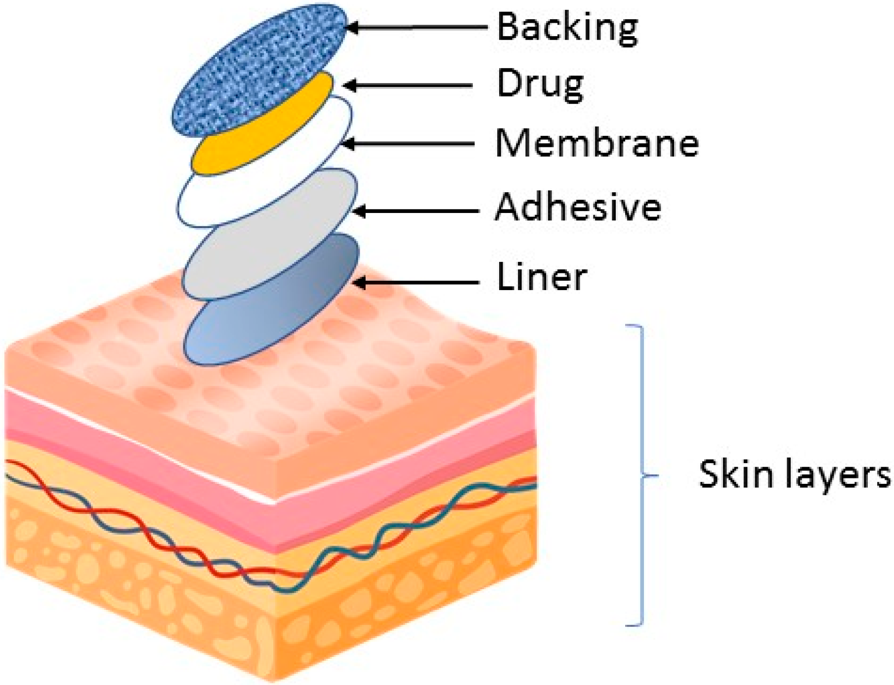
2. Transdermal Patch Design

Transportation of drug across the skin is affected by various factors, such as skin permeability, area, and duration of application, as well as metabolic activity of the skin (i.e., first pass metabolism). In fact, every drug has its unique properties, which can affect transdermal delivery. To achieve adequate skin absorption and penetration, the drug should be non-ionic and relatively lipophilic to cross the skin barrier. Molecules larger than 500 Daltons make it difficult to cross the stratum corneum, and ideally the therapeutic dose of the drug should also be less than 10 mg per day.

3. Basic Component of Transdermal Patch

Transdermal patches typically consist of several layers that are designed to deliver the medication through the skin and into the bloodstream. Figure 1 illustrates the basic component of a medicated patch. The specific composition and structure of the patch may vary depending on the drug being delivered and the desired rate of drug release.
The backing layer is the outermost layer of the patch and serves to protect the other layers from the environment. This layer is usually made of a flexible, waterproof material such as polyethylene or polypropylene. The adhesive layer serves to attach the patch to the skin and keep it in place. It usually consists of a strong, hypoallergenic adhesive that is gentle on the skin. The drug layer contains drugs that are delivered through the skin. It is formulated to release the drugs at a constant rate over a period of time. The rate-controlling membrane serves to control the rate at which the drugs are released from the patch. Membranes are usually made of semi-permeable materials that allow the drugs to pass through the membrane at a controlled rate. Linen acts as a protector for the patch and adhesive. The patch must be removed before being applied to the skin surface.

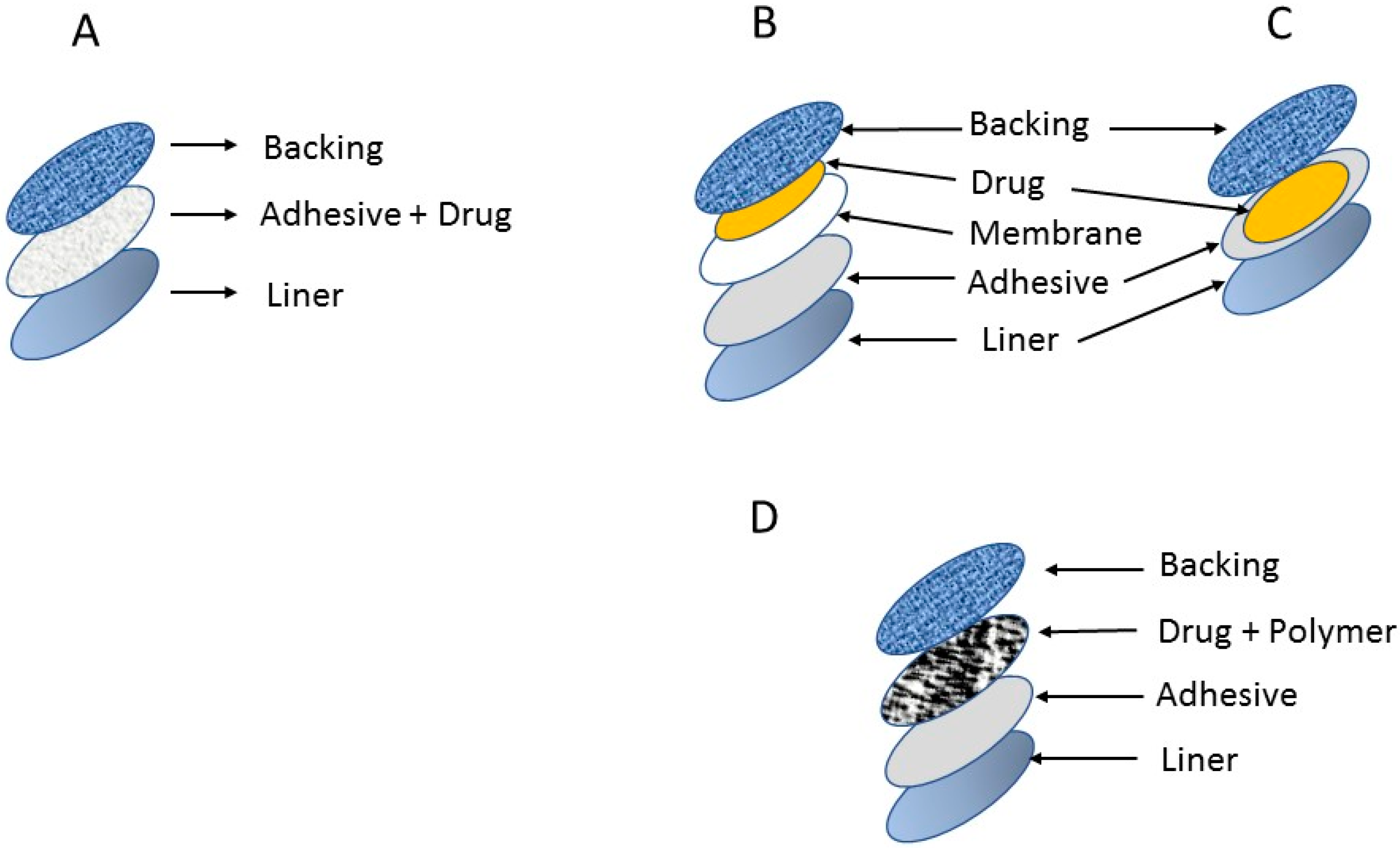
4. Types of Transdermal Patches

In general, there are four main type of transdermal medical patches (drug-in adhesive, reservoir, matrix, and micro-reservoir systems), as shown in Figure 2. Most commercially available patches are categorized as reservoir or matrix systems [56].

4.1. Drug-in-Adhesive System

This is the simplest form of membrane permeation control system. The adhesive layer in this system contains drugs and serves to glue the different layers together. The drug mixture is sandwiched between the liner and backing.

4.2. Reservoir System

In this system, the drug reservoir is held between the backing layer and the rate-controlling membrane, and the drug is released through the microporous rate-controlling membrane. The drug can be in solution, suspension, or gel form, or can be dispersed in a solid polymer matrix within the reservoir chamber.

4.3. Matrix System

Drugs are uniformly dispersed in hydrophilic or lipophilic polymer matrices. The resulting drug-containing polymer is affixed to drug-containing discs of controlled thickness and surface area.

4.4. Micro-Reservoir System

This system is a combination of reservoir and matrix dispersion system. Here, the drug is prepared by first suspending drug solids in an aqueous solution of a water-soluble liquid polymer and then uniformly dispersing the solution in a lipophilic polymer to create thousands of non-leaching microscopic drug reservoirs.

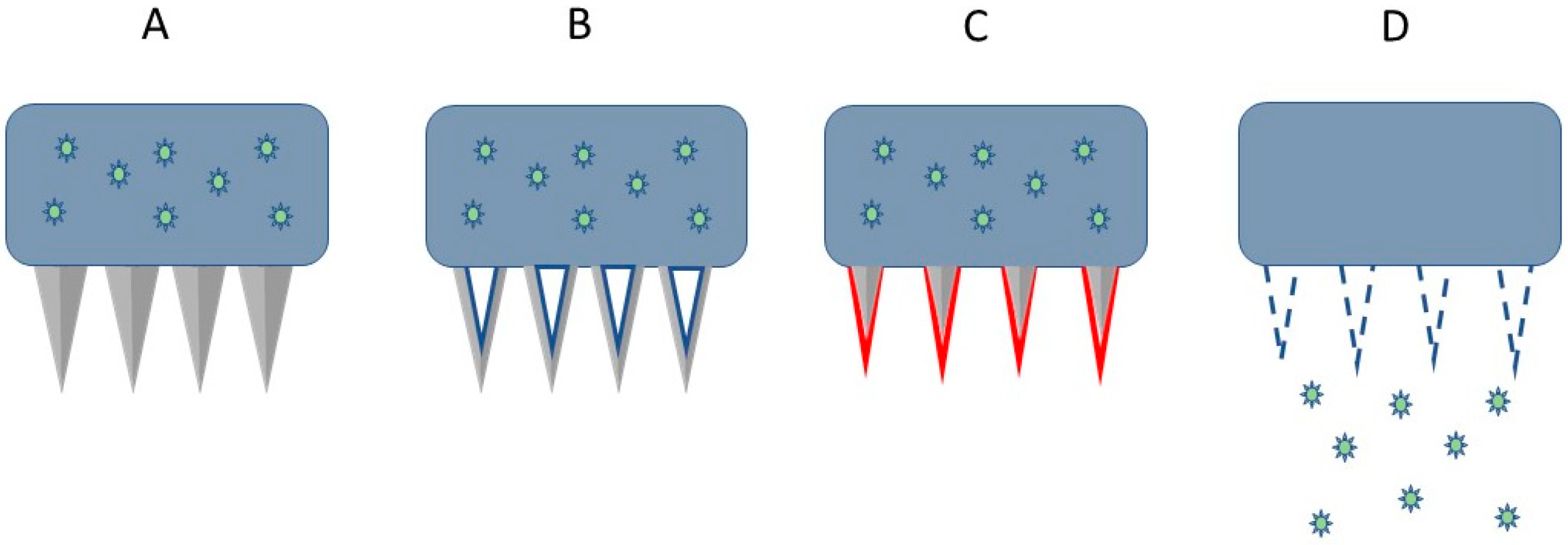
5. Microneedle-Based Patches

There are several types of microneedles, each with unique features and characteristics, as shown in Table 3. Overall, four major types of microneedle-based patches have been developed, namely solid, hollow, dissolving, and coated microneedle (Figure 3). The choice of microneedle type depends on the specific application and requirements of the user.
  • Solid Microneedles: These are the simplest type of microneedles, consisting of solid needles that penetrate the skin to create tiny channels. Solid microneedles are commonly used for drug delivery and cosmetic treatments.
  • Hollow Microneedles: These microneedles have a hollow core that allows for the delivery of fluids or drugs into the skin. Hollow microneedles are often used for transdermal drug delivery and sampling of interstitial fluid.
  • Coated Microneedles: These microneedles have a coating that dissolves upon penetration of the skin, allowing for the release of drugs or other substances. Coated microneedles are often used for transdermal drug delivery.
  • Dissolving Microneedles: These microneedles are made of materials that dissolve in the skin, allowing for the controlled release of drugs or other substances. Dissolving microneedles are often used for vaccines and other drug delivery applications.

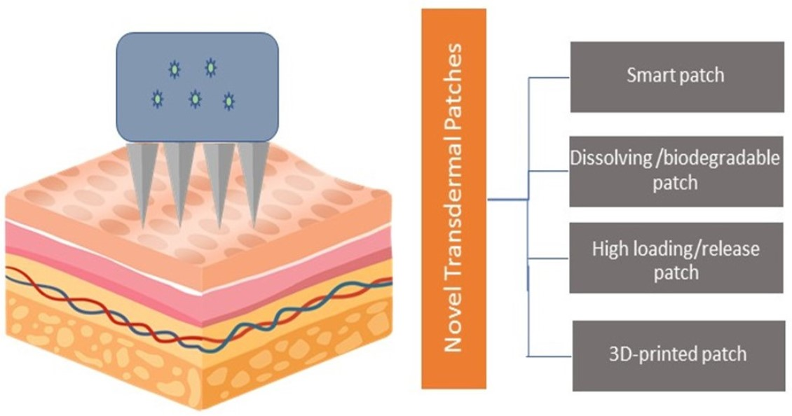
6. Recent Advancement of Transdermal Patch
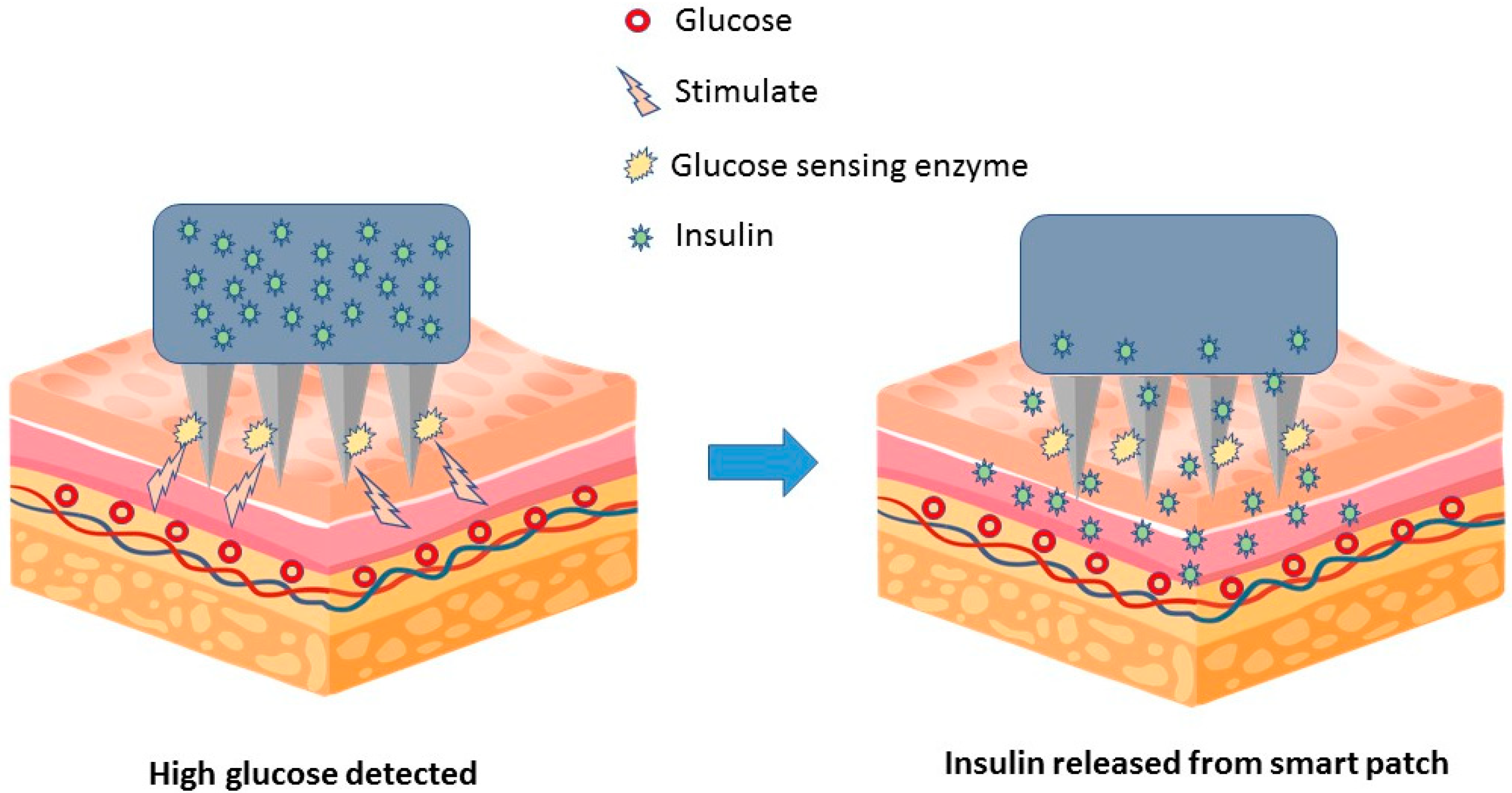
Traditional transdermal patches serve only two purposes: storage and release of drugs. While this method has some advantages, traditional patching has many challenges and drawbacks, for example limited dosage or low release. To date, there have been several advances in the field of transdermal drug delivery. These include the design of novel patches, which include the ability to sense and release drugs accurately, higher loading, and enhanced penetration and release of drugs. Overall, the field of transdermal drug delivery is an active area of research and development, with many exciting new developments on the horizon, as discussed below.

6.1. Smart Patches

Smart patches are equipped with sensors and other technologies that can monitor patient conditions and adjust drug delivery accordingly. In 2014, a group of researchers developed a microneedle-based smart patch sensor platform for painless and continuous intradermal glucose measurement for diabetics. This patch uses a conducting polymer such as poly (3,4-ethylenedioxythiophene) (PEDOT) as an electrical mediator for glucose detection and as an immobilizing agent for the glucose-specific c-enzyme glucose oxidase (GOx) [72]. Further research and development resulted in a smart insulin releasing patch consisting of 121 microneedles containing nanoparticles. The patch painlessly penetrates into the interstitial fluid between subcutaneous skin cells. The nanoparticles in each needle contain insulin and the glucose-sensing enzyme glucose oxidase, which converts glucose into gluconate. These molecules are surrounded by hypoxia-responsive polymers. As shown in Figure 4, increased glucose oxidase activity in response to increased glucose creates an oxygen-depleted environment within the nanoparticles, which is sensed by the hypoxia-responsive polymer, triggering nanoparticle degradation and insulin release [73,74].
Wound healing is a complex and dynamic regenerative process with constantly changing physical and chemical parameters. Its management and monitoring offer great benefits, especially for bedridden patients. Iversen et al. reported an inexpensive, flexible, fully printed smart patch on the skin to measure changes in wound pH and fluid volume. Such bendable sensors can also be easily incorporated into wound dressings. The sensor consists of various electrodes printed on a polydimethylsiloxane (PDMS) substrate for pH and humidity measurements. The generated sensor patch has a sensitivity of 7.1 ohm/pH to the wound pH value. Hydration sensor results showed that the water content of the semi-porous surface can be quantified by the change in resistivity [75].
Besides wound healing, scientists have also developed a smart patch to monitor and treat diabetic foot ulcers (DFU). This system is fabricated from conductive hydrogel patches with a ultra-high transparency polymer network. Importantly, highly transparent conductive hydrogel patches can be used to visually monitor wound healing status, promote haemostasis, improve cell-to-cell communication, prevent wound infection, promote collagen deposition, and improve vascularity. By promoting angiogenesis, it effectively promotes the healing of DFU. In addition, the versatile intelligent patch can also achieve indirect blood glucose monitoring by detecting glucose levels in wounds, and timely detect movements of various sizes of human bodies. Interestingly, this smart patch can monitor chronic wound dressings and treat wounds at the same time [76].
Smart patches are also used to deliver natural compounds such as curcumin. The material consists of paraffin wax and polypropylene glycol as a phase change material (PCM). PCM was combined with graphene-based heating elements obtained by the laser scribing of polyimide films. This arrangement offers a new approach to smart patches whose release can be electronically controlled, and which allows repeated dosing. Emission is induced and terminated by controlled heating of the PCM rather than relying on passive diffusion, and permeation only occurs when the PCM transitions from solid to liquid. Curcumin delivery yields were found to be good and satisfactory [77].

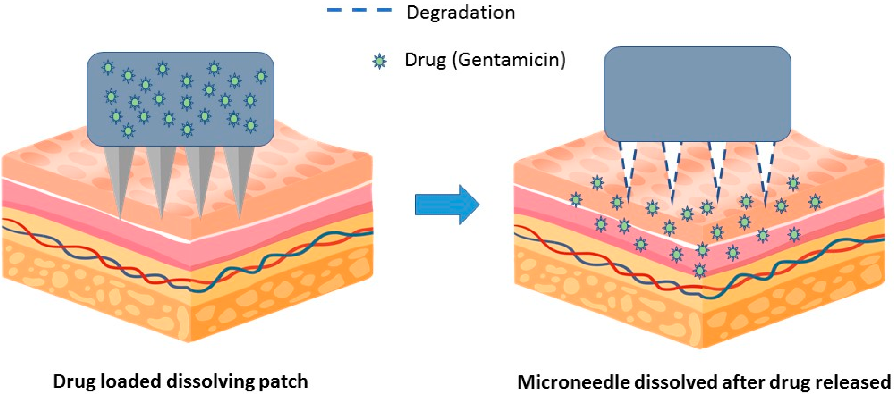
6.2. Dissolving/Degradable Patches

These patches are designed to dissolve on the skin and do not need to be removed and discarded. In general, these patches are made from biodegradable materials that are absorbed by the body after use. In a proof-of-concept paper published in 2019, researchers successfully administered the antibiotic gentamicin via a dissolving patch in a mouse model of bacterial infection [78] (Figure 5). The results showed that a gentamicin-dissolving microarray patch applied to mouse ears could control Klebsiella pneumoniae infection. In addition, mice treated with lysing patches had reduced bacterial burden in nose-associated lymphoid tissue and lungs compared with untreated control.
Dissolving microneedles (MNs) show high efficiency in the delivery of poorly permeable drugs and vaccines. A two-step injection and centrifugation process was used to localize insulin to the needle and achieve efficient transdermal delivery of insulin. The relative pharmacological availability and relative bioavailability (RBA) of insulin from MN patches were 95.6% and 85.7%, respectively. This study demonstrates that the use of dissolving patches for insulin delivery achieves a satisfactory relative bioavailability (RBA) compared to conventional subcutaneous injection, demonstrating the effectiveness of dissolving patches for diabetes treatment [79].
On the other hand, scientists have developed a biodegradable microneedle patch that delivers hyaluronic acid (HA) antigen-peptide conjugates for prophylactic cancer immunotherapy [80]. A cytotoxic T-cell epitope peptide (SI-INFEKL) is conjugated to HA-loaded biodegradable HA microneedle (MN) patches to efficiently deliver antigens to the skin’s immune system. Interestingly, a single transdermal vaccination with an MN patch containing the HA-SIINFEKL conjugate significantly increased tumor growth in B16 melanoma model mice by enhancing antigen-specific cytotoxic T-cells.
Another research group developed a hypotensive biodegradable patch for transdermal delivery of sodium nitroprusside (SNP) in combination with sodium thiosulfate (ST) [81]. Dissolvable microneedles loaded with SNPs and STs were fabricated by centrifugal casting. In this method, SNPs were stably packaged into microneedles and immediately released into the systemic circulation. Antihypertensive microneedle therapy (aH-MN) achieved rapid and strong blood pressure reduction. It met the clinical requirements for blood pressure management in hypertensive emergencies. Concomitant administration of ST effectively suppressed side effects (e.g., organ damage) caused by the continuous intake of SNPs. This study presented an efficient and patient-friendly biodegradable patch for antihypertensive therapy.
Transdermal patches are also commonly used in the cosmetic industry. However, the non-degradable polymers used in cosmetic patches are of concern because they can pollute the environment when disposed irresponsibly in open areas. In one study, biodegradable polylactic acid (PLA) was recommended due to its lack of toxicity. The results showed that the PLA/phycocyanin-alginate composite made with a phycocyanin/alginate ratio of 40/60 at 20 °C for 20 h had the best properties in terms of film flexibility and release properties [82]. Overall, the results are promising but warrant further in vivo or clinical study for further development.

6.3. Three-Dimensional (3D)-Printed Patches

Researchers are using 3D printing technology to create customized transdermal patches that can be tailored to the individual needs of each patient [83]. One good example is the use of a 3D-printed patch for wound healing. In a study by Jang et al., gelatin methacrylate (GelMA) was tested as a viable option with tunable physical properties. GelMA hydrogel incorporating a vascular endothelial growth factor (VEGF)-mimicking peptide was successfully printed using a 3D bio-printer owing to the shear-thinning properties of hydrogel inks. The 3D structure of the hydrogel patch had high porosity and water absorption properties. VEGF peptide, which is slowly released from hydrogel patches, can promote cell viability, proliferation, and tubular structure formation, indicating that the 3D Gel-MA-VEGF hydrogel patch can be used for wound dressing [84].
On the other hand, a three-dimensional (3D) printing technique called continuous liquid interface production (CLIP) was used to design and fabricate transdermal patches. The multifaceted microneedle design increased the surface area compared to the smooth square pyramid design, ultimately resulting in the improved surface coating of model vaccine components (ovalbumin and CpG). In the study, they used fluorescent tags and live animal imaging to assess in vivo charge retention and bioavailability in mice as a function of delivery route. Compared with subcutaneous bolus injection of soluble components, transdermal administration not only resulted in improved skin charge retention, but also improved the activation of immune cells in draining lymph nodes. Moreover, the delivered vaccine elicited a strong humoral immune response with higher total IgG (immunoglobulin G) and a more balanced IgG1/IgG2a repertoire, resulting in dose sparing. Furthermore, it elicited a T-cell response characterized by functional cytotoxic CD8+ T-cells and CD4+ T-cells secreting Th1 (T helper type 1) cytokines. In short, CLIP 3D-printed microneedles coated with vaccine components provide a useful platform for non-invasive, self-administered vaccination [85].
Another group of researchers designed and printed the patch using stereolithography (SLA) technology with a proprietary class I resin. They showed that these patches can be used for transdermal delivery of high molecular weight antibiotics such as rifampicin (M(w) 822.94 g/mol). This drug suffers from gastric chemical instability, reduced bioavailability, and severe hepatotoxicity. The patch was engineered with sub-apical holes present in one-quarter of the needle tip to enhance the mechanical strength and integrity of the patch array. The tips were characterized by optical and electron microscopy to determine print quality and uniformity across the array. The system also underwent mechanical characterization for failure and penetration analysis. The authors systematically evaluated the ex vivo penetration and consequent transport of rifampicin through porcine skin. Moreover, in vivo study of rifampicin administration via the 3D-printed patch demonstrated efficient penetration and desirable bioavailability [86].
Another technique known as powder extrusion (DPE) has emerged as the most viable approach due to its ability to directly process excipients and pharmaceuticals in one step [87]. The study was set to determine whether different grades of ethylene-vinyl acetate (EVA) copolymers could be used as new starting materials for the fabrication of transdermal patches. By choosing two model drugs with different thermal behavior (i.e., ibuprofen and diclofenac sodium), they also wanted to consider the versatility of this EVA excipient in manufacturing patches for custom transdermal therapy. EVA was combined with 30% (w/w) of each model drug. Fourier transform infrared (FT-IR) spectra confirmed that the starting material was effectively incorporated into the final formulation, and thermal analysis revealed that the extrusion process changed the crystalline morphology of the raw polymer, leading to increased crystallization at smaller thicknesses. This study indicated that EVA and direct powder extrusion technology may be promising tools for the fabrication of transdermal patches. By choosing an EVA type with appropriate VA content, it is possible to print drugs with different melting points while maintaining thermal stability. Furthermore, the desired drug release and permeation profiles of drugs can be achieved. In fact, this is an important advantage from the point of view of personalized medicine.
Lim et al. reported the use of 3D printed personalized patches that conform to the skin surface for Acetyl-hexapeptide 3 (AHP-3) delivery. However, commercially available photocurable resins for 3D printing are not suitable for fabricating drug-loaded delivery systems. In this study, two liquid monomers, namely polyethylene glycol diacrylate (PEGDA) and vinylpyrrolidone (VP) in different proportions were used to improve the mechanical strength, polymerization rate, and swelling rate of the final polymer. Optimal drug loading on the resin indicated that AHP-3 remained stable throughout the manufacturing process and had no effect on the physical properties of the final polymer. Using a 3D scanned facial model, a personalized patch was designed in CAD (computer-aided design) software and fabricated in optimized resin using a digital light processing (DLP) 3D printer. In vitro characterization of the prepared transdermal patches showed their ability to penetrate human cadaver skin, and they remained intact after compression. The final polymer was also minimally cytotoxic to human dermal fibroblasts. This is the first study demonstrating personalized patches made using photopolymers and may be a novel approach to improve the transdermal delivery of drugs for effective wrinkle management [88].

6.4. High Loading/Release Patches

Long-acting transdermal drug delivery requires high drug loading and controlled drug release. In order to improve drug-polymer miscibility and achieve controlled drug release, a novel hydroxyphenyl (HP)-modified pressure-sensitive adhesive (PSA) was developed [89]. The results show that the dual-ionic H-bonds between R(3)N and R(2)NH-type drugs and HP-PSA are reversible and relatively strong, unlike ionic and neutral H-bonds. This allowed patches to significantly increase the drug loading from 1.5- to 7-fold and control the drug release rate from 1/5 to 1/2 without changing the overall release profile. Pharmacokinetic results showed that the HP-PSA-based high-load patch achieved sustained drug concentrations in plasma, avoided sudden release, increased area under the concentration-time curve (AUC), and average dwell time by more than 6x, indicating the potential for long-acting drug delivery. In addition, its safety and mechanical properties are met. Mechanistic studies have shown that repulsion of ionic drugs in HP-PSA increases drug loading, and relatively strong interactions can also control drug release. Incomplete hydrogen bond transfer determined its reversibility, making the percentage drug release equal to that of non-functional PSA. In short, the high drug loading efficiency and controlled drug release capability of HP-PSA and its unique interactions will contribute to the development of long-acting transdermal drug delivery systems. Moreover, the construction of double-ionic H-bonds provides further inspiration for various drug delivery systems in non-polar environments.
Pharmaceutical polymers are widely used to inhibit drug recrystallization through strong intermolecular hydrogen and ionic bonding, but at the expense of drug release rates in transdermal patches. To overcome this difficulty, a group of researchers came up with the idea of using a new ionic liquid (drug IL) strategy to increase drug loading [90]. A carboxyl-based pressure sensitive adhesive (PSA) was chosen as a model polymer. The results showed a five-fold increase in PSA drug load. This was caused by the synergistic effect of strong ionic and normal hydrogen bonds formed between the carbonyl groups of the drug and PSA. This study demonstrated an entirely new mechanism of action and provided a powerful tool for the development of high-drug load, high-release patches. In another study, the same group of researchers constructed a high-capacity, high-release transdermal patch with COOH polyacrylate polymer (PA-1) to deliver non-steroidal anti-inflammatory drugs (NSAIDs), namely ibuprofen. The drug load and skin absorption of PA-1 were improved by 2.4-fold and 2.5-fold, respectively. The hydrogen bond formed between the drug (COOH) and PA-1 (COOH) is weakened by repulsive interactions, whereas the enhanced conductivity of PA-1 was confirmed by dielectric spectroscopy, electron paramagnetic resonance (EPR) spectra, four-point probe method, and molecular modeling with the appearance of COO-. In summary, these results showed that ion–ion repulsion by reducing hydrogen bonding can be a viable way to build large-capacity, high-emission patches [91].

7. Potential Application of Transdermal Patches

Previously, we listed a number of therapeutically active drugs marketed as transdermal patches in Table 2. As technology and research have advanced, numerous potential application areas for transdermal patches have been explored, as described below.

7.1. Transdermal Patches for Patches for Vaccination

Researchers are developing transdermal patches that can deliver vaccines through the skin, potentially offering a more convenient and less painful alternative to injections. A good example is the microneedle-based smallpox vaccine patch. When this vaccine patch was applied to mice, neutralizing antibodies were induced from 3 weeks after immunization. Levels were maintained for 12 weeks, and there was a significant increase in IFN-γ secreting cells, suggesting that the transdermal patch could serve as an alternative delivery system for vaccination and preservation [92].
Another research group designed a lytic microneedle patch for influenza vaccination that targets skin antigen-presenting cells. Microneedles were created using a biocompatible polymer that encapsulates an inactivated influenza virus vaccine for insertion and dissolution into the skin within minutes. The patch elicited strong antibody and cell-mediated immune responses in mice that provided complete protection against lethal challenge. The results provide a new technique for simpler and safer vaccination with improved immunogenicity by using a transdermal patch, potentially enabling increased vaccination coverage [93].

7.2. Transdermal Patches for Gene Therapy

Recently, transdermal patches have been investigated as a way of providing gene therapy to deliver genetic material to defective cells [94]. Pioneering studies sought to deliver genes and photothermic agents simultaneously to the cancer cells. For this purpose, transdermal patches co-loaded with p53 DNA and IR820 (a near infrared dye) were prepared by a two-step casting procedure. Hyaluronic acid was first constructed as the matrix, before p53 DNA and IR820 were primarily loaded onto the patches. The patches efficiently penetrated the stratum corneum and rapidly dissolved to release p53 DNA and IR820 at subcutaneous tumor sites. The patch showed an excellent anti-tumor effect in vivo due to the synergistic effect of gene therapy and photothermal agents. These findings demonstrate that a transdermal patch could be a promising strategy for subcutaneous tumor treatment [95].

7.3. Transdermal Patches for Insulin Delivery

Transdermal insulin delivery patches are used to deliver insulin across the skin and into the bloodstream to treat diabetics. Insulin is a hormone secreted by the pancreas that plays a role in regulating blood sugar levels in the body. Diabetics can have high blood sugar levels because they cannot produce enough insulin or use the insulin produced by their body effectively. To date, several new techniques have been reported, including the use of ionic liquids, choline bicarbonate and geranic acid (CAGE), liposome, and nanomaterials to deliver insulin [96,97,98,99,100]. Transdermal patches for insulin delivery can provide a convenient and discreet alternative to traditional methods of insulin delivery, such as injections and insulin pumps [101]. The patches are typically applied to the skin on the abdomen, upper arm, or thigh and are designed to release a consistent dose of insulin over a specific period of time.
Of note, there are several challenges to overcome when developing transdermal patches for insulin delivery. Insulin is a large protein molecule that is not easily absorbed through the skin. To overcome this, researchers have developed a new approach for transdermal protein delivery using a water-swellable spherical double-layered microneedle (MN) patch at the tip. This design allows MNs to mechanically engage soft tissue through selective distal swelling after skin insertion. Furthermore, long-term release of loaded proteins was achieved by passive diffusion through the swollen tips. Insulin-loaded MN patches released 60% insulin upon immersion in saline for 12 h, and approximately 70% of the released insulin appeared to retain its structural integrity. Animal studies have shown that insulin release from swelling MN patches is prolonged, leading to a gradual decrease in blood glucose levels [102]. Overall, transdermal patches for insulin delivery have the potential to provide a convenient and effective method to deliver insulin to diabetic patients. However, further research and development is needed to optimize the efficacy and safety of these products.

7.4. Transdermal Patches for Cardiovascular Diseases

In a heart failure scenario, pharmacokinetics (PK) and pharmacodynamics (PD) are frequently altered to accommodate hypoperfusion systemic conditions due to reduced cardiac ejection fraction [103]. In addition, drug metabolism and metabolite clearance are reduced in the renal failure [104]. Moreover, hypoalbuminemia and hepatic congestion due to heart failure impair drug absorption [105]. Therefore, transdermal patch delivery systems provide a drug delivery solution. For example, propranolol is a nonselective beta-adrenergic blocker. Its hepatic first-pass metabolism is highly altered when taken orally, with a bioavailability of approximately 23% [106,107]. A result of a previous animal study with rabbits showed that oral propranolol gave a Cmax of 56.4 ng/mL within 13.2 min. However, due to liver metabolism involvement, its bioavailability was 12.3% [107]. On the other hand, the transdermal propranolol patch achieved a steady-state plasma concentration (Css) of 9.3 ng/mL after 8 h of initial lag time, recording a bioavailability of 74.8% higher than oral propranolol [107].
On the other hand, Bisono® Tape is a transdermal patch that formulates with bisoprolol as the active ingredient [108] to manage aortic dissection [109], premature ventricular contraction [110], orthostatic hypotension due to heart failure [111], and atrial fibrillation [112]. A comparative study of edematous and non-edematous patients with the Bisono® Tape 4 mg patch showed that the Cmax of the oedema group was 13.3 ng/mL, and the Cmax of the non-oedema group was 17 ng/mL [113]. This study set out to clarify the effect of systemic edema on the absorption of beta-blockers from skin patches in critically ill patients. However, they discovered that blood levels and the heart rate-lowering effects of bisoprolol after application of the bisoprolol skin patch is not affected by systemic edema of the lower extremities.
Another antihypertensive drug that uses transdermal patch delivery is clonidine. Clonidine is an α 2-adrenergic agonist initially used to treat hypertension [114]. It was used for purposes such as attention-deficit hyperactive disorder (ADHD) [115] and drug withdrawal syndrome [116]. The transdermal clonidine patch was introduced in 1983 and was approved by the FDA in 1984 [117,118]. Since then, a comparative study of oral and transdermal clonidine has been conducted [119]. The results showed no difference in Cmax between oral clonidine (0.39 ng/mL) and transdermal clonidine (0.3 ng/mL), but the half-life of transdermal clonidine was longer than oral clonidine (31.9 h vs. 10.8 h) [119]. Furthermore, they showed no difference in the antihypertensive effect [119]. Losartan, an angiotensin II receptor blocker (ARB), is also being developed for transdermal drug delivery. Previously, a rat skin study with proniosome transdermal drug delivery was designed and studied. Transdermal losartan was shown to contribute to a Cmax of 141 ng/mL and 152 ng/mL when taken orally. However, the bioavailability of transdermal losartan is 1.93-fold that of oral losartan [120].
Nitroglycerin is another drug worth mentioning in cardiovascular therapy. Lauder Blanton used nitroglycerin to relieve angina pectoris and first noted drug resistance after repeated doses in 1867 [121]. Ferid Murad found that nitric oxide (NO) from nitroglycerin acts on vascular smooth muscle by activating cyclic guanosine monophosphate (cGMP), resulting in vasodilation [122]. The first transdermal nitroglycerin patch was developed by Gale and Berggren (Patent access US-4615699-A) in 1985. A year later, a two-way crossover study was performed on twenty-five healthy males with Nitro-Dur and another type of nitroglycerin transdermal patch, Nitro-Dur II, which showed an average Cmax of 0.383 ng/mL and 0.432 ng/mL, respectively [123].

7.5. Transdermal Patches for Hormonal Deficiencies and Contraception

Attempts to deliver hormones transdermally can be traced back to 1938, when an attempt was made to apply a testosterone-containing ointment to castrated male guinea pigs. Since then, the cutaneous application of estrone and follicle-stimulating hormone to treat amenorrhea has been investigated. The first estradiol transdermal patch was introduced as a reservoir in 1984. A 0.05 mg/day patch applied twice weekly for 3 weeks showed a mean steady-state plasma estradiol concentration (Css) of 38 ng/L (0.038 ng/mL) [124]. Menorest® was then developed using a transdermal matrix delivery system [125,126]. The matrix transdermal delivery system offers a better pharmacokinetic profile, lesser plasma estradiol fluctuation, and improved local tolerability upon application [127]. Menorest® 50 had a Cmax of 51 pg/mL (0.051 ng/mL) upon its steady state [128]. Then came a new version of the estradiol transdermal matrix patch called Climara. At a nominal dose of 50 µg in 24 h, Climara showed a Cmax of 98 pg/mL (0.098 ng/mL), compared to Menorest’s Cmax (87 pg/mL or 0.087 ng/mL) upon the steady state [129]. However, Menorest has a higher absorption rate and a shorter time to reach the maximum concentration (Tmax) [129].
Ethinylestradiol is another estrogenic drug used for contraception [130]. In November 2001, the FDA approved Ortho Evra™, the first transdermal ethinyl estradiol contraceptive. It is a combination of norelgestromin and ethinyl estradiol [131]. A related pharmacokinetic study showed that transdermal ethinylestradiol reached a Cmax of 58.7 to 71.2 pg/mL, with a minimum half-life of 16.1 upon different sites of the application field [131]. Through early studies on the efficacy of ethinylestradiol transdermal patches, drug compliance was much better compared to oral pills statistically [132].
On the other hand, testosterone has been used to treat male hypogonadism [133]. There are several ways to deliver testosterone, including intravenous testosterone enanthate and transdermal testosterone patches (reservoir and matrix types). Intravenous administration achieved a Cmax of over 1200 ng/L (1.2 ng/mL) 24 h post-dose, with a long half-life of 7–9 days (Drugbank access: DB13944). For reservoir testosterone transdermal patches such as Androderm, Cmax was 765 ng/L (0.765 ng/mL) with a mean Tmax of 8 h at 16 weeks of treatment [134]. Nevertheless, new matrix-type testosterone transdermal patches showed higher testosterone concentrations for delivery after 15–19.5 h (mean Cmax ranged from 4.33 to 6.18 ng/mL) [135]. Once the patch is removed from the skin, the mean half-life of testosterone is 1.3 h [135].

7.6. Transdermal Patches for Central Nervus System (CNS) Disorder

There are advantages in developing transdermal drug delivery systems for central nervous system-related drugs. First, it provides sustained therapeutic dosing at plasma levels. Second, the transdermal drug delivery system exhibited a favorable pharmacological profile and bioavailability. Third, patients tolerate it well, thus reducing systemic side effects [136]. The following discussion covers the pharmacokinetics of methylphenidate, rotigotine, selegiline, asenapine, donepezil, and rivastigmine for transdermal and nondermal approaches.
Methylphenidate, known as Ritalin, has been used to treat ADHD. It blocks presynaptic norepinephrine (NE) and dopamine reuptakes, creating stimulant effects in precortex neurons [137]. Regarding the pharmacokinetics of d-enantiomer methylphenidate, the Tmax for methylphenidate tablets was 2.36 h (Cmax of 18.12 ng/mL) for immediate-release tablets and 1.95 h (Cmax of 20.75 ng/mL) for chewed sustained-release tablets [138]. The elimination half-life of methylphenidate was approximately 5 h (5.33–5.69 h) [138]. Methylphenidate transdermal patches achieved a mean Cmax ranging from 20.0 to 46.5 ng/mL, with a mean Tmax ranging from 7.12 to 8.78 h, based on the size of the patches [139]. Rotigotine is currently incorporated into transdermal patches to treat Parkinson’s disease [140]. This drug demonstrates dose-proportional pharmacokinetics with stable plasma concentrations over 24 h [141]. Through a pilot study, a single dermal dose of rotigotine (4 mg/24 h) had a mean Cmax of 0.56 ng/mL at 19 h and a terminal half-life of 5.3 h [142].
Another medication used to treat Parkinson’s disease is selegiline (brand name Emsam). It is a selective and irreversible monoamine oxidase inhibitor that targets monoamine oxidase B. However, it can inhibit monoamine oxidase A when there is a large dose, causing an increase in cerebral serotonin and norepinephrine [143]. The pharmacokinetics of selegiline tablets in the Parkinson’s disease population have been reviewed previously [144]. The Cmax is 0.9–2.2 ng/mL for 2 × 5 mg tablets after 0.6–0.9 h post oral administration [144,145]. For the transdermal selegiline patch, the Cmax is 2.1 ng/mL for the 1 selegiline transdermal system (STS) patch after 16.5 to 17.3 h post-administration. The elimination half-life for the transdermal selegiline patch is 27.6 to 36.6 h [144,145]. For the pharmacokinetics of selegiline among the major depressive disorder population, the Cmax was 2.162 ng/mL after 18.4 h post application of the transdermal selegiline 6 mg/24 h patch [146]. The elimination half-life was 20.1 h [146]. In addition, asenapine is another drug designed for schizophrenia and the maniac phase of bipolar disorder [8]. For sublingual asenapine in a steady state, the 5 mg BID dosage gave a Cmax of 4.23 ng/mL after 1.75 h. The 10 mg BID dosage gave a Cmax of 6.56 ng/mL after 1.96 h. Both shared a half-life of 17.7 h [9]. On the other hand, the Cmax of the transdermal asenapine patch was 1.14 to 4.68 ng/mL after 12 to 16 h, based on the patch formulation [9]. The half-life for the transdermal asenapine patch was 33.9 h [9].
There are three medications to treat Alzheimer’s disease, namely donepezil, galantamine, and rivastigmine [147]. They are cholinesterase inhibitors that enhance cholinergic transmission in mild to moderate Alzheimer’s disease [148]. Regarding the pharmacokinetics of these drugs, oral donepezil reached a Cmax of 3.2–11.6 ng/mL after 3.2–4.7 h [149]. The elimination half-life of oral donepezil ranged from 53.8 to 82.8 h [149]. The Cmax of the donepezil transdermal patch ranged from 5.24 to 20.36 ng/mL at 74 to 76 h in a dose-dependent manner, with elimination half-lives ranging from 63.77 to 94.07 h [20]. Studies have shown that the rivastigmine patch has a 20% lower Cmax and a 14 times longer Tmax than the oral solution. In this study, rivastigmine 3 mg oral solution produced a Cmax of 7.63 ng/mL in one hour on average. The elimination half-life was also 1.45 h [45,46]. Alternatively, a 9.5 mg/24 h rivastigmine transdermal patch achieved a Cmax of 5.84 ng/mL after a 14.1 h average, with an elimination half-life of 3.02 h [45,46]. For Galantamine, a 10 mg dose of galantamine tablet produced a Cmax of 49.2 ng/mL at 0.88 h and an elimination half-life of 5.68 h [150,151]. Although there are no existing transdermal galantamine patches on the market, several attempts have been made to develop one, for instance a novel patch TAH-8801 from TAHO Pharmaceuticals is currently undergoing Phase III trials. Apart from that, a group of researchers are developing pressure-sensitive adhesive patches to transdermally deliver galantamine continuously [152].

7.7. Transdermal Patches for Infectious Diseases

Advances in transdermal drug delivery approaches unlock the potential for new drug delivery methods. There are currently efforts to try other drugs for transdermal delivery, such as antibiotics and vaccines. As for transdermal antibiotics, the zwitterionic characteristic cephalexin was incorporated into solid lipid nanoparticles (SLNs) to develop a transdermal cephalexin patch. This showed a stable antibiotic effect with minimal antibiotic use [153]. Another approach is to use bacterial cellulose/polycaprolactone (BC/PCL) patches to load amoxicillin, ampicillin, and kanamycin for transdermal delivery development. Such methods can produce a solid bactericidal effect against Staphylococcus aureus and E. coli [154]. Additionally, tetracyclines have been incorporated into hydrogel-forming microarray patches for transdermal delivery systems. An in vivo study in rats determined the Cmax of such an approach and showed 7.40 μg/mL at 24 h compared to the Cmax of oral tetracycline of 5.86 μg/mL at 1 h [155]. A similar approach was applies to vancomycin, in which the Cmax of the rodent model with a hydrogel-forming microarray patch was 3.29 μg/mL at 48 h post-treatment and the Cmax of the rodent model with a dissolving microarray patch showed 1.58 μg/mL at 24 h post-treatment, in comparison to oral with Cmax 3.37 μg/mL and intravenous with Cmax 50.34 μg/mL [156].

8. Conclusions and Future Challenges

Transdermal patch technology is a valuable drug delivery method with many advantages over other delivery routes. Patches can bypass the digestive system and first-pass metabolism to provide continuous dosing of drugs over an extended period of time. They are commonly used to deliver drugs for various indications such as chronic pain, motion sickness, and hormone replacement therapy. In recent years, there have been many advances in transdermal patch technology, including the development of smart, dissolving/biodegradable, high-loading/release and 3D-printed patches. Transdermal patches have the potential to provide a convenient and effective means of drug delivery for a variety of ailments, but some challenges lie ahead, such as the possibility of self-inflicted toxicity due to improper dosing, poor adhesion, low drug penetration, potential trigger for skin irritation, or patch failure. All of this warrants further research and development to optimize the safety and efficacy of this delivery system.

Author Contributions

Conceptualization, W.F.W. and C.Y.L.; writing—review and editing, W.F.W., K.P.A., G.S. and C.Y.L. All authors have read and agreed to the published version of the manuscript.

Funding

The authors would like to acknowledge financial support from the Ministry of Higher Education Malaysia (MOHE) under the Fundamental Research Grant Scheme (FRGS/1/2020/SKK0/TAYLOR/02/6). The work was supported by Singapore Ministry of Education Tier 1 grant to Gautam Sethi. We also acknowledge a research grant from University Malaya (GPF004B-2020).

Institutional Review Board Statement

Not applicable.

Informed Consent Statement

Not applicable.

Data Availability Statement

Not applicable.

Acknowledgments

All the authors of this manuscript are thankful to their respective departments for the successful completion of this review study.

Conflicts of Interest

The authors declare no conflict of interest.

References

  1. Chien, Y.W.; Liu, J.C. Transdermal drug delivery systems. J. Biomater. Appl. 1986, 1, 183–206. [Google Scholar] [CrossRef]
  2. Lasagna, L.; Greenblatt, D.J. More than skin deep: Transdermal drug-delivery systems. N. Engl. J. Med. 1986, 314, 1638–1639. [Google Scholar] [CrossRef]
  3. Berner, B.; John, V.A. Pharmacokinetic characterisation of transdermal delivery systems. Clin. Pharmacokinet. 1994, 26, 121–134. [Google Scholar] [CrossRef]
  4. Kopper, N.W.; Gudeman, J.; Thompson, D.J. Transdermal hormone therapy in postmenopausal women: A review of metabolic effects and drug delivery technologies. Drug Des. Dev. Ther. 2009, 2, 193–202. [Google Scholar] [CrossRef] [PubMed]
  5. Kumar, L.; Verma, S.; Singh, M.; Chalotra, T.; Utreja, P. Advanced Drug Delivery Systems for Transdermal Delivery of Non-Steroidal Anti-Inflammatory Drugs: A Review. Curr. Drug Deliv. 2018, 15, 1087–1099. [Google Scholar] [CrossRef] [PubMed]
  6. Thirunavukkarasu, A.; Nithya, R.; Jeyanthi, J. Transdermal drug delivery systems for the effective management of type 2 diabetes mellitus: A review. Diabetes Res. Clin. Pract. 2022, 194, 109996. [Google Scholar] [CrossRef]
  7. Al Hanbali, O.A.; Khan, H.M.S.; Sarfraz, M.; Arafat, M.; Ijaz, S.; Hameed, A. Transdermal patches: Design and current approaches to painless drug delivery. Acta Pharm. 2019, 69, 197–215. [Google Scholar] [CrossRef] [PubMed]
  8. Musselman, M.; Faden, J.; Citrome, L. Asenapine: An atypical antipsychotic with atypical formulations. Ther. Adv. Psychopharmacol. 2021, 11, 20451253211035269. [Google Scholar] [CrossRef]
  9. Suzuki, K.; Castelli, M.; Komaroff, M.; Starling, B.; Terahara, T.; Citrome, L. Pharmacokinetic Profile of the Asenapine Transdermal System (HP-3070). J. Clin. Psychopharmacol. 2021, 41, 286–294. [Google Scholar] [CrossRef]
  10. Yamashita, T.; Ikeda, T.; Akita, Y. Comparison of heart rate reduction effect and safety between bisoprolol transdermal patch and bisoprolol fumarate oral formulation in Japanese patients with persistent/permanent atrial fibrillation (BISONO-AF study). J. Cardiol. 2019, 73, 386–393. [Google Scholar] [CrossRef]
  11. Transdermal buprenorphine (Butrans) for chronic pain. Med. Lett. Drugs Ther. 2011, 53, 31–32.
  12. James, I.G.; O’Brien, C.M.; McDonald, C.J. A randomized, double-blind, double-dummy comparison of the efficacy and tolerability of low-dose transdermal buprenorphine (BuTrans seven-day patches) with buprenorphine sublingual tablets (Temgesic) in patients with osteoarthritis pain. J. Pain Symptom Manag. 2010, 40, 266–278. [Google Scholar] [CrossRef] [PubMed]
  13. Smyth, M.; Haupt, T.S.; Gregoire, M.C. Retrospective Review of the Use of Transdermal Buprenorphine Patches (Butrans) in a Pediatric Population. J. Palliat. Med. 2020, 23, 1094–1097. [Google Scholar] [CrossRef] [PubMed]
  14. Guo, J.M.; Shi, X.X.; Yang, S.W.; Qian, Q.F.; Huang, Y.; Xie, Y.Q.; Ou, P. Efficacy of clonidine transdermal patch in treatment of moderate to severe tic disorders in children. Chin. J. Contemp. Pediatr. 2017, 19, 786–789. [Google Scholar] [CrossRef]
  15. Kang, H.; Zhang, Y.F.; Jiao, F.Y.; Guo, X.Y.; Gao, X.M. Efficacy of clonidine transdermal patch for treatment of Tourette’s syndrome in children. Chin. J. Contemp. Pediatr. 2009, 11, 537–539. [Google Scholar]
  16. Ke, G.M.; Wang, L.; Xue, H.Y.; Lu, W.L.; Zhang, X.; Zhang, Q.; Guo, H.Y. In vitro and in vivo characterization of a newly developed clonidine transdermal patch for treatment of attention deficit hyperactivity disorder in children. Biol. Pharm. Bull. 2005, 28, 305–310. [Google Scholar] [CrossRef] [PubMed]
  17. Song, P.P.; Jiang, L.; Li, X.J.; Hong, S.Q.; Li, S.Z.; Hu, Y. The Efficacy and Tolerability of the Clonidine Transdermal Patch in the Treatment for Children with Tic Disorders: A Prospective, Open, Single-Group, Self-Controlled Study. Front. Neurol. 2017, 8, 32. [Google Scholar] [CrossRef]
  18. Antihypertensive Patch Italian Study (APIS) Investigators. One year efficacy and tolerability of clonidine administered by the transdermal route in patients with mild to moderate essential hypertension-A multicentre open label study. Clin. Auton. Res. 1993, 3, 379–383. [Google Scholar] [CrossRef]
  19. Cutler, A.J.; Suzuki, K.; Starling, B.; Balakrishnan, K.; Komaroff, M.; Castelli, M.; Meeves, S.; Childress, A.C. Efficacy and safety of dextroamphetamine transdermal system for the treatment of attention-deficit/hyperactivity disorder in children and adolescents: Results from a pivotal phase 2 study. J. Child Adolesc. Psychopharmacol. 2022, 32, 89. [Google Scholar] [CrossRef] [PubMed]
  20. Kim, Y.H.; Choi, H.Y.; Lim, H.S.; Lee, S.H.; Jeon, H.S.; Hong, D.; Kim, S.S.; Choi, Y.K.; Bae, K.S. Single dose pharmacokinetics of the novel transdermal donepezil patch in healthy volunteers. Drug Des. Dev. Ther. 2015, 9, 1419–1426. [Google Scholar] [CrossRef]
  21. Yoon, S.K.; Bae, K.S.; Hong, D.H.; Kim, S.S.; Choi, Y.K.; Lim, H.S. Pharmacokinetic evaluation by modeling and simulation analysis of a donepezil patch formulation in healthy male volunteers. Drug Des. Devel. Ther. 2020, 14, 1729. [Google Scholar] [CrossRef]
  22. Aguirre, W.; Chedraui, P.; Mendoza, J.; Ruilova, I. Gabapentin vs. low-dose transdermal estradiol for treating post-menopausal women with moderate to very severe hot flushes. Gynecol. Endocrinol. 2010, 26, 333–337. [Google Scholar] [CrossRef]
  23. Clemente, C.; Caruso, M.G.; Berloco, P.; Buonsante, A.; Giannandrea, B.; Di Leo, A. alpha-Tocopherol and beta-carotene serum levels in post-menopausal women treated with transdermal estradiol and oral medroxyprogesterone acetate. Horm. Metab. Res. 1996, 28, 558–561. [Google Scholar] [CrossRef]
  24. Zieman, M. The introduction of a transdermal hormonal contraceptive (Ortho Evra/Evra). Fertil. Steril. 2002, 77, S1–S2. [Google Scholar] [CrossRef]
  25. Zieman, M.; Guillebaud, J.; Weisberg, E.; Shangold, G.A.; Fisher, A.C.; Creasy, G.W. Contraceptive efficacy and cycle control with the Ortho Evra/Evra transdermal system: The analysis of pooled data. Fertil. Steril. 2002, 77, S13–S18. [Google Scholar] [CrossRef] [PubMed]
  26. Baumrucker, S.J. Duragesic (transdermal fentanyl) in hospice care. Am. J. Hosp. Palliat. Care 1996, 13, 13–15. [Google Scholar] [CrossRef] [PubMed]
  27. Abramowicz, M.; Zuccotti, G.; Pflomm, J.M.; Daron, S.M.; Houst, B.M.; Zanone, C.E.; Dalton, V.K.; Epstein, E.J.; Hirsch, J.; Juurlink, D.N. A granisetron patch (sancuso). Med. Lett. Drugs Ther. 2008, 50, 103–104; quiz p. 102 following 104. [Google Scholar]
  28. Howell, J.; Smeets, J.; Drenth, H.J.; Gill, D. Pharmacokinetics of a granisetron transdermal system for the treatment of chemotherapy-induced nausea and vomiting. J. Oncol. Pharm. Pract. 2009, 15, 223–231. [Google Scholar] [CrossRef]
  29. Le, T.N.; Adler, M.T.; Ouillette, H.; Berens, P.; Smith, J.A. Observational Case Series Evaluation of the Granisetron Transdermal Patch System (Sancuso) for the Management of Nausea/Vomiting of Pregnancy. Am. J. Perinatol. 2017, 34, 851–855. [Google Scholar] [CrossRef]
  30. Badoi, D.; Crauciuc, E.; Rusu, L.; Luca, V. Therapy with climara in surgical menopause. Rev. Med. Chir. Soc. Med. Nat. Iasi 2012, 116, 828–833. [Google Scholar]
  31. Taggart, W.; Dandekar, K.; Ellman, H.; Notelovitz, M. The effect of site of application on the transcutaneous absorption of 17-beta estradiol from a transdermal delivery system (Climara). Menopause 2000, 7, 364–369. [Google Scholar] [CrossRef] [PubMed]
  32. Desai, M.J.; Siriki, R.; Wang, D. Treatment of pain in Dercum’s disease with Lidoderm (lidocaine 5% patch): A case report. Pain Med. 2008, 9, 1224–1226. [Google Scholar] [CrossRef] [PubMed]
  33. Karmarkar, A.; Lieberman, I. Management of complex regional pain syndrome type II using lidoderm 5% patches. Br. J. Anaesth. 2007, 98, 261–262. [Google Scholar] [CrossRef] [PubMed]
  34. Wokovich, A.M.; Shen, M.; Doub, W.H.; Machado, S.G.; Buhse, L.F. Evaluating elevated release liner adhesion of a transdermal drug delivery system (TDDS): A study of Daytrana methylphenidate transdermal system. Drug Dev. Ind. Pharm. 2011, 37, 1217–1224. [Google Scholar] [CrossRef] [PubMed]
  35. Chen, H.K.; Lan, T.H.; Wu, B.J. A double-blind randomized clinical trial of different doses of transdermal nicotine patch for smoking reduction and cessation in long-term hospitalized schizophrenic patients. Eur. Arch. Psychiatry Clin. Neurosci. 2013, 263, 75–82. [Google Scholar] [CrossRef]
  36. Perng, R.P.; Hsieh, W.C.; Chen, Y.M.; Lu, C.C.; Chiang, S.J. Randomized, double-blind, placebo-controlled study of transdermal nicotine patch for smoking cessation. J. Formos. Med. Assoc. 1998, 97, 547–551. [Google Scholar]
  37. Rich, J.D. Transdermal nicotine patch for smoking cessation. N. Engl. J. Med. 1992, 326, 344–345. [Google Scholar]
  38. Dahlstrom, C.G.; Rasmussen, K.; Bagger, J.P.; Henningsen, P.; Haghfelt, T. Transdermal nitroglycerin (Transiderm-Nitro) in the treatment of unstable angina pectoris. Dan. Med. Bull. 1986, 33, 265–267. [Google Scholar]
  39. Greco, R.; D’Alterio, D.; Schiattarella, M.; Boccia, A.; Greco, L.; Marsico, F. Efficacy of a new transdermal nitroglycerin patch (Deponit 10) for stable angina pectoris. Am. J. Cardiol. 1988, 61, 44E–51E. [Google Scholar] [CrossRef]
  40. Mammen, M.V.; Tripathi, M.; Chandola, H.C.; Tyagi, A.; Bais, P.S.; Sanjeev, O.P. Comparison of Enhancement of Analgesic Effect of Intrathecal Neostigmine by Intrathecal Clonidine and Transdermal Nitroglycerin Patch on Bupivacaine Spinal Anesthesia. Anesth. Essays Res. 2017, 11, 993–997. [Google Scholar] [CrossRef]
  41. Savonitto, S.; Motolese, M.; Agabiti-Rosei, E. Antianginal effect of transdermal nitroglycerin and oral nitrates given for 24 hours a day in 2,456 patients with stable angina pectoris. The Italian Multicenter Study. Int. J. Clin. Pharmacol. Ther. 1995, 33, 194–203. [Google Scholar] [PubMed]
  42. Archer, D.F.; Furst, K.; Tipping, D.; Dain, M.P.; Vandepol, C. A randomized comparison of continuous combined transdermal delivery of estradiol-norethindrone acetate and estradiol alone for menopause. CombiPatch Study Group. Obstet. Gynecol. 1999, 94, 498–503. [Google Scholar] [CrossRef] [PubMed]
  43. Dull, P. Transdermal oxybutynin (oxytrol) for urinary incontinence. Am. Fam. Physician 2004, 70, 2351–2352. [Google Scholar]
  44. Ho, C. Transdermally-delivered oxybutynin (Oxytrol(R) for overactive bladder. Issues Emerg. Health Technol. 2001, 24, 1–4. [Google Scholar]
  45. Kurz, A.; Farlow, M.; Lefevre, G. Pharmacokinetics of a novel transdermal rivastigmine patch for the treatment of Alzheimer’s disease: A review. Int. J. Clin. Pract. 2009, 63, 799–805. [Google Scholar] [CrossRef]
  46. Lefevre, G.; Pommier, F.; Sedek, G.; Allison, M.; Huang, H.L.; Kiese, B.; Ho, Y.Y.; Appel-Dingemanse, S. Pharmacokinetics and bioavailability of the novel rivastigmine transdermal patch versus rivastigmine oral solution in healthy elderly subjects. J. Clin. Pharmacol. 2008, 48, 246–252. [Google Scholar] [CrossRef]
  47. Chatsis, V. Rotigotine transdermal patches (Neupro) for the treatment of Parkinson’s disease. Issues Emerg. Health Technol. 2008, 112, 1–6. [Google Scholar]
  48. Jessen, L.; Kovalick, L.J.; Azzaro, A.J. The selegiline transdermal system (emsam): A therapeutic option for the treatment of major depressive disorder. P T Peer-Rev. J. Formul. Manag. 2008, 33, 212–246. [Google Scholar]
  49. Johnson, P.; Hansen, D.; Matarazzo, D.; Petterson, L.; Swisher, C.; Trappolini, A. Transderm Scop for prevention of motion sickness. N. Engl. J. Med. 1984, 311, 468–469. [Google Scholar] [CrossRef]
  50. Swaminathan, S.K.; Strasinger, C.; Kelchen, M.; Carr, J.; Ye, W.; Wokovich, A.; Ghosh, P.; Rajagopal, S.; Ueda, K.; Fisher, J.; et al. Determination of Rate and Extent of Scopolamine Release from Transderm Scop(R) Transdermal Drug Delivery Systems in Healthy Human Adults. AAPS PharmSciTech 2020, 21, 117. [Google Scholar] [CrossRef]
  51. Bhasin, S.; Storer, T.W.; Asbel-Sethi, N.; Kilbourne, A.; Hays, R.; Sinha-Hikim, I.; Shen, R.; Arver, S.; Beall, G. Effects of testosterone replacement with a nongenital, transdermal system, Androderm, in human immunodeficiency virus-infected men with low testosterone levels. J. Clin. Endocrinol. Metab. 1998, 83, 3155–3162. [Google Scholar] [CrossRef] [PubMed]
  52. De Sanctis, V.; Vullo, C.; Urso, L.; Rigolin, F.; Cavallini, A.; Caramelli, K.; Daugherty, C.; Mazer, N. Clinical experience using the Androderm testosterone transdermal system in hypogonadal adolescents and young men with beta-thalassemia major. J. Pediatr. Endocrinol. Metab. 1998, 11 (Suppl. 3), 891–900. [Google Scholar] [PubMed]
  53. Buch, A.; Shen, L.; Kelly, S.; Sahota, R.; Brezovic, C.; Bixler, C.; Powell, J. Steady-state bioavailability of estradiol from two matrix transdermal delivery systems, Alora and Climara. Menopause 1998, 5, 107–112. [Google Scholar] [CrossRef] [PubMed]
  54. Rozenbaum, H.; Birkhauser, M.; De Nooyer, C.; Lambotte, R.; Pornel, B.; Schneider, H.; Studd, J. Comparison of two estradiol transdermal systems (Oesclim 50 and Estraderm TTS 50). I. Tolerability, adhesion and efficacy. Maturitas 1996, 25, 161–173. [Google Scholar] [CrossRef]
  55. Youngkin, E.Q. Estrogen replacement therapy and the estraderm transdermal system. Nurse Pract. 1990, 15, 19–26, 31. [Google Scholar] [CrossRef]
  56. Wokovich, A.M.; Prodduturi, S.; Doub, W.H.; Hussain, A.S.; Buhse, L.F. Transdermal drug delivery system (TDDS) adhesion as a critical safety, efficacy and quality attribute. Eur. J. Pharm. Biopharm. 2006, 64, 1–8. [Google Scholar] [CrossRef] [PubMed]
  57. Kim, Y.C.; Park, J.H.; Prausnitz, M.R. Microneedles for drug and vaccine delivery. Adv. Drug Deliv. Rev. 2012, 64, 1547–1568. [Google Scholar] [CrossRef] [PubMed]
  58. Li, W.Z.; Huo, M.R.; Zhou, J.P.; Zhou, Y.Q.; Hao, B.H.; Liu, T.; Zhang, Y. Super-short solid silicon microneedles for transdermal drug delivery applications. Int. J. Pharm. 2010, 389, 122–129. [Google Scholar] [CrossRef]
  59. Permana, A.D.; Tekko, I.A.; McCrudden, M.T.C.; Anjani, Q.K.; Ramadon, D.; McCarthy, H.O.; Donnelly, R.F. Solid lipid nanoparticle-based dissolving microneedles: A promising intradermal lymph targeting drug delivery system with potential for enhanced treatment of lymphatic filariasis. J. Control. Release 2019, 316, 34–52. [Google Scholar] [CrossRef]
  60. Cheung, K.; Das, D.B. Microneedles for drug delivery: Trends and progress. Drug Deliv. 2016, 23, 2338–2354. [Google Scholar] [CrossRef]
  61. Ita, K. Transdermal Delivery of Drugs with Microneedles-Potential and Challenges. Pharmaceutics 2015, 7, 90–105. [Google Scholar] [CrossRef] [PubMed]
  62. Ashraf, M.W.; Tayyaba, S.; Nisar, A.; Afzulpurkar, N.; Bodhale, D.W.; Lomas, T.; Poyai, A.; Tuantranont, A. Design, fabrication and analysis of silicon hollow microneedles for transdermal drug delivery system for treatment of hemodynamic dysfunctions. Cardiovasc. Eng. 2010, 10, 91–108. [Google Scholar] [CrossRef] [PubMed]
  63. O’Mahony, C.; Sebastian, R.; Tjulkins, F.; Whelan, D.; Bocchino, A.; Hu, Y.; O’Brien, J.; Scully, J.; Hegarty, M.; Blake, A.; et al. Hollow silicon microneedles, fabricated using combined wet and dry etching techniques, for transdermal delivery and diagnostics. Int. J. Pharm. 2023, 637, 122888. [Google Scholar] [CrossRef]
  64. Ma, Y.; Gill, H.S. Coating solid dispersions on microneedles via a molten dip-coating method: Development and in vitro evaluation for transdermal delivery of a water-insoluble drug. J. Pharm. Sci. 2014, 103, 3621–3630. [Google Scholar] [CrossRef]
  65. Chen, B.Z.; He, M.C.; Zhang, X.P.; Fei, W.M.; Cui, Y.; Guo, X.D. A novel method for fabrication of coated microneedles with homogeneous and controllable drug dosage for transdermal drug delivery. Drug Deliv. Transl. Res. 2022, 12, 2730–2739. [Google Scholar] [CrossRef] [PubMed]
  66. Chen, Y.; Chen, B.Z.; Wang, Q.L.; Jin, X.; Guo, X.D. Fabrication of coated polymer microneedles for transdermal drug delivery. J. Control. Release 2017, 265, 14–21. [Google Scholar] [CrossRef]
  67. Gill, H.S.; Prausnitz, M.R. Coated microneedles for transdermal delivery. J. Control. Release 2007, 117, 227–237. [Google Scholar] [CrossRef]
  68. Dalvi, M.; Kharat, P.; Thakor, P.; Bhavana, V.; Singh, S.B.; Mehra, N.K. Panorama of dissolving microneedles for transdermal drug delivery. Life Sci. 2021, 284, 119877. [Google Scholar] [CrossRef]
  69. Dillon, C.; Hughes, H.; O’Reilly, N.J.; McLoughlin, P. Formulation and characterisation of dissolving microneedles for the transdermal delivery of therapeutic peptides. Int. J. Pharm. 2017, 526, 125–136. [Google Scholar] [CrossRef]
  70. Ita, K. Dissolving microneedles for transdermal drug delivery: Advances and challenges. Biomed. Pharmacother. 2017, 93, 1116–1127. [Google Scholar] [CrossRef]
  71. Lee, J.W.; Park, J.H.; Prausnitz, M.R. Dissolving microneedles for transdermal drug delivery. Biomaterials 2008, 29, 2113–2124. [Google Scholar] [CrossRef] [PubMed]
  72. Invernale, M.A.; Tang, B.C.; York, R.L.; Le, L.; Hou, D.Y.; Anderson, D.G. Microneedle electrodes toward an amperometric glucose-sensing smart patch. Adv. Healthc. Mater. 2014, 3, 338–342. [Google Scholar] [CrossRef] [PubMed]
  73. Veiseh, O.; Langer, R. Diabetes: A smart insulin patch. Nature 2015, 524, 39–40. [Google Scholar] [CrossRef] [PubMed]
  74. Yu, J.; Zhang, Y.; Ye, Y.; DiSanto, R.; Sun, W.; Ranson, D.; Ligler, F.S.; Buse, J.B.; Gu, Z. Microneedle-array patches loaded with hypoxia-sensitive vesicles provide fast glucose-responsive insulin delivery. Proc. Natl. Acad. Sci. USA 2015, 112, 8260–8265. [Google Scholar] [CrossRef] [PubMed]
  75. Iversen, M.; Monisha, M.; Agarwala, S. Flexible, Wearable and Fully-printed Smart Patch for pH and Hydration Sensing in Wounds. Int. J. Bioprinting 2022, 8, 447. [Google Scholar] [CrossRef]
  76. Liu, H.; Li, Z.; Che, S.; Feng, Y.; Guan, L.; Yang, X.; Zhao, Y.; Wang, J.; Zvyagin, A.V.; Yang, B.; et al. A smart hydrogel patch with high transparency, adhesiveness and hemostasis for all-round treatment and glucose monitoring of diabetic foot ulcers. J. Mater. Chem. B 2022, 10, 5804–5817. [Google Scholar] [CrossRef]
  77. Gilpin, V.; Surandhiran, D.; Scott, C.; Devine, A.; Cundell, J.H.; Gill, C.I.R.; Pourshahidi, L.K.; Davis, J. Lasered Graphene Microheaters Modified with Phase-Change Composites: New Approach to Smart Patch Drug Delivery. Micromachines 2022, 13, 1132. [Google Scholar] [CrossRef]
  78. Rodgers, A.M.; McCrudden, M.T.C.; Courtenay, A.J.; Kearney, M.C.; Edwards, K.L.; Ingram, R.J.; Bengoechea, J.; Donnelly, R.F. Control of Klebsiella pneumoniae Infection in Mice by Using Dissolving Microarray Patches Containing Gentamicin. Antimicrob. Agents Chemother. 2019, 63, e02612-18. [Google Scholar] [CrossRef]
  79. Lee, I.C.; Lin, W.M.; Shu, J.C.; Tsai, S.W.; Chen, C.H.; Tsai, M.T. Formulation of two-layer dissolving polymeric microneedle patches for insulin transdermal delivery in diabetic mice. J. Biomed. Mater. Res. Part A 2017, 105, 84–93. [Google Scholar] [CrossRef]
  80. Kim, H.; Seong, K.Y.; Lee, J.H.; Park, W.; Yang, S.Y.; Hahn, S.K. Biodegradable Microneedle Patch Delivering Antigenic Peptide-Hyaluronate Conjugate for Cancer Immunotherapy. ACS Biomater. Sci. Eng. 2019, 5, 5150–5158. [Google Scholar] [CrossRef]
  81. Li, Y.; Liu, F.; Su, C.; Yu, B.; Liu, D.; Chen, H.J.; Lin, D.A.; Yang, C.; Zhou, L.; Wu, Q.; et al. Biodegradable Therapeutic Microneedle Patch for Rapid Antihypertensive Treatment. ACS Appl. Mater. Interfaces 2019, 11, 30575–30584. [Google Scholar] [CrossRef] [PubMed]
  82. Adli, S.A.; Ali, F.; Azmi, A.S.; Anuar, H.; Nasir, N.A.M.; Hasham, R.; Idris, M.K.H. Development of Biodegradable Cosmetic Patch Using a Polylactic Acid/Phycocyanin-Alginate Composite. Polymers 2020, 12, 1669. [Google Scholar] [CrossRef] [PubMed]
  83. Economidou, S.N.; Pissinato Pere, C.P.; Okereke, M.; Douroumis, D. Optimisation of Design and Manufacturing Parameters of 3D Printed Solid Microneedles for Improved Strength, Sharpness, and Drug Delivery. Micromachines 2021, 12, 117. [Google Scholar] [CrossRef]
  84. Jang, M.J.; Bae, S.K.; Jung, Y.S.; Kim, J.C.; Kim, J.S.; Park, S.K.; Suh, J.S.; Yi, S.J.; Ahn, S.H.; Lim, J.O. Enhanced wound healing using a 3D printed VEGF-mimicking peptide incorporated hydrogel patch in a pig model. Biomed. Mater. 2021, 16, 045013. [Google Scholar] [CrossRef]
  85. Caudill, C.; Perry, J.L.; Iliadis, K.; Tessema, A.T.; Lee, B.J.; Mecham, B.S.; Tian, S.; DeSimone, J.M. Transdermal vaccination via 3D-printed microneedles induces potent humoral and cellular immunity. Proc. Natl. Acad. Sci. USA 2021, 118, e2102595118. [Google Scholar] [CrossRef] [PubMed]
  86. Yadav, V.; Sharma, P.K.; Murty, U.S.; Mohan, N.H.; Thomas, R.; Dwivedy, S.K.; Banerjee, S. 3D printed hollow microneedles array using stereolithography for efficient transdermal delivery of rifampicin. Int. J. Pharm. 2021, 605, 120815. [Google Scholar] [CrossRef]
  87. Maurizii, G.; Moroni, S.; Khorshid, S.; Aluigi, A.; Tiboni, M.; Casettari, L. 3D-printed EVA-based patches manufactured by direct powder extrusion for personalized transdermal therapies. Int. J. Pharm. 2023, 635, 122720. [Google Scholar] [CrossRef] [PubMed]
  88. Lim, S.H.; Kathuria, H.; Amir, M.H.B.; Zhang, X.; Duong, H.T.T.; Ho, P.C.; Kang, L. High resolution photopolymer for 3D printing of personalised microneedle for transdermal delivery of anti-wrinkle small peptide. J. Control. Release 2021, 329, 907–918. [Google Scholar] [CrossRef] [PubMed]
  89. Zhang, S.; Liu, C.; Song, Y.; Ruan, J.; Quan, P.; Fang, L. High drug-loading and controlled-release hydroxyphenyl-polyacrylate adhesive for transdermal patch. J. Control. Release 2023, 353, 475–489. [Google Scholar] [CrossRef]
  90. Yang, D.; Liu, C.; Piao, H.; Quan, P.; Fang, L. Enhanced Drug Loading in the Drug-in-Adhesive Transdermal Patch Utilizing a Drug-Ionic Liquid Strategy: Insight into the Role of Ionic Hydrogen Bonding. Mol. Pharm. 2021, 18, 1157–1166. [Google Scholar] [CrossRef]
  91. Yang, D.; Liu, C.; Quan, P.; Fang, L. Molecular mechanism of high capacity-high release transdermal drug delivery patch with carboxyl acrylate polymer: Roles of ion-ion repulsion and hydrogen bond. Int. J. Pharm. 2020, 585, 119376. [Google Scholar] [CrossRef]
  92. Choi, I.J.; Cha, H.R.; Hwang, S.J.; Baek, S.K.; Lee, J.M.; Choi, S.O. Live Vaccinia Virus-Coated Microneedle Array Patches for Smallpox Vaccination and Stockpiling. Pharmaceutics 2021, 13, 209. [Google Scholar] [CrossRef] [PubMed]
  93. Sullivan, S.P.; Koutsonanos, D.G.; Del Pilar Martin, M.; Lee, J.W.; Zarnitsyn, V.; Choi, S.O.; Murthy, N.; Compans, R.W.; Skountzou, I.; Prausnitz, M.R. Dissolving polymer microneedle patches for influenza vaccination. Nat. Med. 2010, 16, 915–920. [Google Scholar] [CrossRef]
  94. Singh, P.; Muhammad, I.; Nelson, N.E.; Tran, K.T.M.; Vinikoor, T.; Chorsi, M.T.; D’Orio, E.; Nguyen, T.D. Transdermal delivery for gene therapy. Drug Deliv. Transl. Res. 2022, 12, 2613–2633. [Google Scholar] [CrossRef] [PubMed]
  95. Xu, Q.; Li, X.; Zhang, P.; Wang, Y. Rapidly dissolving microneedle patch for synergistic gene and photothermal therapy of subcutaneous tumor. J. Mater. Chem. B 2020, 8, 4331–4339. [Google Scholar] [CrossRef]
  96. Islam, M.R.; Uddin, S.; Chowdhury, M.R.; Wakabayashi, R.; Moniruzzaman, M.; Goto, M. Insulin Transdermal Delivery System for Diabetes Treatment Using a Biocompatible Ionic Liquid-Based Microemulsion. ACS Appl. Mater. Interfaces 2021, 13, 42461–42472. [Google Scholar] [CrossRef]
  97. Jorge, L.R.; Harada, L.K.; Silva, E.C.; Campos, W.F.; Moreli, F.C.; Shimamoto, G.; Pereira, J.F.B.; Oliveira, J.M., Jr.; Tubino, M.; Vila, M.; et al. Non-invasive Transdermal Delivery of Human Insulin Using Ionic Liquids: In vitro Studies. Front. Pharmacol. 2020, 11, 243. [Google Scholar] [CrossRef]
  98. Lin, X.; Choi, D.; Hong, J. Insulin particles as building blocks for controlled insulin release multilayer nano-films. Mater. Sci. Eng. C Mater. Biol. Appl. 2015, 54, 239–244. [Google Scholar] [CrossRef] [PubMed]
  99. Maciel, V.B.V.; Yoshida, C.M.P.; Pereira, S.; Goycoolea, F.M.; Franco, T.T. Electrostatic Self-Assembled Chitosan-Pectin Nano- and Microparticles for Insulin Delivery. Molecules 2017, 22, 1707. [Google Scholar] [CrossRef]
  100. Tanner, E.E.L.; Ibsen, K.N.; Mitragotri, S. Transdermal insulin delivery using choline-based ionic liquids (CAGE). J. Control. Release 2018, 286, 137–144. [Google Scholar] [CrossRef]
  101. Sugumar, V.; Ang, K.P.; Alshanon, A.F.; Sethi, G.; Yong, P.V.C.; Looi, C.Y.; Wong, W.F. A Comprehensive Review of the Evolution of Insulin Development and Its Delivery Method. Pharmaceutics 2022, 14, 1406. [Google Scholar] [CrossRef] [PubMed]
  102. Seong, K.Y.; Seo, M.S.; Hwang, D.Y.; O’Cearbhaill, E.D.; Sreenan, S.; Karp, J.M.; Yang, S.Y. A self-adherent, bullet-shaped microneedle patch for controlled transdermal delivery of insulin. J. Control. Release 2017, 265, 48–56. [Google Scholar] [CrossRef] [PubMed]
  103. Ogawa, R.; Stachnik, J.M.; Echizen, H. Clinical pharmacokinetics of drugs in patients with heart failure: An update (part 1, drugs administered intravenously). Clin. Pharmacokinet. 2013, 52, 169–185. [Google Scholar] [CrossRef]
  104. Lainscak, M.; Vitale, C.; Seferovic, P.; Spoletini, I.; Cvan Trobec, K.; Rosano, G.M. Pharmacokinetics and pharmacodynamics of cardiovascular drugs in chronic heart failure. Int. J. Cardiol. 2016, 224, 191–198. [Google Scholar] [CrossRef] [PubMed]
  105. Mangoni, A.A.; Jarmuzewska, E.A. The influence of heart failure on the pharmacokinetics of cardiovascular and non-cardiovascular drugs: A critical appraisal of the evidence. Br. J. Clin. Pharmacol. 2019, 85, 20–36. [Google Scholar] [CrossRef]
  106. Ahad, A.; Aqil, M.; Kohli, K.; Sultana, Y.; Mujeeb, M.; Ali, A. Interactions between novel terpenes and main components of rat and human skin: Mechanistic view for transdermal delivery of propranolol hydrochloride. Curr. Drug Deliv. 2011, 8, 213–224. [Google Scholar] [CrossRef]
  107. Corbo, M.; Liu, J.C.; Chien, Y.W. Bioavailability of propranolol following oral and transdermal administration in rabbits. J. Pharm. Sci. 1990, 79, 584–587. [Google Scholar] [CrossRef]
  108. Matsuoka, H.; Kuwajima, I.; Shimada, K.; Mitamura, H.; Saruta, T. Comparison of efficacy and safety between bisoprolol transdermal patch (TY-0201) and bisoprolol fumarate oral formulation in Japanese patients with grade I or II essential hypertension: Randomized, double-blind, placebo-controlled study. J. Clin. Hypertens. 2013, 15, 806–814. [Google Scholar] [CrossRef]
  109. Hara, T.; Yagi, S.; Akaike, M.; Sata, M. Transdermal patch of bisoprolol for the treatment of hypertension complicated with aortic dissection. Int. J. Cardiol. 2015, 198, 220–221. [Google Scholar] [CrossRef]
  110. Shinohara, M.; Fujino, T.; Koike, H.; Kitahara, K.; Kinoshita, T.; Yuzawa, H.; Suzuki, T.; Fukunaga, S.; Kobayashi, K.; Aoki, J.; et al. Assessment of a novel transdermal selective beta1-blocker, the bisoprolol patch, for treating frequent premature ventricular contractions in patients without structural heart disease. J. Cardiol. 2017, 70, 212–219. [Google Scholar] [CrossRef]
  111. Kiuchi, S.; Hisatake, S.; Kabuki, T.; Oka, T.; Dobashi, S.; Fujii, T.; Sano, T.; Ikeda, T. Bisoprolol transdermal patch improves orthostatic hypotension in patients with chronic heart failure and hypertension. Clin. Exp. Hypertens. 2020, 42, 539–544. [Google Scholar] [CrossRef] [PubMed]
  112. Yasui, T.; Oka, T.; Shioyama, W.; Oboshi, M.; Fujita, M. Bisoprolol transdermal patch treatment for patients with atrial fibrillation after noncardiac surgery: A single-center retrospective study of 61 patients. SAGE Open Med. 2020, 8, 7817. [Google Scholar] [CrossRef] [PubMed]
  113. Takahashi, Y.; Sonoo, T.; Nakano, H.; Naraba, H.; Hashimoto, H.; Nakamura, K. The influence of edema on the bisoprolol blood concentration after bisoprolol dermal patch application: A case-control study. Medicine 2021, 100, e27354. [Google Scholar] [CrossRef] [PubMed]
  114. Onesti, G.; Bock, K.D.; Heimsoth, V.; Kim, K.E.; Merguet, P. Clonidine: A new antihypertensive agent. Am. J. Cardiol. 1971, 28, 74–83. [Google Scholar] [CrossRef] [PubMed]
  115. Groom, M.J.; Cortese, S. Current Pharmacological Treatments for ADHD. Curr. Top. Behav. Neurosci. 2022, 57, 19–50. [Google Scholar] [CrossRef] [PubMed]
  116. Gossop, M. Clonidine and the treatment of the opiate withdrawal syndrome. Drug Alcohol Depend. 1988, 21, 253–259. [Google Scholar] [CrossRef]
  117. Popli, S.; Stroka, G.; Ing, T.S.; Daugirdas, J.T.; Norusis, M.J.; Hano, J.E.; Gandhi, V.C. Transdermal clonidine for hypertensive patients. Clin. Ther. 1983, 5, 624–628. [Google Scholar]
  118. Elliott, W.J.; Prisant, L.M. Drug delivery systems for antihypertensive agents. Blood Press. Monit. 1997, 2, 53–60. [Google Scholar] [CrossRef]
  119. Fujimura, A.; Ebihara, A.; Ohashi, K.; Shiga, T.; Kumagai, Y.; Nakashima, H.; Kotegawa, T. Comparison of the pharmacokinetics, pharmacodynamics, and safety of oral (Catapres) and transdermal (M-5041T) clonidine in healthy subjects. J. Clin. Pharmacol. 1994, 34, 260–265. [Google Scholar] [CrossRef]
  120. Thakur, R.; Anwer, M.K.; Shams, M.S.; Ali, A.; Khar, R.K.; Shakeel, F.; Taha, E.I. Proniosomal transdermal therapeutic system of losartan potassium: Development and pharmacokinetic evaluation. J. Drug Target. 2009, 17, 442–449. [Google Scholar] [CrossRef]
  121. Marsh, N.; Marsh, A. A short history of nitroglycerine and nitric oxide in pharmacology and physiology. Clin. Exp. Pharmacol. Physiol. 2000, 27, 313–319. [Google Scholar] [CrossRef]
  122. Nicholls, M. Nitric oxide discovery Nobel Prize winners. Eur. Heart J. 2019, 40, 1747–1749. [Google Scholar] [CrossRef] [PubMed]
  123. Noonan, P.K.; Gonzalez, M.A.; Ruggirello, D.; Tomlinson, J.; Babcock-Atkinson, E.; Ray, M.; Golub, A.; Cohen, A. Relative bioavailability of a new transdermal nitroglycerin delivery system. J. Pharm. Sci. 1986, 75, 688–691. [Google Scholar] [CrossRef] [PubMed]
  124. Balfour, J.A.; Heel, R.C. Transdermal estradiol. A review of its pharmacodynamic and pharmacokinetic properties, and therapeutic efficacy in the treatment of menopausal complaints. Drugs 1990, 40, 561–582. [Google Scholar] [CrossRef]
  125. Le Roux, Y.; Borg, M.L.; Sibille, M.; Thebault, J.; Renoux, A.; Douin, M.J.; Djebbar, F.; Dain, M.P. Bioavailability Study of Menorest(R), a New Estrogen Transdermal Delivery System, Compared with a Transdermal Reservoir System. Clin. Drug Investig. 1995, 10, 172–178. [Google Scholar] [CrossRef]
  126. Marty, J.P. Menorest: Technical development and pharmacokinetic profile. Eur. J. Obstet. Gynecol. Reprod. Biol. 1996, 64, S29–S33. [Google Scholar] [CrossRef]
  127. Muller, P.; Botta, L.; Ezzet, F. Bioavailability of estradiol from a new matrix and a conventional reservoir-type transdermal therapeutic system. Eur. J. Clin. Pharmacol. 1996, 51, 327–330. [Google Scholar] [CrossRef]
  128. Reginster, J.Y.; Albert, A.; Deroisy, R.; Colette, J.; Vrijens, B.; Blacker, C.; Brion, N.; Caulin, F.; Mayolle, C.; Regnard, A.; et al. Plasma estradiol concentrations and pharmacokinetics following transdermal application of Menorest 50 or Systen (Evorel) 50. Maturitas 1997, 27, 179–186. [Google Scholar] [CrossRef] [PubMed]
  129. Andersson, T.L.; Stehle, B.; Davidsson, B.; Hoglund, P. Bioavailability of estradiol from two matrix transdermal delivery systems: Menorest and Climara. Maturitas 2000, 34, 57–64. [Google Scholar] [CrossRef]
  130. Zhang, H.; Cui, D.; Wang, B.; Han, Y.H.; Balimane, P.; Yang, Z.; Sinz, M.; Rodrigues, A.D. Pharmacokinetic drug interactions involving 17alpha-ethinylestradiol: A new look at an old drug. Clin. Pharmacokinet. 2007, 46, 133–157. [Google Scholar] [CrossRef]
  131. Abrams, L.S.; Skee, D.M.; Natarajan, J.; Wong, F.A.; Anderson, G.D. Pharmacokinetics of a contraceptive patch (Evra/Ortho Evra) containing norelgestromin and ethinyloestradiol at four application sites. Br. J. Clin. Pharmacol. 2002, 53, 141–146. [Google Scholar] [CrossRef]
  132. Dittrich, R.; Parker, L.; Rosen, J.B.; Shangold, G.; Creasy, G.W.; Fisher, A.C.; Ortho Evra/Evra 001 Study, G. Transdermal contraception: Evaluation of three transdermal norelgestromin/ethinyl estradiol doses in a randomized, multicenter, dose-response study. Am. J. Obstet. Gynecol. 2002, 186, 15–20. [Google Scholar] [CrossRef] [PubMed]
  133. Bhasin, S.; Brito, J.P.; Cunningham, G.R.; Hayes, F.J.; Hodis, H.N.; Matsumoto, A.M.; Snyder, P.J.; Swerdloff, R.S.; Wu, F.C.; Yialamas, M.A. Testosterone Therapy in Men with Hypogonadism: An Endocrine Society Clinical Practice Guideline. J. Clin. Endocrinol. Metab. 2018, 103, 1715–1744. [Google Scholar] [CrossRef] [PubMed]
  134. Dobs, A.S.; Meikle, A.W.; Arver, S.; Sanders, S.W.; Caramelli, K.E.; Mazer, N.A. Pharmacokinetics, efficacy, and safety of a permeation-enhanced testosterone transdermal system in comparison with bi-weekly injections of testosterone enanthate for the treatment of hypogonadal men. J. Clin. Endocrinol. Metab. 1999, 84, 3469–3478. [Google Scholar] [CrossRef] [PubMed]
  135. Raynaud, J.P.; Aumonier, C.; Gualano, V.; Betea, D.; Beckers, A. Pharmacokinetic study of a new testosterone-in-adhesive matrix patch applied every 2 days to hypogonadal men. J. Steroid Biochem. Mol. Biol. 2008, 109, 177–184. [Google Scholar] [CrossRef] [PubMed]
  136. Priano, L.; Gasco, M.R.; Mauro, A. Transdermal treatment options for neurological disorders: Impact on the elderly. Drugs Aging 2006, 23, 357–375. [Google Scholar] [CrossRef]
  137. Verghese, C.; Abdijadid, S. Methylphenidate. In StatPearls; NCBI Bookshelf: Treasure Island, FL, USA, 2023. [Google Scholar]
  138. Kimko, H.C.; Cross, J.T.; Abernethy, D.R. Pharmacokinetics and clinical effectiveness of methylphenidate. Clin. Pharmacokinet. 1999, 37, 457–470. [Google Scholar] [CrossRef] [PubMed]
  139. Pierce, D.; Dixon, C.M.; Wigal, S.B.; McGough, J.J. Pharmacokinetics of methylphenidate transdermal system (MTS): Results from a laboratory classroom study. J. Child Adolesc. Psychopharmacol. 2008, 18, 355–364. [Google Scholar] [CrossRef] [PubMed]
  140. Frampton, J.E. Rotigotine Transdermal Patch: A Review in Parkinson’s Disease. CNS Drugs 2019, 33, 707–718. [Google Scholar] [CrossRef]
  141. Elshoff, J.P.; Braun, M.; Andreas, J.O.; Middle, M.; Cawello, W. Steady-state plasma concentration profile of transdermal rotigotine: An integrated analysis of three, open-label, randomized, phase I multiple dose studies. Clin. Ther. 2012, 34, 966–978. [Google Scholar] [CrossRef]
  142. Elshoff, J.P.; Cawello, W.; Andreas, J.O.; Mathy, F.X.; Braun, M. An update on pharmacological, pharmacokinetic properties and drug-drug interactions of rotigotine transdermal system in Parkinson’s disease and restless legs syndrome. Drugs 2015, 75, 487–501. [Google Scholar] [CrossRef] [PubMed]
  143. Chrisp, P.; Mammen, G.J.; Sorkin, E.M. Selegiline. A review of its pharmacology, symptomatic benefits and protective potential in Parkinson’s disease. Drugs Aging 1991, 1, 228–248. [Google Scholar] [CrossRef] [PubMed]
  144. Mahmood, I. Clinical pharmacokinetics and pharmacodynamics of selegiline. An update. Clin. Pharmacokinet. 1997, 33, 91–102. [Google Scholar] [CrossRef] [PubMed]
  145. Barrett, J.S.; Hochadel, T.J.; Morales, R.J.; Rohatagi, S.; DeWitt, K.E.; Watson, S.K.; DiSanto, A.R. Pharmacokinetics and safety of a selegiline transdermal system relative to single-dose oral administration in the elderly. Am. J. Ther. 1996, 3, 688–698. [Google Scholar] [CrossRef]
  146. Frampton, J.E.; Plosker, G.L. Selegiline transdermal system: In the treatment of major depressive disorder. Drugs 2007, 67, 257–265. [Google Scholar] [CrossRef]
  147. Birks, J. Cholinesterase inhibitors for Alzheimer’s disease. Cochrane Database Syst. Rev. 2006, 2006, CD005593. [Google Scholar] [CrossRef]
  148. Marucci, G.; Buccioni, M.; Ben, D.D.; Lambertucci, C.; Volpini, R.; Amenta, F. Efficacy of acetylcholinesterase inhibitors in Alzheimer’s disease. Neuropharmacology 2021, 190, 108352. [Google Scholar] [CrossRef]
  149. Rogers, S.L.; Friedhoff, L.T. Pharmacokinetic and pharmacodynamic profile of donepezil HCl following single oral doses. Br. J. Clin. Pharmacol. 1998, 46 (Suppl. 1), 1–6. [Google Scholar] [CrossRef]
  150. Bickel, U.; Thomsen, T.; Weber, W.; Fischer, J.P.; Bachus, R.; Nitz, M.; Kewitz, H. Pharmacokinetics of galanthamine in humans and corresponding cholinesterase inhibition. Clin. Pharmacol. Ther. 1991, 50, 420–428. [Google Scholar] [CrossRef]
  151. Farlow, M.R. Clinical pharmacokinetics of galantamine. Clin. Pharmacokinet. 2003, 42, 1383–1392. [Google Scholar] [CrossRef]
  152. Ameen, D.; Michniak-Kohn, B. Development and in vitro evaluation of pressure sensitive adhesive patch for the transdermal delivery of galantamine: Effect of penetration enhancers and crystallization inhibition. Eur. J. Pharm. Biopharm. 2019, 139, 262–271. [Google Scholar] [CrossRef] [PubMed]
  153. Nasrollahzadeh, M.; Ganji, F.; Taghizadeh, S.M.; Vasheghani-Farahani, E.; Mohiti-Asli, M. Drug in adhesive transdermal patch containing antibiotic-loaded solid lipid nanoparticles. J. Biosci. Bioeng. 2022, 134, 471–476. [Google Scholar] [CrossRef]
  154. Altun, E.; Yuca, E.; Ekren, N.; Kalaskar, D.M.; Ficai, D.; Dolete, G.; Ficai, A.; Gunduz, O. Kinetic Release Studies of Antibiotic Patches for Local Transdermal Delivery. Pharmaceutics 2021, 13, 613. [Google Scholar] [CrossRef] [PubMed]
  155. Zhao, L.; Vora, L.K.; Kelly, S.A.; Li, L.; Larraneta, E.; McCarthy, H.O.; Donnelly, R.F. Hydrogel-forming microarray patch mediated transdermal delivery of tetracycline hydrochloride. J. Control. Release 2023, 356, 196–204. [Google Scholar] [CrossRef]
  156. Ramadon, D.; Permana, A.D.; Courtenay, A.J.; McCrudden, M.T.C.; Tekko, I.A.; McAlister, E.; Anjani, Q.K.; Utomo, E.; McCarthy, H.O.; Donnelly, R.F. Development, Evaluation, and Pharmacokinetic Assessment of Polymeric Microarray Patches for Transdermal Delivery of Vancomycin Hydrochloride. Mol. Pharm. 2020, 17, 3353–3368. [Google Scholar] [CrossRef] [PubMed]
Figure 1. Basic component of a transdermal medical patch.
Medicina 59 00778 g001
Figure 2. Types of transdermal patches: (A) drug-in adhesive system; (B) Reservoir system; (C) matrix system; (D) Micro-reservoir system.
Medicina 59 00778 g002
Figure 3. The microneedle-based patch: (A) solid; (B) hollow; (C) coated; (D) dissolving.
Medicina 59 00778 g003
Figure 4. The microneedle-based patch painlessly penetrates into the skin layer. The smart patch contains insulin and the glucose-sensing enzyme glucose oxidase, which converts glucose into gluconate. Higher glucose oxidase activity in response to increased glucose triggers nanoparticle degradation and insulin release.
Medicina 59 00778 g004
Figure 5. The microneedles in these patches are made out of biodegradable materials. After gentamicin has been released from the patch, the microneedle dissolve on the skin.
Medicina 59 00778 g005
Table 1. Pros and cons of using transdermal patches.
AdvantagesDisadvantages
Continuous dosing, multi-day treatmentLimited type of medication
Bypass the digestive systemSkin irritation
Avoid first-pass metabolismInconsistent absorption
Can be terminated anytimePatch failure
Less invasiveLimited dosing option
Table 2. A summary of transdermal patches/products and their unique features.
DrugsIndication Product NameDuration of ApplicationReference
AsenapineMania, bipolar disorderSecuado®24 h[8,9]
BisoprololAtrial fibrillationBisono®24 h[10]
BuprenorphineManagement of painButrans®7 days[11,12,13]
ClonidineHypertension,
Tic disorder,
Tourette syndrome,
Attention deficit hyperactivity disorder (ADHD)
Catapres-TTS®7 days[14,15,16,17,18]
DextroamphetamineADHDXelstrym®Up to 9 h[19]
DonepezilAlzheimer diseaseAdlarity®7 days[20,21]
EstrogenPostmenstrual
syndrome
Fematrix®7 days[22,23]
Ethinyl EstradiolPrevent pregnancyOrtho Evra®7 days[24,25]
FentanylModerate/severe
pain
Duragesic®72 hours[26]
GranisetronAnti-emeticSancuso®Up to 7 days[27,28,29]
Levonorgestrel,
Estradiol
Postmenstrual
syndrome
Climara Pro®7 days[30,31]
LidocaineTreatment of painLidoderm®
Dermalid®
up to 3 times daily for no more than 12 hours[32,33]
MethylphenidateADHDDaytrana®Up to 9 days[34]
NicotineSmoking cessationHabitrol®,
Nicoderm®
Nicoderm CQ®
Nicorette®
24 h
16 h
[35,36,37]
NitroglycerinAngina pectoris
Relieve pain after surgery
Minitran®
Nitro-dur®
12–14 h[38,39,40,41]
Norethindrone EstradiolSymptoms of menopauseCombipatch®3–4 days[42]
OxybutyninOveractive bladderOxytrol®3–4 days[43,44]
RivastigmineAlzheimer diseaseExelon®24 h[45,46]
RotigotineParkinson’s diseaseNeupro®24 h[47]
SelegilineDepressionEmsam®24 h[48]
ScopolamineMotion sicknessTransderm-scop®72 h[49,50]
TestosteroneHypogonadism in
males
Androderm®24 h[51,52]
17-β-EstradiolPostmenstrual
syndrome and
osteoporosis
Alora®
Climara®
Estraderm®
Vivelle-Dot ®
Vivella®
Menostar®
Minivelle®
3–4 days
7 days
3–4 days
3–4 days
3–4 days
7 days
3–4 days
[53,54,55]
Table 3. Microneedle types with their unique features.
TypeMaterialStructureUseDoseDelivery RateReferences
SolidSilicon,
Metal,
Polymer
SimpleCan be reuseSmall doseFast[57,58,59]
HollowSiliconSimpleCan be reuseLarge dose Fast[60,61,62,63]
CoatedPolymer, Sugar, LipidsComplexSingleMore precise dosingFast[64,65,66,67]
DissolvingPolymerComplexSingleMore precise dosingSlow[68,69,70,71]
Disclaimer/Publisher’s Note: The statements, opinions and data contained in all publications are solely those of the individual author(s) and contributor(s) and not of MDPI and/or the editor(s). MDPI and/or the editor(s) disclaim responsibility for any injury to people or property resulting from any ideas, methods, instructions or products referred to in the content.

