
Primary Melanoma Histopathologic Predictors of Sentinel Lymph Node Positivity: A Proposed Scoring System for Risk Assessment and Patient Selection in a Clinical Setting

 , ,
, ,  , , , , ,
  ,
,
Abstract
:1. Introduction
2. Materials and Methods
2.1. Patient Selection, Inclusion and Exclusion Criteria
2.2. Extracted Patient Data
2.3. Statistical Analysis
3. Results
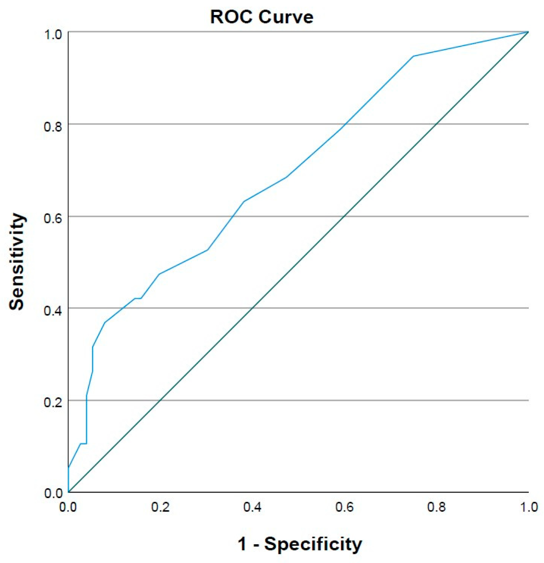
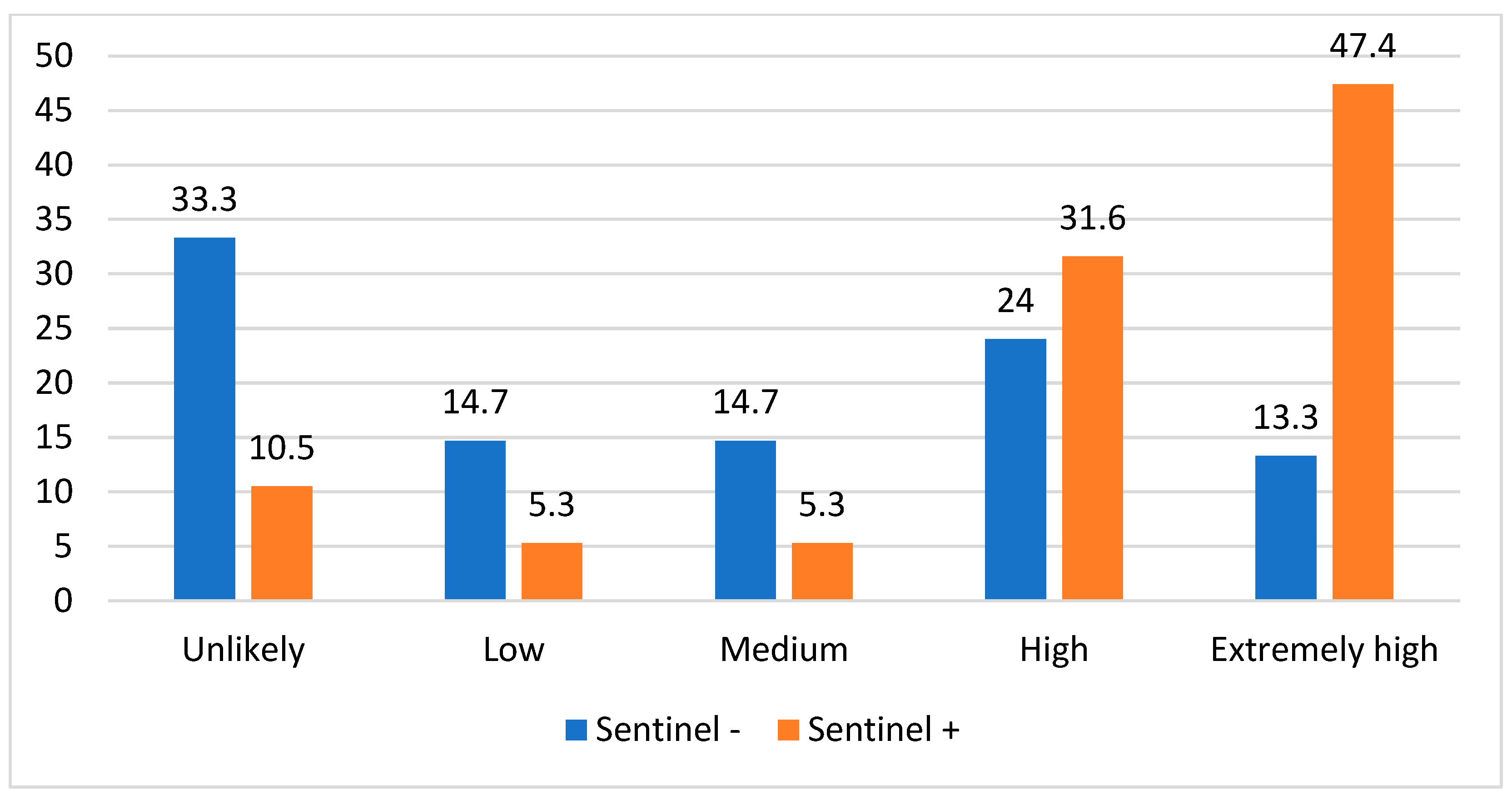
The Scoring System
4. Discussion
Study Limitations
5. Conclusions
Author Contributions
Funding
Institutional Review Board Statement
Informed Consent Statement
Data Availability Statement
Conflicts of Interest
References
- Matthews, N.H.; Li, W.-Q.; Qureshi, A.A.; Weinstock, M.A.; Cho, E. Epidemiology of Melanoma. In Cutaneous Melanoma: Etiology and Therapy; Ward, W.H., Farma, J.M., Eds.; Codon Publications: Brisbane, AU, USA, 2017; ISBN 978-0-9944381-4-0. [Google Scholar]
- American Cancer Society Key Statistics for Melanoma. Available online: https://www.cancer.org/cancer/types/melanoma-skin-cancer/about/key-statistics.html (accessed on 29 September 2023).
- Bobircă, F.; Tebeică, T.; Pumnea, A.; Dumitrescu, D.; Alexandru, C.; Banciu, L.; Popa, I.L.; Bobircă, A.; Leventer, M.; Pătrașcu, T. The Characteristics of Sentinel Lymph Node Biopsy in Cutaneous Melanoma and the Particularities for Elderly Patients—Experience of a Single Clinic. Diagnostics 2023, 13, 926. [Google Scholar] [CrossRef] [PubMed]
- Aitken, J.F.; Elwood, M.; Baade, P.D.; Youl, P.; English, D. Clinical Whole-Body Skin Examination Reduces the Incidence of Thick Melanomas. Int. J. Cancer 2010, 126, 450–458. [Google Scholar] [CrossRef]
- Ferlay, J.; Soerjomataram, I.; Dikshit, R.; Eser, S.; Mathers, C.; Rebelo, M.; Parkin, D.M.; Forman, D.; Bray, F. Cancer Incidence and Mortality Worldwide: Sources, Methods and Major Patterns in GLOBOCAN 2012. Int. J. Cancer 2015, 136, E359–E386. [Google Scholar] [CrossRef] [PubMed]
- Balch, C.M.; Gershenwald, J.E.; Soong, S.-J.; Thompson, J.F.; Atkins, M.B.; Byrd, D.R.; Buzaid, A.C.; Cochran, A.J.; Coit, D.G.; Ding, S.; et al. Final Version of 2009 AJCC Melanoma Staging and Classification. J. Clin. Oncol. Off. J. Am. Soc. Clin. Oncol. 2009, 27, 6199–6206. [Google Scholar] [CrossRef] [PubMed]
- Gershenwald, J.E.; Scolyer, R.A.; Hess, K.R.; Sondak, V.K.; Long, G.V.; Ross, M.I.; Lazar, A.J.; Faries, M.B.; Kirkwood, J.M.; McArthur, G.A.; et al. Melanoma Staging: Evidence-Based Changes in the American Joint Committee on Cancer Eighth Edition Cancer Staging Manual. CA. Cancer J. Clin. 2017, 67, 472–492. [Google Scholar] [CrossRef]
- Balch, C.M.; Gershenwald, J.E. Clinical Value of the Sentinel-Node Biopsy in Primary Cutaneous Melanoma. N. Engl. J. Med. 2014, 370, 663–664. [Google Scholar] [CrossRef]
- Huang, H.; Fu, Z.; Ji, J.; Huang, J.; Long, X. Predictive Values of Pathological and Clinical Risk Factors for Positivity of Sentinel Lymph Node Biopsy in Thin Melanoma: A Systematic Review and Meta-Analysis. Front. Oncol. 2022, 12, 817510. [Google Scholar] [CrossRef]
- Coit, D.G.; Thompson, J.A.; Albertini, M.R.; Barker, C.; Carson, W.E.; Contreras, C.; Daniels, G.A.; DiMaio, D.; Fields, R.C.; Fleming, M.D.; et al. Cutaneous Melanoma, Version 2.2019, NCCN Clinical Practice Guidelines in Oncology. J. Natl. Compr. Cancer Netw. JNCCN 2019, 17, 367–402. [Google Scholar] [CrossRef]
- Wong, S.L.; Faries, M.B.; Kennedy, E.B.; Agarwala, S.S.; Akhurst, T.J.; Ariyan, C.; Balch, C.M.; Berman, B.S.; Cochran, A.; Delman, K.A.; et al. Sentinel Lymph Node Biopsy and Management of Regional Lymph Nodes in Melanoma: American Society of Clinical Oncology and Society of Surgical Oncology Clinical Practice Guideline Update. J. Clin. Oncol. Off. J. Am. Soc. Clin. Oncol. 2018, 36, 399–413. [Google Scholar] [CrossRef]
- Conic, R.R.Z.; Ko, J.; Damiani, G.; Funchain, P.; Knackstedt, T.; Vij, A.; Vidimos, A.; Gastman, B.R. Predictors of Sentinel Lymph Node Positivity in Thin Melanoma Using the National Cancer Database. J. Am. Acad. Dermatol. 2019, 80, 441–447. [Google Scholar] [CrossRef]
- Yonick, D.V.; Ballo, R.M.; Kahn, E.; Dahiya, M.; Yao, K.; Godellas, C.; Shoup, M.; Aranha, G.V. Predictors of Positive Sentinel Lymph Node in Thin Melanoma. Am. J. Surg. 2011, 201, 324–327, discussion 327–328. [Google Scholar] [CrossRef] [PubMed]
- Mitteldorf, C.; Bertsch, H.P.; Jung, K.; Thoms, K.-M.; Schön, M.P.; Tronnier, M.; Kretschmer, L. Sentinel Node Biopsy Improves Prognostic Stratification in Patients with Thin (pT1) Melanomas and an Additional Risk Factor. Ann. Surg. Oncol. 2014, 21, 2252–2258. [Google Scholar] [CrossRef] [PubMed]
- Murali, R.; Haydu, L.E.; Quinn, M.J.; Saw, R.P.M.; Shannon, K.; Spillane, A.J.; Stretch, J.R.; Thompson, J.F.; Scolyer, R.A. Sentinel Lymph Node Biopsy in Patients with Thin Primary Cutaneous Melanoma. Ann. Surg. 2012, 255, 128–133. [Google Scholar] [CrossRef] [PubMed]
- Paek, S.C.; Griffith, K.A.; Johnson, T.M.; Sondak, V.K.; Wong, S.L.; Chang, A.E.; Cimmino, V.M.; Lowe, L.; Bradford, C.R.; Rees, R.S.; et al. The Impact of Factors beyond Breslow Depth on Predicting Sentinel Lymph Node Positivity in Melanoma. Cancer 2007, 109, 100–108. [Google Scholar] [CrossRef] [PubMed]
- White, R.L.; Ayers, G.D.; Stell, V.H.; Ding, S.; Gershenwald, J.E.; Salo, J.C.; Pockaj, B.A.; Essner, R.; Faries, M.; Charney, K.J.; et al. Factors Predictive of the Status of Sentinel Lymph Nodes in Melanoma Patients from a Large Multicenter Database. Ann. Surg. Oncol. 2011, 18, 3593–3600. [Google Scholar] [CrossRef]
- Sassen, S.; Shaw, H.M.; Colman, M.H.; Scolyer, R.A.; Thompson, J.F. The Complex Relationships between Sentinel Node Positivity, Patient Age, and Primary Tumor Desmoplasia: Analysis of 2303 Melanoma Patients Treated at a Single Center. Ann. Surg. Oncol. 2008, 15, 630–637. [Google Scholar] [CrossRef]
- Gershenwald, J.E.; Thompson, W.; Mansfield, P.F.; Lee, J.E.; Colome, M.I.; Tseng, C.H.; Lee, J.J.; Balch, C.M.; Reintgen, D.S.; Ross, M.I. Multi-Institutional Melanoma Lymphatic Mapping Experience: The Prognostic Value of Sentinel Lymph Node Status in 612 Stage I or II Melanoma Patients. J. Clin. Oncol. Off. J. Am. Soc. Clin. Oncol. 1999, 17, 976–983. [Google Scholar] [CrossRef]
- Leiter, U.; Stadler, R.; Mauch, C.; Hohenberger, W.; Brockmeyer, N.; Berking, C.; Sunderkötter, C.; Kaatz, M.; Schulte, K.-W.; Lehmann, P.; et al. Complete Lymph Node Dissection versus No Dissection in Patients with Sentinel Lymph Node Biopsy Positive Melanoma (DeCOG-SLT): A Multicentre, Randomised, Phase 3 Trial. Lancet Oncol. 2016, 17, 757–767. [Google Scholar] [CrossRef]
- Faries, M.B.; Thompson, J.F.; Cochran, A.J.; Andtbacka, R.H.; Mozzillo, N.; Zager, J.S.; Jahkola, T.; Bowles, T.L.; Testori, A.; Beitsch, P.D.; et al. Completion Dissection or Observation for Sentinel-Node Metastasis in Melanoma. N. Engl. J. Med. 2017, 376, 2211–2222. [Google Scholar] [CrossRef]
- Keung, E.Z.; Gershenwald, J.E. The Eighth Edition American Joint Committee on Cancer (AJCC) Melanoma Staging System: Implications for Melanoma Treatment and Care. Expert Rev. Anticancer. Ther. 2018, 18, 775–784. [Google Scholar] [CrossRef]
- Santos, F.D.M.D.; da Silva, F.C.; Pedron, J.; Furian, R.D.; Fortes, C.; Bonamigo, R.R. Association between Tumor-Infiltrating Lymphocytes and Sentinel Lymph Node Positivity in Thin Melanoma. An. Bras. Dermatol. 2019, 94, 47–51. [Google Scholar] [CrossRef] [PubMed]
- Rodriguez Otero, J.C.; Dagatti, M.S.; Fernandez Bussy, R.; Bergero, A.; Gorosito, M.; Staffieri, R.; Villavicencio, R.; Batalles, S.M.; Pezzotto, S.M. Sentinel Lymph Node Biopsy in Patients with Thick Primary Cutaneous Melanoma. World J. Oncol. 2019, 10, 112–117. [Google Scholar] [CrossRef] [PubMed]
- Swetter, S.M.; Thompson, J.A.; Albertini, M.R.; Barker, C.A.; Baumgartner, J.; Boland, G.; Chmielowski, B.; DiMaio, D.; Durham, A.; Fields, R.C.; et al. NCCN Guidelines® Insights: Melanoma: Cutaneous, Version 2.2021. J. Natl. Compr. Cancer Netw. 2021, 19, 364–376. [Google Scholar] [CrossRef]
- Ribero, S.; Osella-Abate, S.; Sanlorenzo, M.; Balagna, E.; Senetta, R.; Fierro, M.T.; Macripò, G.; Macrì, L.; Sapino, A.; Quaglino, P. Sentinel Lymph Node Biopsy in Thick-Melanoma Patients (N = 350): What Is Its Prognostic Role? Ann. Surg. Oncol. 2015, 22, 1967–1973. [Google Scholar] [CrossRef] [PubMed]
- White, I.; Fortino, J.; Curti, B.; Vetto, J. Clinical Impact of Sentinel Lymph Node Biopsy in Patients with Thick (>4 Mm) Melanomas. Am. J. Surg. 2014, 207, 702–707, discussion 707. [Google Scholar] [CrossRef]
- Jokic, S.; Markovic, I.; Bukumiric, Z.; Jokic, V.; Rakovic, M.; Tripkovic, J.; Stojiljkovic, D.; Spurnic, I.; Jevric, M.; Matic, M.; et al. Predictors of Sentinel Lymph Node Status of Cutaneous Melanoma in Serbian Patients. J. BUON Off. J. Balk. Union Oncol. 2018, 23, 468–474. [Google Scholar]
- Dunne, J.A.; Wormald, J.C.R.; Steele, J.; Woods, E.; Odili, J.; Powell, B.W.E.M. Is Sentinel Lymph Node Biopsy Warranted for Desmoplastic Melanoma? A Systematic Review. J. Plast. Reconstr. Aesthetic Surg. 2017, 70, 274–280. [Google Scholar] [CrossRef]
- Cheraghlou, S.; Ugwu, N.; Girardi, M. Sentinel Lymph Node Biopsy Positivity in Patients with Acral Lentiginous and Other Subtypes of Cutaneous Melanoma. JAMA Dermatol. 2022, 158, 51–58. [Google Scholar] [CrossRef]
- Kato, J.; Hida, T.; Kamiya, T.; Horimoto, K.; Sato, S.; Sawada, M.; Minowa, T.; Handa, T.; Komatsu, S.; Uhara, H. Relationships between Tumor Thickness and the Risk of Sentinel Node Metastasis in Acral and Non-Acral Melanoma. Int. J. Dermatol. 2023, 62, e573–e575. [Google Scholar] [CrossRef]
- Balch, C.M.; Thompson, J.F.; Gershenwald, J.E.; Soong, S.-J.; Ding, S.; McMasters, K.M.; Coit, D.G.; Eggermont, A.M.M.; Gimotty, P.A.; Johnson, T.M.; et al. Age as a Predictor of Sentinel Node Metastasis among Patients with Localized Melanoma: An Inverse Correlation of Melanoma Mortality and Incidence of Sentinel Node Metastasis among Young and Old Patients. Ann. Surg. Oncol. 2014, 21, 1075–1081. [Google Scholar] [CrossRef]
- Balch, C.M.; Soong, S.; Gershenwald, J.E.; Thompson, J.F.; Coit, D.G.; Atkins, M.B.; Ding, S.; Cochran, A.J.; Eggermont, A.M.M.; Flaherty, K.T.; et al. Age as a Prognostic Factor in Patients with Localized Melanoma and Regional Metastases. Ann. Surg. Oncol. 2013, 20, 3961–3968. [Google Scholar] [CrossRef] [PubMed]
- Featherston, C.; Nardi, W.S.; Tomé, F.R.; Quildrian, S.D. Role of Sentinel Lymph Node Biopsy for Cutaneous Melanoma in Elderly Patients: Preliminary Results in a Latin-American Population. Ecancermedicalscience 2021, 15, 1167. [Google Scholar] [CrossRef] [PubMed]
- Moody, J.A.; Ali, R.F.; Carbone, A.C.; Singh, S.; Hardwicke, J.T. Complications of Sentinel Lymph Node Biopsy for Melanoma-A Systematic Review of the Literature. Eur. J. Surg. Oncol. J. Eur. Soc. Surg. Oncol. Br. Assoc. Surg. Oncol. 2017, 43, 270–277. [Google Scholar] [CrossRef] [PubMed]


| Variables | n = 101 (100%) |
|---|---|
| Age (mean ± SD) | 56.26 ±15.1 |
| Age groups: | |
| <40 | 17 (16.8%) |
| 40–60 | 38 (37.6%) |
| >60 | 46 (45.5%) |
| Sex: | |
| Male | 60 (59.4%) |
| Female | 41 (40.6%) |
| Localization of primary melanoma: | |
| Head and neck | 16 (16.2%) |
| Torso | 40 (40.4%) |
| Upper extremities | 21 (21.2%) |
| Lower extremities | 20 (20.2%) |
| Acral | 2 (2.0%) |
| Melanoma subtype: | |
| Superficial spreading | 58 (59.2%) |
| Nodular | 30 (30.6%) |
| Lentigo maligna | 3 (3.1%) |
| Acral | 2 (2.0%) |
| Other | 5 (5.1%) |
| Breslow thickness in mm (median, 25th–75th percentile) | 2.20 (1.23–4.50) |
| Breslow thickness: | |
| <1 mm | 14 (14.7%) |
| 1.01–2 mm | 29 (30.5%) |
| 2.01–4 mm | 26 (27.4%) |
| >4 mm | 26 (27.4%) |
| Clark level: | |
| II | 5 (5.3%) |
| III | 34 (35.8%) |
| IV | 50 (52.6%) |
| V | 6 (6.3%) |
| pT staging: | |
| T1–T2 | 42 (44.7%) |
| T3–T4 | 52 (55.3%) |
| Mitotic index rate (median, 25th–75th percentile): | 3.00 (1.00–6.00) |
| Ulceration present: | |
| Yes | 39 (41.1%) |
| No | 56 (58.9%) |
| Variables | n = 101 (100%) |
|---|---|
| Nodal metastasis present: | |
| Yes | 19 (18.8%) |
| No | 82 (81.2%) |
| SLN localization: | |
| Axilla | 54 (53.5%) |
| Groin | 26 (25.7%) |
| Head & Neck | 17 (16.8%) |
| Interval Nodes | 4 (4.0%) |
| Diameter of SLN metastasis in mm (median, 25th–75th percentile) | 1.20 (0.80–2.60) |
| Microanatomic SLN metastasis localization: | |
| Subcapsular | 3 (16.7%) |
| Parenchymal | 8 (44.4%) |
| Both structures | 7 (38.9%) |
| Extracapsular spreading of metastatic deposits present | |
| Yes | 2 (11.1%) |
| No | 16 (88.9%) |
| Variables | SLN Positive | SLN Negative | p Value |
|---|---|---|---|
| n = 19 (18.8%) | n = 82 (81.2%) | ||
| Age (mean ± SD) | 56.21 ± 13.48 | 56.27 ± 15.52 | |
| Age groups: | 0.548 | ||
| <40 | 2 (10.5%) | 15 (18.3%) | |
| 41–60 | 9 (47.4%) | 29 (35.4%) | |
| >60 | 8 (42.1%) | 38 (46.3%) | |
| Sex: | 0.160 | ||
| Male | 14 (73.7%) | 46 (56.1%) | |
| Female | 5 (26.3%) | 36 (43.9%) | |
| Primary melanoma localization: | 0.912 | ||
| Head and Neck | 3 (15.8%) | 13 (16.3%) | |
| Torso | 9 (47.4%) | 31 (38.8%) | |
| Upper extremities | 4 (21.1%) | 17 (21.3%) | |
| Lower extremities | 3 (15.8%) | 17 (21.3%) | |
| Acral | 0 (0.0%) | 2 (2.5%) | |
| Melanoma subtype: | 0.208 | ||
| Superficial Spreading | 8 (42.1%) | 50 (63.3%) | |
| Nodular | 9 (47.4%) | 21 (26.6%) | |
| Lentigo maligna | 0 (0.0%) | 3 (3.8%) | |
| Acral | 0 (0.0%) | 2 (2.5%) | |
| Others | 2 (10.5%) | 3 (3.8%) | |
| Breslow thickness (median, 25th–75th percentile) | 3.80 (2.90–7.00) | 1.97 (1.20–3.27) | 0.002 |
| Breslow thickness: | 0.032 | ||
| <1 mm | 1 (5.3%) | 13 (17.1%) | |
| 1.01–2 mm | 2 (10.5%) | 27 (35.5%) | |
| 2.01–4 mm | 7 (36.8%) | 19 (25.0%) | |
| >4 mm | 9 (47.4%) | 17 (22.4%) | |
| Clark level: | 0.390 | ||
| II | 0 (0.0%) | 5 (6.6%) | |
| III | 5 (26.3%) | 29 (38.2%) | |
| IV | 13 (68.4%) | 37 (48.7%) | |
| V | 1 (5.3%) | 5 (6.6%) | |
| pT staging: | 0.005 | ||
| T1–T2 | 3 (15.8%) | 39 (52.0%) | |
| T3–T4 | 16 (84.2%) | 36 (48.0%) | |
| Mitotic index rate (median, 25th–75th percentile) | 5.00 (2.00–12.00) | 2.00 (0.25–5.00) | 0.009 |
| Ulceration present: | 0.007 | ||
| Yes | 13 (68.4%) | 26 (34.2%) | |
| No | 6 (31.6%) | 50 (65.8%) |
| Predictors for SLN Positivity | Sn | PPV | Sp | NPV |
|---|---|---|---|---|
| Breslow thickness | 84.2 | 30.8 | 52.6 | 93.0 |
| Mitotic index | 63.2 | 29.3 | 61.8 | 87.0 |
| Presence of ulceration | 68.4 | 33.3 | 65.8 | 89.3 |
| pT3-4 | 84.2 | 30.8 | 50.2 | 92.9 |
| Nodular subtype | 47.4 | 30.3 | 73.4 | 85.3 |
| Variable | OR | 95% CI for OR | p |
|---|---|---|---|
| Univariate analysis | |||
| Nodular subtype | 2.486 | 0.888–6.960 | 0.078 |
| Breslow thickness | 5.926 | 1.595–22.023 | 0.008 |
| Mitotic index rate | 2.778 | 0.981–7.866 | 0.054 |
| Ulceration present | 4.167 | 1.419–12.235 | 0.009 |
| pT3-4 stage | 5.778 | 1.553–21.493 | 0.009 |
| Multivariate analysis | |||
| pT3-4 stage | 5.778 | 1.553–21.493 | 0.009 |
Disclaimer/Publisher’s Note: The statements, opinions and data contained in all publications are solely those of the individual author(s) and contributor(s) and not of MDPI and/or the editor(s). MDPI and/or the editor(s) disclaim responsibility for any injury to people or property resulting from any ideas, methods, instructions or products referred to in the content. |
© 2023 by the authors. Licensee MDPI, Basel, Switzerland. This article is an open access article distributed under the terms and conditions of the Creative Commons Attribution (CC BY) license (https://creativecommons.org/licenses/by/4.0/).
Share and Cite
Jeremić, J.; Radenović, K.; Jurišić, M.; Suđecki, B.; Marinković, M.; Mihaljević, J.; Radosavljević, I.; Jovanović, M.; Stojanović, M.; Milić, N.; et al. Primary Melanoma Histopathologic Predictors of Sentinel Lymph Node Positivity: A Proposed Scoring System for Risk Assessment and Patient Selection in a Clinical Setting. Medicina 2023, 59, 1921. https://doi.org/10.3390/medicina59111921
Jeremić J, Radenović K, Jurišić M, Suđecki B, Marinković M, Mihaljević J, Radosavljević I, Jovanović M, Stojanović M, Milić N, et al. Primary Melanoma Histopathologic Predictors of Sentinel Lymph Node Positivity: A Proposed Scoring System for Risk Assessment and Patient Selection in a Clinical Setting. Medicina. 2023; 59(11):1921. https://doi.org/10.3390/medicina59111921
Chicago/Turabian StyleJeremić, Jelena, Kristina Radenović, Milana Jurišić, Branko Suđecki, Milana Marinković, Jovan Mihaljević, Ivan Radosavljević, Milan Jovanović, Marina Stojanović, Nataša Milić, and et al. 2023. "Primary Melanoma Histopathologic Predictors of Sentinel Lymph Node Positivity: A Proposed Scoring System for Risk Assessment and Patient Selection in a Clinical Setting" Medicina 59, no. 11: 1921. https://doi.org/10.3390/medicina59111921
APA StyleJeremić, J., Radenović, K., Jurišić, M., Suđecki, B., Marinković, M., Mihaljević, J., Radosavljević, I., Jovanović, M., Stojanović, M., Milić, N., Pavlović, V., Stojičić, M., Inić, Z., & Jović, M. (2023). Primary Melanoma Histopathologic Predictors of Sentinel Lymph Node Positivity: A Proposed Scoring System for Risk Assessment and Patient Selection in a Clinical Setting. Medicina, 59(11), 1921. https://doi.org/10.3390/medicina59111921